Assessing the Biomechanical, Kinematic, and Force Distribution Properties of the Foot Following Tarsometatarsal Joint Arthrodesis: A Systematic Review
Abstract
1. Introduction
2. Materials and Methods
2.1. Search Parameters
2.2. Inclusion/Exclusion Criteria
2.3. Study Definitions
2.4. Data Extraction
2.5. Statistical Analysis
3. Results
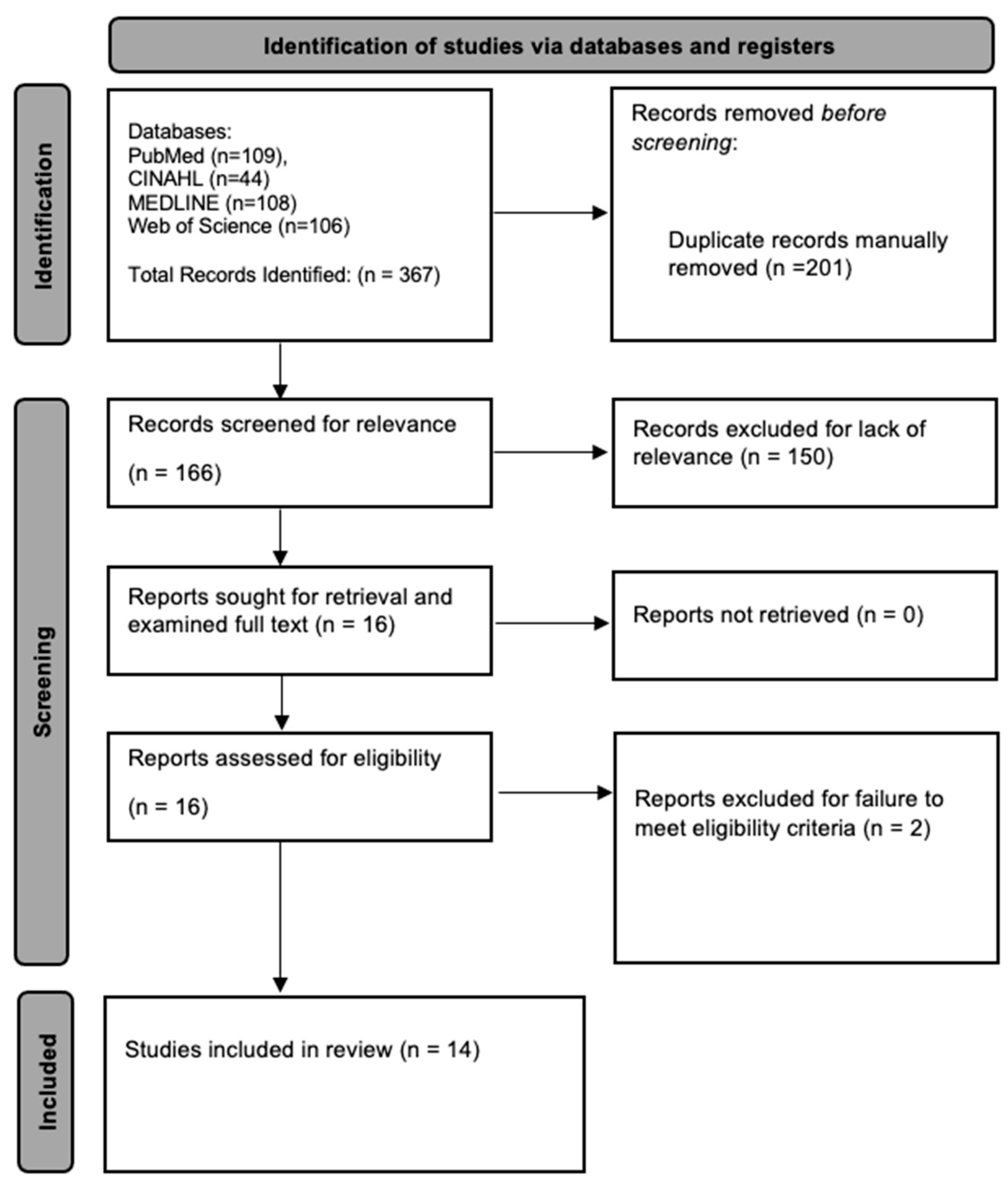
3.1. Preliminary Search Findings
3.2. Study Demographics
3.3. Single TMT Joint Arthrodesis
3.4. Multilevel TMT Arthrodesis
3.5. Kinetic Behavior and Force Distribution of Foot Following Arthrodesis
4. Discussion
5. Conclusions
Author Contributions
Funding
Institutional Review Board Statement
Informed Consent Statement
Data Availability Statement
Conflicts of Interest
References
- Bevilacqua, N.J. Tarsometatarsal Arthrodesis for Lisfranc Injuries. Clin. Podiatr. Med. Surg. 2017, 34, 315–325. [Google Scholar] [CrossRef]
- Weatherford, B.M.; Anderson, J.G.; Bohay, D.R. Management of Tarsometatarsal Joint Injuries. J. Am. Acad. Orthop. Surg. 2017, 25, 469–479. [Google Scholar] [CrossRef]
- Han, P.F.; Zhang, Z.L.; Chen, C.L.; Han, Y.C.; Wei, X.C.; Li, P.C. Comparison of primary arthrodesis versus open reduction with internal fixation for Lisfranc injuries: Systematic review and meta-analysis. J. Postgrad. Med. 2019, 65, 93–100. [Google Scholar] [PubMed]
- Myerson, M.S.; Cerrato, R.A. Current management of tarsometatarsal injuries in the athlete. J. Bone Jt. Surg. Am. 2008, 90, 2522–2533. [Google Scholar]
- Ouzounian, T.J.; Shereff, M.J. In vitro determination of midfoot motion. Foot Ankle 1989, 10, 140–146. [Google Scholar] [CrossRef] [PubMed]
- Henning, J.A.; Jones, C.B.; Sietsema, D.L.; Bohay, D.R.; Anderson, J.G. Open reduction internal fixation versus primary arthrodesis for lisfranc injuries: A prospective randomized study. Foot Ankle Int. 2009, 30, 913–922. [Google Scholar] [CrossRef] [PubMed]
- Catanzariti, A.R.; Mendicino, R.W. Technical considerations in tarsometatarsal joint arthrodesis. J. Am. Podiatr. Med. Assoc. 2005, 95, 85–90. [Google Scholar] [CrossRef]
- Boffeli, T.J.; Pfannenstein, R.R.; Thompson, J.C. Combined medial column primary arthrodesis, middle column open reduction internal fixation, and lateral column pinning for treatment of Lisfranc fracture-dislocation injuries. J. Foot Ankle Surg. 2014, 53, 657–663. [Google Scholar] [CrossRef]
- Kapoor, C.; Patel, A.; Jhaveri, M.; Golwala, P.; Jhaveri, M.R. Post-traumatic Arthritis of the Tarsometatarsal Joint Complex: A Case Report. Cureus 2016, 8, e923. [Google Scholar] [CrossRef]
- Myerson, M.S.; Fisher, R.T.; Burgess, A.R.; Kenzora, J.E. Fracture dislocations of the tarsometatarsal joints: End results correlated with pathology and treatment. Foot Ankle 1986, 6, 225–242. [Google Scholar] [CrossRef]
- Rao, S.; Nawoczenski, D.A.; Baumhauer, J.F. Midfoot Arthritis: Nonoperative Options and Decision Making for Fusion. Tech. Foot Ankle Surg. 2008, 7, 188–195. [Google Scholar] [CrossRef]
- Goossens, M.; De Stoop, N. Lisfranc’s fracture-dislocations: Etiology, radiology, and results of treatment. A review of 20 cases. Clin. Orthop. Relat. Res. 1983, 176, 154–162. [Google Scholar] [CrossRef]
- Sheibani-Rad, S.; Coetzee, J.C.; Giveans, M.R.; DiGiovanni, C. Arthrodesis versus ORIF for Lisfranc fractures. Orthopedics 2012, 35, e868–e873. [Google Scholar] [CrossRef] [PubMed]
- Arntz, C.T.; Veith, R.G.; Hansen, S.T., Jr. Fractures and fracture-dislocations of the tarsometatarsal joint. J. Bone Jt. Surg. Am. 1988, 70, 173–181. [Google Scholar] [CrossRef]
- Ly, T.V.; Coetzee, J.C. Treatment of primarily ligamentous Lisfranc joint injuries: Primary arthrodesis compared with open reduction and internal fixation. A prospective, randomized study. J. Bone Jt. Surg. Am. 2006, 88, 514–520. [Google Scholar] [CrossRef] [PubMed]
- Burchard, R.; Massa, R.; Soost, C.; Richter, W.; Dietrich, G.; Ohrndorf, A.; Christ, H.-J.; Fritzen, C.-P.; Graw, J.A.; Schmitt, J. Biomechanics of common fixation devices for first tarsometatarsal joint fusion-a comparative study with synthetic bones. J. Orthop. Surg. Res. 2018, 13, 176. [Google Scholar] [CrossRef] [PubMed]
- Knutsen, A.R.; Fleming, J.F.; Ebramzadeh, E.; Ho, N.C.; Warganich, T.; Harris, T.G.; Sangiorgio, S.N. Biomechanical Comparison of Fixation Devices for First Metatarsocuneiform Joint Arthrodesis. Foot Ankle Spec. 2017, 10, 322–328. [Google Scholar] [CrossRef] [PubMed]
- Koroneos, Z.A.; Manto, K.M.; Martinazzi, B.J.; Stauch, C.; Bifano, S.M.; Kunselman, A.R.; Lewis, G.S.; Aynardi, M. Biomechanical Comparison of Fiber Tape Device Versus Transarticular Screws for Ligamentous Lisfranc Injury in a Cadaveric Model. Am. J. Sports Med. 2022, 50, 3299–3307. [Google Scholar] [CrossRef]
- Ettinger, S.; Hemmersbach, L.C.; Schwarze, M.; Stukenborg-Colsman, C.; Yao, D.; Plaass, C.; Claassen, L. Biomechanical Evaluation of Tarsometatarsal Fusion Comparing Crossing Lag Screws and Lag Screw With Locking Plate. Foot Ankle Int. 2022, 43, 77–85. [Google Scholar] [CrossRef]
- Yu, X.; Pang, Q.J.; Yu, G.R. The injuries to the fourth and fifth tarsometatarsal joints: A review of the surgical management by internal fixation, arthrodesis and arthroplasty. Pak. J. Med. Sci. 2013, 29, 687–692. [Google Scholar] [CrossRef]
- Wu, G.; Gu, S.; Yu, G.; Yin, F. Effect of different fusion types on kinematics of midfoot lateral column: A comparative biomechanical study. Ann. Transl. Med. 2019, 7, 665. [Google Scholar] [CrossRef]
- Prieto-Diaz, C.; Anderle, M.R.; Brinker, L.Z.; Allard, R.; Leasure, J. Biomechanical Comparison of First Tarsometatarsal Arthrodesis Constructs Over Prolonged Cyclic Testing. Foot Ankle Orthop. 2019, 4, 2473011419892240. [Google Scholar] [CrossRef] [PubMed]
- Ho, N.C.; Sangiorgio, S.N.; Cassinelli, S.; Shymon, S.; Fleming, J.; Agrawal, V.; Ebramzadeh, E.; Harris, T.G. Biomechanical comparison of fixation stability using a Lisfranc plate versus transarticular screws. Foot Ankle Surg. 2019, 25, 71–78. [Google Scholar] [CrossRef] [PubMed]
- Perez, H.R.; Reber, L.K.; Christensen, J.C. Effects on the metatarsophalangeal joint after simulated first tarsometatarsal joint arthrodesis. J. Foot Ankle Surg. 2007, 46, 242–247. [Google Scholar] [CrossRef]
- Kim, J.; Hoffman, J.; Steineman, B.; Eble, S.K.; Roberts, L.E.; Ellis, S.J.; Drakos, M.C. Kinematic Analysis of Sequential Partial-Midfoot Arthrodesis in Simulated Gait Cadaver Model. Foot Ankle Int. 2022, 43, 1587–1594. [Google Scholar] [CrossRef] [PubMed]
- Pope, E.J.; Takemoto, R.C.; Kummer, F.J.; Mroczek, K.J. Midfoot fusion: A biomechanical comparison of plantar planting vs intramedullary screws. Foot Ankle Int. 2013, 34, 409–413. [Google Scholar] [CrossRef] [PubMed]
- Marks, R.M.; Parks, B.G.; Schon, L.C. Midfoot fusion technique for neuroarthropathic feet: Biomechanical analysis and rationale. Foot Ankle Int. 1998, 19, 507–510. [Google Scholar] [CrossRef]
- Nadaud, J.P.; Parks, B.G.; Schon, L.C. Plantar and calcaneocuboid joint pressure after isolated medial column fusion versus medial and lateral column fusion: A biomechanical study. Foot Ankle Int. 2011, 32, 1069–1074. [Google Scholar] [CrossRef]
- Aiyer, A.; Russell, N.A.; Pelletier, M.H.; Myerson, M.; Walsh, W.R. The Impact of Nitinol Staples on the Compressive Forces, Contact Area, and Mechanical Properties in Comparison to a Claw Plate and Crossed Screws for the First Tarsometatarsal Arthrodesis. Foot Ankle Spec. 2016, 9, 232–240. [Google Scholar] [CrossRef]
- Shen, V.C.; Bumgardner, C.; Actis, L.; Park, J.; Moyer, D.; May-Nikstaitis, K.; Li, X. In Situ Deformation of First Tarsometatarsal Arthrodesis Implants with Digital Image Correlation: A Cadaveric Study. JOM 2022, 74, 3357–3366. [Google Scholar] [CrossRef]
- Grant, W.P.; Garcia-Lavin, S.; Sabo, R. Beaming the columns for Charcot diabetic foot reconstruction: A retrospective analysis. J. Foot Ankle Surg. 2011, 50, 182–189. [Google Scholar] [CrossRef] [PubMed]
- Wukich, D.K.; Liu, G.T.; Raspovic, K.; Vicenzi, F. Biomechanical Performance of Charcot-Specific Implants. J. Foot Ankle Surg. 2021, 60, 440–447. [Google Scholar] [CrossRef] [PubMed]
- Balu, A.R.; Baumann, A.N.; Tsang, T.; Talaski, G.M.; Anastasio, A.T.; Walley, K.C.; Adams, S.B. Evaluating the Biomechanical Integrity of Various Constructs Utilized for First Metatarsophalangeal Joint Arthrodesis: A Systematic Review. Materials 2023, 16, 6562. [Google Scholar] [CrossRef]
- Fraser, T.W.; Miles, D.T.; Huang, N.; Davis, F.B.; Dunlap, B.D.; Doty, J.F. Radiographic Outcomes, Union Rates, and Complications Associated With Plantar Implant Positioning for Midfoot Arthrodesis. Foot Ankle Orthop. 2021, 6, 24730114211027115. [Google Scholar] [CrossRef] [PubMed]
- Klos, K.; Wilde, C.H.; Lange, A.; Wagner, A.; Gras, F.; Skulev, H.K.; Mückley, T.; Simons, P. Modified Lapidus arthrodesis with plantar plate and compression screw for treatment of hallux valgus with hypermobility of the first ray: A preliminary report. Foot Ankle Surg. 2013, 19, 239–244. [Google Scholar] [CrossRef]
- Bayam, L.; Ryan, P.; Bilal, M.; Fayyaz, I.; Drampalos, E. Early Results and Patient-Reported Outcome Measures (PROMS) of an Intraosseous Device for Arthrodesis of the First Tarso-Metatarsal (TMT) Joint. Indian J. Orthop. 2022, 56, 895–901. [Google Scholar] [CrossRef]

| First Author (Year) | Full Title | Model Used (Cadaver, Synthetic Bone Model, etc.) | Number of Construct | HPrimary Fusion Joint | Fixation Method(s) | Diastasis Mean (mm) | Plantar Gapping (mm) | Displacement (mm) | Average Diastasis Decrease (mm) | Ultimate Failure Load (N) | Axial Stiffness (N/mm) |
|---|---|---|---|---|---|---|---|---|---|---|---|
| Amiethab Aiyer (2015) [29] | The Impact of Nitinol Staples on the Compressive Forces, Contact Area, and Mechanical Properties in Comparison to a Claw Plate and Crossed Screws for the First Tarsometatarsal Arthrodesis | Synthetic Bone | 20 | 1 TMT | Single Shape Memory Staple, Double Shape Memory Staple, Claw Plate, Crossed Screws | Single Staple: 0.95 Double Staple: 0.69 Claw Plate: 2.61 Crossed Screws: 0.6 | Single Staple: 206.78 Double Staple: 226.24 Claw Plate: 184.47 Crossed Screws: 347.85 | ||||
| Ashleen R. Knutsen (2017) [17] | Biomechanical Comparison of Fixation Devices for First Metatarsocuneiform Joint Arthrodesis | Synthetic Bone | 32 | 1 TMT | IO Fixation, Crossed Screws, Dorsal Plate | IO Fix: 2.8 Crossed Screws: 0.4 Dorsal Plate: 5.5 | IO Fix: 460 Crossed Screws: 430 Dorsal Plate: 110 | IO Fix: 4 Crossed Screws: 10 Dorsal Plate: 5.9 | |||
| Cynthia Prieto-Diaz (2019) [22] | Biomechanical Comparison of First Tarsometatarsal Arthrodesis Constructs Over Prolonged Cyclic Testing | Cadaver | 40 | 1 TMT | Intramedullary fixation, medial plate with crossing screw, crossed screws | Intramedullary 4 hole plate: 0.14 Intramedullary 3 hole plate: 0.17 Medial Plate: 0.23 Crossing Screws: 0.54 | |||||
| Ernest Pope (2013) [26] | Midfoot Fusion: A Biomechanical Comparison of Plantar Planting vs. Intramedullary Screws | Cadaver | 7 | 1/2/3/4/5 TMT | Plantar Plating, Intramedullary Screws | Plantar Plate: 344 Intramedullary Screws: 313 | Plantar Plate: 9.7 Intramedullary Screws: 11.2 | ||||
| Genbin Wu (2019) [21] | Effect of Different Fusion Types on Kinematics of Midfoot Lateral Column: A Comparative Biomechanical Study | Cadaver | 10 | 4/5 TMT | 3.5-mm Fully Threaded Screws | ||||||
| Hugo Perez (2007) [24] | Effects on the Metatarsophalangeal Joint After Simulated First Tarsometatarsal Joint Arthrodesis | Cadaver | 5 | 1 TMT | K-wires | ||||||
| Jaeyoung Kim (2022) [25] | Kinematic Analysis of Sequential Partial-Midfoot Arthrodesis in Simulated Gait Cadaver Model | Cadaver | 10 | 1/2/3 TMT | Dorsal Lisfranc Plate | ||||||
| Joshua Nadaud (2011) [28] | Plantar and Calcaneocuboid Joint Pressure After Isolated Medial Column Fusion Versus Medial and Lateral Column Fusion: A Biomechanical Study | Cadaver | 12 | 1/2/3/4/5 TMT | 4.5-mm Fully Threaded Screws | ||||||
| Nathan C. Ho (2019) [23] | Biomechanical Comparison of Fixation Stability Using a Lisfranc Plate Versus Transarticular Screws | Cadaver | 13 pairs | 1/2 TMT | Dorsal Plate, Crossed Screws | ||||||
| Rene Burchard (2018) [16] | Biomechanics of Common Fixation Devices For First Tarsometatarsal Joint Fusion—A Comparative Study With Synthetic Bones | Synthetic Bone | 9 | 1 TMT | Dorsal Locking Plate, Plantar Locking Plate, IO Fixation | Dorsal Plate: 0.06 Plantar Plate: 0.03 IO Fix: 0.08 | Dorsal Plate: 324 Plantar Plate: 377 IO Fix: 173 | ||||
| Richard Marks (1998) [27] | Midfoot Fusion Technique for Neuroarthropathic Feet: Biomechanical Analysis and Rationale | Cadaver | 8 pairs | 1 TMT | Plantar Plate, Crossed Screws | Plantar Plate: 0.2 Crossed Screws: 0.89 | Plantar Plate: 2917 Crossed Screws: 1480 | ||||
| Sarah Ettinger (2022) [19] | Biomechanical Evaluation of Tarsometatarsal Fusion Comparing Crossing Lag Screws and Lag Screw With Locking Plate | Cadaver | 30 | 1/2/3 TMT | Locking Plate with Lag Screw, Crossed Screws | ||||||
| VC Shen (2022) [30] | In Situ Deformation of First Tarsometatarsal Arthrodesis Implants with Digital Image Correlation: A Cadaveric Study | Cadaver | 6 pairs | 1 TMT | Locking Plate with Lag Screw, Shape-Memory Staples | Locking Plate: 3.1 Nitinol Staples: 3.9 | |||||
| Zachary A. Koroneos (2022) [18] | Biomechanical Comparison of Fiber Tape Device Versus Transarticular Screws for Ligamentous Lisfranc Injury in a Cadaveric Model | Cadaver | 8 pairs | Medial Cuneiform—2nd Metatarasal | Crossed Screws, Interosseous Fiber Tape | Crossed Screws: 0.68 Fiber Tape: 0.9 | Crossed Screws: −2.61 Fiber Tape: −2.81 |
Disclaimer/Publisher’s Note: The statements, opinions and data contained in all publications are solely those of the individual author(s) and contributor(s) and not of MDPI and/or the editor(s). MDPI and/or the editor(s) disclaim responsibility for any injury to people or property resulting from any ideas, methods, instructions or products referred to in the content. |
© 2024 by the authors. Licensee MDPI, Basel, Switzerland. This article is an open access article distributed under the terms and conditions of the Creative Commons Attribution (CC BY) license (https://creativecommons.org/licenses/by/4.0/).
Share and Cite
Balu, A.R.; Baumann, A.N.; Burkhead, D.; Talaski, G.M.; Anastasio, A.T.; Walley, K.C.; Adams, S.B. Assessing the Biomechanical, Kinematic, and Force Distribution Properties of the Foot Following Tarsometatarsal Joint Arthrodesis: A Systematic Review. Appl. Sci. 2024, 14, 765. https://doi.org/10.3390/app14020765
Balu AR, Baumann AN, Burkhead D, Talaski GM, Anastasio AT, Walley KC, Adams SB. Assessing the Biomechanical, Kinematic, and Force Distribution Properties of the Foot Following Tarsometatarsal Joint Arthrodesis: A Systematic Review. Applied Sciences. 2024; 14(2):765. https://doi.org/10.3390/app14020765
Chicago/Turabian StyleBalu, Abhinav Reddy, Anthony N. Baumann, Daniel Burkhead, Grayson M. Talaski, Albert T. Anastasio, Kempland C. Walley, and Samuel B. Adams. 2024. "Assessing the Biomechanical, Kinematic, and Force Distribution Properties of the Foot Following Tarsometatarsal Joint Arthrodesis: A Systematic Review" Applied Sciences 14, no. 2: 765. https://doi.org/10.3390/app14020765
APA StyleBalu, A. R., Baumann, A. N., Burkhead, D., Talaski, G. M., Anastasio, A. T., Walley, K. C., & Adams, S. B. (2024). Assessing the Biomechanical, Kinematic, and Force Distribution Properties of the Foot Following Tarsometatarsal Joint Arthrodesis: A Systematic Review. Applied Sciences, 14(2), 765. https://doi.org/10.3390/app14020765


