Article

Synthesis of Si-Fe Chondrule-like Dust Analogues in RF Discharge Plasmas

by
Akdaulet Baikaliyev
1,
Assan Abdirakhmanov
2,*,†,
Sagi Orazbayev
1,2,
Yerbolat Ussenov
2,‡,
Alexander Brodsky
3,
Madi Aitzhanov
1,
Nazym Akhanova
2,
Merlan Dosbolayev
1,
Maratbek Gabdullin
2,
Tlekkabul Ramazanov
1,2 and
Didar Batryshev
2,*
1
Faculty of Physics and Technology, Al-Farabi Kazakh National University, Almaty 050040, Kazakhstan
2
Laboratory of Alternative Energy and Nanotechnology, Kazakh-British Technical University, Almaty 050000, Kazakhstan
3
Sokolsky Institute of Fuel, Catalysis and Electrochemistry, Almaty 050010, Kazakhstan
*
Authors to whom correspondence should be addressed.
Current address: Plasma-Surface Interaction Chemistry Laboratory, Mons University, 7000 Mons, Belgium.
Current address: Princeton Plasma Physics Laboratory, Princeton University, Princeton, NJ 08544, USA.
Appl. Sci. 2024, 14(19), 8714; https://doi.org/10.3390/app14198714
Submission received: 11 August 2024 / Revised: 20 September 2024 / Accepted: 21 September 2024 / Published: 27 September 2024
(This article belongs to the Section Nanotechnology and Applied Nanosciences)

Abstract

:
Chondrules are tiny particles that occur in stony meteorites and are considered as the building blocks of early asteroids and planets. It is believed that they were formed by the fast heating of the dust in the solar nebula. To date, there is no lab-scale experimental study of the formation of chondrules from the initial gas phase precursors following fast heating and crystallisation. The motivation of this work is a pre-trial study of the formation of chnodrule-like particles. The formation of meteorites in the space environment is associated with the aggregation of small particles or molecular clouds under the influence of shock waves or high-energy gas discharges in the solar nebula. In this work, the properties of product formation at the nanoscale-level were investigated using different feedstock materials which are the dominant elements in the meteorite. The structural and morphological properties of the synthesised Si-Fe nanomaterials were analysed by scanning/transmission electron microscopy (SEM/TEM), and chemical composition was analysed by X-ray energy-dispersive spectroscopy (EDS). The identification of crystalline phases was carried out by X-ray diffraction (XRD), whereas the presence of an Fe-Si system in the synthesised particles was demonstrated by Mössbauer spectroscopy. The obtained materials were exposed to the relatively high-energy pulsed plasma beam on the substrate with the aim to emulate the possible fast heating and melting of the formed nanoparticles. The formation steps of growing synthetic (engineered) chondro-like particles and nanostructures in laboratory conditions is discussed.

1. Introduction

Chondrules are small spherical objects of varying sizes found in many primitive meteorites that constitute about 80% of meteorites found on Earth [1,2]. Although most chondrules have diameters in the sub-millimeter range, a few millimeter-sized chondrules have also been discovered [3]. Chondrules are primarily composed of silicate minerals from the olivine ( Mg , Fe ) 2 [ Si 2 O 4 ] and pyroxene ( Mg , Fe ) 2 [ Si 2 O 6 ] groups surrounded by feldspathic material that may be glassy or crystalline. The elemental composition of chondrules is similar to that of the Sun, except for light elements such as helium and hydrogen. Chondrules, along with inclusions rich in calcium and aluminum, are among the oldest materials in our solar system. Most models of planets and planetesimals consider chondrules to be the building blocks of celestial bodies in the Solar System [4]. The process of chondrule formation is presumably based on the coagulation of small particles and/or molecular clouds due to shock waves or the impact of high-energy gas discharges (lightning) in the solar nebula [5,6]. However, the vast majority of models and primitive experiments aimed at studying the formation of chondrules do not account for the presence of the so-called “dust plasma” as a formation medium and the presence of a charge in the particles themselves [7]. Although the properties of the early solar nebula are not very well-known, it is now understood that the gas component was also partially ionised. Thus, the early solar nebula, including the environment where chondrules formed, can be considered as a “dusty plasma”. Dusty plasma (or complex plasma) is a gas-discharge plasma consisting of electrons, ions, and neutral atoms, in which the micro- and macroparticles of a solid are present [8,9,10]. Dust is quite widespread in the universe [11] and represents the majority of the solid matter in it. The gas component of matter in space is often ionised due to hard UV radiation (e.g., from the Sun) and high-energy cosmic particles, so dust coexists with plasma and forms “dust plasma” [12]. Typically, the strongest forces acting on dust particles near stars or planets are gravity or radiation pressure. However, in a dusty plasma, the dust particles tend to be charged and are, therefore, also affected by electric and magnetic fields, often in subtle and unexpected ways.
Despite extensive research conducted over more than 150 years, the processes leading to the formation of chondrules remain controversial. The texture of chondrules indicates that they were formed due to crystallisation during the process of melt condensation. A realistic model of chondrule formation must correspond to their extreme microstructure, high temperatures (1000–1600 K), and high cooling rates. Chondrules are proposed to form directly from the protoplanetary cloud surrounding the Sun through the condensation of matter and the accretion of dust, with intermediate heating [13]. This heating source is considered to be lightning in the solar nebula. As noted, a small number of studies, including computer simulations, have explored the formation of chondrules in the presence of gas discharges, especially in a plasma-dust environment. Horany et al. [14] first modeled the propagation of hot matter and its role in the formation of distant chondrules. Their calculations showed that surface flow energy could cause rapid heating and cooling, supporting the hypothesis that chondrules form from lightning discharges in the nebula. Computational studies [15,16] modeled interactions between chondrules and surrounding disks, with and without charge, in chondrule-forming particles. The authors concluded that charge and low turbulence intensify disk particle movement around chondrules, emphasizing the role of charge in chondrule formation. The study in [17] explored the hypothesis that dust aggregates transform into meteorite chondrules under nebular discharges. Using silicate and metal dust subjected to electric discharges (120–500 V) in 10 to 105 Pa, the authors concluded that lightning likely creates smaller crystallites—chondrule precursors—due to its destructive effects. Morlock et al. [18] investigated chondrule formation by nebula shock waves, using hot plasma to simulate shock waves in the protoplanetary medium. Significant progress includes the ISS-based EXCISS experiment [19], where forsterite particles were exposed to solar arc discharges in microgravity, showing smoothness under discharge influence and confirming the system’s full functionality.
The dusty plasma properties (in particular, the dust nanoparticles synthesised from the gas phase in the plasma medium) have been studied as an analogue to the interstellar dust [20]. Most of the research focused on the formation and various properties of carbonaceous dust particles [21]. The composition and structure of carbonaceous interstellar dust analogues have been explored using the plasma-polymerised nanoparticles in Ar and C 2 H 2 mixtures in RF discharge plasmas [22]. The researchers produced carbonaceous nanoparticles and analysed their infrared spectra. The results showed a strong 3.4 µm aliphatic band, weak OH and carbonyl bands, and traces of aromatic compounds, similar to the characteristics of diffuse interstellar medium dust. These plasma-grown particles are found to be a strong candidate for laboratory studies aiming to replicate the spectra of diffuse interstellar dust medium. In another work [23], interstellar (IS) dust analogues made from amorphous hydrogenated carbon (a-C:H) were produced via plasma deposition using CH 4 and He mixtures. These samples were analysed by secondary electron microscopy, IR spectroscopy, and UV–visible reflectivity, alongside DFT calculations. Experimental data showed high hydrogen content (≈50%) but differing sp2/sp3 carbon ratios (1.5 from IR and 0.25 from reflectivity). The produced analogues exhibited infrared spectra similar to those observed in interstellar dust.
Although significant effort has been devoted to the lab-scale growth of carbon-based analogues of interstellar dust in gas discharge plasma, the synthesis of silicate-based (chondrule-like) particles has not been explored. Here, we report the formation of dust particles in RF discharge plasma using a mixture of Ar gas with ferrocene and HMDSO vapour. HMDSO serves as a source of SiO x molecules, while ferrocene provides iron-based molecules. The RF discharge plasma effectively dissociates precursor molecules through energetic electrons, UV photons, and Ar metastable atoms. The subsequent plasma polymerisation and condensation of these molecules simulate dust particle nucleation and accretion pathways.

2. Materials and Methods

2.1. Experimental Setup

The experimental setup for the synthesis of Si-Fe nanoparticles, schematically shown in Figure 1a, was upgraded [24]. In a stainless steel vacuum vessel, an Ar-Fe-HMDSO plasma was generated between 12 cm diameter electrode discs with RF discharge. Radiofrequency energy was applied to an electrode at the top of the chamber, which was electrically isolated from the grounded side and bottom walls of the chamber. In all experiments, Ar (99.9 percent purity) was continuously supplied to the discharge chamber using a Bronkhorst mass-flow controller (Ruurlo, The Netherlands), and the vapour supply of HMDSO (Sigma-Aldrich, St. Louis, MO, USA, NMR grade, 99 percent purity) and ferrocene was controlled using a needle valve. The pressure was kept constant during the experiments using a pumping system. The argon flow rate was 50 sccm, and the total working pressure was determined by adding the partial pressures of HMDSO and ferrocene vapours.
Once synthesised, the Fe-Si films formed in the RF discharge, as an additional experiment was tested for interaction with a high-energy pulsed plasma beam. The exposure to the highly energetic plasma beam induces a fast heating of the deposited Fe-Si nanopowders on the substrate surface. The schematic of the pulsed plasma beam setup is shown in Figure 1b, and the detailed characterisation of the device is given in [25]. The plasma beams are formed in a coaxial pulsed plasma acceleration device with its own self-generating magnetic field. Table 1 summarises the main operating parameters of the pulsed plasma beam accelerator. After plasma beam irradiation, the morphology of the samples was studied using a Quanta 3D 200i SEM (FEI company, Hillsboro, OR, USA), while the mineralogy was investigated using Rigaku MiniFlex 600 X-ray diffractometer (Tokyo, Japan).

2.2. Liquid/Solid Feedstock

The HMDSO in the liquid state is filled in a bubbler and supplied to the chamber volume without heating because of its high vapour pressure at room temperature. The formed vapour is injected through the connecting tubes to the near-electrode layer (active plasma zone). To validate the vapour in the interelectrode space where the plasma is ignited, a vapour trace from the liquid was tested using a solid-state laser beam.
Ferrocen powder in a bubbler needs to vaporise because this organic compound turns to vapour at room temperature. In this order, iron vapour from the solid powder was generated by a heating element with a gradual step from 20 °C to 200 °C to avoid the abrupt appearance of soot in the wall. The concentration of iron vapour in the gas stream, i.e., the initial mass measured, was adjusted by changing the argon flow.

2.3. Synthesis

In the first stage of the experiment, plasma is first ignited in precursor vapour mixtures. In the second stage, the generated products are deposited on the substrate and, through cooling, are extracted for further material characterisation.
The plasma duration is optimised to achieve maximum particle formation. All processes are automated using LabView 2015. The plasma is ignited for 60 s, providing sufficient time for reaction and particle formation, and then switched off for 60 s to allow the particles to settle on the substrate. This cycle is repeated 300 times to produce a significant number of particles for further research. A parametric study (cycle, power, etc.) was carried out for detailed investigation. However, no relationship between iron and silicon composition ratio was found in the material characterisation. Only under the following optimum conditions is repeatability observed: 50 sccm of Ar and 30–60 W discharge power.

3. Sample Characterisation

The following different techniques [26,27] were used to study the mechanisms of formation in the plasma reaction and for physical interpretations to characterise the synthesised samples in the substrate (quartz, aluminum, or copper, depending on the technique applied). For chemical composition analysis, the nanoparticles were synthesised on the surface of aluminium substrate. Afterward, the peak of Al was subtracted from the spectrum.

3.1. SEM, TEM, and Elemental Analysis

The morphological properties of the synthesised iron-containing materials were studied by scanning electron microscopy (SEM, Quanta 3D 200i, FEI Inc., Valley City, ND, USA) with a spatial resolution of 3 nm at an operating voltage of 30 kV in secondary electron imaging (SEI) mode. Chemical composition analysis was assessed by energy-dispersive X-ray spectrometry (EDS, EDAX, Mahwah, NJ, USA) coupled with SEM. In SEM-equipped EDS, atoms on a surface are excited by an electron beam, emitting X-rays of specific energies [28].
The morphology of the nanoparticles was analysed by transmission electron microscopy (TEM, JEM-1400 plus, JEOL Inc., Tokyo, Japan). For SEM analysis, the synthesised particles are deposited on the quartz glass, whereas, for TEM, they are deposited on the surface of copper grid (Ted Pella Inc., Redding, CA, USA, TEM Grids, #12563-CU).

3.2. Crystallographic Phase Identification

The mineralogical composition of crystallographic phase identification [29] was carried out on a Rigaku MiniFlex 600 X-ray spectrometer with copper radiation (CuK- α ) under the following conditions: X-ray tube voltage 40 kV, tube current 15 mA, goniometer movement step 2 θ = 0.02°. The phase analysis was performed using the PCPDFWIN programme (version 13) with the diffraction database PDF-2.

3.3. Mössbauer Spectroscopy

The identification of iron in the sample was carried out using a MS-1104Em Mössbauer spectrometer (Kazan, Russia) with a source of cobalt isotope Co-57 in Rh matrix, with an activity of 100 mCi. The Mössbauer spectroscopy method is widely used, mainly in mineralogy, to study the valence state of iron, which occurs in nature as Fe 0 (metal), Fe 2 + , and Fe 3 + ; the type of coordination polyhedron occupied by iron atoms (trigonal, tetrahedral, octahedral, etc.); and the iron oxide phases based on their magnetic properties. In our case, this method was applied to identify iron and its compounds in the obtained sample materials [30].

4. Results and Discussion

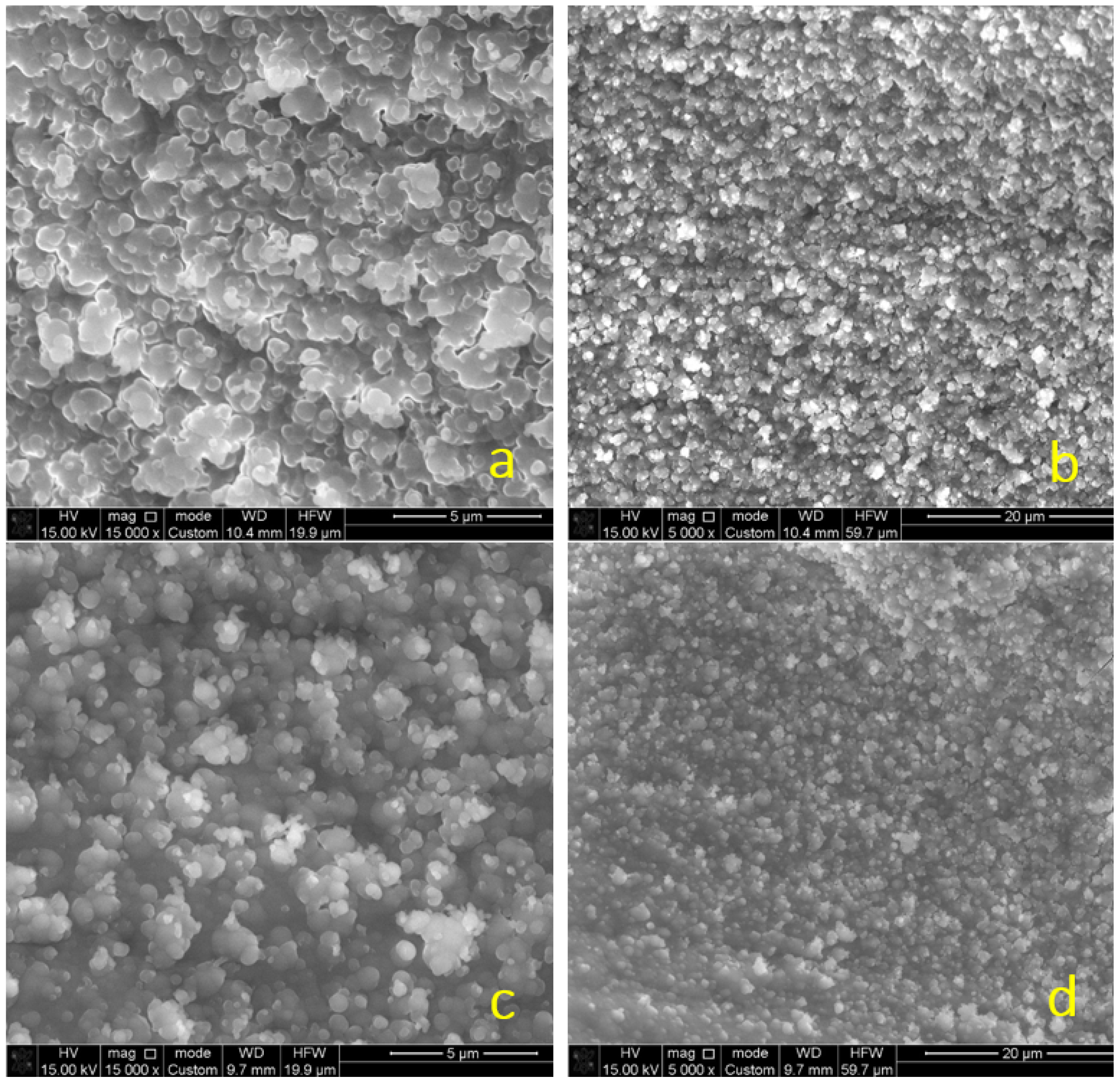
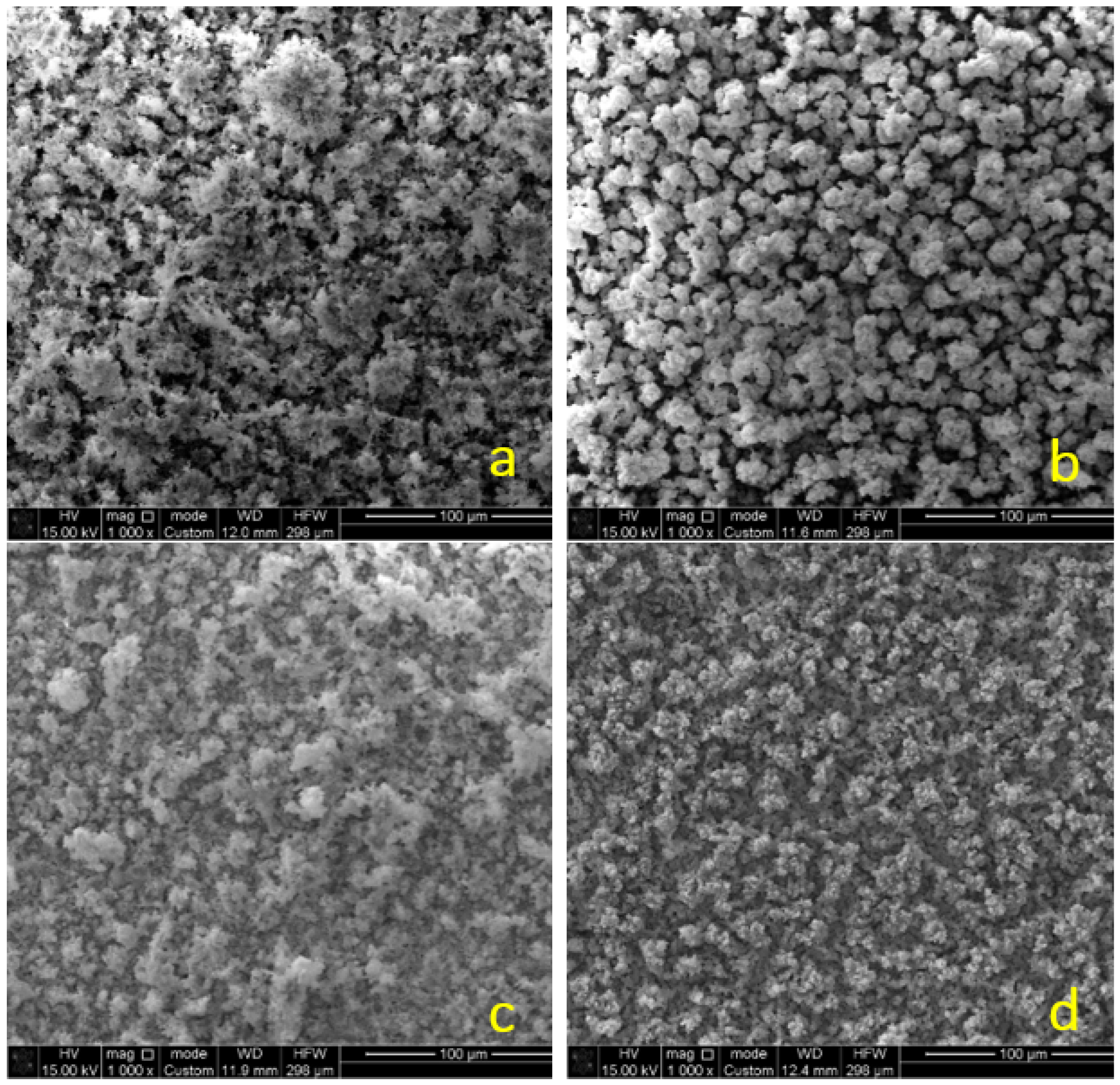
The results of the identification carried out using a scanning electron microscope (SEM) show that the obtained particles have mainly spherical shapes and they form non-uniform agglomerates. The surface of the agglomerates is heterogeneous and their sizes range up to 700 nm (Figure 2). The mainly spherical shape of particles without well-developed edges suggests that the particles’ structures are amorphous. During the collection, the particles and agglomerated clusters settled on the substrate surface, forming a thin porous film. The simple visual analysis shows that the density of such agglomerated clusters is low for the samples collected at 30 W (Figure 2a,b) plasma power (in contrast to the 60 W, Figure 2c,d), indicating the higher production growth yield of nanoparticles. No other significant differences were found in the structure and morphology of nanoparticles synthesised at different RF discharge powers.
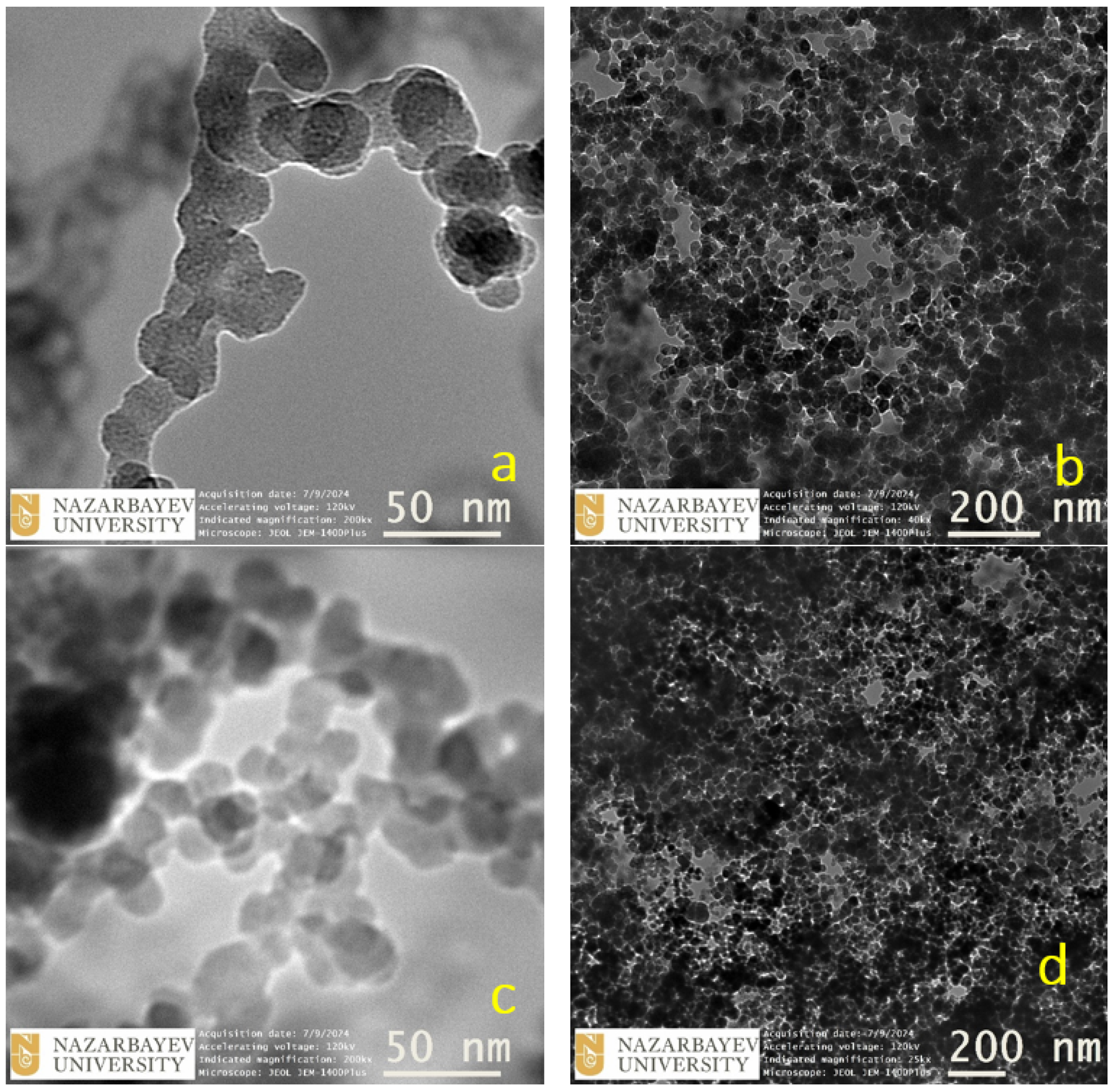
The morphology of the obtained structures was analysed by TEM. The samples were placed onto a copper mesh. Figure 3a,b shows bright-field TEM images of the sample at different magnifications and indicates that it is mainly composed of spherically shaped nanoparticles that form chain-like agglomerated structures. The mean diameter of a single nanoparticle is in the range of 20–30 nm. The same trends can be seen in Figure 3c,d for the particles obtained at 60 W. There is no significant difference between Figure 3a,b and Figure 3c,d.
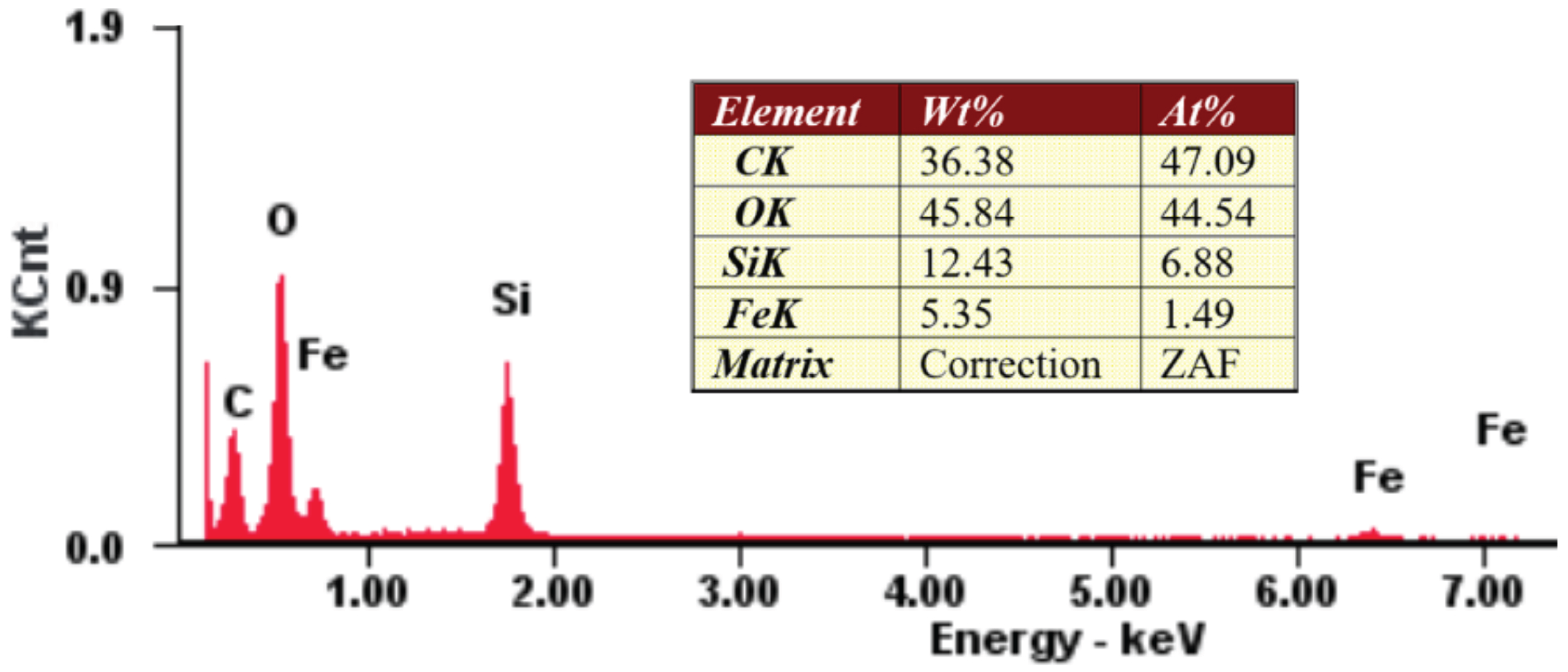
Figure 4 shows the signal intensities at certain energies of different chemical elements presented in the synthesised particles and deposited from the vapour in the plasma environment, in which a high percentage of carbon and oxygen elements were observed. Chemical analysis shows a high carbon content, 36 ± 4 wt. % ( 47 ± 5 at. %), and the rest is oxygen, which either originates from the HMDSO ( C 6 H 18 OSi 2 ) structure or from moisture, which is usually present in the argon flow and difficult to avoid even in a vacuum system. Oxygen impurity has been found in different studies in the growth of plasma-polymerised films using a mixture and Ar in vacuum systems. Also, as expected, in the deposit structure, 12 ± 2 wt.% ( 6 ± 1 at.%) silicon and 5 ± 1 wt.% ( 2 ± 1 at.%) iron are present in the deposited structure.
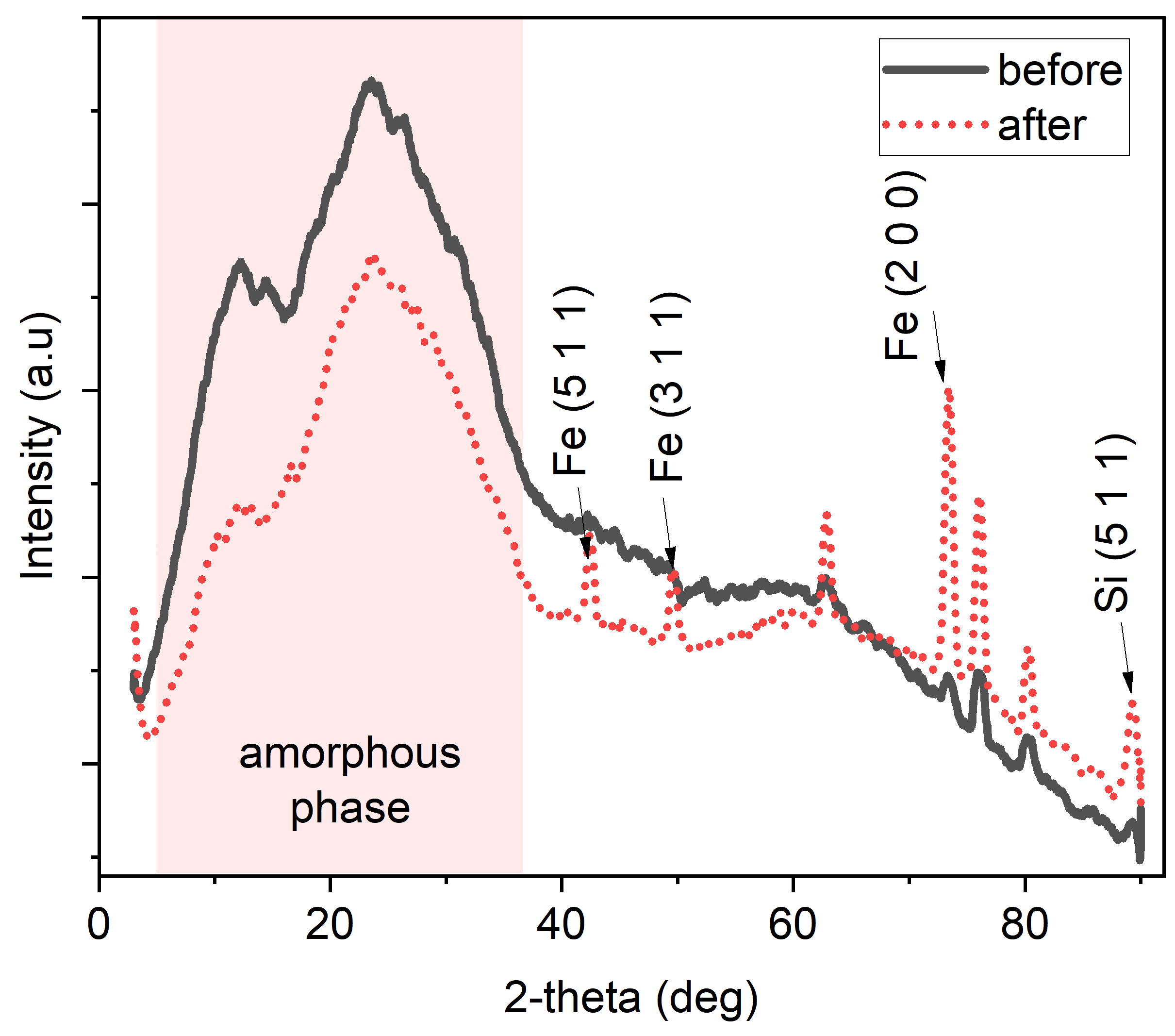
Figure 5 shows the XRD spectra of synthesised particles at 60 W (the same trend is observed at 30 W) both before and after irradiation in the plasma accelerator (see Table 1). The obtained signals are interpreted using the versatile PDXL2 software package (PDF2 Release 2016 RDB), which provides various analysis tools such as automatic phase identification, quantitative analysis, crystallite size analysis, lattice constant refinement, Rietveld analysis, ab initio structure determination, etc. (Rigaku). PDXL2 supports various diffraction databases such as ICDD PDF-2, PDF-4+, COD, and ICSD. In our case, XRD was used with the following parameters: scan rate/duration time—10.0000 deg/min, step width—0.0200°, scan range—3–90°, registration range—2 θ .
The diffraction pattern before irradiation indicates that the synthesised particles primarily exhibit an amorphous phase (the broad and diffuse peak between 2–25° 2-theta), with small and weak peaks. After irradiation, a significant transformation occurs: there is a marked increase in the intensity of several distinct peaks, and additional peaks emerge, corresponding to elements such as iron (PDF card No: 00-052-0513), silicon (PDF card No: 00-047-1187), and other unidentified phases. This alteration can be attributed to the interaction with high-energy plasma flux. Plasma irradiation is known to induce structural modifications, such as crystallisation or phase transformation, due to the energetic bombardment of plasma species, leading to the formation of crystalline phases or enhancing the crystallinity of existing phases. Figure 6 also shows changes in the morphology of synthesised particles before and after irradiation with pulsed plasma. It can be seen that plasma irradiation causes a more intensive agglomeration of clusters (Figure 6c,d).
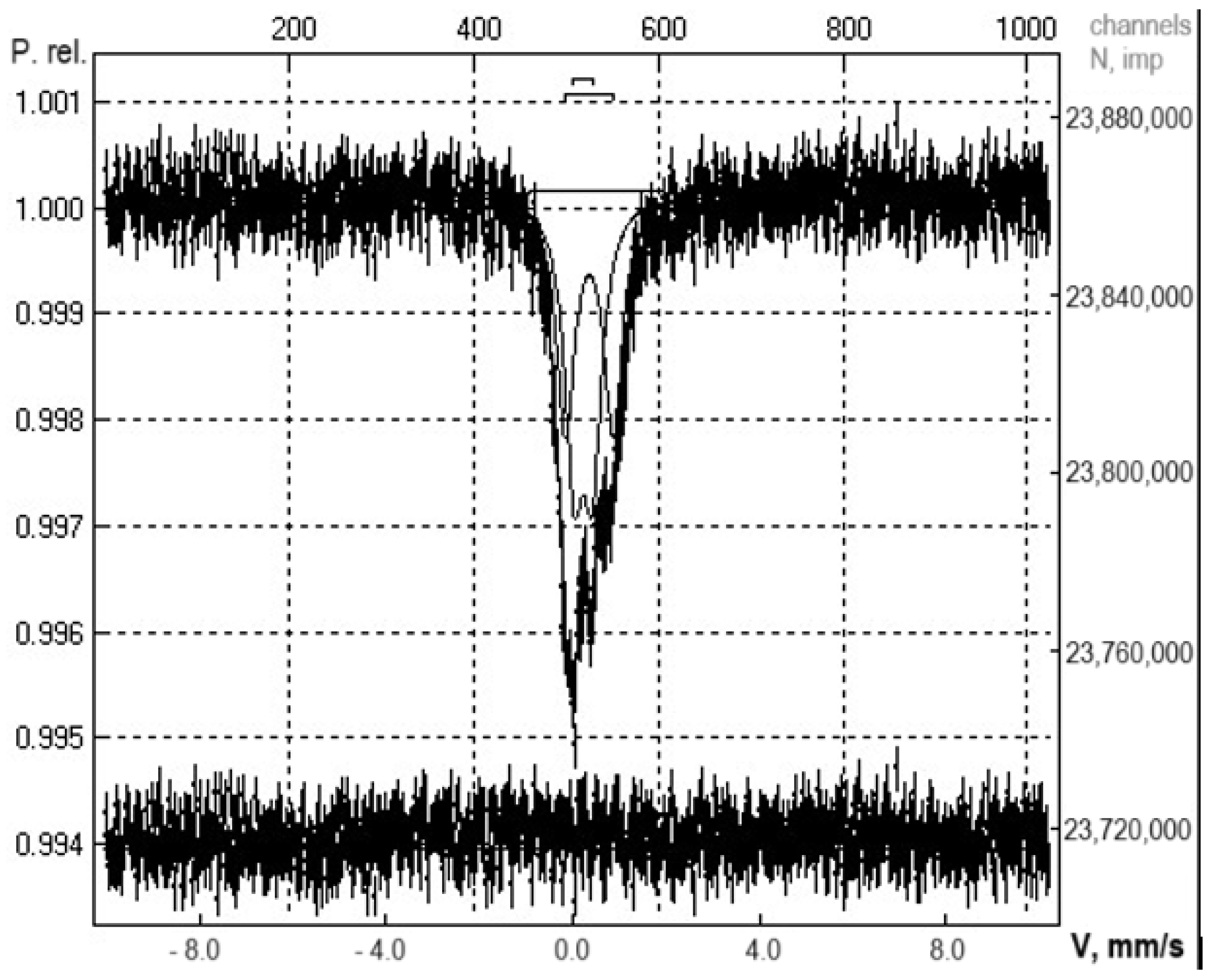
The Mössbauer spectrum of the sample shows the presence of some paramagnetic states of iron (Figure 7). Based on the literature data [31,32], it can be concluded that the sample is an Fe-Si system. The crystal structure of the F e S i 2 phase ( β - FeSi 2 —low-temperature; α - FeSi 2 —high-temperature) corresponds to two non-equivalent positions of Fe atoms (I and II), whose environments are characterised by low symmetry. The occupancies of these positions are approximately the same. The Mössbauer spectrum of β - FeSi 2 at room temperature can be decomposed into two quadrupole doublets of approximately equal intensity, corresponding to two crystallographically non-equivalent positions of iron atoms. The high-temperature phase α - FeSi 2 has a larger quadrupole splitting. Probably, there is a case of a superposition of spectra from these two modifications of phases. The relative content of Fe in positions I and II according to Mössbauer spectroscopy is close, which agrees well with the occupancy of these positions.
Thus, the obtained result suggests that the non-thermal plasma environment with gas-phase organosilicon and metallocene (e.g., Fe) molecules can be used as the lab-scale platform to study the formation of the chondrule-like astrophysical dust particles from the gas phase. Rapid heating sources, such as the pulsed plasma beam or pulsed arc discharge, along with high-energy laser sources can also be utilised to emulate the rapid melting and cooling of the dust particles in the interstellar medium.

5. Conclusions and Future Prospects

This research analyses the nanoscale properties of Si-Fe nanomaterials using SEM coupled with energy-dispersive spectroscopy, TEM, Mössbauer spectroscopy, and XRD (before/after) exposure to a high-energy pulsed plasma beam to simulate rapid heating. In non-thermal RF discharge, vapour from feedstock moves into a plasma region, where it fragments and nucleates into particles, forming larger powder particles. Elemental Fe inclusion was confirmed through EDS analysis. The results suggest that a non-thermal plasma environment can serve as a lab-scale platform to study the formation of chondrule-like particles using rapid heating sources to mimic interstellar conditions. We believe that the data obtained open new perspectives and provide valuable insights into the initial formation process of chondrule particles. Future work could focus on creating more complex chondrule-like structures by incorporating precursors of other common chondrite elements such as magnesium, copper, and aluminum.

6. Patents

An experimental setup for obtaining chondrule-like particles was used in the patent for utility model #9522 entitled ‘Method for production of chondro-like particles’ (Qazpatent, RSE ‘National Institute of Intellectual Property’, Kazakhstan).

Author Contributions

Conceptualisation, A.A., S.O., Y.U. and D.B.; data curation, A.B. (Akdaulet Baikaliyev), A.A. and D.B.; investigation, A.B. (Akdaulet Baikaliyev), A.A., S.O., A.B. (Alexander Brodsky), M.A. and M.D.; methodology, A.A., S.O., Y.U. and D.B.; supervision, M.G., T.R. and D.B.; visualisation, A.B. (Akdaulet Baikaliyev), N.A. and A.A.; writing—original draft, A.A. and Y.U.; writing—review and editing, D.B. All authors have read and agreed to the published version of the manuscript.

Funding

This research was funded by Program of the Committee of Science of the Ministry Higher Education of the Republic of Kazakhstan, grant number no. AP14870870.

Institutional Review Board Statement

Not applicable.

Informed Consent Statement

Not applicable.

Data Availability Statement

The raw data supporting the conclusions of this article will be made available by the authors on request.

Acknowledgments

The authors are very grateful for the analysis of the samples by TEM, which was performed at the Nazarbayev University’s facility (Kazakhstan).

Conflicts of Interest

The authors declare no conflicts of interest.

References

  1. Russel, S.; Connolly, H.C., Jr.; Krot, A.N. Chondrules: Records of protoplanetary disk processes. In Cambridge Planetary Science; Cambridge University Press: Cambridge, UK, 2018; Volume 22. [Google Scholar]
  2. Whipple, F.L. Chondrules: Suggestion concerning the origin. Science 1966, 153, 54–56. [Google Scholar] [CrossRef] [PubMed]
  3. Floyd, C.J.; Benito, S.; Martin, P.-E.; Jenkins, L.E.; Dunham, E.; Daly, L.; Lee, M.R. Chondrule sizes within the CM carbonaceous chondrites and measurement methodologies. Meteorit. Planet. Sci. 2024, 47. [Google Scholar] [CrossRef]
  4. Bollard, J.; Connelly, J.N.; Whitehouse, M.J.; Pringle, E.A.; Bonal, L.; Jørgensen, J.K.; Nordlund, Å.; Moynier, F.; Bizzarro, M. Early formation of planetary building blocks inferred from Pb isotopic ages of chondrules. Sci. Adv. 2017, 3, e1700407. [Google Scholar] [CrossRef]
  5. Connelly, J.N.; Bizzarro, M.; Krot, A.N.; Nordlund, A.; Wielandt, D.; Ivanova, M.A. The Absolute Chronology and Thermal Processing of Solids in the Solar Protoplanetary Disk. Science 2012, 338, 651–655. [Google Scholar] [CrossRef] [PubMed]
  6. Alexander, C.M.O.; Grossman, J.N.; Ebel, D.S.; Ciesla, F.J. The formation conditions of chondrules and chondrites. Science 2008, 320, 1617–1619. [Google Scholar] [CrossRef] [PubMed]
  7. Mann, I. Interplanetary medium—A dusty plasma. Adv. Space Res. 2008, 41, 160–167. [Google Scholar] [CrossRef]
  8. Morfill, G.E.; Ivlev, A.V. Complex plasmas: An interdisciplinary research field. Rev. Mod. Phys. 2009, 81, 1353. [Google Scholar] [CrossRef]
  9. Merlino, R.; Gore, J. Dusty Plasmas in the Laboratory, Industry, and Space. Phys. Today 2007, 57, 32. [Google Scholar] [CrossRef]
  10. Beckers, J.; Berndt, J.; Block, D.; Bonitz, M.; Bruggeman, P.J.; Couëdel, L.; Delzanno, G.L.; Feng, Y.; Gopalakrishnan, R.; Greiner, F.; et al. Physics and applications of dusty plasmas: The Perspectives 2023. Phys. Plasmas 2023, 30, 120601. [Google Scholar]
  11. Draine, B.T. Interstellar Dust Grains. Annu. Rev. Astron. Astrophys. 2003, 41, 142–149. [Google Scholar] [CrossRef]
  12. Goertz, C.K. Dusty plasmas in the solar system. Rev. Geophys. 1989, 27, 271–292. [Google Scholar] [CrossRef]
  13. Herbst, W.; Greenwood, J.P. A new mechanism for chondrule formation: Radiative heating by hot planetesimals. Icarus 2016, 267, 364–367. [Google Scholar] [CrossRef]
  14. Horányi, M.; Morfill, G.; Goertz, C.K.; Levy, E.H. Chondrule formation in lightning discharge. Icarus 1995, 114, 174–185. [Google Scholar] [CrossRef]
  15. Xiang, C.; Carballido, A.; Hanna, R.D.; Matthews, L.S.; Hyde, T.W. The initial structure of chondrule dust rims I: Electrically neutral grains. Icarus 2019, 321, 99–111. [Google Scholar] [CrossRef]
  16. Xiang, C.; Carballido, A.; Matthews, L.S.; Hyde, T.W. The initial structure of chondrule dust rims II: Charged grains. Icarus 2021, 354, 114053. [Google Scholar] [CrossRef]
  17. Güttler, C. TExposing metal and silicate charges to electrical discharges: Did chondrules form by nebular lightning? Icarus 2008, 195, 504–510. [Google Scholar] [CrossRef]
  18. Morlok, A. Chondrules born in plasma? Simulation of gas–grain interaction using plasma arcs with applications to chondrule and cosmic spherule formation. Meteorit. Planet. Sci. 2012, 47, 2269–2280. [Google Scholar] [CrossRef]
  19. Spahr, D.; Koch, T.E.; Merges, D.; Beck, A.A.; Bohlender, B.; Carlsson, J.M.; Christ, O.; Fujita, S.; Genzel, P.T.; Kerscher, J.; et al. A chondrule formation experiment aboard the ISS: Experimental set-up and test experiments. Icarus 2020, 350, 113898. [Google Scholar] [CrossRef]
  20. Herrero, V.J. Structure and evolution of interstellar carbonaceous dust. Insights from the laboratory. Front. Astron. Space Sci. 2022, 9, 1083288. [Google Scholar] [CrossRef]
  21. Roy, A.; Surendra, V.S.; Ramachandran, R.; Meka, J.K.; Gupta, S.; Janardhan, P.; Rajasekhar, B.N.; Hill, H.; Bhardwaj, A.; Mason, N.J.; et al. Interstellar Carbonaceous Dust and Its Formation Pathways: From an Experimental Astrochemistry Perspective. J. Indian Inst. Sci. 2023, 103, 919–938. [Google Scholar] [CrossRef]
  22. Kovacevic, E.; Stefanović, I.; Berndt, J.; Pendleton, Y.J.; Winter, J. A Candidate Analog for Carbonaceous Interstellar Dust: Formation by Reactive Plasma Polymerization. Astrophys. J. 2005, 623, 242. [Google Scholar] [CrossRef]
  23. Pelaez, R.J.; Maté, B.; Tanarro, I.; Molpeceres, G.; Jiménez-Redondo, M.; Timón, V.; Escribano, R.; Herrero, V.J. Plasma generation and processing of interstellar carbonaceous dust analogs. Plasma Sources Sci. Technol. 2018, 27, 035007. [Google Scholar] [CrossRef] [PubMed]
  24. Batryshev, D.; Yerlanuly, Y.; Gabdullin, M.T.; Ramazanov, T.S. Investigation of Synthesis of Carbon Nanowalls by the Chemical Vapor Deposition Method in the Plasma of a Radio Frequency Capacitive Discharge. IEEE Trans. Plasma Sci. 2019, 47, 3044–3046. [Google Scholar] [CrossRef]
  25. Dosbolayev, M.K.; Tazhen, A.B.; Ramazanov, T.S.; Ussenov, Y.A. Investigation of dust formation during changes in the structural and surface properties of plasma-irradiated materials. Nucl. Mater. Energy 2022, 33, 101300. [Google Scholar] [CrossRef]
  26. Ortiz Ortega, E.; Hosseinian, H.; Rosales López, M.J.; Rodríguez Vera, A.; Hosseini, S. Characterization Techniques for Morphology Analysis. In Material Characterization Techniques and Applications; Springer: Singapore, 2022; Volume 19. [Google Scholar]
  27. Newbury, D.E.; Williams, D.B. The electron microscope: The materials characterization tool of the millennium. Acta Mater. 2000, 48, 323–346. [Google Scholar] [CrossRef]
  28. Palani, N.; Elangovan, R.D. Chapter 21—Microbial-Mediated Copper Nanoparticles Synthesis, Characterization, and Applications; Elsevier: Amsterdam, The Netherlands, 2022; Volume 26. [Google Scholar]
  29. Marinangeli, L.; Pompilio, L.; Baliva, A.; Billotta, S.; Bonanno, G.; Domeneghetti, M.C.; Fioretti, A.M.; Menozzi, O.; Nestola, F.; Piluso, E.; et al. Development of an ultra-miniaturised XRD/XRF instrument for the in situ mineralogical and chemical analysis of planetary soils and rocks: Implication for archaeometry. Rend. Fis. Acc. Lincei 2015, 26, 529–537. [Google Scholar] [CrossRef]
  30. Kopcewicz, B.; Kopcewicz, M.; Pietruczuk, A. The Mössbauer study of atmospheric iron-containing aerosol in the coarse and PM2.5 fractions measured in rural site. Chemosphere 2015, 131, 9–16. [Google Scholar] [CrossRef]
  31. Litvinov, V.S.; Karakishev, S.D.; Ovchinnikov, V.V. Nuclear Gamma Resonance Spectroscopy of Alloys; Metallurgy Publishing House: Moscow, Russia, 1982; Volume 143. (In Russian) [Google Scholar]
  32. Chekin, V.V. Mössbauer Spectroscopy of Iron, Gold and Tin Alloys; Energoatom Publishing: Kyiv, Ukraine, 1981; Volume 106. (In Russian) [Google Scholar]
Figure 1. Experimental setup: (a) RF discharge plasma (red line means heating); (b) Plasma accelerator.
Applsci 14 08714 g001
Figure 2. SEM images (with different scales) of samples at 30 W (a,b) and 60 W (c,d).
Applsci 14 08714 g002
Figure 3. TEM images (with different scales) of samples synthesised with a power of 30 W (a,b) and 60 W (c,d).
Applsci 14 08714 g003
Figure 4. Elemental composition of the sample.
Applsci 14 08714 g004
Figure 5. X-ray diffraction analysis before/after irradiation via the plasma accelerator.
Applsci 14 08714 g005
Figure 6. SEM pictures of synthesised films before (a,c) and after (b,d) irradiation at different scales.
Applsci 14 08714 g006
Figure 7. Mössbauer spectra of the synthesised particles.
Applsci 14 08714 g007
Table 1. Operating parameters of the plasma accelerator [25].
Operating ParametersValues
Accelerator voltage, U4 kV
Discharge current, I m a x 0.1 MA
Pulse duration, τ 150 μs
Pressure, p0.06 torr
Disclaimer/Publisher’s Note: The statements, opinions and data contained in all publications are solely those of the individual author(s) and contributor(s) and not of MDPI and/or the editor(s). MDPI and/or the editor(s) disclaim responsibility for any injury to people or property resulting from any ideas, methods, instructions or products referred to in the content.