Article

Limitations in Maximum Intensity Front Crawl in Swimmers with Down Syndrome

by
Giampiero Merati
1,2,
Damiano Formenti
1,
Claudio Gandola
1,
Paolo Castiglioni
1,2,
Linda Casalini
1,3,
Athos Trecroci
4,
Luca Cavaggioni
5,
Pietro Luigi Invernizzi
4,
Umberto Menichino
6 and
Raffaele Scurati
4,*
1
Department of Biotechnology and Life Sciences, Università degli Studi dell’Insubria, 21100 Varese, Italy
2
IRCCS Fondazione don Carlo Gnocchi, 20148 Milano, Italy
3
Italian Federation of Intellectual and Relational Disability Sports (FISDIR), 00191 Rome, Italy
4
Department of Biomedical Sciences for Health, Università degli Studi di Milano, 20133 Milan, Italy
5
Obesity Unit, Laboratory of Nutrition and Obesity Research, Department of Endocrine and Metabolic Diseases, IRCCS Istituto Auxologico Italiano, 20145 Milan, Italy
6
Independent Researcher, 21047 Saronno, Italy
*
Author to whom correspondence should be addressed.
Appl. Sci. 2024, 14(18), 8387; https://doi.org/10.3390/app14188387
Submission received: 30 July 2024 / Revised: 9 September 2024 / Accepted: 12 September 2024 / Published: 18 September 2024
(This article belongs to the Special Issue Advances in Assessment of Physical Performance)

Abstract

:
Individuals with Down Syndrome exhibit deficits in muscle strength and cardiovascular adaptation, which limit athletic performance. We compared a maximum-intensity 50 m front crawl test between competitive male swimmers with Down Syndrome (SDS; n = 11; 26.5 ± 5.6 years; m ± SD) and a control group of swimmers (CNT; n = 11; 27.1 ± 4.0 years) with similar training routines (about 5 h/week). Wearable sternal sensors measured their heart rate and 3D accelerometry. The regularity index Sample Entropy (SampEn) was calculated using the X component of acceleration. The total times (SDS: 58.91 ± 13.68 s; CNT: 32.55 ± 3.70 s) and stroke counts (SDS: 66.1 ± 9.6; CNT: 51.4 ± 7.4) were significantly higher in the SDS group (p < 0.01). The heart rate was lower in the SDS group during immediate (SDS: 129 ± 15 bpm; CNT: 172 ± 11 bpm) and delayed recovery (30 s, SDS: 104 ± 23 bpm; CNT: 145 ± 21 bpm; 60 s, SDS: 79 ± 27 bpm; CNT: 114 ± 27 bpm) (p < 0.01 for all the comparisons). The SampEn of sternal acceleration showed no differences between the groups and between 0–25 m and 25–50 m. Body pitch correlated strongly with performance in the SDSs (R2 = 0.632, p < 0.01), but during the first 25 m only. The high-intensity front crawl performances differed between the SDS and CNT athletes in terms of time, biomechanics, and training adaptation, suggesting the need for tailored training to improve swimming efficiency in SDSs.

1. Introduction

Down Syndrome (DS), a common form of intellectual and relational disability, is a genetic disorder with an estimated incidence ranging from 1 in 1000 to 1 in 1200 live births [1]. A lower metabolic rate, endocrine abnormalities, poor muscle tone, reduced physical activity, and often inadequate nutrition combine to create the typical picture of fragility that often accompanies such individuals, making them susceptible to higher incidences of cardiovascular disease, diabetes, osteoporosis, and obesity [2,3]. Among the different sports activities, swimming seems to counteract the tendency toward obesity in persons with DS and helps to increase strength, speed, and balance [4,5].
Regarding the muscular system, individuals with DS show typical muscle hypotonia and joint hyper-flexibility due to ligament laxity [6,7]. This hyper-flexibility affects muscle strength and power, as well as gait and motor development, usually leading to delayed reflexes, arthritis, early-onset osteoporosis (resulting in long-bone fractures), increased fatigue, scoliosis, excessive dorsal kyphosis, and both lumbar and cervical hyper-lordosis [8]. In addition, children with DS often experience delays in sensory processing patterns and motor skills [9]. Therefore, habilitative and rehabilitative interventions tailored to enhance motor and cognitive skills are crucial [10]. Indeed, with appropriate support, individuals with DS can achieve high levels of autonomy, as reported in a recent systematic review [11].
As physical activity is so important, engaging in sports positively influences aerobic parameters, muscular strength, body composition, and psychological well-being in persons with DS, also increasing their overall daily physical activity level [12]. In addition, sports provide opportunities for socialization, personal growth, improved self-esteem, and the prevention of depression [13]. Sports activities like dance, martial arts, and gymnastics are beneficial for enhancing skills such as balance, coordination, and agility. These tasks require complex motor planning and bilateral synchronization, which can enhance both cognitive and physical abilities. Likewise, engaging in outdoor activities like cycling and hiking offers a variety of sensory stimuli that enhance sensory processing and motor control. Apart from individual sports, participation in team sports also has great benefits for persons with DS. Playing soccer or basketball can improve physical fitness and foster social abilities, including communication and teamwork. Moreover, adaptive modalities such as yoga and Pilates can augment core strength, stability, and mindfulness, thus promoting both physical and mental well-being. Overall, diverse sport-related possibilities assist the development of motor and cognitive abilities in persons with DS, with the final aim of promoting increased autonomy and social integration [11].
Certain precautions should, however, be considered, such as monitoring cardiovascular activity during strength training, using appropriate equipment, and adjusting exercise intensity and frequency. Exercises and activities that involve hyperextension should be avoided to prevent hernias, dislocations, strains, and sprains. Instead, exercises that strengthen the stabilizing muscles and stimulate proprioception while maintaining joint integrity are recommended. Monitoring sports activity in water is more challenging than on land, and very little data are available on sports activity at competitive levels in athletes with DS [14]. It is important to obtain these data to understand (a) whether there are safety reasons for not reaching the maximal levels of cardiovascular activation in athletes with DS and (b) to assess the variations between athletes with and without DS of equal competitive level to better target training activity in swimmers with DS.
The objective of this study was to evaluate the biomechanical and bioenergetic characteristics of maximum-intensity front crawl swimming in competitive athletes with DS. While there is a substantial body of the scientific literature on the biomechanics of swimming in typically developed individuals, studies specifically focusing on individuals with DS in this context are very rare [14]. Furthermore, most biomechanical studies are not accompanied by a parallel bioenergetic evaluation, which estimates cardiovascular effort. We therefore analyzed and compared the front crawl swimming performances of swimmers with DS (SDSs) and athletes with typical development (CNT). This comparison was made possible through a biomechanical assessment using sternal 3D accelerometry and video recordings and a cardiovascular evaluation using an underwater wearable ECG sensor. The findings may provide valuable insights to coaches and trainers regarding training optimization in this sport.

2. Materials and Methods

2.1. Subjects

A total of 22 individuals, all males, voluntarily participated in this study: 11 SDS and 11 CNT athletes. Participants with DS were selected from competitive swimmers with this syndrome from three teams in northern Italy belonging to the C21 FISDIR category (national level) of the Italian Paralympic Sports Federation for Intellectual and Relational Disabilities. Participants in the CNT group were competitive swimmers from the Italian Swimming Federation (FIN) and were randomly selected from a panel of swimming teams at the same swimming centers. To achieve a power of 0.80, we calculated the required sample size using the G*Power software (version 3.1.9.4, Universitat Kiel, Germany) based on the 50 m front crawl performance times and HRmax, determining a minimum of 3 and 10 participants per group, respectively.
The entire group of swimmers (n = 22) had an average age of 26.9 ± 4.7 years (M ± SD) and trained 3.4 ± 0.7 times per week, with each session lasting approximately 1 h and 30 min.
All athletes or their parents provided written informed consent to participate in this study, which was conducted in accordance with the Declaration of Helsinki and approved by the Ethics Committee of the University of Insubria, Varese, Italy (protocol code: 0035483; date of approval: 12 March 2024).

2.2. Measurements and Experimental Procedures

Body measurements included height, body mass, and body mass index (BMI), which was calculated. Table 1 displays the basic anthropometric characteristics and BMI for the SDS and CNT groups.
The experimental protocol consisted of a timed 50 m front crawl at maximal intensity, starting in the water and requiring a turn or touch at 25 m. A single lightweight (18 g) wearable device (Faros® 180 waterproof ECG, Bittium Corp., Oulu, Finland) simultaneously measured 3D-accelerometry and electrocardiography and recorded data on an internal memory board. The sternal 3D-accelerometry (sampling frequency: 100 Hz; dynamic range ± 16 g; precision 14-bit, IP class. IP67) provided information about external load, whereas the V5-lead electrocardiography (sampling frequency, 250 Hz) served as an indicator of exercise intensity, through the subsequent calculation of heart rate (HR) and high-resolution underwater videos (sampling frequency: 60 Hz). Standard ECG electrodes by FIAB® (Doctor Shop, Milan, Italy) were used, covered with a plastic sheet to keep them dry during the swim test. Both accelerometric and ECG data were recorded in standard EDF (European Data Format) files. Figure 1 displays a typical accelerometer recording obtained with the described setup, indicating the positioning and reference axes of the inertial measurement unit (IMU) used.
A GoPro® Hero action camera was used for the underwater video recordings. The camera was positioned 20 cm below the water level and 12.5 m from the pool edge. Each swimmer passed in the first lane, less than two meters from the camera. The image for analysis was reconstructed by capturing the still image corresponding to the passing at 12.5 m of the central point of the line joining the rotation centers of the shoulders and hips.

2.3. Data Analysis

After acquiring data from both groups of swimmers, the data were analyzed offline using two different software programs: (i) EDF Browser (version 1.84, freeware software by Teuniz van Beelen) for biomechanical data analysis; (ii) Kubios® HRV Premium (version 4.0, Kubios Oy, Finland) for HR analysis. HR values were derived from the ECG signal using an efficient peak detector based on the validated Pan Tomkins algorithm [15].
The body pitch (i.e., the angle of the swimmer’s body in relation to the horizontal plane of the water) was assessed using the photographic frame on the sagittal plane of the underwater video during the swimmer’s transition to the middle of the first length (i.e., at 12.5 m) and the middle of the second length (i.e., at 37.5 m). The straight line passing through the centers of rotation of the shoulder and pelvis was taken as the tilt line of the swimmer’s body, and its inclination was measured using the digital goniometer of the Kinovea open-source software (version 2023.1.2, under GPL v2 license).
The data calculated from the 3D accelerometric recordings included (i) length time, which was the difference between the times of the maximal acceleration peaks on the x-axis (direction of motion) caused by the contact with the edge of the pool (Figure 1); (ii) stroke count (visually counted), which was the number of negative acceleration peaks on the y-axis (parallel to the biacromial line) within a length of time; and (iii) propulsion regularity. For propulsion regularity, the x-axis signal was first downsampled to 10 Hz to better focus on stroke variations, and then the regularity index Sample Entropy was applied (see the Data Analysis Section). Validation studies have shown that 3D axial accelerometers can accurately and reliably measure swimming parameters [16].
Finally, a regularity index was applied to the accelerometer signal to assess the potential degradation of coordination in the horizontal propulsion axis (corresponding to the x-axis of the sternal accelerometer). The original x-axis signal was first downsampled to 10 Hz to better focus on stroke variations and then cut for individual analysis between 0–25 m and 25–50 m. The chosen regularity index was the Sample Entropy (SampEn), calculated using the original method by Richman and Moorman [17] with the freeware software PyBios (version 4.3, 2021) [18].

2.4. Statistical Analysis

Data were expressed as the mean ± standard deviation (m ± SD) unless otherwise specified. The anthropometric characteristics, training volumes (hours/week), performance data (length times and stroke numbers), and SampEn values were all normally distributed in both groups of swimmers (p = ns at the Lilliefors test for all parameters). Therefore, comparisons between the SDS and CNT groups were performed by the unpaired Student’s t-test; effect sizes were calculated by Cohen’s d with 0.2, 0.5, and 0.8, indicating a small, medium, and large effect, respectively [19]. Student’s paired t-test was used to compare SampEn values between 0–25 m and 25–50 m. Differences in SampEn and body pitch between 0–25 m and 25–50 m, and in HR between SDS and CNT groups in 4 periods of the swimming test (before swimming at rest [REST], immediately after the end of the test [50 m END], and after 30 s and 60 s recovery [30 s REC and 60 s REC]) were tested by a two-way ANOVA for repeated measures, with post-hoc multiple comparisons by the two-stage linear step-up procedure of Benjamini, Krieger, and Yekutieli [20]. When the assumption of sphericity was violated due to limited sample sizes, the Geisser–Greenhouse correction was applied. Linear regression analysis between the body pitch angle and length time was performed, excluding possible outliers in the performance data (length time) using Tukey’s test (which identified two outliers in the SDS group and one outlier in the CNT group).
The level of statistical significance was set at p < 0.05. Statistical analyses were conducted using Prism® software (version 10.0, GraphPad Software, Boston, MA, USA).

3. Results

3.1. Anthropometric Characteristics and Performance Data

The athletes were well matched in age, but as expected, the SDS group had a significantly lower height and body mass than the CNT group (p < 0.01), with large effect sizes. However, the BMI was within the normal range in both groups (Table 1).
Table 2 shows the comparison of the chronometric (lap times) and mechanical (stroke counts) variables between the SDS and CNT groups in the maximum-intensity 50 m front crawl swimming test. SDS swimmers had significantly higher total and partial times (p < 0.01), as well as total and partial numbers of strokes (p < 0.01). Lap times were almost double in SDS athletes, with large effect sizes for all comparisons between groups.

3.2. Exercise Intensity

Figure 2 displays the HR at rest, at the end of the swimming test (50 m END), and during recovery after 30 s (30 s REC) and 60 s (60 s REC) from the end of the test, separately for the SDS and CNT groups. The repeated measures two-way ANOVA revealed a significant effect of group (SDS vs. CNT, p < 0.001) and time (p < 0.001), as well as a significant time x group interaction (p < 0.001). In the multiple comparisons test, the difference between resting HR values was slightly larger than the statistical significance level (p = 0.065). In contrast, HR values immediately at the end of the test and at 30 and 60 s of recovery were significantly lower in the SDS group compared to the CNT group (p < 0.001).

3.3. Acceleration Regularity

To investigate the hypothesis of a progressive reduction in the coordination of the athletic gesture, the regularity of the accelerometric signal between 0–25 m and 25–50 m was quantified using the SampEn index. In the SDS group, SampEn was 2.10 ± 0.51 in the first length (0–25 m) and 1.89 ± 0.24 in the second length (25–50 m). In the CNT group, SampEn was 2.10 ± 0.53 in the first length and 2.01 ± 0.50 in the second length. The two-way ANOVA showed no significant effects of time (significance of differences between lengths: p = 0.161), groups (significance of differences between groups: p = 0.72), or their interaction (p = 0.59).

3.4. Body Pitch

Table 3 compares the angles of body pitch relative to the water plane between the two groups at each length. The two-way ANOVA showed that time has no effect (differences between lengths: p = 0.83), but the difference between groups is highly significant (p = 0.0004), with no interaction between lengths and groups (p = 0.19). In the post-hoc analysis, the differences between groups were significant for both the 0–25 m (p = 0.016) and the 25–50 m (p = 0.0002) lengths.
In particular, SDS swimmers had higher body pitch angles in both lengths.
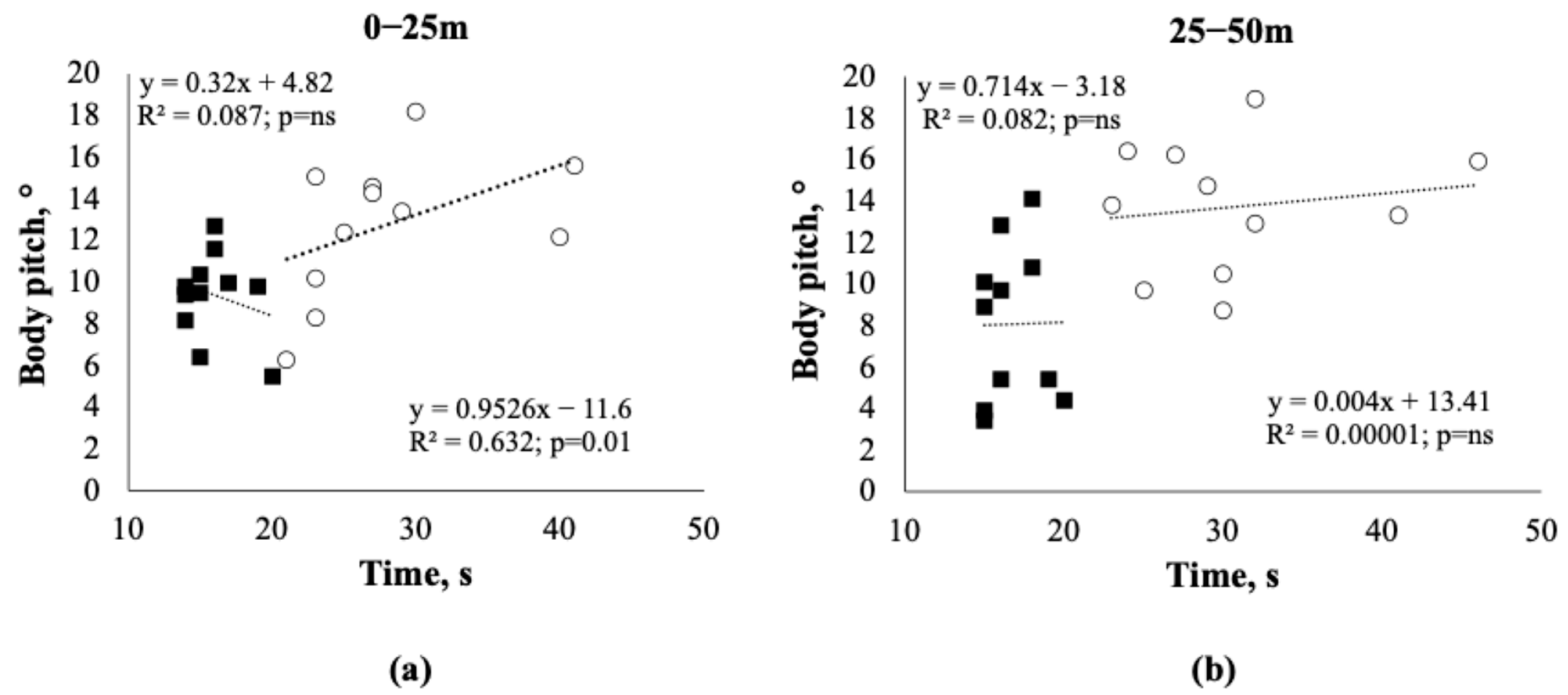
Figure 3 displays the relationship between the body pitch angle and lap time, as estimated through linear regression analysis. This analysis was performed separately for the 0–25 m and 25–50 m segments of the 50 m swimming test. The regression results were not significant in the CNT group, but they were positive and statistically significant in the first length for the SDS group. However, in the second length, the significant relationship was completely lost for the SDS athletes (p = ns).

4. Discussion

The aim of this study was to evaluate several biomechanics and bioenergetics features of maximum intensity front crawl in swimmers with Down Syndrome (SDS) and compare them with those of competitive swimmers of the same age and weekly training volume.
Regarding the anthropometric characteristics of the athletes enrolled (Table 1), we observed that the swimmers in the SDS group, as expected from previous literature data, had significantly lower body masses and heights than the CNT athletes did [21]. In contrast, the BMI calculated in our DS swimmers did not indicate overweight values. These findings suggest that chronic training volume positively influenced the body composition of adult DS athletes in the present study, confirming previous data [5,22,23].
From a functional perspective, this study did not directly evaluate any specific strength component. However, the typical deficit in voluntary muscle strength in individuals with DS is already well-known and documented in the scientific literature (see Padia et al. for a recent review on grip strength in DS patients [24]). The presumed strength deficit, along with the significant height deficit and consequently shorter upper limbs, likely contributed to the differences in the biomechanical and chronometric characteristics of maximum-intensity front crawl swimming between the two groups. Indeed, the lap times and stroke counts were significantly higher in both lengths for the SDS group, indicating lower propulsive forces and a biomechanical disadvantage for this group. At present, discriminating which biomechanical or kinematic factors are the most relevant for generating lower swimming speeds in athletes with DS is difficult, and this topic deserves further study.
Furthermore, the horizontal body position in water may have played a relevant role in the propulsion biomechanics of individuals with DS. The body pitch was significantly greater in the SDS group’s 0–25 m and 25–50 m lengths, with no interaction between length and group (Table 3). This could be attributed to two main factors: a lower propulsive capacity of the lower limbs in DS swimmers, who are unable to maintain the longitudinal axis in a horizontal position, leading to decreased buoyancy, and a lower ability of the abdominal muscles to maintain trunk alignment with a reduced inclination relative to the water surface [14]. In addition, motor dysfunctions (asymmetries, rhythm discontinuity, breathing pattern) due to the nature of the syndrome may also have affected the body position while swimming. We compared body pitch with lap times in both groups to further examine whether the trunk inclination could have impacted swimming propulsion. We found they were positively correlated, but only at 0–25 m in the SDS group, meaning that higher angles corresponded to slower times. The high linear regression coefficient indicated an association greater than 60% between the angle of inclination in water and lap time in SDS athletes. Notably, the body pitch remains greater at 25–50 m for the swimmers in the SDS group and remains low for those in the CNT swimmer group. However, interestingly, the relationship between the angle and swimming speed was lost entirely at 25–50 m for the SDS group. This may suggest that those swimmers with DS who can maintain a low body pitch at 0–25 m through more efficient propulsion also lose this ability at 25–50 m. Indeed, the associations among arm propulsive forces, lower limb kicking action, and pitch angle remain rather elusive in swimming. Recent work has indicated that such a relationship varies with swimming velocity, as kicking raises the legs in low-velocity ranges and suppresses leg sinking in high-velocity ranges, reducing the hand propulsive force required to achieve a given swimming velocity [25]. Considering these recent data, given the slow swimming speed of DS athletes, the fatigue of large muscle groups such as the gluteus maximus and quadriceps femoris likely contributed significantly to the reduced lifting of the lower limbs in the second lap, thus further reducing the athlete’s overall propulsive capacity.
In addition to the possible loss of propulsive force, coordination issues arise with central or peripheral fatigue. To further assess this coordinative aspect, we evaluated an index of gesture regularity, SampEn, applied to the propulsive alternating accelerations along the x-axis of movement. Although the irregularity indices tended to decrease in the second lap compared with the first lap in the SDS athletes, this trend did not reach statistical significance. Therefore, it may be hypothesized that speed reduction, especially observed in the second lap, does not seem to be attributable to some coordination deficits causing gross variations in swimming gestures. Therefore, SDS athletes lose propulsion but not coordination, once again indicating that the overall strength deficit and rapid loss of strength due to progressive fatigue may be the main causes of propulsion difficulties in maximum-intensity front crawl swimming between SDS and CNT athletes.
Beyond these peripheral muscle deficits, however, it is also conceivable that the muscular apparatus may also have failed to express its maximum aerobic capacity in the SDS group because of a possible central cardiovascular deficit. For this reason, we also decided to measure exercise intensity, i.e., the HR. As the swimming test was required at a maximal intensity, we would have expected the highest involvement of the exercise intensity, which did not differ significantly between the two groups. In contrast, both the maximal and the “recovery” loads at 30 and 60 s post-test were significantly lower in the SDS athletes (Figure 1). Interestingly, the interaction time × group in the ANOVA was significant, suggesting that the cardiovascular system recruitment during the swimming test in this group was submaximal or may be deficient compared with that in the CNT group. Indeed, HR peak frequencies are not elevated in many studies conducted on individuals with DS, likely reflecting a potential autonomic nervous system dysfunction [26]. The “chronotropic incompetence” hypothesis has been explored in a study comparing the differences in cardiorespiratory capacity between adults with DS and those with other intellectual disabilities [27]. The authors reported that the lower peak HR of the DS swimmers explained their lower levels of aerobic capacity. These results support the hypothesis that limited cardiac output at peak exercise likely explains the low work capacity in individuals with DS. This led to speculations of possible relationships between attenuated sympathetic response to exercise (i.e., autonomic dysfunction), chronotropic incompetence, and low VO2peak in this population [28]. Some studies have also highlighted a deficiency in peripheral oxygen utilization in individuals with DS, most likely caused by mitochondrial disorders related to the syndrome [29]. Additionally, the extreme variability of the resting HR should be noted, given the presence of marked bradycardia in at least 3 out of the 11 SDS athletes investigated. The presence of resting bradycardia has also been documented in the literature in individuals with DS and requires particular attention, especially in endurance athletes, as bradycardic states, particularly at night, can lead to significant cardiac rhythm disorders and ventricular perfusion issues [30]. Athletes with DS and marked bradycardia at rest do not have the selective advantage of normal endurance athletes, as they cannot “release” their HR to the maximum level during exertion [31]. This was also observed in our group of athletes with DS, in which the swimmers with the most bradycardic resting conditions also experienced relative bradycardia compared with the other CNT and SDS individuals. From a practical viewpoint, this hypokinesia of the cardiovascular response to exertion should be carefully evaluated during medical preparticipation screening (which has been mandatory in Italy since 1982). Coaches should be informed of any symptoms (such as fainting, weakness, prolonged fatigue) that may be related to signs of “cardiac low output” during the athlete’s competitive season [32]. Interestingly, as clearly shown in Figure 2, the slope of HR recovery after the maximal swim test was identical for both groups. Since this rapid HR recovery (after 30 and 60 s) depends largely on the reactivation ability of the parasympathetic system, we can conclude that in our DS athletes, aerobic training by swimming induced benefits to the autonomic nervous system comparable to those of athletes with typical development. This translates into an undoubted cardiovascular advantage since, as widely demonstrated, the speed of post-test HR recovery correlates directly with cardiovascular and overall mortality risk [33].
Some limitations of the study need to be acknowledged. First, this investigation involved a limited number of participants, which might need to be more representative of the larger population of swimmers with DS. In addition, all the participants were male; therefore, our findings cannot be translated to female athletes with DS, who may exhibit different physiological and biomechanical characteristics. Moreover, the study did not directly measure specific components of muscle strength, given that muscle strength deficits are well documented in individuals with DS; however, a direct strength assessment would have provided a clearer understanding of how this capacity contributes to the observed differences in swimming performance. Finally, we did not provide detailed information on the usual nutritional intake in DS vs. CNT swimmers or on environmental factors such as water temperature, pool conditions, or swimmer fatigue outside the testing period. We know that these factors could have significantly influenced both the physical and performance metrics observed.
Our results may have some practical implications for the training of DS swimmers. For coaches, it is important to know HR values at rest and during exercise to optimize training programming with appropriate loads and recoveries. It would be optimal to perform the 50 m maximal swimming tests 3–4 times during the season, monitoring the HR with a waterproof HR monitor to adjust the programming phases and assess the athletes’ progress. Monitoring the variations in data obtained from the comparison between HR and lap time helps to estimate an athlete’s effort. For example, high HR and lap times may indicate a fatigue state. Conversely, a low HR despite high lap times may indicate bradycardia or a lack of motivational effort. For SDS individuals presenting with very bradycardic resting HR values at the beginning of the season, purely aerobic training (which may further lead to developing resting bradycardia) and initiating endurance races might not be advisable. Therefore, coaches should consider focusing on medium- and high-intensity intermittent work with moderate- to short-duration repetitions [34,35]. In addition, because of the difficulty in maintaining SDS individuals’ attention during training sessions, coaches should manage various attentional focuses [36,37].
Finally, from a biomechanical viewpoint, improving the inclination of the longitudinal axis of the body in the water (pitch angle) may be an additional target of the coaches of swimmers with DS, given the specific relationship observed between this angle and lap time. A more horizontal position would reduce the frontal drag toward advancing in the water. Furthermore, attempting to reduce the stroke count per length by improving and lengthening the movement of the upper limbs and increasing the propulsion of the lower limbs could lead to greater swimming efficiency. From a speculative point of view, strength training should receive appropriate consideration to improve the swimming performance of SDS athletes [38].
In conclusion, high-intensity front crawl performance differed between SDS and CNT athletes in terms of time, biomechanics, and training adaptation, suggesting a need for tailored training to improve swimming efficiency in SDS athletes.

Author Contributions

Conceptualization, G.M., C.G. and D.F.; methodology, G.M., P.C., U.M. and C.G.; software, G.M.; formal analysis, C.G.; investigation, C.G., L.C. (Linda Casalini), L.C. (Luca Cavaggioni), U.M. and D.F.; data curation, C.G. and P.C.; writing—original draft preparation, G.M., R.S., C.G. and D.F.; writing—review and editing, P.C., L.C. (Linda Casalini), L.C. (Luca Cavaggioni) and A.T.; supervision, P.L.I.; funding acquisition, G.M. and P.C. All authors have read and agreed to the published version of the manuscript.

Funding

G.M. and P.C. were funded by the Italian Ministry of Health (Ricerca Corrente RC2024).

Institutional Review Board Statement

This study was conducted in accordance with the Declaration of Helsinki and approved by the Ethics Committee of the University of Insubria, Varese, Italy (Prot. N. 0035483, 12 March 2024).

Informed Consent Statement

Informed consent was obtained from all subjects involved in the study.

Data Availability Statement

The data supporting the main findings of this study are available on reasonable request with access granted to researchers meeting the criteria for access to confidential data. The data repository is Zenodo, at https://zenodo.org/records/13133045 (URL accessed on 13 September 2024).

Acknowledgments

The authors thank all the participants and their swimming societies (Ice Club Como SSD, Osha Como ASD, and Briantea84).

Conflicts of Interest

The authors declare no conflicts of interest.

References

  1. Irving, C.; Basu, A.; Richmond, S.; Burn, J.; Wren, C. Twenty-Year Trends in Prevalence and Survival of Down Syndrome. Eur. J. Hum. Genet. 2008, 16, 1336–1340. [Google Scholar] [CrossRef] [PubMed]
  2. Grohmann, D.; Wellsted, D.; Mengoni, S.E. Definition, Assessment and Management of Frailty for People with Intellectual Disabilities: A Scoping Review. J. Appl. Res. Intellect. Disabil. 2024, 37, e13219. [Google Scholar] [CrossRef] [PubMed]
  3. Capone, G.T.; Chicoine, B.; Bulova, P.; Stephens, M.; Hart, S.; Crissman, B.; Videlefsky, A.; Myers, K.; Roizen, N.; Esbensen, A.; et al. Co-Occurring Medical Conditions in Adults with Down Syndrome: A Systematic Review toward the Development of Health Care Guidelines. Am. J. Med. Genet. A 2018, 176, 116–133. [Google Scholar] [CrossRef] [PubMed]
  4. Boer, P.H. The Effect of 8 Weeks of Freestyle Swim Training on the Functional Fitness of Adults with Down Syndrome. J. Intellect. Disabil. Res. 2020, 64, 770–781. [Google Scholar] [CrossRef] [PubMed]
  5. Querido, A.; Costa, M.J.; Araújo, D.; Sampaio, A.R.; Vilas-Boas, J.P.; Corredeira, R.; Daly, D.J.; Fernandes, R.J. Swimmers with Down Syndrome Are Healthier and Physically Fit than Their Untrained Peers. Healthcare 2023, 11, 482. [Google Scholar] [CrossRef]
  6. Berghof, R.; Carstens, C. Hip joint problems in patients with Down’s syndrome. Z. Orthop. Ihre Grenzgeb. 1992, 130, 136–141. [Google Scholar] [CrossRef]
  7. Livingstone, B.; Hirst, P. Orthopedic Disorders in School Children with Down’s Syndrome with Special Reference to the Incidence of Joint Laxity. Clin. Orthop. Relat. Res. 1986, 207, 74–76. [Google Scholar] [CrossRef]
  8. LaCombe, J.M.; Roper, R.J. Skeletal Dynamics of Down Syndrome: A Developing Perspective. Bone 2020, 133, 115215. [Google Scholar] [CrossRef]
  9. Brugnaro, B.H.; Pauletti, M.F.; Lima, C.R.G.; Verdério, B.N.; Fonseca-Angulo, R.I.; Romão-Silva, B.; de Campos, A.C.; Rosenbaum, P.; Rocha, N.A.C.F. Relationship between Sensory Processing Patterns and Gross Motor Function of Children and Adolescents with Down Syndrome and Typical Development: A Cross-Sectional Study. J. Intellect. Disabil. Res. 2024, 68, 358–368. [Google Scholar] [CrossRef]
  10. Büyükçelik, N.M.; Yiğit, S.; Turhan, B. An Investigation of the Effects of Dual-Task Balance Exercises on Balance, Functional Status and Dual-Task Performance in Children with Down Syndrome. Dev. Neurorehabil 2023, 26, 320–327. [Google Scholar] [CrossRef]
  11. Muñoz-Llerena, A.; Ladrón-de-Guevara, L.; Medina-Rebollo, D.; Alcaraz-Rodríguez, V. Impact of Physical Activity on Autonomy and Quality of Life in Individuals with Down Syndrome: A Systematic Review. Healthcare 2024, 12, 181. [Google Scholar] [CrossRef] [PubMed]
  12. Izquierdo-Gomez, R.; Shields, N. Organised and Non-Organised Activities Contribute to Overall Physical Activity Levels in Adolescents and Young Adults with Down Syndrome: A Cross-Sectional Study. J. Intellect. Disabil. Res. 2024, 68, 997–1006. [Google Scholar] [CrossRef] [PubMed]
  13. Merzbach, V.; Jewiss, M.; Scruton, A.; Gordon, D. The Effects of Prescribed Physical and Cognitive Exercise on Life Satisfaction, Self-Efficacy and Mood States in Adults with Down Syndrome: The MinDSets Study. Int. J. Environ. Res. Public Health 2024, 21, 610. [Google Scholar] [CrossRef] [PubMed]
  14. Marques-Aleixo, I.; Querido, A.; Figueiredo, P.; Vilas-Boas, J.P.; Corredeira, R.; Daly, D.; Fernandes, R.J. Intracyclic Velocity Variation and Arm Coordination Assessment in Swimmers with Down Syndrome. Adapt. Phys. Act. Q. 2013, 30, 70–84. [Google Scholar] [CrossRef]
  15. Pan, J.; Tompkins, W.J. A Real-Time QRS Detection Algorithm. IEEE Trans. Biomed. Eng. 1985, 32, 230–236. [Google Scholar] [CrossRef]
  16. Ganzevles, S.; Vullings, R.; Beek, P.J.; Daanen, H.; Truijens, M. Using Tri-Axial Accelerometry in Daily Elite Swim Training Practice. Sensors 2017, 17, 990. [Google Scholar] [CrossRef]
  17. Richman, J.S.; Moorman, J.R. Physiological Time-Series Analysis Using Approximate Entropy and Sample Entropy. Am. J. Physiol. Heart Circ. Physiol. 2000, 278, H2039–H2049. [Google Scholar] [CrossRef]
  18. Silva, L.E.V.; Fazan, R.; Marin-Neto, J.A. PyBioS: A Freeware Computer Software for Analysis of Cardiovascular Signals. Comput. Methods Programs Biomed. 2020, 197, 105718. [Google Scholar] [CrossRef]
  19. Cohen, J. Statistical Power Analysis for the Behavioral Sciences, 2nd ed.; Routledge: New York, NY, USA, 1988; ISBN 978-0-203-77158-7. [Google Scholar]
  20. Benjamini, Y.; Hochberg, Y. Controlling the False Discovery Rate: A Practical and Powerful Approach to Multiple Testing. J. R. Stat. Soc. Ser. B Stat. Methodol. 1995, 57, 289–300. [Google Scholar] [CrossRef]
  21. Villani, E.R.; Onder, G.; Marzetti, E.; Coelho-Junior, H.; Calvani, R.; Di Paola, A.; Carfì, A. Body Composition Parameters and Sarcopenia in Adults with Down Syndrome: A Case-Control Study. Aging Clin. Exp. Res. 2024, 36, 81. [Google Scholar] [CrossRef]
  22. Yamanaka, E.; Inayama, T.; Ohkawara, K.; Kojima, M.; Nakada, T.; Kita, I. Effects of Substituting Sedentary Time with Physical Activity on Body Mass Index in Japanese Adults with Down Syndrome: A Cross-Sectional Study. Heliyon 2024, 10, e29294. [Google Scholar] [CrossRef] [PubMed]
  23. González-Agüero, A.; Vicente-Rodríguez, G.; Moreno, L.A.; Guerra-Balic, M.; Ara, I.; Casajús, J.A. Health-Related Physical Fitness in Children and Adolescents with Down Syndrome and Response to Training. Scand. J. Med. Sci. Sports 2010, 20, 716–724. [Google Scholar] [CrossRef] [PubMed]
  24. Padia, N.; Bose, M.; Parab, S. Determinants of Hand Function in Children and Adolescent with Down Syndrome—A Scoping Review. J. Hand Ther. 2023, 36, 622–631. [Google Scholar] [CrossRef] [PubMed]
  25. Kadi, T.; Washino, S.; Tsunokawa, T.; Narita, K.; Mankyu, H.; Murai, A.; Tamaki, H. Role of Kicking Action in Front Crawl: The Inter-Relationships between Swimming Velocity, Hand Propulsive Force and Trunk Inclination. Sports Biomech. 2024, 2303361. [Google Scholar] [CrossRef]
  26. Fernhall, B.; Pitetti, K.H.; Rimmer, J.H.; McCubbin, J.A.; Rintala, P.; Millar, A.L.; Kittredge, J.; Burkett, L.N. Cardiorespiratory Capacity of Individuals with Mental Retardation Including Down Syndrome. Med. Sci. Sports Exerc. 1996, 28, 366–371. [Google Scholar] [CrossRef]
  27. Mendonca, G.V.; Pereira, F.D.; Fernhall, B. Reduced Exercise Capacity in Persons with Down Syndrome: Cause, Effect, and Management. Ther. Clin. Risk Manag. 2010, 6, 601–610. [Google Scholar] [CrossRef]
  28. Iellamo, F.; Galante, A.; Legramante, J.M.; Lippi, M.E.; Condoluci, C.; Albertini, G.; Volterrani, M. Altered Autonomic Cardiac Regulation in Individuals with Down Syndrome. Am. J. Physiol. Heart Circ. Physiol. 2005, 289, H2387–H2391. [Google Scholar] [CrossRef]
  29. Boonman, A.J.N.; Schroeder, E.C.; Hopman, M.T.E.; Fernhall, B.O.; Hilgenkamp, T.I.M. Cardiopulmonary Profile of Individuals with Intellectual Disability. Med. Sci. Sports Exerc. 2019, 51, 1802–1808. [Google Scholar] [CrossRef]
  30. Sinton, J.W.; Cooper, D.S.; Wiley, S. Down Syndrome and the Autonomic Nervous System, an Educational Review for the Anesthesiologist. Paediatr. Anaesth. 2022, 32, 609–616. [Google Scholar] [CrossRef]
  31. Hilgenkamp, T.I.M.; Baynard, T. Do Individuals with Intellectual Disability Have a Lower Peak Heart Rate and Maximal Oxygen Uptake? J. Appl. Res. Intellect. Disabil. 2018, 31, 785–791. [Google Scholar] [CrossRef]
  32. Oviedo, G.R.; Carbó-Carreté, M.; Guerra-Balic, M.; Tamulevicius, N.; Esquius, L.; Guàrdia-Olmos, J.; Javierre, C. Hemodynamic and Cardiorespiratory Responses to Submaximal and Maximal Exercise in Adults with Down Syndrome. Front. Physiol. 2022, 13, 905795. [Google Scholar] [CrossRef] [PubMed]
  33. Qiu, S.; Cai, X.; Sun, Z.; Li, L.; Zuegel, M.; Steinacker, J.M.; Schumann, U. Heart Rate Recovery and Risk of Cardiovascular Events and All-Cause Mortality: A Meta-Analysis of Prospective Cohort Studies. J. Am. Heart Assoc. 2017, 6, e005505. [Google Scholar] [CrossRef] [PubMed]
  34. Boer, P.H.; Moss, S.J. Effect of Continuous Aerobic vs. Interval Training on Selected Anthropometrical, Physiological and Functional Parameters of Adults with Down Syndrome. J. Intellect. Disabil. Res. 2016, 60, 322–334. [Google Scholar] [CrossRef] [PubMed]
  35. Rosety-Rodriguez, M.; Bernardi, M.; Elosegui, S.; Rosety, I.; Diaz, A.J.; Rosety, M.A.; Brenes, F.; Oliva-Pascual-Vaca, A.; Alvero-Cruz, J.R.; Ordonez, F.J. A Short-Term Resistance Training Circuit Improved Antioxidants in Sedentary Adults with Down Syndrome. Oxid. Med. Cell. Longev. 2021, 2021, 8811153. [Google Scholar] [CrossRef]
  36. Chiviacowsky, S.; Wulf, G.; Avila, L.T.G. An External Focus of Attention Enhances Motor Learning in Children with Intellectual Disabilities. J. Intellect. Disabil. Res. 2013, 57, 627–634. [Google Scholar] [CrossRef]
  37. Cavaggioni, L.; Ardigò, L.P.; Castiglioni, P.; Trecroci, A.; Casalini, L.; Formenti, D.; Merati, G. A Crossover Study on Attentional Focus and Gross Motor Performance in Individuals with Down Syndrome. PLoS ONE 2024, 19, e0305267. [Google Scholar] [CrossRef]
  38. Cowley, P.M.; Ploutz-Snyder, L.L.; Baynard, T.; Heffernan, K.S.; Jae, S.Y.; Hsu, S.; Lee, M.; Pitetti, K.H.; Reiman, M.P.; Fernhall, B. The Effect of Progressive Resistance Training on Leg Strength, Aerobic Capacity and Functional Tasks of Daily Living in Persons with Down Syndrome. Disabil. Rehabil. 2011, 33, 2229–2236. [Google Scholar] [CrossRef]
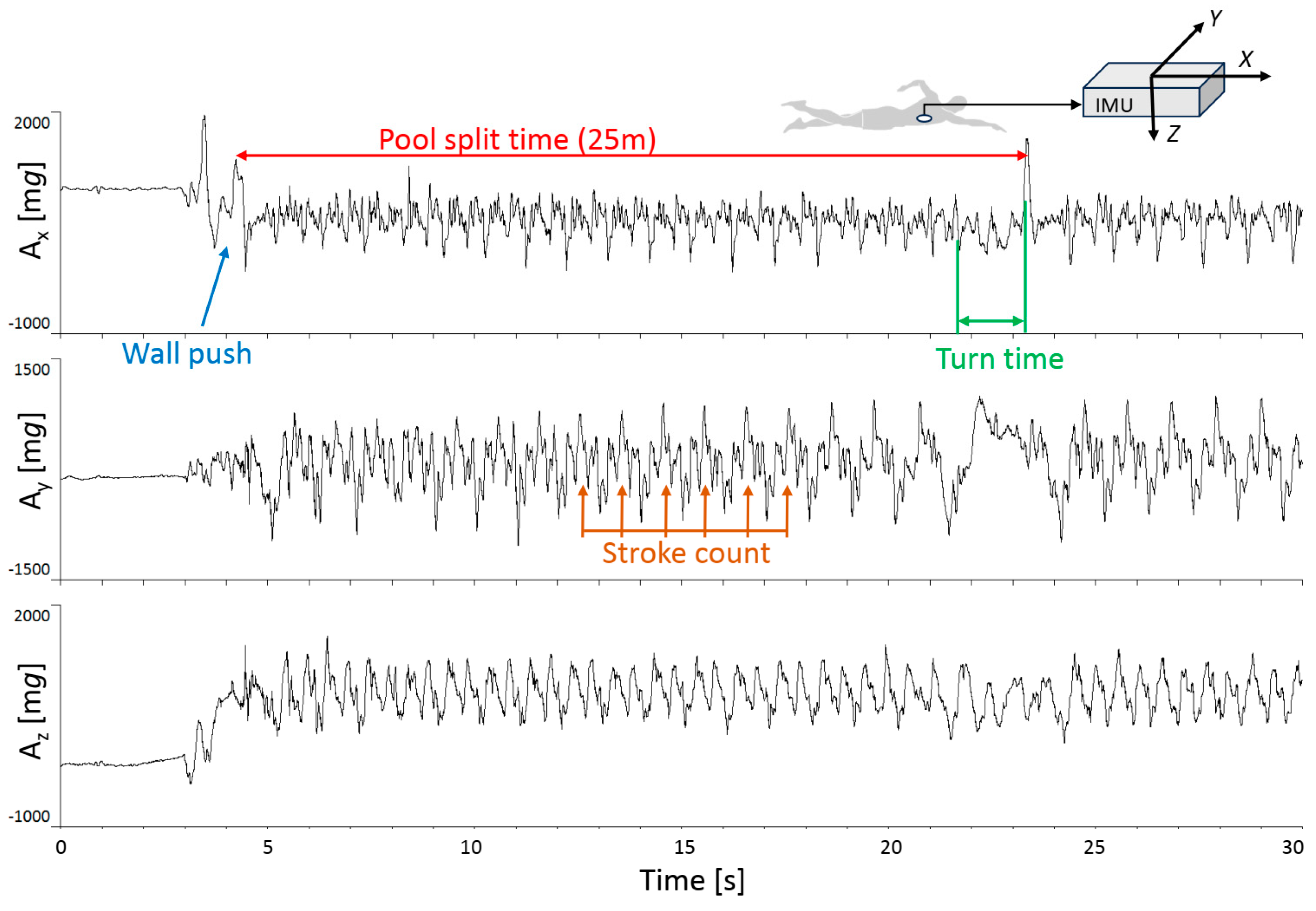
Figure 1. Recording of the triaxial accelerometry (Faros 180®; sampling frequency: 100 Hz, values expressed in mg: 1 mg = 9.80665 × 10⁻³ m/s2) from a representative subject. The accelerometer was placed on the swimmer’s sternum, with the reference orientation of the 3 axes indicated in the upper right corner of the figure. From these traces, length times and stroke counts were obtained; the SampEn regularity index was evaluated on the acceleration of the longitudinal axis of motion (X component).
Applsci 14 08387 g001
Figure 2. HR values at rest (REST), immediately after the 50 m front crawl test (50 m END), and after 30 (30 s REC) and 60 s (60 s REC) of post-test recovery. Data are presented as the mean ± SD. Black squares and continuous line: CNT group; White circles and dashed line: SDS group. The * indicates differences between the SDS vs. CNT groups significant at p < 0.001 in the post-hoc analysis of two-way ANOVA.
Applsci 14 08387 g002
Figure 3. Relationships, estimated through simple linear regression analysis, between the angle of inclination relative to the plane (body pitch) of the water and the lap time, separately for the first (0–25 m, panel (a)) and second length (25–50 m, panel (b)) of the 50 m swimming test. Black squares: CNT group; white circles: SDS group.
Applsci 14 08387 g003
Table 1. Anthropometric and training volume data (m ± SD) in the two groups of swimmers enrolled.
SDS (n = 11)CNT (n = 11)p 1Effect Size
Age (years)26.6 ± 5.627.1 ± 4.00.8280.09
Height (cm)157 ± 5179 ± 6<0.014.09
Body mass (kg)58.0 ± 7.881.3 ± 13.2<0.012.15
BMI (kg‧m−2)23.5 ± 2.725.1 ± 2.90.1880.58
Weekly training (hours)5.3 ± 1.44.9 ± 0.70.4000.37
1 Unpaired t-test.
Table 2. Chronometric and mechanical variables (m ± SD) between SDS and CNT group.
SDS (n = 11)CNT (n = 11)p 1Effect Size
Time 0–25 m (s)28.09 ± 6.7215.91 ± 2.02<0.012.45
Time 25–50 m (s)30.82 ± 7.0516.64 ± 1.80<0.012.75
Total time (s)58.91 ± 13.6832.55 ± 3.70<0.012.63
Stroke number 0–25 m33.18 ± 4.3825.64 ± 3.47<0.011.91
Stroke number 25–50 m32.91 ± 5.4325.82 ± 4.09<0.011.47
Total stroke number66.09 ± 9.6451.45 ± 7.41<0.011.70
1 Unpaired t-test.
Table 3. Body pitch angle (°) (m ± SD) between the SDS and CNT group.
SDS (n = 11)CNT (n = 11)p 1
Body pitch 0–25 m (°)12.78 ± 3.439.39 ± 2.080.016
Body pitch 25–50 m (°)13.73 ± 3.148.08 ± 3.750.0002
1 Two-way ANOVA for repeated measure, post-hoc analysis.
Disclaimer/Publisher’s Note: The statements, opinions and data contained in all publications are solely those of the individual author(s) and contributor(s) and not of MDPI and/or the editor(s). MDPI and/or the editor(s) disclaim responsibility for any injury to people or property resulting from any ideas, methods, instructions or products referred to in the content.