Article

Aromatic Amines in Organic Synthesis Part III; p-Aminocinnamic Acids and Their Methyl Esters

by
Marek Pietrzak
* and
Beata Jędrzejewska
Faculty of Chemical Technology and Engineering, Bydgoszcz University of Science and Technology, Seminaryjna 3, 85-326 Bydgoszcz, Poland
*
Author to whom correspondence should be addressed.
Appl. Sci. 2024, 14(14), 6032; https://doi.org/10.3390/app14146032
Submission received: 4 June 2024 / Revised: 1 July 2024 / Accepted: 8 July 2024 / Published: 10 July 2024
(This article belongs to the Special Issue Advances in Organic Synthetic Chemistry)

Abstract

:
Fifteen amine derivatives of cinnamic acid were synthesized by reaction of the corresponding benzaldehydes and malonic acid. The selected acids were then converted into methyl esters. Three esterification methods were tested with (1) thionyl chloride in methanol, (2) sulfuric acid in methanol, and (3) dimethyl sulfate in acetone. The latter method turned out to be the best, both in terms of reaction efficiency and product purity. The chemical structure and purity of all the synthesized compounds were verified by elemental analysis, 1H and 13C NMR, and IR spectroscopy. The cinnamic acids and their esters, thanks to an extensive system of conjugated double bonds compared to analogous benzoic acids, can be used to obtain dyes for various applications, including non-linear optics and optoelectronics. Therefore, their basic spectroscopic properties are presented as well.

1. Introduction

Chemical compounds containing an amino group (or its derivative) are widely used in many fields of science. They are intensively studied because the electron-donating properties of the amino group influence their biological activity and spectroscopic and electrochemical properties [1]. Particular attention is paid to organic compounds with amino groups in the preparation of dyes [2], spectroscopic probes [3,4,5], solar cells [6], polymerization photoinitiators [7,8], and drugs [9].
Our research group and others have investigated the influence of different amino groups on the spectroscopic and electron-donating properties of various classes of dyes, such as azomethine [10], stilbazolium [11], hemicyanine [12,13,14], styrylquinolinium [15], oxazolone [16,17], biphenyl [18], BODIPY [19,20,21,22,23], chalcone [24,25], phenothiazine [26,27], anthracene [28,29], and pyrazolone [30,31], for many years. The application of dyes in modern technologies based on multi-photon absorption, spectroscopic probes, and sensors requires compounds with an extended system of conjugated double bonds. Such compounds are synthesized using either p-aminocinnamic aldehydes or their esters instead of p-aminobenzaldehydes (or their esters).
The esters of p-aminocinnamic acid derivatives can be synthesized by applying several methods. The most common procedure is based on the general Knoevenagel reaction. In the first step, the corresponding cinnamic acid is prepared, which is then esterified [32,33]. The esters can also be obtained from the appropriate benzaldehydes [34] or benzoyl bromides [35] and iodides [36] using the Wittig or Heck reactions, respectively.
In this paper, we describe the synthesis of fifteen derivatives of p-aminocinnamic acids and show the optimal way to transform them into methyl esters. The esters of p-aminocinnamic acids were synthesized in a two-step reaction, starting with easily accessible benzaldehydes and applying the first of the above-mentioned methods. The aldehydes were synthesized according to the procedure given by Gawinecki et al. [37]. Only p-(dibutylamino)benzaldehyde was prepared by applying the methodology given by van den Berg and co-workers [38]. The basic spectral properties of the synthesized compounds in two solvents of different polarities (methanol and ethyl acetate) are also described.

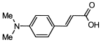
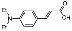
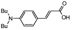
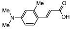
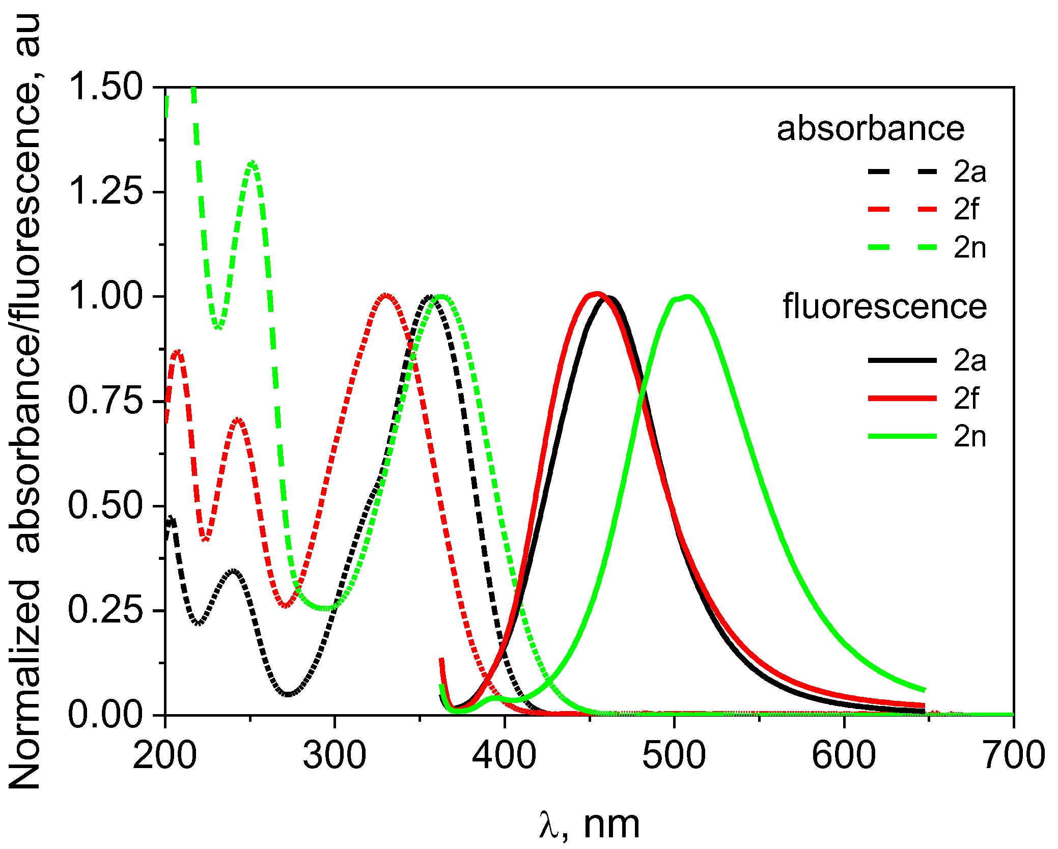
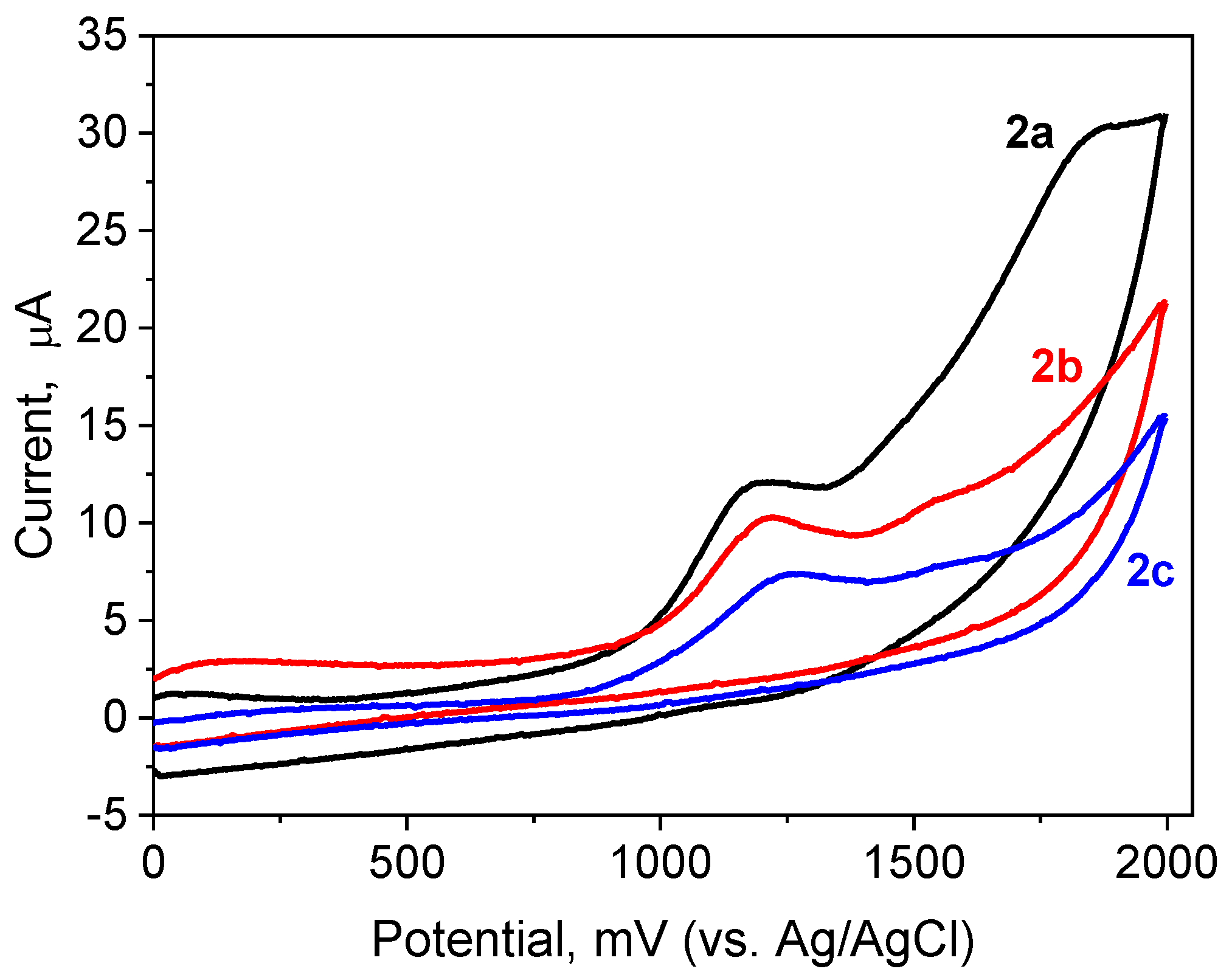
2. Results and Discussion

The amine methyl esters of cinnamic acid derivatives were prepared according to the synthetic routes outlined in Figure 1.
In the first method (Method A), the Knoevenagel condensation was used, and the corresponding cinnamic acids were obtained from amine derivatives of benzoic or naphthoic aldehydes. Six of these acids (2c, 2d, 2e, 2f, 2g, and 2k) have not been described in the literature yet. The reaction yield of cinnamic acid synthesis varies between 58% and 88%. A significantly lower yield of 29% was obtained for the 4-(dimethylamino)-2,6-dimethylcinnamic acid, probably due to steric hindrance. The purity of the synthesized acids was very high, and they were used in the next stage without further purification.
In the second stage, selected cinnamic acids were converted into methyl esters. Four of these compounds (3h, 3i, 3l, 3n) are new and have not been described in the literature so far. In search of the most effective esterification method, the process was carried out using three different reagents, i.e., thionyl chloride in methanol (Method B), sulfuric acid in methanol (Method C), and dimethyl sulfate in acetone (Method D). All these methods proved to be relatively effective, with reaction yields of 65–93% and high-purity products. The best, both in terms of the ease of carrying out the reaction (room temperature, no volatile by-products) and the highest yield, was the reaction with dimethyl sulfate (Method D). The yields of the reaction were greater than 75%, and the obtained esters were of high purity.
The structure and purity of all the synthesized acids and esters were confirmed by spectroscopic methods, namely 1H NMR, 13C NMR, and IR (see spectra in Supplementary File), and by elemental analysis. The NMR spectra showed all signals from protons and carbons present in the product molecules, as described in the Experimental part. As can be seen, the proton signal of the carboxyl group occurs at about 12 ppm, whereas the characteristic signals from the protons of the methine group are in the range of 8.3 to 5.9 ppm. The coupling constant of these protons (3JH,H = 15.8 Hz) indicates the trans configuration. The signal from the carbon of the carboxyl group is in the range of 168–169 ppm and is shifted by about 1–2 ppm in relation to the analogous benzoic acids [39]. After the esterification, the proton signal of the carboxyl group disappears, while the methyl signal appears at approx. 3.7 ppm. The analysis of the IR spectra confirms the presence of the carboxyl group in the tested acids thanks to the characteristic, strong signal at frequencies from 1653 cm−1 (2m) to 1704 cm−1 (2j). In most cases, these values are 5–20 cm−1 higher than for analogous benzoic acids [39]. After esterification, this signal shifts to the range of 1702–1716 cm−1.
Figure 2 shows the 1H NMR spectra of 3-(1-methyl-1,2,3,4-tetrahydroquinolin-6-yl)acrylic acid (2l) in DMSO-d6, its methyl ester, and the analogous benzoic acid to illustrate the shift in signal position associated with the elongation of the π electron system. Comparing these three spectra, the position of the proton signal of the carboxyl group changes little. For both the benzoic acid and the cinnamic acid derivative, it is at approx. 11.9 ppm. The protons of the methine group give signals at 7.41 and 6.15 ppm, with a coupling constant of approximately 16 Hz, indicating a trans configuration. Moreover, there is a visible shift in the signals coming from the protons of the aromatic ring, located in the ortho position in relation to the methine group, by about 0.2–0.4 ppm towards lower δ values (they are shielded more strongly than in benzoic acid). On the other hand, methylation of acid 2l has no obvious effect on the NMR spectrum, except for the disappearance of the proton signal of the carboxyl group (11.9 ppm) and the appearance of the proton signal of the methyl group (3.7 ppm).
Since the synthesized compounds can be used to obtain different groups of dyes with extended conjugated double-bond systems, their basic spectroscopic properties were investigated. The physicochemical data are presented in Table 1. The measurements were carried out in three solvents with significantly different properties, i.e., in 1,4-dioxane (1,4-Dx), methyl acetate (EtOAc), and methanol (MeOH). The UV-Vis spectra of the tested p-aminocinnamic acids indicate the presence of the main absorption band in the range 332–375 nm in 1,4-dioxane, 329–374 nm in ethyl acetate, and 330–382 nm in methanol. This band is related to the π→π* transition. The compound 2m shows the highest bathochromic shift with an absorption maximum of 374 nm for ethyl acetate and 382 nm for methanol. This is attributed to the stiffening of the alkylamino group, which facilitates the delocalization of electrons throughout the molecule. The opposite effect is observed for compound 2f. It has the most blue-shifted absorption band among the analyzed acids (absorption maximum at 329 nm in ethyl acetate and 330 nm in methanol). In this case, the presence of the methyl group in the ortho position with respect to the amino group forces the twisting of the amino group with respect to the phenyl ring, which makes it difficult to delocalize the electrons of the amino group. The molar extinction coefficients of the p-aminocinnamic acids are in the range from 17,200 M−1cm−1 and 16,500 M−1cm−1 to 41,800 M−1cm−1 and 32,400 M−1cm−1 in EtOAc and MeOH, respectively. These values are similar to analogous benzoic acids [39] but, at the same time, are about 30% lower than cinnamaldehydes [40].
Figure 3 shows an example of normalized electronic absorption and emission spectra in methanol for selected p-aminocinnamic acids (2a, 2f, 2n). The absorption and fluorescence spectra of all synthesized acids and esters in 1,4-dioxane, ethyl acetate, and methanol are presented in the Supplementary File.
The fluorescence spectra of the tested acids show a sharp band in the range of 413–464 nm in 1,4-dioxane, 426–478 nm in EtOAc, and 437–508 nm in MeOH (see Figures S10–S12 in Supplementary File). While the bathochromic shift of the absorption spectra with increasing solvent polarity is insignificant and amounts to a maximum of several nm, the shift of the fluorescence spectra is even more than 40 nm. This indicates a better stabilization of the excited state of the compounds in a more polar environment and suggests an increase in the dipole moment upon excitation.
By comparing the spectral properties of the cinnamic acids (Table 1) with the corresponding benzoic acids [39], we can assess the effect of an additional vinyl group on the absorption properties of these compounds. The electronic absorption spectra of the tested cinnamic acids, compared to the corresponding benzoic acids, are redshifted by about 40–55 nm. Only in the case of acids 2f and 2o is the shift smaller and equals about 30 nm regardless of the solvent polarity. Comparing the spectra of cinnamic acids to analogous cinnamic aldehydes [40], a shift towards shorter wavelengths by 16–23 nm in ethyl acetate and 27–36 nm in methanol is observed. The fluorescence spectra are also blue-shifted by 11–21 nm in both solvents used.
The fluorescence quantum yield of the synthesized acids and esters is low, and it does not exceed 3.5% in non-polar (1,4-Dioxane), polar aprotic (EtOAc), and protic (MeOH) solvents, which proves that the deactivation of the S1 excited state occurs mainly by non-radiative processes. The only exception is compound 2o, for which the fluorescence quantum yield is 59.9% in 1,4-dioxane, 74% in ethyl acetate, 70.8% in DMF, and 61% in methanol. The results suggest that the modification of the amino group does not significantly affect the fluorescence quantum yield of the cinnamic acids. However, the slightly higher fluorescence intensity in polar solvents, both protic and aprotic (see Table 1 and Table 2), may be attributed to the protic nature of the solvent and/or high polarization character (dielectric constant of 32.66 for MeOH and 36.71 for DMF). This feature contributes to the stabilization of the excited states and the charge or dipole of the molecule [41,42] and molecular symmetry, preventing non-radiative decay [43,44,45]. Compound 2e showed the lowest fluorescence intensity regardless of the solvent. Probably, the presence of a methyl group in the ortho position in relation to the central double bond (meta position in relation to the amino group) causes the molecule to twist and leads to a coplanar conformation, which reduces the probability of radiative transitions. On the other hand, the ϕFl values for compound 2o in 1,4-dioxane, EtOAc, DMF, and MeOH with respect to 2a are over 71-, 120-, 34- and 89-fold higher, despite the same electron-donating (NMe2) and -withdrawing (COOH) groups. In fact, the electronic character of the N,N-dimethylamino group in 2o is significantly altered by the elongation of the conjugated π-system by introducing an additional aromatic ring into the molecule, which stiffens the molecule and lowers the HOMO-LUMO energy gap [46] compared to 2a. As a consequence, the fluorescence intensity increases.
The literature data for other naphthalene derivatives with a dimethylamine group: 6-(dimethylamino)-2-naphtoic acid (ϕfl = 0.53) [47], 6-(dimethylamino)-2-naphthaldehyde (ϕfl = 0.84) [46], and 2-(dimethylamino)-naphthalene (ϕfl = 0.89) [48] indicate that high fluorescence intensity is a feature of naphthalene derivatives substituted into position 2 with a dimethylamine group. In the case of derivatives substituted into position 1, the intensity is much lower and does not exceed several percent [48,49].
According to Lewis et al. [49] and Suzuki et al. [48] the structure of the compounds based on 2-substituted naphthalene is flattened with a low torsion angle for C-NMe2 due to the lack of unbonded interaction with the H8 hydrogen atom. In the case of 1-dialkylamino-substituted naphthalene, the optimized conformation of the amino group corresponds to a structure having a twisted lone pair orbital relative to the carbon 2pπ orbital in the naphthalene ring. The lack of repulsion between the alkyl substituents on the nitrogen atom and the perihydrogen atom in the naphthalene ring counteracts the rotation of the amino group to the plane of the naphthalene ring, while simultaneously shifting the charge from the alkylamino group towards the electron-withdrawing substituent, which results in a redshift of the 2o absorption band. This is consistent with the MO calculation results [46].
Figure S19 shows the calculated molecular structure and electron distribution of the HOMO and LUMO energy levels of the 2a and 2o acids. A comparison of the electron distribution in the frontier molecular orbitals (MOs) reveals that HOMO–LUMO excitation moved the electron distribution from the dimethylamine group towards the withdrawing substituent via the aromatic π system, indicating a strong migration of the intramolecular charge transfer character of the acids.
The HOMO and LUMO energies for compound 2a were computed to be −5.552 and −1.771 eV, respectively. Similarly, the energies of the HOMO and LUMO of 2o are −5.410 and −2.079 eV, respectively. These results show that the naphthalene ring separating the electron-donating and the electron-withdrawing groups caused a decrease in the energy level of the highest occupied molecular orbital (HOMO) and an increase in the lowest unoccupied molecular orbital (LUMO). Thus, the HOMO–LUMO energy gap of 2a is higher than that of 2o. As a result, the electron transfer from HOMO to LUMO in 2o is relatively easier than in 2a. Therefore, compared to 2a, a bathochromic shift is observed in the electronic absorption spectrum of 2o.
Figure 4 shows the absorption and fluorescence spectra of 2a and 2o in 1,4-dioxane and DMF at 293 K. It can be seen that changing the solvent from nonpolar 1,4-dioxane to polar DMF almost does not affect the position and shape of the absorption spectra. In contrast, the fluorescence maxima of the compounds in DMF significantly redshift compared to those in 1,4-Dx (Figure 4), resulting in larger Stokes shifts in the polar solvent (Table 1 and Table 2). This effect is greater for the naphthalene derivative than for the benzene one. Based on the literature data, the redshifts in the fluorescence maxima of the aminocinnamic acids in polar solvents may result from a bulk relaxation of the solvent dipoles about the solute’s excited-state dipole moment with some contribution of an intramolecular reorganization of the amino group [48].
The excited state lifetimes were determined by deconvolution of the fluorescence decay curves recorded using a single photon counting method, which are shown in Figure 5.
The fluorescence lifetime (τfl) and fluorescence quantum yield (ϕfl) of 3-[6-(dimethylamino)naphthalen-2-yl]acrylic acid (2o) and 4-dimethylaminocinnamic acid (2a) are listed in Table 2. The values of τfl and ϕf are found to be affected by the number of aromatic rings separating the electron-donating and electron-withdrawing substituents and their position, as well as solvent polarity. In the three solvents tested (DMF, EtOAc, and 1,4-Dx), 2a gives smaller τfl values than those of 2a. In particular, decreases in average τfl can be recognized for them in the nonpolar solvent (1,4-Dx), and the ϕf values of these compounds become smaller. Since both compounds have the same substituents, the remarkable increase in τfl and ϕf is limited to the aromatic π system, i.e., the presence of a naphthalene ring substituted by a dimethylamino group in the two position, which is responsible for the planar structure of compound 2o even in the ground state. This plays an important role in the promotion of the radiative deactivation processes of its excited state.
The radiative rate constants (kr = ϕfl τ−1) for the two compounds in three solvents of different polarities are collected in Table 2. The values of kr for 2o are higher than the values for 2a. The dominance of radiative processes in the deactivation of the 2o excited state results in an increase in the fluorescence quantum yield. Moreover, the increase in fluorescence lifetime is mainly due to the decrease in non-radiative rate constants, internal conversion, and intersystem crossing in case 2o compared to 2a,. for which non-emissive processes dominate [49].
The oxidation potentials of the tested aminocinnamic acids were determined based on cyclic voltammetry. The measurements were performed to show the influence of the acid structure on the possibility of electron loss. Examples of cyclic voltammograms are presented in Figure 6, whereas the oxidation potentials for all compounds tested are summarized in Table 3.
The analysis of cyclic voltammograms of the tested compounds indicates that their electrochemical oxidation is irreversible. The position of the oxidation peaks depends on the amino substituents. For example, the values of the oxidation potential increase with the elongation of the aliphatic substituents at the amino group (2a is methyl groups, 2b is ethyl groups, and 2c is butyl groups). The lowest value is observed for compound 2j, which is a morpholine derivative. This means that 2j has the greatest tendency to lose electrons. The oxidation potentials of all the tested acids range from 963 to 1285 mV, with the highest value of 1285 mV for compound 2f with a methyl substituent in the ortho position to the N,N-dimethylamino group responsible for its coplanar conformation and the decoupling effect between the dimethylamino group and the electron-accepting part of the molecule [50]. Moreover, the oxidation potentials of cinnamic acids containing cyclic terminal groups, i.e., pyrrolidine, piperidine, and morpholine, depend on the change in aromatic ring–nitrogen resonance interaction as the saturated heterocycle is varied. In general, the extent of overlap of the nitrogen lone-pair electrons with the π-electrons of the aromatic ring in N-phenyl-substituted saturated heterocycles may depend upon the molecular conformation, the nature of the heterocycle, the angle of rotation of the aromatic ring about the N-C(phenyl) bond, and any p-substituent present [51]. The transformation of cinnamic acids into the corresponding methyl esters leads to an increase in oxidation potentials, e.g., from 993 and 1084 mV for 2h and 2i to 1306 and 1210 mV for 3h and 3i, respectively. This effect is probably related to the decrease in the dipole moment when the COOH group is replaced with COOMe (Δμg = 0.58 D for 2h→3h and Δμg = 1.07 D for 2i→3i). Furthermore, alkane bridges between Cortho and Namino, e.g., in julolidine derivatives, cause the nitrogen atom to reveal weaker electron-donor properties than that in the 1-pyrrolidino group (EOX = 1171 mV vs. 993 mV). This is consistent with the σR0 substituent constants of the 1-N{2,6-[(CH2)3]2} (−0.59) and N(CH2)4 (−0.63) amino groups [52]. The modification of cinnamic acids with stiffened electron-donating substituents leads to a planar conformation of the molecules, which increases the probability of charge separation and leads to a decrease in oxidation potentials. The importance of the electronic effect of amino groups is also confirmed by the linear correlation of EOX values with the corresponding σR0 substituent constants [52,53,54] (see Figure S13 in Supplementary File). Cinnamic acids, which bear a stronger electron-releasing substituent, have a lower oxidation potential.

3. Materials and Methods
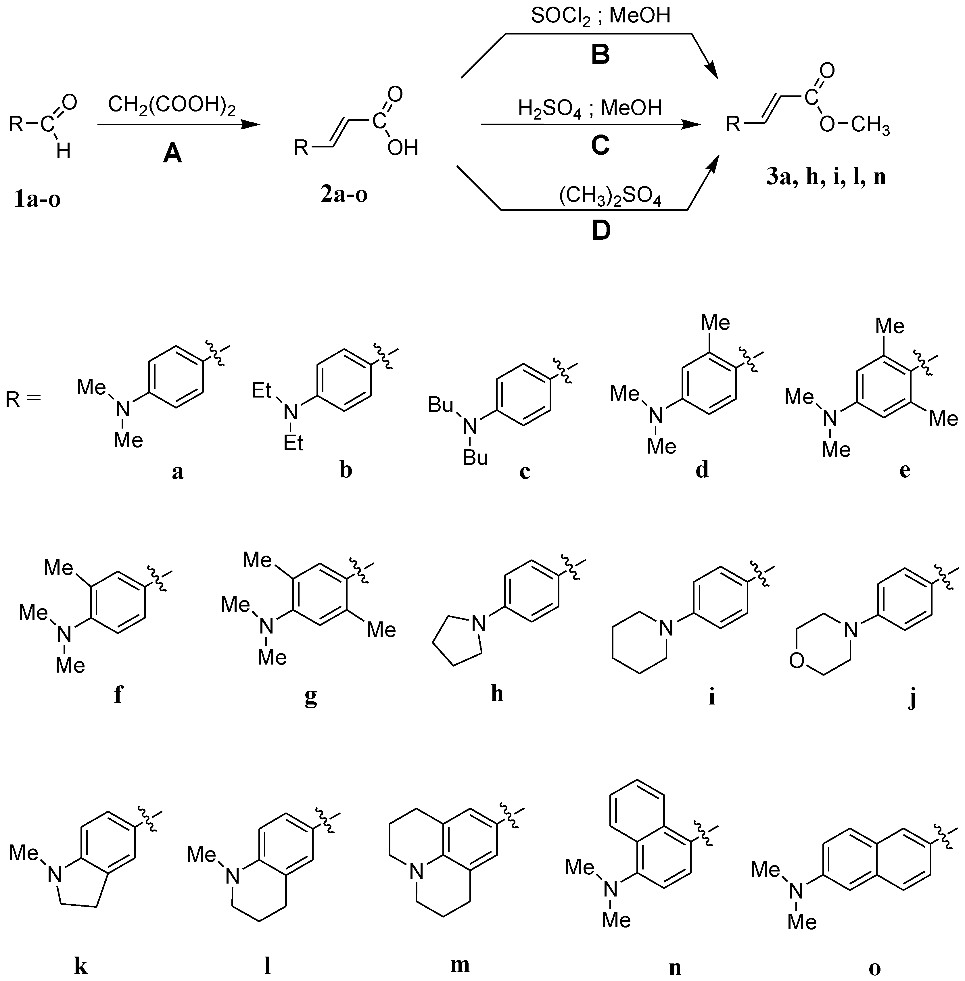
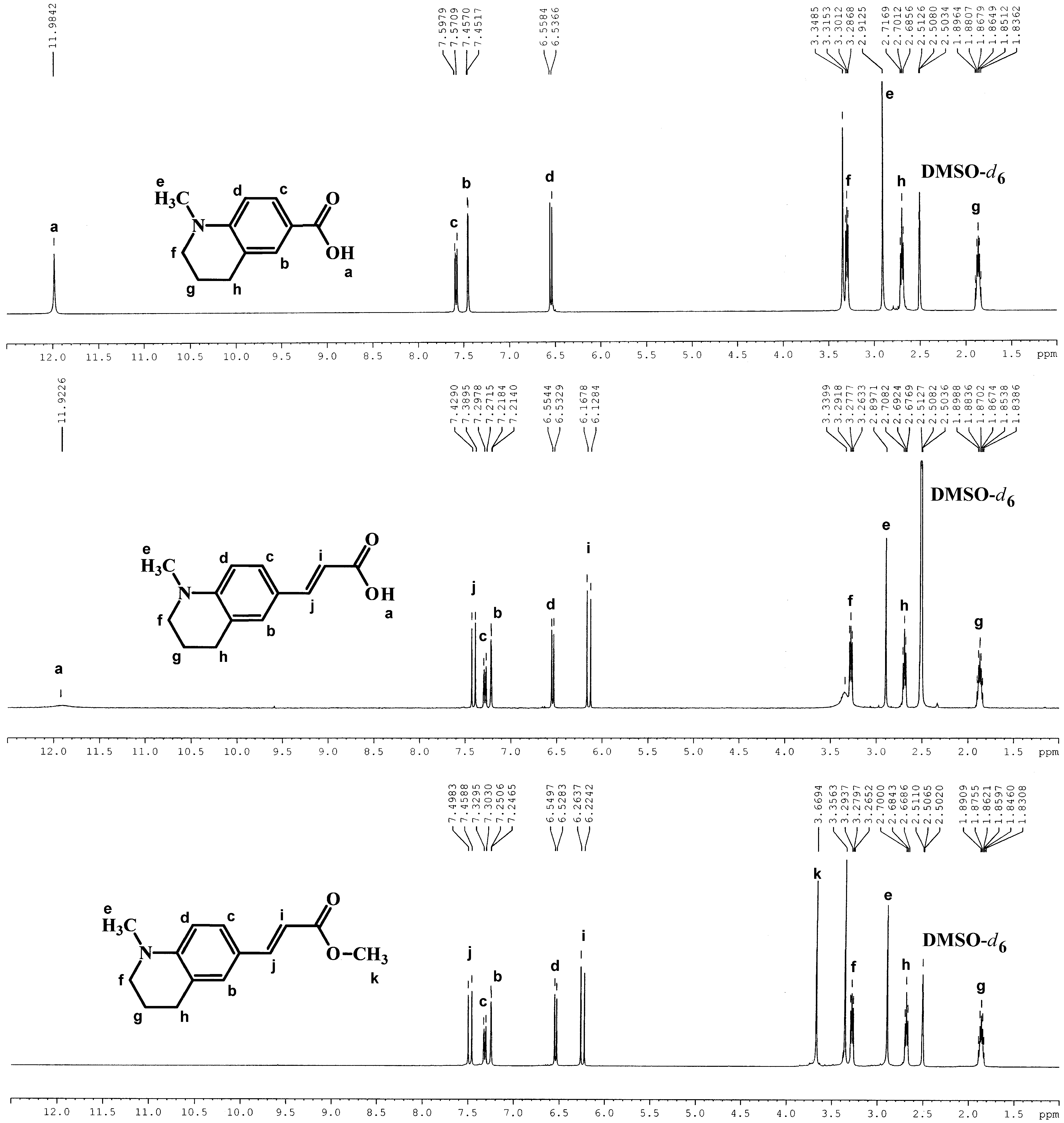
All starting reagents and solvents were purchased from Aldrich Chemical Co. (Milwaukee, MI, USA). The melting points were determined with the Büchi melting point apparatus MP-1. The 1H (400 MHz) and 13C (100 MHz) NMR spectra were recorded on a Bruker AscentTM 400 NMR spectrometer (Bruker, Billerica, MA, USA). Dimethylsulfoxide (DMSO-d6) was used as the solvent and tetramethylsilane as the internal standard. Elemental analysis was carried out by an Elementar Vario MACRO apparatus (Elementar Americas Inc., Ronkonkoma, NY, USA). The IR spectra were recorded on a Bruker spectrophotometer Vector 22 in the range of 400–4500 cm−1 by the KBr pellet technique. The UV-Vis absorption and emission spectra were recorded on a Shimadzu UV-Vis Multispec-1501 spectrophotometer (Spectralab Scientific Inc., Markham, ON, Canada) and a Hitachi F-7100 spectrofluorometer (Hitachi High-Tech Corporation, Tokyo, Japan), respectively. The fluorescence quantum yield (FQY; ϕ) was calculated according to Equation (1) using coumarin I in ethanol as a reference [55].
ϕ s = ϕ r e f I s A r e f I r e f A s n s 2 n r e f 2
where I is the integrated intensity (area) (in units of photons) and n is the refractive index. The absorbances (A) of both the sample (s) and reference (ref) solution at an excitation wavelength (350 nm) was ca. 0.1.
The fluorescence lifetimes were measured using an Edinburgh Instruments single-photon counting system (FLS920P Spectrometers (StellarNet Inc., Keystone, FL, USA)). The apparatus utilizes a picosecond diode laser for the excitation-generating pulses of about 55 ps at 375 nm. The dyes were studied at dilute solution (~0.2 in a 10 mm cell). The fluorescence decays were fitted to double-exponential functions. The average lifetime, τav, is calculated as
τ a v = Σ τ i α i Σ α i
where αi and τi are the amplitudes and lifetimes.
An Electrochemical Analyzer EA9C MTM Cracow (Wieliczka, Poland) was used to determine the oxidation potential (Eox) of the synthesized compounds. Measurements were made by cyclic voltammetry using anhydrous acetonitrile with 0.1 M of tetrabutylammonium perchlorate as a supporting electrolyte. The scan rate was 300 mV/s. A typical three-electrode setup, containing a platinum 1 mm electrode as the working electrode and platinum and Ag/AgCl as the auxiliary and reference electrodes, respectively, was applied for the measurements.
DFT calculations were carried out using the B3LYP/6-311+G(2d,2p) method. Calculations were carried out with the Gaussian 03 program [56].

3.1. Synthesis

The routes leading to the preparation of derivatives of p-aminocinnamic esters are shown in Figure 1.

3.1.1. Synthesis of the Amine Derivatives of Cinnamic Acid

For Method A, pyridine (15 mL), malonic acid (25 mmol), and the appropriate benzaldehyde (20 mmol) were placed in a flask. Then piperidine (20 drops) was added, and the solution was heated at 80 °C for 20 h under stirring. The resulting mixture was poured into 120 mL of ice water, and 5 mL of saturated NaHCO3 was added. The precipitate was filtered off. Then, the crude precipitate was added to 200 mL of cold 1% NaOH, and after 1 min of mixing, it was filtered. The filtrate was neutralized to pH = 7 with 10% hydrochloric acid. The product of the reaction was filtered off and dried.

3.1.2. Synthesis of the Methyl p-(Dimethylamino)cinnamate

For Method B, thionyl chloride (21 mmol) was added dropwise to a solution of p-(dimethylamino)cinnamic acid (15 mmol) in anhydrous methanol within 2 h under vigorous stirring. After 5 h, most of the methanol was distilled off, and the residue was poured onto 70 mL of ice water. Saturated NaHCO3 was added while stirring until the CO2 bubbles were stopped to volatilize. The precipitate was filtered and recrystallized from methanol.
For Method C, concentrated H2SO4 (4 g) was added dropwise to anhydrous methanol (30 mL). Then p-(dimethylamino)cinnamic acid (15 mmol) was put into the flask. The mixture was heated and held at gentle reflux for 20 h. Most of the methanol was distilled off, and the residue was poured onto 80 mL of ice water. Next, KHCO3 was added in portions until the CO2 bubbles were stopped to volatilize. The precipitate was filtered and recrystallized from methanol.
For Method D, a total of 40 mL of acetone, 15 mmol of p-(dimethylamino)cinnamic acid, 20 mmol of dimethyl sulfate, and 21 mmol of potassium carbonate were put into a flask and stirred at r.t. for 24 h. The resulting mixture was filtered, and the filtrate was concentrated by evaporation to 15 mL and poured onto 100 mL of ice water. The precipitate was filtered off and dried. The crude product was crystallized from methanol.
4-Dimethylaminocinnamic acid (2a)
Applsci 14 06032 i001
The compound was obtained as a brown solid; yield: 71%; mp 221 °C.
1H NMR (400 MHz, DMSO-d6) σ (ppm): 11.96 (s, 1H, COOH), 7.50-7.46 (m, 3H), 6.70 (d, J = 8.9 Hz, 2H), 6.22 (d, J = 15.8 Hz, 1H, -CH =), 2.97 (s, 6H, N(CH3)2).
13C NMR (100 MHz, DMSO-d6) σ (ppm): 168.3 (COOH), 151.6 (C), 144.7 (CH), 129.8 (CH), 121.6 (C), 113.0 (CH), 111.8 (CH), 39.8 (N(CH3)2).
IR (KBr): 1679, 1605, 1529, 1372, 1231, 1203, 1191, 816.
Anal. Calcd for C11H13NO2: C, 69.09; H, 6.85; N, 7.33. Found: C, 69.01; H, 6.74; N, 7.28.
3-[4-(Diethylamino)phenyl]acrylic acid (2b)
Applsci 14 06032 i002
The compound was obtained as a yellow solid; yield: 80%; mp 184 °C.
1H NMR (400 MHz, DMSO-d6) σ (ppm): 11.91 (s, 1H, COOH), 7.47–7.43 (m, 3H), 6.65 (d, J = 9.0 Hz, 2H), 6.17 (d, J = 15.8 Hz, 1H, -CH =), 3.38 (q, J = 6.9 Hz, 4H, NCH2), 1.10 (t, J = 7.0 Hz, 6H, CH3).
13C NMR (100 MHz, DMSO-d6) σ (ppm): 168.8 (COOH), 149.4 (C), 145.2 (CH), 130.5 (CH), 121.1 (C), 112.7 (CH), 111.5 (CH), 44.2 (NCH2), 12.9 (CH3).
IR (KBr): 1670, 1658, 1598, 1356, 1261, 1208, 1184, 818.
Anal. Calcd for C13H17NO2: C, 71.21; H, 7.82; N, 6.39. Found: C, 71.15; H, 7.70; N, 6.32.
3-[4-(Dibuthylamino)phenyl]acrylic acid (2c)
Applsci 14 06032 i003
The compound was obtained as a pale-yellow solid; yield: 68%; mp 138.5–140.5 °C.
1H NMR (400 MHz, DMSO-d6) σ (ppm): 11.91 (s, 1H, COOH), 7.47–7.43 (m, 3H), 6.62 (d, J = 8.9 Hz, 2H), 6.16 (d, J = 15.8 Hz, 1H, -CH=), 3.31 (q, J = 7.6 Hz, 4H, NCH2), 1.50 (m, 4H), 1.35 (m, 4H), 0.92 (t, J = 7.3 Hz, 6H, CH3).
13C NMR (100 MHz, DMSO-d6) σ (ppm): 168.7 (COOH), 149.8 (C), 145.1 (CH), 130.4 (CH), 121.1 (C), 112.7 (CH), 111.6 (CH), 50.3 (NCH2), 29.5 (CH2), 20.1 (CH2), 14.3 (CH3).
IR (KBr): 1670, 1578, 1525, 1403, 1204, 1185, 819.
Anal. Calcd for C17H25NO2: C, 74.15; H, 9.15; N, 5.09. Found: C, 74.10; H, 9.06; N, 5.11.
3-[4-(Dimethylamino)-2-methylphenyl]acrylic acid (2d)
Applsci 14 06032 i004
The compound was obtained as a pale-yellow solid; yield: 63%; mp 176 °C.
1H NMR (400 MHz, DMSO-d6) σ (ppm): 12.00 (s, 1H, COOH), 7.71 (d, J = 15.8 Hz, 1H, -CH=), 7.56 (d, J = 8.6 Hz, 1H), 6.59–6.54 (m, 3H), 6.18 (d, J = 15.7 Hz, 1H, -CH=), 2.95 (s, 6H, N(CH3)2), 2.34 (s, 3H, ArCH3).
13C NMR (100 MHz, DMSO-d6) σ (ppm): 168.8 (CHO), 151.8 (C), 141.7 (CH), 139.1 (C), 128.2 (CH), 120.7 (C), 114.3 (CH), 113.7 (CH), 110.6 (CH), 40.1 (N(CH3)2), 20.3 (ArCH3).
IR (KBr): 1680, 1586, 1514, 1366, 1290, 1099, 827.
Anal. Calcd for C12H15NO2: C, 70.23; H, 7.37; N, 6.83. Found: C, 70.15; H, 7.27; N, 6.79.
3-[4-(Dimethylamino)-2,6-dimethylphenyl]acrylic acid (2e)
Applsci 14 06032 i005
The compound was obtained as a pale-yellow solid; yield: 29%; mp 176 °C.
1H NMR (400 MHz, DMSO-d6) σ (ppm): 12.12 (s, 1H, COOH), 7.75 (d, J = 16.2 Hz, 1H, -CH=), 6.47 (s, 1H), 5.93 (d, J = 16.2 Hz, 1H, -CH=), 2.93 (s, 6H, N(CH3)2), 2.32 (s, 6H, ArCH3).
13C NMR (100 MHz, DMSO-d6) σ (ppm): 168.7 (COOH), 150.6 (C), 142.3 (CH), 139.2 (C), 120.8 (C), 119.3 (CH), 112.7 (CH), 40.1 (N(CH3)2), 22.5 (ArCH3).
IR (KBr): 1672, 1581, 1364, 1313, 1219, 1201, 1144, 822.
Anal. Calcd for C13H17NO2: C, 71.21; H, 7.82; N, 6.39. Found: C, 71.25; H, 7.78; N, 6.45.
3-[4-(Dimethylamino)-3-methylphenyl]acrylic acid (2f)
Applsci 14 06032 i006
The compound was obtained as a light-gray solid; yield: 69%; mp 129 °C.
1H NMR (400 MHz, DMSO-d6) σ (ppm): 12.21 (s, 1H, COOH), 7.51–7.44 (m, 3H), 7.04 (d, J = 5.1 Hz, 1H), 6.37 (d, J = 15.9 Hz, 1H, -CH=), 2.72 (s, 6H, N(CH3)2), 2.29 (s, 3H, ArCH3).
13C NMR (100 MHz, DMSO-d6) σ (ppm): 168.3 (COOH), 154.5 (C), 144.4 (CH), 131.4 (CH), 131.3 (C), 128.3 (C), 127.5 (CH), 118.7 (CH), 117.0 (CH), 43.9 (N(CH3)2), 19.0 (ArCH3).
IR (KBr): 1684, 1621, 1602, 1506, 1420, 1279, 1232, 1109, 982, 827.
Anal. Calcd for C12H15NO2: C, 70.23; H, 7.37; N, 6.83. Found: C, 70.15; H, 7.31; N, 6.81.
3-[4-(Dimethylamino)-2,5-dimethylphenyl]acrylic acid (2g)
Applsci 14 06032 i007
The compound was obtained as a brown solid; yield: 61%; mp 151 °C.
1H NMR (400 MHz, DMSO-d6) σ (ppm): 12.21 (s, 1H, COOH), 7.73 (d, J = 15.8 Hz, 1H, -CH=), 7.50 (s, 1H), 6.82 (s, 1H), 6.28 (d, J = 15.8 Hz, 1H, -CH=), 2.67 (s, 6H, N(CH3)2), 2.33 (s, 3H, ArCH3), 2.23 (s, 3H, ArCH3).
13C NMR (100 MHz, DMSO-d6) σ (ppm): 168.3 (COOH), 154.6 (C), 141.5 (CH), 136.2 (C), 129.7 (CH), 128.8 (C), 126.3 (C), 120.2 (CH), 117.4 (CH), 43.7 (N(CH3)2), 19.5 (CH3), 18.7 (CH3).
IR (KBr): 1682, 1599, 1508, 1318, 1236, 1211, 1075, 984, 859.
Anal. Calcd for C13H17NO2: C, 71.21; H, 7.82; N, 6.39. Found: C, 71.14; H, 7.73; N, 6.30.
3-[4-(Pyrrolidin-1-yl)phenyl]acrylic acid (2h)
Applsci 14 06032 i008
The compound was obtained as a light-brown solid; yield: 73%; mp 261 °C.
1H NMR (400 MHz, DMSO-d6) σ (ppm): 11.90 (s, 1H, COOH), 7.49 − 7.45 (m, 3H), 6.53 (d, J = 8.8 Hz, 2H), 6.18 (d, J = 15.8 Hz, 1H, -CH=), 3.28 (t, J = 6.5 Hz, 4H, NCH2), 1.95 (m, 4H, CH2).
13C NMR (100 MHz, DMSO-d6) σ (ppm): 168.7 (COOH), 149.5 (C), 145.4 (CH), 130.3 (CH), 121.4 (C), 112.7 (CH), 112.1 (CH), 47.7 (NCH2), 25.4 (CH2).
IR (KBr): 1673, 1588, 1526, 1391, 1261, 1179, 815.
Anal. Calcd for C13H15NO2: C, 71.87; H, 6.96; N, 6.45. Found: C, 71.88; H, 6.87; N, 6.40.
3-[4-(Piperidin-1-yl)phenyl]acrylic acid (2i)
Applsci 14 06032 i009
The compound was obtained as a pale-yellow solid; yield: 72%; mp 223 °C.
1H NMR (400 MHz, DMSO-d6) σ (ppm): 12.03 (s, 1H, COOH), 7.50–7.45 (m, 3H), 6.91 (d, J = 9.0 Hz, 2H), 6.25 (d, J = 15.8 Hz, 1H, -CH=), 3.26 (t, J = 5.0 Hz, 4H, NCH2), 1.58 (m, 6H).
13C NMR (100 MHz, DMSO-d6) σ (ppm): 168.6 (COOH), 152.8 (C), 144.7 (CH), 130.1 (CH), 123.7 (C), 114.9 (CH), 114.4 (CH), 48.7 (NCH2), 25.4 (CH2), 24.4 (CH2).
IR (KBr): 1666, 1602, 1519, 1241, 1188, 1126, 817.
Anal. Calcd for C14H17NO2: C, 72.71; H, 7.41; N, 6.06. Found: C, 72.72; H, 7.30; N, 6.03.
3-[4-(Morpholin-4-yl)phenyl]acrylic acid (2j)
Applsci 14 06032 i010
The compound was obtained as a pink solid; yield: 88%; mp 247 °C.
1H NMR (400 MHz, DMSO-d6) σ (ppm): 12.09 (s, 1H, COOH), 7.54-7.38 (m, 3H), 6.95 (d, J = 8.9 Hz, 2H), 6.30 (d, J = 15.9 Hz, 1H, -CH=), 3.74 (t, J = 4.9 Hz, 4H, NCH2), 3.21 (t, J = 4.9 Hz, 4H, OCH2).
13C NMR (100 MHz, DMSO-d6) σ (ppm): 168.5 (COOH), 152.8 (C), 144.6 (CH), 130.0 (CH), 124.9 (C), 115.2 (CH), 114.7 (CH), 66.4 (OCH2), 47.8 (NCH2).
IR (KBr): 1704, 1604, 1517, 1174, 1112, 820, 618.
Anal. Calcd for C13H15NO3 C, 66.94; H, 6.48; N, 6.01. Found: C, 67.02; H, 6.36; N, 6.09.
3-(1-Methyl-2,3-dihydro-1H-indol-5-yl)acrylic acid (2k)
Applsci 14 06032 i011
The compound was obtained as a brown solid; yield: 58%; mp 151 °C.
1H NMR (400 MHz, DMSO-d6) σ (ppm): 11.93 (s, 1H, COOH), 7.45 (d, J = 15.8 Hz, 1H, -CH=), 7.37 (s, 1H), 7.29 (d, J = 8.1 Hz, 1H), 6.46 (d, J = 8.1 Hz, 1H), 6.17 (d, J = 15.8 Hz, 1H, -CH=), 3.38 (t, J = 8.2 Hz, 2H, NCH2), 2.91 (t, J = 8.3 Hz, 2H ArCH2), 2.77 (s, 3H, NCH3).
13C NMR (100 MHz, DMSO-d6) σ (ppm): 168.8 (COOH), 155.6 (C), 145.4 (CH), 131.1 (C), 130.5 (CH), 123.7 (CH), 123.6 (C), 113.2 (CH), 106.3 (CH), 55.3 (NCH2), 35.1 (NCH3), 27.9 (ArCH2).
IR (KBr): 1676, 1592, 1517, 1294, 1230, 1201.
Anal. Calcd for C12H13NO2: C, 70.92; H, 6.45; N, 6.89. Found: C, 70.84; H, 6.41; N, 6.82.
3-(1-Methyl-1,2,3,4- tetrahydroquinolin-6-yl)acrylic acid (2l)
Applsci 14 06032 i012
The compound was obtained as a brown solid; yield: 65%; mp 158 °C.
1H NMR (400 MHz, DMSO-d6) σ (ppm): 11.92 (s, 1H, COOH), 7.41 (d, J = 15.8 Hz, 1H, -CH=), 7.28 (d, J = 8.5 Hz, 1H), 7.22 (s, 1H), 6.54 (d, J = 8.6 Hz, 1H), 6.15 (d, J = 16.1 Hz, 1H, -CH=), 3.28 (t, J = 5.7 Hz, 2H, NCH2), 2.90 (s, 3H, NCH3), 2.69 (t, J = 6.2 Hz, 2H, ArCH2), 1.87 (m, 2H).
13C NMR (100 MHz, DMSO-d6) σ (ppm): 168.7 (COOH), 148.6 (C), 145.3 (CH), 129.0 (CH), 128.8 (CH), 122.5 (CH), 121.7 (C), 112.7 (CH), 110.6 (CH), 50.9 (NCH2), 38.9 (NCH3), 27.6 (ArCH2), 21.9 (CH2).
IR (KBr): 1672, 1658, 1599, 1522, 1321, 1269, 1206.
Anal. Calcd for C13H15NO2: C, 71.87; H, 6.96; N, 6.45. Found: C, 71.81; H, 6.87; N, 6.38.
3-(9-Julolidyl)acrylic acid (2m)
Applsci 14 06032 i013
The compound was obtained as a light-brown solid; yield: 64%; mp 178 °C.
1H NMR (400 MHz, DMSO-d6) σ (ppm): 11.86 (s, 1H, COOH), 7.34 (d, J = 15.8 Hz, 1H, -CH=), 7.01 (s, 2H), 6.08 (d, J = 15.7 Hz, 1H, -CH=), 3.19 (t, J = 5.7 Hz, 4H, NCH2), 2.66 (t, J = 6.3 Hz, 4H, ArCH2), 1.85 (m, 4H, CH2).
13C NMR (100 MHz, DMSO-d6) σ (ppm): 168.8 (COOH), 145.4 (CH), 144.9 (C), 127.7 (CH), 120.94 (C), 120.90 (C), 112.2 (CH), 49.6 (CH2), 27.5 (CH2), 21.6 (CH2).
IR (KBr): 1653, 1592, 1513, 1312, 1256, 1160.
Anal. Calcd for C15H17NO2: C, 74.05; H, 7.04; N, 5.76. Found: C, 73.98; H, 6.97; N, 5.72.
3-[4-(Dimethylamino)naphthalen-1-yl]acrylic acid (2n)
Applsci 14 06032 i014
The compound was obtained as a yellow solid; yield: 61%; mp 166 °C.
1H NMR (400 MHz, DMSO-d6) σ (ppm): 12.42 (s, 1H, COOH), 8.33 (d, J = 15.7 Hz, 1H, -CH=), 8.20–8.17 (m, 2H), 7.90 (d, J = 8 Hz, 1H), 7.63–7.55 (m, 2H), 7.12 (d, J = 8Hz, 1H), 6.50 (d, J = 15.6 Hz, 1H, -CH=), 2.88 (s, 6H, N(CH3)2).
13C NMR (100 MHz, DMSO-d6) σ (ppm): 168.2 (COOH), 153.2 (C), 140.7 (CH), 132.7 (C), 128.1 (C), 127.4 (CH), 126.3 (CH), 125.7 (CH), 125.3 (CH), 125.2 (C), 123.8 (CH), 119.7 (CH), 114.0 (C), 45.0 (N(CH3)2).
IR (KBr): 1676, 1611, 1570, 1417, 1338, 1212, 770.
Anal. Calcd for C15H15NO2: C, 74.67; H, 6.27; N, 5.81. Found: C, 74.51; H, 6.32; N, 5.88.
3-[6-(Dimethylamino)naphthalen-2-yl]acrylic acid (2o)
Applsci 14 06032 i015
The compound was obtained as a yellow solid; yield: 63%; mp 251 °C.
1H NMR (400 MHz, DMSO-d6) σ (ppm): 12.16 (s, 1H, COOH), 7.94 (s, 1H), 7.76 (d, J = 9.1 Hz, 1H), 7.69-7.62 (m, 3H), 7.24 (d, J = 9.1Hz, 1H), 6.95 (s, 1H), 6.50 (d, J = 15.9 Hz, 1H, -CH=), 3.04 (s, 6H, N(CH3)2).
13C NMR (100 MHz, DMSO-d6) σ (ppm): 168.5 (COOH), 149.7 (C), 144.7 (CH), 136.2 (C), 130.2 (CH), 129.9 (CH), 128.0 (C), 127.0 (CH), 126.0 (C), 124.6 (CH), 117.5 (CH), 116.9 (CH), 105.8 (CH), 40.5 (N(CH3)2).
IR (KBr): 1683, 1612, 1599, 1428, 1312, 1202, 1176, 842.
Anal. Calcd for C15H15NO2: C, 74.67; H, 6.27; N, 5.81. Found: C, 74.58; H, 6.39; N, 5.84.
Methyl 3-[4-(dimethylamino)phenyl]acrylate (3a)
Applsci 14 06032 i016
The compound was obtained as a pale-yellow solid; yield: 73% (method B), 76% (method C), 82% (method D); mp 137 °C.
1H NMR (400 MHz, DMSO-d6) σ (ppm): 7.57–7.52 (m, 3H), 6.71 (d, J = 8.9 Hz, 2H), 6.32 (d, J = 15.9 Hz, 1H, -CH=), 3.68 (s, 3H, OCH3), 2.98 (s, 6H, N(CH3)2).
13C NMR (100 MHz, DMSO-d6) σ (ppm): 167.8 (COO), 152.2 (C), 145.7 (CH), 130.4 (CH), 121.8 (C), 112.2 (CH), 111.9 (CH), 51.5 (OCH3), 40.1 (N(CH3)2).
IR (KBr): 1702, 1605, 1529, 1187, 1170, 815.
Anal. Calcd for C12H15NO2: C, 70.23; H, 7.37; N, 6.83. Found: C, 70.16; H, 7.31; N, 6.77.
Methyl 3-[4-(pyrrolidin-1-yl)phenyl]acrylate (3h)
Applsci 14 06032 i017
The compound was obtained as a yellow solid; yield: 71% (method B), 72% (method C), 83% (method D); mp 146 °C.
1H NMR (400 MHz, DMSO-d6) σ (ppm): 7.56–7.50 (m, 3H), 6.54 (d, J = 8.8 Hz, 2H), 6.28 (d, J = 15.8 Hz, 1H, -CH=), 3.67 (s, 3H, OCH3), 3.28 (t, J = 6.5 Hz, 4H, NCH2), 1.96 (m, 4H, CH2).
13C NMR (100 MHz, DMSO-d6) σ (ppm): 167.8 (COO), 149.7 (C), 145.9 (CH), 130.6 (CH), 121.2 (C), 112.1 (CH), 111.2 (CH), 51.5 (OCH3), 47.7 (NCH2), 25.4 (CH2).
IR (KBr): 1708, 1701, 1605, 1527, 1393, 1184, 1164, 985, 814.
Anal. Calcd for C14H17NO2: C, 72.71; H, 7.41; N, 6.06. Found: C, 72.65; H, 7.38; N, 6.01.
Methyl 3-[4-(piperidin-1-yl)phenyl]acrylate (3i)
Applsci 14 06032 i018
The compound was obtained as a light-pink–yellow solid; yield: 84% (method B), 81% (method C), 93% (method D); mp 101 °C.
1H NMR (400 MHz, DMSO-d6) σ (ppm): 7.56–7.51 (m, 3H), 6.91 (d, J = 8.9 Hz, 2H), 6.35 (d, J = 15.9 Hz, 1H, -CH=), 3.68 (s, 3H, OCH3), 3.29 (t, J = 5.0 Hz, 4H, NCH2), 1.57 (m, 6H).
13C NMR (100 MHz, DMSO-d6) σ (ppm): 167.7 (COO), 153.0 (C), 145.2 (CH), 130.4 (CH), 123.4 (C), 114.8 (CH), 112.9 (CH), 51.6 (OCH3), 48.7 (NCH2), 25.4 (CH2), 24.4 (CH2);
IR (KBr): 1708, 1625, 1604, 1518, 1190, 1170, 1127, 985, 816.
Anal. Calcd for C15H19NO2: C, 73.45; H, 7.81; N, 5.71. Found: C, 73.37; H, 7.75; N, 5.77.
Methyl 3-(1-methyl-1,2,3,4-tetrahydroquinolin-6-yl)acrylate (3l)
Applsci 14 06032 i019
The compound was obtained as a brown solid; yield: 73% (method B), 71% (method C), 88% (method D); mp 72 °C.
1H NMR (400 MHz, DMSO-d6) σ (ppm): 7.48 (d, J = 15.8 Hz, 1H, -CH=), 7.30 (d, J = 8.5 Hz, 1H), 7.25 (s, 1H), 6.54 (d, J = 8.6 Hz, 1H), 6.25 (d, J = 15.8 Hz, 1H, -CH=), 3.67 (s, 3H, OCH3), 3.28 (t, J = 5.7 Hz, 2H, NCH2), 2.91 (s, 3H, NCH3), 2.68 (t, J = 6.1 Hz, 2H, ArCH2), 1.87 (m, 2H).
13C NMR (100 MHz, DMSO-d6) σ (ppm): 167.8 (COO), 148.8 (C), 145.9 (CH), 129.2 (CH), 128.9 (CH), 122.6 (C), 121.5 (C), 111.2 (CH), 110.6 (CH), 51.4 (OCH3), 50.8 (NCH2), 38.8 (NCH3), 27.6 (ArCH2), 21.9 (CH2).
IR (KBr): 1708, 1597, 1524, 1324, 1210, 1157, 1004, 845, 799.
Anal. Calcd for C14H17NO2: C, 72.71; H, 7.41; N, 6.06. Found: C, 72.60; H, 7.32; N, 6.10.
Methyl 3-[4-(Dimethylamino)naphthalen-1-yl]acrylate (3n)
Applsci 14 06032 i020
The compound was obtained as a dark yellow oil; yield: 67% (method B), 65% (method C), 75% (method D).
1H NMR (400 MHz, DMSO-d6) σ (ppm): 8.40 (d, J = 15.7 Hz, 1H, -CH=), 8.19–8.17 (m, 2H), 7.92 (d, J = 8.2 Hz, 1H), 7.63–7.54 (m, 2H), 7.10 (d, J = 8 Hz, 1H), 6.59 (d, J = 15.6 Hz, 1H, -CH=), 3.65 (s, 3H, OCH3), 2.87 (s, 6H, N(CH3)2).
13C NMR (100 MHz, DMSO-d6) σ (ppm): 167.3 (COO), 153.5 (C), 141.3 (CH), 132.7 (C), 128.0 (C), 127.4 (CH), 126.5 (CH), 125.7 (CH), 125.3 (CH), 124.8 (C), 123.8 (CH), 118.1 (CH), 113.9 (CH), 51.9 (OCH3), 44.9 (N(CH3)2).
IR (KBr): 1716, 1626, 1573, 1435, 1391, 1310, 1192, 1157, 1043, 765.
Anal. Calcd for C16H17NO2: C, 75.28; H, 6.71; N, 5.49. Found: C, 75.31; H, 6.73; N, 5.37.

4. Conclusions

Fifteen p-aminocinnamic acids were prepared by a reaction of the corresponding p-aminobenzaldehydes with malonic acid (Method A). In the second step, the selected acids were converted into methyl esters. In search of the most effective synthesis route, three methods were tested, with thionyl chloride in methanol (Method B), with sulfuric acid in methanol (Method C), and with dimethyl sulfate in acetone (Method D). The desired products were obtained in a moderate-to-high yield of 65–93% and as high-purity products. The simplest reaction route for the preparation of the esters, which gave the highest yields, was Method D. It should also be noted that six of the synthesized acids (2c, 2d, 2e, 2f, 2g, and 2k) and four esters (3h, 3i, 3l, and 3n) have not been described in the literature so far.
It is clear from the electronic absorption spectra that the introduction of an additional double bond to the acid molecule redshifts the band maximum by approx. 50 nm relative to the corresponding benzoic acids. In general, the spectroscopic properties of the synthesized compounds depend on the amine substituent present in the phenyl ring and the solvent used. The amine group also influences the oxidation potentials of the tested cinnamic acids. The values of the oxidation potentials, determined by cyclic voltammetry, vary from 963 mV to 1306 mV depending on the acid structure.
The best photophysical properties have compound 2o with a naphthalene ring substituted in position 2 with an N,N-dimethylamino group. It is characterized by a high molar absorption coefficient, large Stokes shift, high fluorescence quantum yield, and relatively long fluorescence lifetime compared to other acids.

Supplementary Materials

The following supporting information can be downloaded at: https://www.mdpi.com/article/10.3390/app14146032/s1, 1H NMR, 13C NMR, IR, UV-Vis, and fluorescence spectroscopy data for all tested compounds.

Author Contributions

Conceptualization, M.P. and B.J.; methodology, M.P.; validation, M.P.; formal analysis, M.P. and B.J.; investigation, M.P.; writing—original draft preparation, M.P.; writing—review and editing, B.J.; supervision, B.J. All authors have read and agreed to the published version of the manuscript.

Funding

This research was supported by the National Science Center of Poland, Decision No. DEC-2012/07/B/ST4/01417.

Institutional Review Board Statement

Not applicable.

Informed Consent Statement

Not applicable.

Data Availability Statement

The original contributions presented in the study are included in the article/Supplementary Material, further inquiries can be directed to the corresponding author.

Conflicts of Interest

The authors declare no conflicts of interest. The funders had no role in the design of the study; in the collection, analyses, or interpretation of data; in the writing of the manuscript; or in the decision to publish the results.

References

  1. Malkin, J. Photophysical and Photochemical Properties of Aromatic Compounds; CRC: Boca Raton, FL, USA, 1992. [Google Scholar]
  2. Li, X.; Gao, X.; Shi, W.; Ma, H. Design Strategies for Water-Soluble Small Molecular Chromogenic and Fluorogenic Probes. Chem. Rev. 2014, 114, 590–659. [Google Scholar] [CrossRef] [PubMed]
  3. Lavis, L.D.; Raines, R.T. Bright Ideas for Chemical Biology. ACS Chem. Biol. 2008, 3, 142–155. [Google Scholar] [CrossRef]
  4. Costero, A.M.; Bañuls, M.J.; Aurell, M.J.; Ochando, L.E.; Doménech, A. Cation and anion fluorescent and electrochemical sensors derived from 4,4′-substituted biphenyl. Tetrahedron 2005, 61, 10309–10320. [Google Scholar] [CrossRef]
  5. Krawczyk, P.; Jędrzejewska, B.; Pietrzak, M.; Janek, T. Synthesis, spectroscopic, physicochemical properties and binding site analysis of 4-(1H-phenanthro [9,10-d]-imidazol-2-yl)-benzaldehyde fluorescent probe for imaging in cell biology: Experimental and theoretical study. J. Photochem. Photobiol. B Biol. 2016, 164, 112–122. [Google Scholar] [CrossRef] [PubMed]
  6. Chen, S.-L.; Yang, L.-N.; Li, Z.-S. How to design more efficient organic dyes for dye-sensitized solar cells? Adding more sp2-hybridized nitrogen in the triphenylamine donor. J. Power Sources 2013, 223, 86–93. [Google Scholar] [CrossRef]
  7. Cook, W.D.; Chen, F. Enhanced photopolymerization of dimethacrylates with ketones, amines, and iodonium salts: The CQ system. J. Polym. Sci. Part A Polym. Chem. 2011, 49, 5030–5041. [Google Scholar] [CrossRef]
  8. Chen, H.; Vahdati, M.; Xiao, P.; Dumur, F.; Lalevée, J. Water-Soluble Visible Light Sensitive Photoinitiating System Based on Charge Transfer Complexes for the 3D Printing of Hydrogels. Polymers 2021, 13, 3195. [Google Scholar] [CrossRef] [PubMed]
  9. Müller, K.; Faeh, C.; Diederich, F. Fluorine in pharmaceuticals: Looking beyond intuition. Science 2007, 317, 1881–1886. [Google Scholar] [CrossRef] [PubMed]
  10. Georgiev, A.; Kostadinov, A.; Ivanov, D.; Dimov, D.; Stoyanov, S.; Nedelchev, L.; Nazarova, D.; Yancheva, D. Synthesis, spectroscopic and TD-DFT quantum mechanical study of azo-azomethine dyes. A laser induced trans-cis-trans photoisomerization cycle. Spectrochim. Acta Part A 2018, 192, 263–274. [Google Scholar] [CrossRef]
  11. Hubenova, Y.; Todorova, M.; Bakalska, R.; Mitov, M. Photophysical and Electrochemical Properties of Newly Synthesized Stilbazolium Dyes. ChemElectroChem 2022, 9, e202200918. [Google Scholar] [CrossRef]
  12. Luo, P.; Wang, M.; Liu, W.; Liu, L.; Xu, P. Activity-Based Fluorescent Probes Based on Hemicyanine for Biomedical Sensing. Molecules 2022, 27, 7750. [Google Scholar] [CrossRef]
  13. Mustroph, H. Hemicyanine dyes. Phys. Sci. Rev. 2023, 8, 1367–1379. [Google Scholar] [CrossRef]
  14. Mustroph, H. Merocyanine dyes. Phys. Sci. Rev. 2022, 7, 143–158. [Google Scholar] [CrossRef]
  15. Saady, A.; Varon, E.; Jacob, A.; Shav-Tal, Y.; Fischer, B. Applying styryl quinolinium fluorescent probes for imaging of ribosomal RNA in living cells. Dyes Pigment. 2020, 174, 107986. [Google Scholar] [CrossRef]
  16. Albelwi, F.F.; Al-anazi, M.; Naqvi, A.; Hritani, Z.M.; Okasha, R.M.; Afifi, T.H.; Hagar, M. Novel oxazolones incorporated azo dye: Design, synthesis photophysical-DFT aspects and antimicrobial assessments with In-silico and In-vitro surveys. J. Photochem. Photobiol. 2021, 7, 100032. [Google Scholar] [CrossRef]
  17. Rodrigues, C.A.B.; Mariz, I.F.A.; Maçôas, E.M.S.; Afonso, C.A.M.; Martinho, J.M.G. Two-photon absorption properties of push–pull oxazolones derivatives. Dyes Pigment. 2012, 95, 713–722. [Google Scholar] [CrossRef]
  18. Józefowicz, M.; Heldt, J.R.; Bajorek, A.; Pączkowski, J. Spectroscopic properties of ethyl 5-(4-dimethylaminophenyl)-3-amino-2,4-dicyanobenzoate. Chem. Phys. 2009, 363, 88–99. [Google Scholar] [CrossRef]
  19. Oliden-Sánchez, A.; Alvarado-Martínez, E.; Ramírez-Ornelas, D.E.; Vázquez, M.A.; Avellanal-Zaballa, E.; Bañuelos, J.; Peña-Cabrera, E. Extended BODIPYs as Red–NIR Laser Radiation Sources with Emission from 610 nm to 750 nm. Molecules 2023, 28, 4750. [Google Scholar] [CrossRef] [PubMed]
  20. Rybczyński, P.; Bousquet, M.H.E.; Kaczmarek-Kędziera, A.; Jędrzejewska, B.; Jacquemin, D.; Ośmiałowski, B. Controlling the fluorescence quantum yields of benzothiazole-difluoroborates by optimal substitution. Chem. Sci. 2022, 13, 13347–13360. [Google Scholar] [CrossRef]
  21. Kage, Y.; Kang, S.; Mori, S.; Mamada, M.; Adachi, C.; Kim, D.; Furuta, H.; Shimizu, S. An Electron-Accepting aza-BODIPY-Based Donor–Acceptor–Donor Architecture for Bright NIR Emission. Chem. Eur. J. 2021, 27, 5259–5267. [Google Scholar] [CrossRef]
  22. Shi, Z.; Han, X.; Hu, W.; Bai, H.; Peng, B.; Ji, L.; Fan, Q.; Li, L.; Huang, W. Bioapplications of small molecule Aza-BODIPY: From rational structural design to in vivo investigations. Chem. Soc. Rev. 2020, 49, 7533–7567. [Google Scholar] [CrossRef]
  23. Kaur, P.; Singh, K. Recent advances in the application of BODIPY in bioimaging and chemosensing. J. Mater. Chem. C 2019, 7, 11361–11405. [Google Scholar] [CrossRef]
  24. Donaire-Arias, A.; Poulsen, M.L.; Ramón-Costa, J.; Montagut, A.M.; Estrada-Tejedor, R.; Borrell, J.I. Synthesis of Chalcones: An Improved High-Yield and Substituent-Independent Protocol for an Old Structure. Molecules 2023, 28, 7576. [Google Scholar] [CrossRef] [PubMed]
  25. Pietrzak, M.; Józefowicz, M.; Bajorek, A.; Heldt, J.R. Experimental and Theoretical Studies of the Spectroscopic Properties of Chalcone Derivatives. J. Fluoresc. 2017, 27, 537–549. [Google Scholar] [CrossRef] [PubMed]
  26. Khadieva, A.; Rayanov, M.; Shibaeva, K.; Piskunov, A.; Padnya, P.; Stoikov, I. Towards Asymmetrical Methylene Blue Analogues: Synthesis and Reactivity of 3-N′-Arylaminophenothiazines. Molecules 2022, 27, 3024. [Google Scholar] [CrossRef]
  27. Tiravia, M.; Sabuzi, F.; Valentini, F.; Conte, V.; Galloni, P. 3-Morpholino-7-[N-methyl-N-(4′-carboxyphenyl)amino]phenothiazinium Chloride. Molbank 2022, 2022, M1493. [Google Scholar] [CrossRef]
  28. Teng, C.; Yang, X.; Yang, C.; Li, S.; Cheng, M.; Hagfeldt, A.; Sun, L. Molecular Design of Anthracene-Bridged Metal-Free Organic Dyes for Efficient Dye-Sensitized Solar Cells. J. Phys. Chem. C 2010, 114, 9101–9110. [Google Scholar] [CrossRef]
  29. Nagy, M.; Fiser, B.; Szőri, M.; Vanyorek, L.; Viskolcz, B. Optical Study of Solvatochromic Isocyanoaminoanthracene Dyes and 1,5-Diaminoanthracene. Int. J. Mol. Sci. 2022, 23, 1315. [Google Scholar] [CrossRef] [PubMed]
  30. Demirçalı, A.; Karcı, F.; Sari, F. Synthesis and absorption properties of five new heterocyclic disazo dyes containing pyrazole and pyrazolone and their acute toxicities on the freshwater amphipod Gammarus roeseli. Color. Technol. 2021, 137, 280–291. [Google Scholar] [CrossRef]
  31. Szukalski, A.; Stottko, R.; Krawczyk, P.; Sahraoui, B.; Jędrzejewska, B. Application of the pyrazolone derivatives as effective modulators in the opto-electronic networks. J. Photochem. Photobiol. A Chem. 2023, 437, 114482. [Google Scholar] [CrossRef]
  32. Luo, Y.; Qiu, K.-M.; Lu, X.; Liu, K.; Fu, J.-Y.; Zhu, H.-L. Synthesis, biological evaluation, and molecular modeling of cinnamic acyl sulfonamide derivatives as novel antitubulin agents. Biorg. Med. Chem. 2011, 19, 4730–4738. [Google Scholar] [CrossRef]
  33. Abdel-Atty, M.M.; Farag, N.A.; Kassab, S.E.; Serya, R.A.T.; Abouzid, K.A.M. Design, synthesis, 3D pharmacophore, QSAR, and docking studies of carboxylic acid derivatives as Histone Deacetylase inhibitors and cytotoxic agents. Bioorg. Chem. 2014, 57, 65–82. [Google Scholar] [CrossRef] [PubMed]
  34. El-Batta, A.; Jiang, C.; Zhao, W.; Anness, R.; Cooksy, A.L.; Bergdahl, M. Wittig Reactions in Water Media Employing Stabilized Ylides with Aldehydes. Synthesis of α,β-Unsaturated Esters from Mixing Aldehydes, α-Bromoesters, and Ph3P in Aqueous NaHCO3. J. Org. Chem. 2007, 72, 5244–5259. [Google Scholar] [CrossRef] [PubMed]
  35. Baud, M.G.J.; Leiser, T.; Meyer-Almes, F.-J.; Fuchter, M.J. New synthetic strategies towards psammaplin A, access to natural product analogues for biological evaluation. Org. Biomol. Chem. 2011, 9, 659–662. [Google Scholar] [CrossRef]
  36. Handy, S.T. One-Pot Halogenation-Heck Coupling Reactions in Ionic Liquids. Synlett 2006, 2006, 3176–3178. [Google Scholar] [CrossRef]
  37. Gawinecki, R.; Andrzejak, S.; Puchala, A. Efficiency of the Vilsmeier-Haack method in the synthesis of p-aminobenzaldehydes. Org. Prep. Proced. Int. 1998, 30, 455–460. [Google Scholar] [CrossRef]
  38. van den Berg, O.; Sengers, W.G.F.; Jager, W.F.; Picken, S.J.; Wübbenhorst, M. Dielectric and Fluorescent Probes To Investigate Glass Transition, Melt, and Crystallization in Polyolefins. Macromolecules 2004, 37, 2460–2470. [Google Scholar] [CrossRef]
  39. Pietrzak, M.; Jędrzejewska, B.; Mądrzejewska, D.; Bajorek, A. Convenient Synthesis of p-Aminobenzoic Acids and their Methyl Esters. Org. Prep. Proced. Int. 2017, 49, 45–52. [Google Scholar] [CrossRef]
  40. Pietrzak, M.; Jędrzejewska, B. Aromatic Amines in Organic Synthesis. Part II. p-Aminocinnamaldehydes. Molecules 2021, 26, 4360. [Google Scholar] [CrossRef]
  41. Pereira, A.R.; Freitas, V.D.; Mateus, N.; Oliveira, J. Functionalization of 7-Hydroxy-pyranoflavylium: Synthesis of New Dyes with Extended Chromatic Stability. Molecules 2022, 27, 7351. [Google Scholar] [CrossRef]
  42. Divac, V.M.; Šakić, D.; Weitner, T.; Gabričević, M. Solvent effects on the absorption and fluorescence spectra of Zaleplon: Determination of ground and excited state dipole moments. Spectrochim. Acta Part A 2019, 212, 356–362. [Google Scholar] [CrossRef] [PubMed]
  43. Tamulis, A.; Tamuliene, J.; Balevicius, M.L.; Rinkevicius, Z.; Tamulis, V. Quantum Mechanical Studies of Intensity in Electronic Spectra of Fluorescein Dianion and Monoanion Forms. Struct. Chem. 2003, 14, 643–648. [Google Scholar] [CrossRef]
  44. Batistela, V.R.; da Costa Cedran, J.; Moisés de Oliveira, H.P.; Scarminio, I.S.; Ueno, L.T.; Eduardo da Hora Machado, A.; Hioka, N. Protolytic fluorescein species evaluated using chemometry and DFT studies. Dyes Pigment. 2010, 86, 15–24. [Google Scholar] [CrossRef]
  45. Pilla, V.; Gonçalves, A.C.; Dos Santos, A.A.; Lodeiro, C. Lifetime and Fluorescence Quantum Yield of Two Fluorescein-Amino Acid-Based Compounds in Different Organic Solvents and Gold Colloidal Suspensions. Chemosensors 2018, 6, 26. [Google Scholar] [CrossRef]
  46. Koo, J.Y.; Heo, C.H.; Shin, Y.-H.; Kim, D.; Lim, C.S.; Cho, B.R.; Kim, H.M.; Park, S.B. Readily Accessible and Predictable Naphthalene-Based Two-Photon Fluorophore with Full Visible-Color Coverage. Chem. Eur. J. 2016, 22, 14166–14170. [Google Scholar] [CrossRef] [PubMed]
  47. Kazama, A.; Imai, Y.; Okayasu, Y.; Yamada, Y.; Yuasa, J.; Aoki, S. Design and Synthesis of Cyclometalated Iridium(III) Complexes—Chromophore Hybrids that Exhibit Long-Emission Lifetimes Based on a Reversible Electronic Energy Transfer Mechanism. Inorg. Chem. 2020, 59, 6905–6922. [Google Scholar] [CrossRef] [PubMed]
  48. Suzuki, K.; Tanabe, H.; Tobita, S.; Shizuka, H. Solvent-Dependent Radiationless Transitions of Excited 1-Aminonaphthalene Derivatives. J. Phys. Chem. A 1997, 101, 4496–4503. [Google Scholar] [CrossRef]
  49. Lewis, F.D.; Hougland, J.L.; Markarian, S.A. Formation and Anomalous Behavior of Aminonaphthalene−Cinnamonitrile Exciplexes. J. Phys. Chem. A 2000, 104, 3261–3268. [Google Scholar] [CrossRef]
  50. Bajorek, A.; Trzebiatowska, K.; Jędrzejewska, B.; Pietrzak, M.; Gawinecki, R.; Pączkowski, J. Developing of Fluorescence Probes Based on Stilbazolium Salts for Monitoring Free Radical Polymerization Processes. II. J. Fluoresc. 2004, 14, 295–307. [Google Scholar] [CrossRef]
  51. Beach, S.F.; Hepworth, J.D.; Sawyer, J.; Hallas, G.; Marsden, R.; Mitchell, M.M.; Ibbitson, D.A.; Jones, A.M.; Neal, G.T. Dipole moments of some N-phenyl-substituted derivatives of pyrrolidine, piperidine, morpholine, and thiomorpholine. J. Chem. Soc. Perkin Trans. 2 1984, 2, 217–221. [Google Scholar] [CrossRef]
  52. Gawinecki, R.; Kolehmainen, E.; Kauppinen, R. 1H and 13C NMR studies of para-substituted benzaldoximes for evaluation of the electron donor properties of substituted amino groups. J. Chem. Soc. Perkin Trans. 2 1998, 1, 25–30. [Google Scholar] [CrossRef]
  53. Pelmus, M.; Ungureanu, E.-M.; Stanescu, M.D.; Tarko, L. Electrochemical and QSPR studies of several hydroxy- and amino-polysubstituted benzenes constituents of useful compounds. J. Appl. Electrochem. 2020, 50, 851–862. [Google Scholar] [CrossRef]
  54. Hansch, C.; Leo, A.; Taft, R.W. A survey of Hammett substituent constants and resonance and field parameters. Chem. Rev. 1991, 91, 165–195. [Google Scholar] [CrossRef]
  55. Olmsted, J. Calorimetric determinations of absolute fluorescence quantum yields. J. Phys. Chem. 1979, 83, 2581–2584. [Google Scholar] [CrossRef]
  56. Frisch, M.J.; Trucks, G.W.; Schlegel, H.B.; Scuseria, G.E.; Robb, M.A.; Cheeseman, J.R.; Montgomery, J.; Vreven, T.; Kudin, K.N.; Burant, J.C.; et al. Gaussian 03, Revision B.05; Gaussian, Inc.: Pittsburgh, PA, USA, 2003. [Google Scholar]
Figure 1. Synthesis of the derivatives of p-aminocinnamic acids and their esters.
Applsci 14 06032 g001
Figure 2. 1H NMR spectra of 1-methyl-1,2,3,4-tetrahydroquinoline-6-carboxylic acid, 3-(1-methyl-1,2,3,4-tetrahydroquinolin-6-yl)acrylic acid (2l), and methyl 3-(1-methyl-1,2,3,4-tetrahydroquinolin-6-yl)acrylate (3l) in DMSO-d6.
Applsci 14 06032 g002
Figure 3. Normalized electronic absorption and emission spectra of selected aminocinnamic acids in methanol.
Applsci 14 06032 g003
Figure 4. Electronic absorption (A) and normalized emission spectra (B) of 2a and 2o aminocinnamic acids in 1,4-dioxane and DMF.
Applsci 14 06032 g004
Figure 5. The fluorescence decay curves of compounds 2a and 2o recorded for 1,4-dioxane and DMF, respectively; λEX = 735 nm, IRF—instrument response function.
Applsci 14 06032 g005
Figure 6. Cyclic voltammograms of compounds 2a, 2b, and 2c in anhydrous acetonitrile. Solution contained tetra-n-butylammonium perchlorate as the supporting electrolyte and was deoxygenated prior to analysis by purging with helium for 15 min. Scan rate was 300 mVs−1.
Applsci 14 06032 g006
Table 1. Absorption maxima wavelength (λmax abs; nm), molar absorption coefficient (εmax; 103 M−1cm−1), fluorescence maxima wavelength (λmax fl; nm), fluorescence quantum yield (ϕfl; %), and Stokes shift (ΔνSt.; cm−1) in ethyl acetate and methanol.
Abbr.1,4-DioxaneEthyl AcetateMethanol
λmax absλmax flϕflΔνλmax absεmaxλmax flϕflΔνλmax absεmaxλmax flϕflΔν
2a3534140.84417435528.94260.61469535626.94610.696398
2b3634180.96362636034.34260.62430436631.24621.295677
2c3664181.15339936436.54270.93405336729.74651.755743
2d3594380.52502435841.84330.45483836533.94630.675799
2e3524130.16419635223.74320.30526135722.34370.175128
2f3324383.48728932920.54390.31761633018.44540.648277
2g3384431.58701233619.34451.80729033616.54590.847975
2h3624200.89381536133.24290.66439136132.44631.076103
2i3514201.47468134825.54301.28548035224.94671.626996
2j3414161.21528734122.84251.06579634127.54661.677866
2k3604441.69525535727.24431.63543836522.54671.225984
2l3654391.44461836531.44341.29435637125.64651.365449
2m3754452.14419537427.24442.38421538231.34692.044856
2n3654640.57584536220.44780.39670436516.85080.677712
2o36645059.9510037017.247173.8579636618.548961.26873
3a3574170.81403035631.24260.74461636429.94671.276059
3h3654190.83353136431.44290.81416337134.24701.515678
3i3544241.56466435219.54321.18526135832.24732.066791
3l3674391.31446936732.24341.29420637527.24771.785702
3n3664650.61581736524.14780.48647736615.65111.007753
Table 2. Photophysical data a for compounds 2a and 2o.
No.Solventλmax absλmax flΔνϕfl τ 1 f l τ 2 f l τ a v f l χ2krknrkr/knrfos
εmaxα1α2
2a1,4-Dx35341441740.840.0740.5210.082.1941.0712.60.00850.619
27.498.951.05
EtOAc35542646950.610.080.8960.092.4190.67311.00.00610.625
28.998.71.3
DMF35744856902.060.1272.4550.141.7001.487.040.0210.646
27.499.480.52
2o1,4-Dx366450510059.91.6842.5061.951.0963.060.2051.490.466
20.967.0732.93
EtOAc370471579673.81.7772.4542.091.0023.520.1252.820.401
17.253.0546.95
DMF373499684270.81.8362.6732.531.1102.800.1152.420.539
22.416.9383.07
a Absorption (λmax abs; nm), fluorescence maxima (λmax fl; nm), shift (Δν; cm−1), maximum extinction coefficient (εmax; 103 M−1cm−1), fluorescence quantum yield (ϕfl; %), fluorescence lifetime (τ; ns), component amplitudes (α; %) and correlation coefficients (χ2), radiative (kr; 108 s−1) and nonradiative (knr; 109 s−1) rate constants, and fos oscillator strength.
Table 3. The measured oxidation potential of the tested compounds.
Abbr.Eox (mV)Abbr.Eox (mV)Abbr.Eox (mV)Abbr.Eox (mV)
2a11322f12852k10723a1183
2b12132g12642l11503h1306
2c12612h9932m1171 3i1210
2d11922i10842n11473l1240
2e11772j9632o10663n1171
Disclaimer/Publisher’s Note: The statements, opinions and data contained in all publications are solely those of the individual author(s) and contributor(s) and not of MDPI and/or the editor(s). MDPI and/or the editor(s) disclaim responsibility for any injury to people or property resulting from any ideas, methods, instructions or products referred to in the content.

