Article

Characterization of Mesenchymal Stromal Cells after Serum Starvation for Extracellular Vesicle Production

1
Unit of Nephrology, Dialysis, Transplantation, Fondazione IRCCS Policlinico San Matteo, 27100 Pavia, Italy
2
Department of Medical and Surgical, Science, Alma Mater Studiorum, Università di Bologna, 40126 Bologna, Italy
3
Cell Factory and Center for Advanced Therapies, Fondazione IRCCS Policlinico San Matteo, 27100 Pavia, Italy
4
Pediatric Hematology Oncology, Fondazione IRCCS Policlinico San Matteo, 27100 Pavia, Italy
5
Department of Internal Medicine and Therapeutics, University of Pavia, 27100 Pavia, Italy
6
Center for the Study of Myelofibrosis, Fondazione IRCCS Policlinico San Matteo, 27100 Pavia, Italy
*
Author to whom correspondence should be addressed.
Appl. Sci. 2024, 14(13), 5821; https://doi.org/10.3390/app14135821
Submission received: 24 May 2024 / Revised: 28 June 2024 / Accepted: 2 July 2024 / Published: 3 July 2024
(This article belongs to the Section Applied Biosciences and Bioengineering)

Abstract

:

Featured Application

Mesenchymal stromal cell derived-extracellular vesicles (MSC-EVs) are considered an alter-native approach to cell therapies. They are derived from in vitro starved cells, and we demon-strated that they maintain their functional properties. For this reason, we support the idea that MSCs after starvation, otherwise discarded, could be collected and used as therapeutic agents.

Abstract

It has been demonstrated that mesenchymal stromal cells (MSCs) act by releasing bioactive molecules, among these are extracellular vesicles (EVs). The MSC-EVs are considered a convenient alternative to cell therapy, showing several functional characteristics of their origin cells. EVs can be collected from conditioned in vitro cultured MSCs. Different processes have been developed to induce in vitro EV release, and these approaches have been demonstrated to also influence MSC potentialities. This study aimed to investigate the effect of serum starvation on MSC characteristics. The morphology, phenotype, differentiation capacity, immunomodulatory ability, and metabolic state were maintained by MSCs cultured under starvation. To evaluate basic ultrastructural characteristics of cells and EVs, Transmission Electron Microscopy (TEM) analysis was performed on MSCs after 12, 24, and 48 h starvation, demonstrating that 24 h starvation was the best time for MSC structure preservation. Further studies are needed to support the hypothesis that MSCs after starvation could still be considered as therapeutic agents.

1. Introduction

The therapeutic efficacy of mesenchymal stromal cells (MSCs) is well documented, and it has been closely associated with the release of bioactive molecules, and among these molecules, extracellular vesicles (EVs) are considered to be of great interest. They are reported to be as effective, or even more, as their parental cells [1,2], showing adequate advantages. Due to their small dimensions, they can cross the epithelium and blood barrier, due to the absence of nuclei, and they show a major safety profile [3], and they are not influenced by the surrounding environment. These characteristics make EVs good candidates for clinical approaches, and several applications of EVs in different settings are reported [2,3,4]. EVs can be collected by in vitro cultured MSCs as a result of stress conditions, such as starvation [3]. Little data are available regarding the MSC functional activity after starvation and culture condition restoration. It has been demonstrated that the complete starvation of MSCs may act as preconditioning, influencing the cellular metabolic state, and may enhance the MSC survival in vivo, promoting quiescence or decreasing metabolic demands [5]. During the in vitro culture, MSC glucose and oxygen consumption rise considerably, and this result in them having a short lifespan after transplantation [6,7,8].
It has been reported that, under long-term starvation, MSCs employ gluconeogenesis, β-oxidation, and autophagy in order to overcome the in vitro poor metabolic conditions [9,10,11,12,13]. In particular, it has been observed that high amounts of growth factors, such as IGFBP2, IGFBP3, IGFBP6, HGF, TGFβ2, PDGFα, VEGFR2, and IGF2, are secreted for adapting to the in vitro harsh conditions [9,10]. Moreover, concomitant starvation and hypoxia have been described to induce MSC quiescence and to enhance the autophagy process that induces lipid β-oxidation as an alternative energy source [12].
Considering the growing interest in the clinical application of MSC-EVs and taking into account the above reported observations, we would like to understand the fate of MSCs after starvation for EV collection. For this reason, we investigated the MSC functional properties after growth factor deprivation.
The aim of the study was to investigate the serum starvation impact on features of MSCs, otherwise discarded after EV collection.
To achieve our purpose, after starvation, we added complete medium, and at confluence, MSCs were detached and evaluated for morphology, phenotype, differentiation capacity, immunomodulatory ability, and metabolic state and compared to non-starved MSCs, while Transmission Electron Microscopy (TEM) analysis was performed on MSCs after starvation to reveal the basic ultrastructural characteristics of cells and EVs.

2. Materials and Methods

2.1. Mesenchymal Stromal Cell Isolation, Expansion, and Starvation

We thawed three different MSC lines at P3, previously expanded and cryopreserved from bone marrow of adult healthy donors (HDs) of hematopoietic stem cells. HDs provided written informed consent for the use of BM leftovers for research purposes.
HD-MSCs were isolated according to a previously published protocol [14]. Briefly, bone marrow samples were centrifuged on density gradient to obtain mononuclear cells (MNCs). Isolation was performed in D-MEM + GlutaMAX (Gibco, Waltham, MA, USA) supplemented with 5% platelet lysed (Macopharma, Tourcoing, France) at 160,000 cells/cm2 and cultured at 37 °C and 5% CO2. Culture medium was replaced twice a week. At ≥80% confluence, MSCs were detached by Tryspin EDTA (Euroclone, Pero, Italy) and replated at 4000 cells/cm2 or cryopreserved for further expansion. After thawing, MSCs were counted using eosin 2% (Sigma-Aldrich, Burlington, MA, USA), plated in two different T175 media, and cultured to reach 70% confluence, and one medium was refreshed to reach a ≥80% confluence (MSCs), while the other medium was serum deprived for 12, 24, and 48 h (S-MSCs).

2.2. Characterization of MSCs

MSCs cultured in normal culture conditions (MSCs) and after 24 h starvation and restoration (S-MSCs) were characterized according to the criteria defined by International Society for Cellular Therapy (ISCT) [15], by plastic adhesion, morphology, immunophenotype, osteogenic and adipogenic differentiation, senescence, and karyotyping. MTT (3-[4,5-dimethylthiazol-2-yl]-2,5 diphenyl tetrazolium bromide) (Sigma-Aldrich, Burlington, MA, USA) assay and immunomodulation assays were also performed in order to evaluate their functionality.

2.2.1. Flow Cytometry

CD73 FITC, CD90 BV421, and CD105 APC as positive markers and CD34 Pe, CD19 Pe Cy-7, CD45 V500, and HLA-DR APC-H7 (all from Becton Dickinson (BD, Milan, Italy) were evaluated by flow cytometry [14]. Briefly, cells (1 × 10⁵ cell/tube) were incubated for 20 min at +4 °C with conjugated monoclonal antibodies. After washing, 10,000 events were acquired by FACS Canto II (BD). Analysis was performed by Diva software v.9.0. (BD). The cells were gated (gate A) for physical parameters (FSC for cell sizes and SSC for cell complexity), and the percentage of positive cells was calculated within this gate.

2.2.2. Differentiation Assay

Osteogenic and adipogenic differentiation capacity of MSCs and S-MSCs was evaluated, as previously described [14].
For osteogenic differentiation, the induction medium was αMEM, 10% FBS, 10−7 M dexamethasone, 50 mg/mL L-ascorbic acid, and 5 mM β-glycerol phosphate (all from Sigma-Aldrich); for adipogenic differentiation, the induction medium was the same as that for osteogenic supplemented with 100 mg/mL insulin, 50 mM isobutyl methylxanthine (Sigma-Aldrich), and 0.5 mM indomethacin (MP Biomedica, Illkirch, France).
In both protocols, the medium was changed twice a week, and differentiation was evaluated after 21 days.
In vitro osteogenic differentiation was evidenced by phosphatase alkaline activity stained in blue/violet by BCIP/NBT and calcium deposition stained by Alizarin Red S (both from Sigma-Aldrich). In vitro adipogenic differentiation was evidenced by the appearance of fat droplets stained with Oil Red O (Bio Optica, Milan, Italy).

2.2.3. Senescence

MSCs and S-MSCs were expanded to reach the passage of senescence, evaluated by β-galactosidase (SA-β-gal) staining kit (Cell Signaling Technology, Danvers, MA, USA), according to the manufacturer’s instructions, and evaluated by direct light microscopy.

2.2.4. MSC Immunomodulation Capacity

The in vitro immunomodulation capacity of MSC and S-MSC was evaluated on Phytohemagglutinin (PHA)-activated peripheral blood mononuclear cells (PBMCs), as previously described [12]. PBMCs (100,000 PBMCs/well) were cultured in the presence or absence of PHA (4 µg/mL, Sigma-Aldrich). These two conditions were tested at different MSCs:PBMCs ratios (1:2, 1:20, and 1:200). After 48 h incubation at 37 °C and 5% CO2, cultures were pulsed with 3H-thymidine (1µCi/well, Perkin Elmer, Waltham, MA, USA) and harvested after 18 h. 3H-thymidine incorporation was measured by gamma counter (PerkinElmer, Hopkinton, MA, USA). Experiments were performed in triplicate, and results were expressed as a stimulation index (SI = cpm stimulated/cpm unstimulated).

2.2.5. MSC Metabolic Activity

MSCs and S-MSCs, after detachment, were evaluated for metabolic activity. A 500 µL volume of MTT solution was added to 300,000 MSCs and incubated at 37 °C and 5% CO2 for 3 h. After centrifugation, 600 µL isopropanol + HCl 0.1% were added to cause cellular lysis. A 200 µL volume of the supernatant was transferred to 96 well/plate. Optical density was read at 570 nm with a photometer (Sunrise, Tecan, Männedorf Switzerland).

2.2.6. Cytogenetic Analysis by Conventional Karyotype

MSCs and S-MSCs were analyzed by conventional karyotyping. Colcemid (IrvineSci-entific, Santa Ana, CA, USA) was added at 1 µg/mL final concentration and incubated for 3 h. The cells were then fixed and spread according to standard procedures. Metaphases of cells were GTG-banded and karyotyped in accordance with the International System for Human Cytogenetic Nomenclature (ISCN) recommendations. At least 20 metaphases were analyzed for each sample.

2.3. Transmission Electron Microscopy (TEM)

TEM was performed by standard technique. Detached MSCs and S-MSCs after 12, 24, and 48 h starvation were centrifuged at 1000 rpm for 10 min, and pellets were fixed in glutaraldehyde 2.5% and cacodylate sodium buffer, pH 7.4, for 90 min at room temperature, and then rinsed in cacodylate sodium buffer overnight and post-fixed in 1% aqueous osmium (OsO4) for 90 min at room temperature. Dehydration was performed at increasing ethanol concentrations (50% to 100%), and then, the samples were embedded in epoxy resin (Epon 812, Electron Microscopy Science, Hatfield, PA, USA). Semi-thin sections were stained with toluidine blue. Observations and micrographs of counterstained thin sections were performed on a Jeol JEM (Tokio, Japan) operating at 100 kV. The observations were carried out at several positions across each grid to avoid biased selections; ten grids were observed for each sample.

2.4. Statistical Analysis

For MTT assay, data were checked for normality using the Shapiro–Wilk test. Since the normality assumption was rejected, the Mann–Whitney U test was applied to compare the two independent groups.
A two-way mixed model ANOVA was applied to examine the immunomodulatory effects between groups (MSC vs. S-MSC) and different ratios. All analyses considered significance at p < 0.05. Bonferroni p-value correction for multiple comparisons (between the three different ratios) was applied.

3. Results

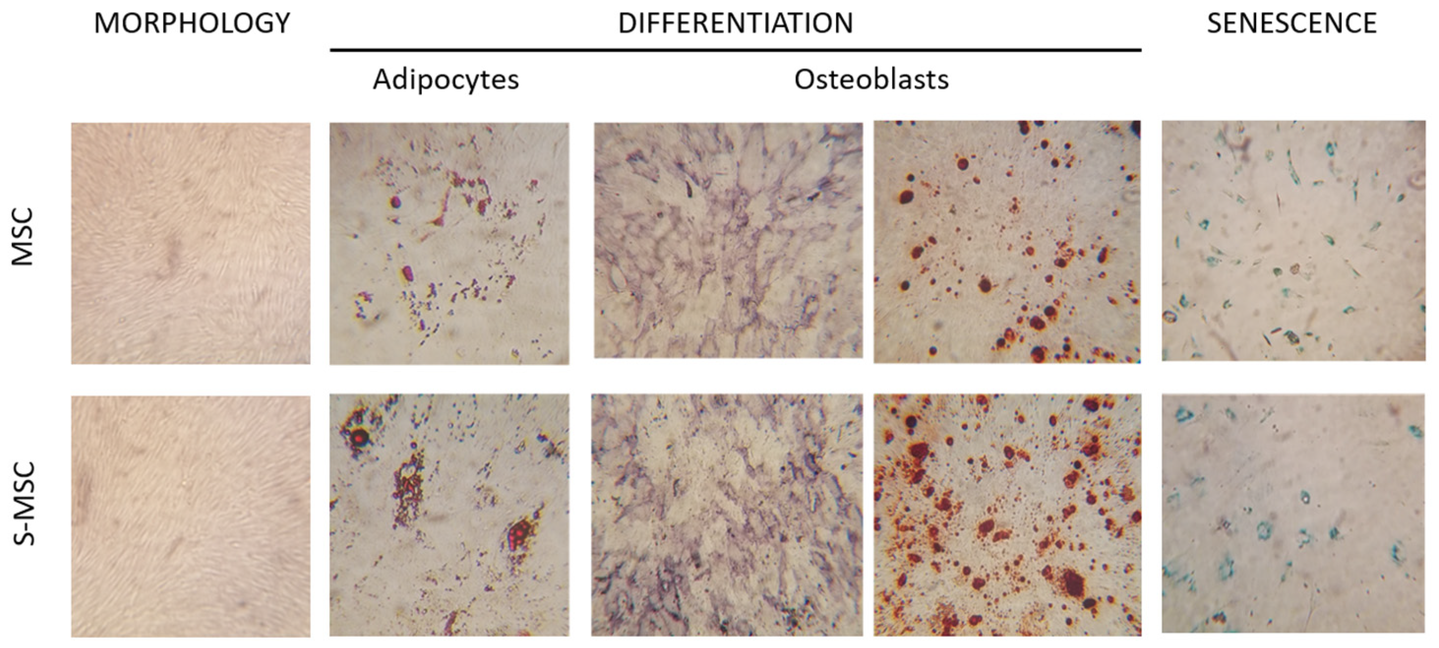
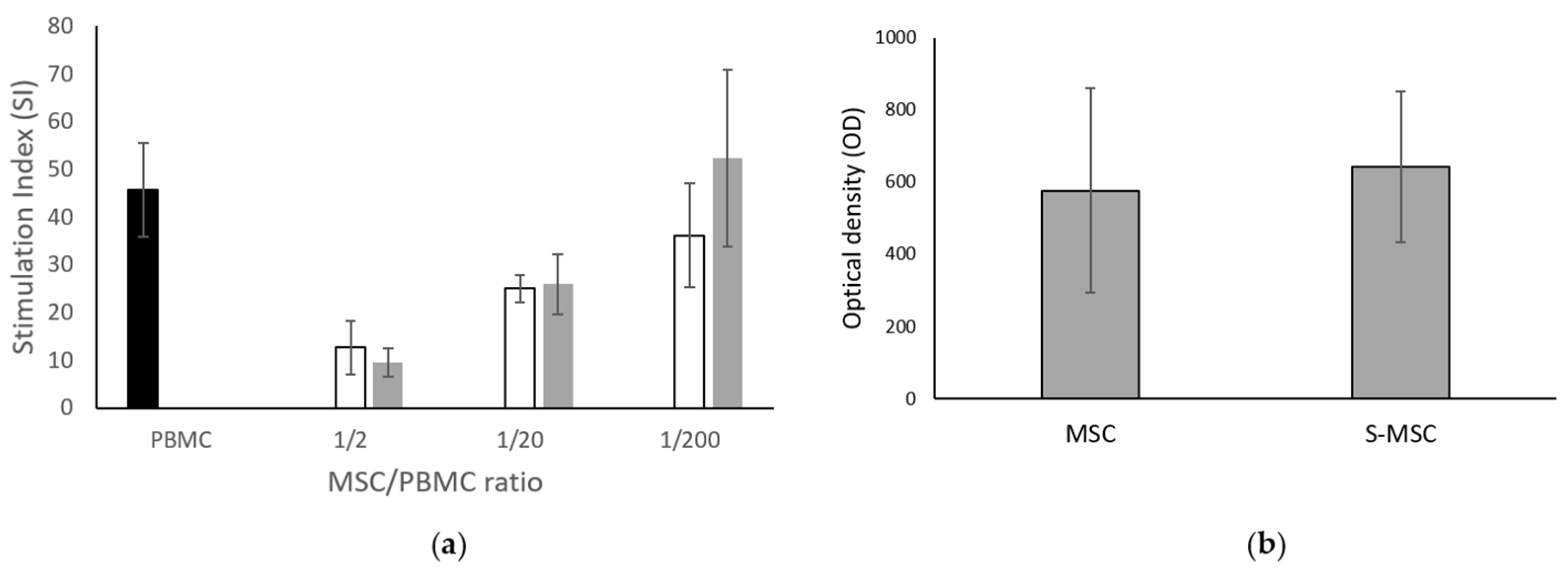
3.1. MSC and S-MSC Characterization

After culture condition restoration post 24 h starvation, S-MSCs like MSCs were plastic adherent and showed the typical spindle-shaped morphology (Figure 1). They maintained the in vitro differentiation capacity towards adipocytes and osteoblasts as demonstrated by the appearance of lipid droplets and the detection of phosphatase alkaline activity and calcium depositions as their corresponding MSCs (Figure 1). S-MSCs entered senescence at P10, medium passage, (range 8–11) similar to MSC (P11, range 8–12).
S-MSCs expressed ≥95% of CD90, CD73, and CD105 surface antigens and ≤5% of CD34, CD45, CD19, and HLA-DR molecules as MSCs (Figure 2).
Moreover, S-MSCs maintained the in vitro immunomodulatory properties as those shown by their counterparts, without any significant difference between the groups. The capacity to downregulate the immune response, in particular, the activation of T lymphocytes by a polyclonal activator, is evidenced by the reduction in the stimulation index (Figure 3a).
For the metabolic activity detected by the MTT assay, no significant difference was found between the two groups (mean value for MSCs and S-MSCs: 576 ± 283 and 641 ± 209, respectively), as described in Figure 3b.

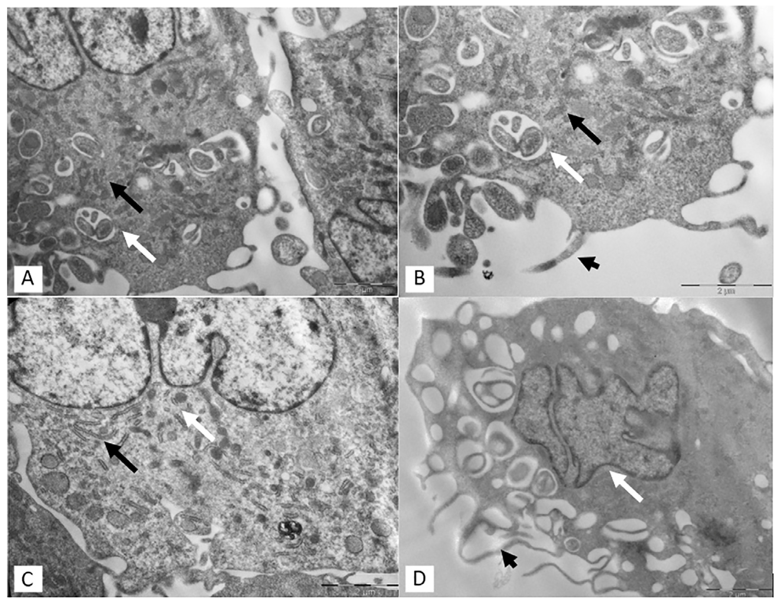
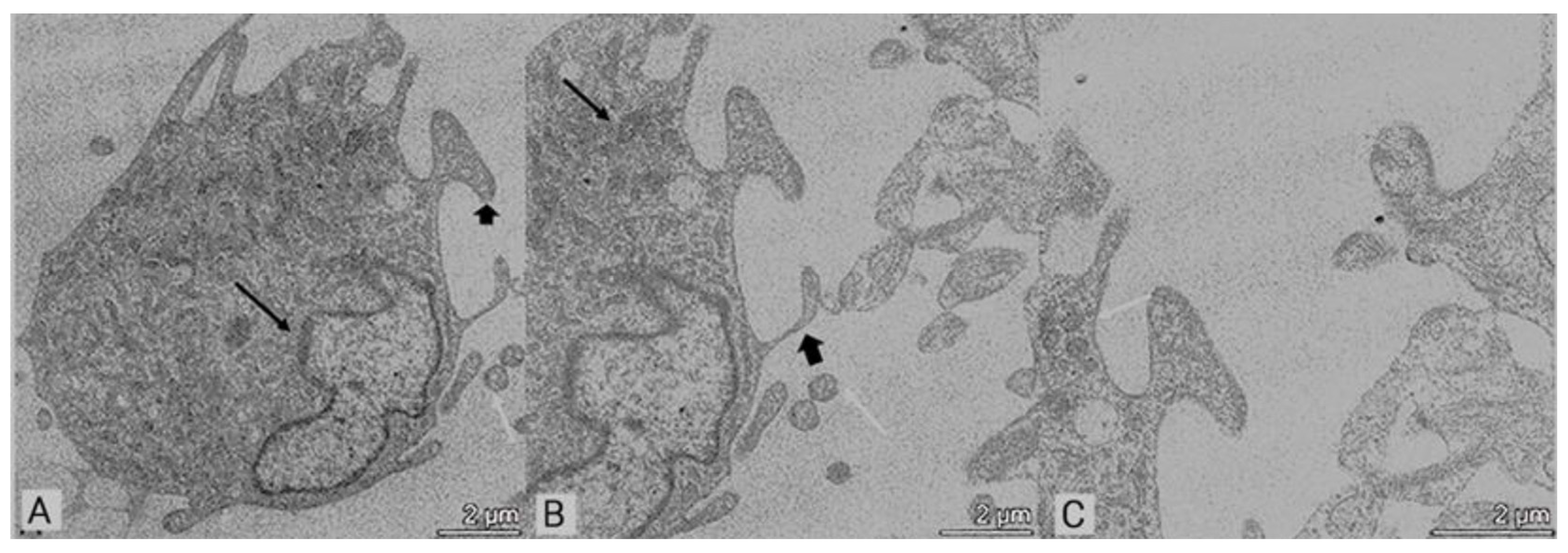
3.2. Transmission Electron Microscopy (TEM)

In this study, TEM was considered as a standard imaging method for observing nanosized samples, allowed to reveal inner cell and EV structures. The TEM images of non-starved MSCs showed polymorphic and large euchromatic nuclei with prominent nucleoli, visible in Figure 4A,C,D. In cytoplasm, mainly alongside the nucleus, well-developed dilated cisternae of rough endoplasmic reticulum (RER), formed by broad vesicles, were observed (Figure 4A,B). Multivesicular bodies (MVBs) loaded with intraluminal vesicles were in appreciable number inside the cell cytoplasm, as shown in Figure 4A, and closely packed-vesicles were detectable in Figure 4B. Protrusions of the plasma membranes, filopodia, were detectable in Figure 4B,D. The presence of wide vacuoles and vesicles gave the periphery of the cytoplasm a multilocular appearance (Figure 4D).
In order to evaluate the cellular structure and EV release, we performed TEM analysis after 12, 24, and 48 h of starvation (Figure 5). It was possible to highlight the budding process of cell membranes that released EVs with the typical morphology characterized by a lipid bilayer covering a specific payload of biomolecules, and a dilated RER was also observable.
Ultrastructural differences due to an increase in the starvation time were detectable, such as EV heterogeneous populations in terms of size and density (Figure 5). After 12 h starvation (Figure 5A), the amount of EVs was lower, and EVs seem to be less preserved compared to those observed after 24 h (Figure 5B). The same results were observed in cells starved for 48 h (Figure 5C). Moreover, after 48 h starvation, EVs seemed “wilted”, and in the area observed, a significant cell membrane blebbing was evident. In non-starved condition, EV release was not evaluable in MSCs, as already shown in Figure 4.
In order to evaluate if it is possible to restore S-MSCs after starvation, we added complete medium for further 24 h, and the restoration of culture conditions allowed us to find restored cells with detectable RER, mitochondria, and nucleus (Figure 6A). MVBs are smaller and less numerous with respect to those observed in MSCs, as described in Figure 4. Multiple projection extending from plasma membrane and some EVs in the extracellular space were also detectable (Figure 6B,C). However, the 48 h starvation exhibited detrimental results, demonstrated by “ghosts” outside the plasma membrane (Figure 7A) and by mitochondria with enlarged cristae (Figure 7B). No EVs were detectable inside cell cytoplasm or in the extracellular space.

4. Discussion

It is known that MSCs act by paracrine components represented by bioactive molecules that are secreted in response to the surrounding microenvironment. Over the last decade, the role of MSC secretoma, with particular attention provided to extracellular vesicles (EVs), has emerged as a convenient alternative to cell therapy. Several functions of EVs have been demonstrated, such as the capability to regulate the balance between pro-inflammatory and anti-inflammatory cytokines [16] and to modulate fibrosis and tissue regeneration after damage [17]. Different preconditioning strategies, such as oxygen percentage and nutrient composition of medium, can affect the metabolic activities of MSCs and the characteristics of the produced and released biomolecules [5,6,7,8]. For example, EVs derived from MSCs cultured under hypoxia showed an increased tube formation capacity, becoming more potent in angiogenesis compared to EVs obtained under normoxic conditions [18]. In addition, EVs from hypoxic MSCs regulated miRNA and proteins involved in extracellular matrix–receptor interaction, focal adhesion, leukocyte transendothelial migration, protein digestion, absorption, and metabolic pathways [19]. Moreover, EVs secreted by MSCs after serum starvation showed immunomodulatory capacity, eliciting the switch from M1 to M2 phenotype of bone marrow-derived macrophages [16]. Similar results were reported for EVs released by MSCs cultured in hypoxia and in presence of TNFα and IL1α, to mimic the inflammatory state of the injured environment. The EVs obtained under these culture conditions modulated the macrophages, exerting an anti-inflammatory activity [20]. Moreover, it has been reported that EVs from umbilical cord-derived MSCs (UC-MSCs), cultured in serum-free conditions, were loaded with increased levels of regeneration-related cytokines, compared to EVs derived from UC-MSCs cultured in normal conditions [21]. Even if recent studies related to enhanced EV production by the application of mechanical stimuli, including flow and stretching, in bioreactors to engineered tissues seeded with stem cells [22], or commercially accessible track-etched membranes to generate EVs containing therapeutic mRNAs by cellular nanoelectroporation [23], we underline the safety and accessibility of starvation as a more suitable method for EV bioproduction.
On the other hand, all the preconditioning culture conditions described for EV release have been demonstrated to promote and increase proliferation, viability, and potency of MSCs, favoring the expression of cytoprotective genes, as well as improving the secretion of reparative factors [24,25]. In particular, it has been demonstrated that MSCs in the absence of oxygen can survive using anaerobic ATP production. Further observations, such as the rapid death of MSCs following glucose deprivation but the slower depletion of cellular ATP under hypoxic conditions, suggested that glycolysis has specific pro-survival functions. Moreover, exposed MSCs maintained the differentiation capacity, highlighting their metabolic flexibility, which makes them able to survive in an ischemic environment [26]. Recently, it has been reported that adipose tissue-derived MSCs exposed to different stress conditions, such as hypoxia, starvation, or TNFα treatment, exhibited an increased immunomodulatory potency if maintained in starvation compared to the other culture conditions [6]. Since it has been reported that the in vivo transplanted MSCs are exposed to an ischemic habitat characterized by nutrient deprivation, the in vitro “starvation” could be considered the reproduction of the in vivo post-transplantation condition and a way to positively modify the MSC functions acting on immunomodulatory [6], antitumoral [7], and differentiation potential [12,13]. Our results fit well in this context, and our observations are in agreement with data reported by other authors. MSCs cultured under starvation maintained all the characteristics such as morphology, antigen surface expression, and immunomodulatory and differentiation capacities. The increasing trend in the metabolic activity observed in S-MSCs compared to MSCs indicated that, despite the induced reduction in basal respiration and ATP production, they still respond to changes in energy demand. This metabolic phenotype correlates with the obtained evidence that rarefied and enlarged mitochondrial cristae upon starvation are observed under electron microscope. The results obtained by TEM showed that 24 h starvation seems to be the best time for MSC structure preservation and for in vivo transfer of their contents to the target cells.
The results of our study underling the maintenance of MSC capacities after the starvation to induce EV release could suggest the possibility to still consider starved MSCs as pharmaceutical products.
However, further studies are needed to support our hypothesis and a deep investigation on how starvation can influence the different cell paracrine activities is fundamental to provide new insights for their therapeutic use.

Author Contributions

Conceptualization, A.L.A.; methodology, E.L.; investigation, S.C. and C.V.; data curation, A.C.; writing—original draft preparation, C.V. and S.C.; writing—review and editing, M.G., M.Z., and T.R.; visualization, M.A.G.; supervision, M.A.A. and P.C.; funding acquisition, M.A.A. and T.R. All authors have read and agreed to the published version of the manuscript.

Funding

This research was supported by a grant, grant number 80380, to MAA from Fondazione IRCCS Policlinico San Matteo Pavia, POR-FERS 2526393 Force4cure to PC from Regione Lombardia, and by a grant, grant number 08054221, to TR from Fondazione IRCCS Policlinico San Matteo Pavia.

Institutional Review Board Statement

The healthy bone marrow donors, included in the protocol code 2011002429 (date of approval: 6 June 2011), provided their written informed consent for the use of leftovers for research purposes.

Informed Consent Statement

Informed consent was obtained from all subjects involved in the study.

Data Availability Statement

Data is contained within the article.

Conflicts of Interest

The authors declare no conflicts of interest.

References

  1. Keshtkar, S.; Azarpira, N.; Ghahremani, M.H. Mesenchymal stem cell-derived extracellular vesicles: Novel frontiers in regenerative medicine. Stem Cell Res. Ther. 2018, 9, 63. [Google Scholar] [CrossRef] [PubMed]
  2. Lotfy, A.; AboQuella, N.M.; Wang, H. Mesenchymal stromal/stem cell (MSC)-derived exosomes in clinical trials. Stem Cell Res. Ther. 2023, 14, 66. [Google Scholar] [CrossRef]
  3. Tang, Y.; Zhou, Y.; Li, H.J. Advances in mesenchymal stem cell exosomes: A review. Stem Cell Res. Ther. 2021, 12, 71. [Google Scholar] [CrossRef]
  4. Manzoor, T.; Saleem, A.; Farooq, N.; Dar, L.A.; Nazir, J.; Saleem, S.; Ismail, S.; Gugjoo, M.B.; Shiekh, P.A.; Ahmad, S.M. Extracellular vesicles derived from mesenchymal stem cells—A novel therapeutic tool in infectious diseases. Inflamm. Regen. 2023, 43, 17. [Google Scholar] [CrossRef]
  5. Patel, D.B.; Gray, K.M.; Santharam, Y.; Lamichhane, T.N.; Stroka, K.M.; Jay, S.M. Impact of cell culture parameters on production and vascularization bioactivity of mesenchymal stem cell-derived extracellular vesicles. Bioeng. Trans. Med. 2017, 2, 170–179. [Google Scholar] [CrossRef]
  6. Vu, B.T.; Le, H.T.; Nguyen, K.N.; Van Pham, P. Hypoxia, Serum starvation, and TNF-a can modify the immunomodulation potency of human adipose-derived stem cells. In Advances in Mesenchymal Stem Cells and Tissue Engineering; Springer: Berlin/Heidelberg, Germany, 2021. [Google Scholar]
  7. Jung, P.Y.; Ryu, H.; Rhee, K.J.; Hwang, S.; Lee, C.G.; Gwon, S.Y.; Kim, J.; Kim, J.; Yoo, B.S.; Baik, S.K.; et al. Adipose tissue-derived mesenchymal stem cells cultured at high density express IFN-b and TRAIL and suppress the growth of H460 human lung cancer cells. Cancer Lett. 2019, 202, 440–441. [Google Scholar] [CrossRef]
  8. Haraszti, R.A.; Miller, R.; Dubuke, M.L.; Rockwell, H.E.; Coles, A.H.; Sapp, E.; Didiot, M.C.; Echeverria, D.; Stoppato, M.; Sere, Y.Y.; et al. Serum deprivation of mesenchymal stem cells improves exosome activity and alters lipid and protein composition. iScience 2019, 16, 230–241. [Google Scholar] [CrossRef]
  9. Oskowitz, A.; McFerrin, H.; Gutschow, M.; Carter, M.L.; Pochampally, R. Serum-deprived human multipotent mesenchymal stromal cells (MSCs) are highly angiogenic. Stem Cell Res. 2011, 6, 215–225. [Google Scholar] [CrossRef] [PubMed]
  10. Bakopoulou, A.; Kritis, A.; Andreadis, D.; Papachristou, E.; Leyhausen, G.; Koidis, P.; Geurtsen, W.; Tsiftsoglou, A. Angiogenic potential and secretome of human apical papilla mesenchymal stem cells in various stress microenvironments. Stem Cells Dev. 2015, 24, 2496–2512. [Google Scholar] [CrossRef] [PubMed]
  11. Hahm, J.; Kim, J.; Park, J. Strategies to enhance extracellular vesicle production. Tissue Eng. Regen. Med. 2021, 18, 513–524. [Google Scholar] [CrossRef]
  12. Binder, B.Y.; Sagun, J.E.; Leach, J.K. Reduced serum and hypoxic culture conditions enhance the osteogenic potential of human mesenchymal stem cells. Stem Cell Rev. Rep. 2015, 11, 387–393. [Google Scholar] [CrossRef]
  13. Kim, J.H.; Shin, S.H.; Li, T.Z.; Suh, H. Influence of in vitro biomimicked stem cell ‘niche’ for regulation of proliferation and differentiation of human bone marrow-derived mesenchymal stem cells to myocardial phenotypes: Serum starvation without aid of chemical agents and prevention of spontaneous stem cell transformation enhanced by the matrix environment. J. Tissue Eng. Regen. Med. 2016, 10, E1–E13. [Google Scholar]
  14. Avanzini, M.A.; Bernardo, M.E.; Cometa, A.M.; Perotti, C.; Zaffaroni, N.; Novara, F.; Visai, L.; Moretta, A.; Del Fante, C.; Villa, R.; et al. Generation of mesenchymal stromal cells in the presence of platelet lysate: A phenotypic and functional comparison of umbilical cord blood- and bone marrow-derived progenitors. Haematologica 2009, 94, 1649–1660. [Google Scholar] [CrossRef]
  15. Dominici, M.; Le Blanc, K.; Mueller, I.; Slaper-Cortenbach, I.; Marini, F.; Krause, D.; Deans, R.; Keating, A.; Prockop, D.; Horwitz, E. Minimal criteria for defining multipotent mesenchymal stromal cells. The International Society for Cellular Therapy position statement. Cytotherapy 2006, 8, 315–317. [Google Scholar] [CrossRef]
  16. Lo Sicco, C.; Reverberi, D.; Balbi, C.; Ulivi, V.; Principi, E.; Pascucci, L.; Becherini, P.; Bosco, M.C.; Varesio, L.; Franzin, C.; et al. Mesenchymal Stem Cell-Derived Extracellular Vesicles as Mediators of Anti-Inflammatory Effects: Endorsement of Macrophage Polarization. Stem Cells Transl. Med. 2017, 6, 1018–1028. [Google Scholar] [CrossRef]
  17. Magarotto, F.; Sgrò, A.; Dorigo Hochuli, A.H.; Andreetta, M.; Grassi, M.; Saggioro, M.; Nogara, L.; Tolomeo, A.M.; Francescato, R.; Collino, F.; et al. Muscle functional recovery is driven by extracellular vesicles combined with muscle extracellular matrix in a volumetric muscle loss murine model. Biomaterials 2021, 269, 120653. [Google Scholar] [CrossRef]
  18. Almeria, C.; Weiss, R.; Roy, M.; Tripisciano, C.; Kasper, C.; Weber, V.; Egger, D. Hypoxia Conditioned Mesenchymal Stem Cell-Derived Extracellular Vesicles Induce Increased Vascular Tube Formation in vitro. Front. Bioeng. Biotechnol. 2019, 7, 292. [Google Scholar] [CrossRef]
  19. Gregorius, J.; Wang, C.; Stambouli, O.; Hussner, T.; Qi, Y.; Tertel, T.; Börger, V.; Mohamud Yusuf, A.; Hagemann, N.; Yin, D.; et al. Small extracellular vesicles obtained from hypoxic mesenchymal stromal cells have unique characteristics that promote cerebral angiogenesis, brain remodeling and neurological recovery after focal cerebral ischemia in mice. Basic. Res. Cardiol. 2021, 116, 40. [Google Scholar] [CrossRef]
  20. Gorgun, C.; Africano, C.; Ciferri, M.C.; Bertola, N.; Reverberi, D.; Quarto, R.; Ravera, S.; Tasso, R. Preconditioned Mesenchymal Stromal Cell-Derived Extracellular Vesicles (EVs) Counteract Inflammaging. Cells 2022, 11, 3695. [Google Scholar] [CrossRef]
  21. Kim, J.Y.; Rhim, W.K.; Seo, H.J.; Lee, J.Y.; Park, C.G.; Han, D.K. Comparative Analysis of MSC-Derived Exosomes Depending on Cell Culture Media for Regenerative Bioactivity. Tissue Eng. Regen. Med. 2021, 18, 355–367. [Google Scholar] [CrossRef]
  22. Guo, S.; Debbi, L.; Zohar, B.; Samuel, R.; Arzi, R.S.; Fried, A.I.; Carmon, T.; Shevach, D.; Redenski, I.; Schlachet, I.; et al. Stimulating Extracellular Vesicles Production from Engineered Tissues by Mechanical Forces. Nano Lett. 2021, 21, 2497–2504. [Google Scholar] [CrossRef] [PubMed]
  23. Ma, Y.; Sun, L.; Zhang, J.; Chiang, C.; Pan, J.; Wang, X.; Kwak, K.J.; Li, H.; Zhao, R.; Rima, X.Y.; et al. Exosomal mRNAs for Angiogenic–Osteogenic Coupled Bone Repair. Adv. Sci. 2023, 10, 2302622. [Google Scholar] [CrossRef] [PubMed]
  24. Silva, L.H.A.; Antunes, M.A.; Dos Santos, C.C.; Weiss, D.J.; Cruz, F.F.; Rocco, P.R.M. Strategies to improve the therapeutic effects of mesenchymal stromal cells in respiratory diseases. Stem Cell Res. Ther. 2018, 9, 45. [Google Scholar] [CrossRef]
  25. Moya, A.; Larochette, N.; Paquet, J.; Deschepper, M.; Bensidhoum, M.; Izzo, V.; Kroemer, G.; Petite, H.; Logeart-Avramoglou, D. Quiescence Preconditioned Human Multipotent Stromal Cells Adopt a Metabolic Profile Favorable for Enhanced Survival under Ischemia. Stem Cells 2017, 35, 181–196. [Google Scholar] [CrossRef] [PubMed]
  26. Mylotte, L.A.; Duffy, A.M.; Murphy, M.; O’Brien, T.; Samali, A.; Barry, F.; Szegezdi, E. Metabolic flexibility permits mesenchymal stem cell survival in an ischemic environment. Stem Cells 2008, 26, 1325–1336. [Google Scholar] [CrossRef]
Figure 1. MSC and S-MSC characterization. Morphology, differentiation, and senescence of representative MSC and S-MSC samples.
Applsci 14 05821 g001
Figure 2. Flow cytometer analysis of MSCs and S-MSCs. Gate A defines MSCs according to their physical parameters (FSC-A and SSC-A). MSC and S-MSC stained with specific antibodies were positive for CD73, CD105, and CD90 and negative for CD34, CD19, HLA-DR, and CD45.
Applsci 14 05821 g002
Figure 3. MSC functionality. (a) PHA-activated PBMC modulation by MSCs and S-MSCs at different ratios. Black bar= PHA-activated PBMC alone; white bars = PHA-activated PBMC in presence of MSCs at different ratios; gray bars= PHA-activated PBMC in presence of S-MSCs at different ratios. Results are expressed as SI = cpm stimulated/cpm unstimulated. (b) Mitochondrial activity evaluated by MTT assay in MSC and S-MSC. Results are expressed as OD. No statistical significance was observed.
Applsci 14 05821 g003
Figure 4. Representative TEM images of non-starved MSCs. (AC) the cisternae of rough endoplasmic reticulum (RER) are clearly detectable (black arrows). In (A,B), different MVBs with a characteristic cup shape in the cytoplasm are contained within a membrane boundary (white arrows). (B,D) the protrusion of the plasmatic membrane (filopodia) is evidenced by arrowhead. In (C,D), a convoluted nucleus (black arrows) and some MVBs in (D) (white arrows) are also detectable. Bar = 2 µm.
Applsci 14 05821 g004
Figure 5. Representative TEM of S-MSCs releasing EVs after different times of starvation. (A) After 12 h starvation, EVs are detectable near the plasmatic membrane (arrow), and a nucleus with a prominent nucleolus is also visible (arrowhead); (B) after 24 h starvation, EVs protrude from the plasmatic membrane into the extracellular space; the heterogeneous population of vesicles is visible (arrow), and a dilated RER and some vacuoles are observed in the cytoplasm (arrowhead); (C,D) after 48 h starvation, EVs seem to be wilted, and there is a significant membrane blebbing (arrow); a RER with dilatated cisternae is also detectable (arrowhead). Bar = 2 µm.
Applsci 14 05821 g005
Figure 6. Representative TEM images showing 24 h S-MSCs after restoration with complete medium. (AC) show a well-conserved cellular morphology with a prominent nucleus (arrow); multiple projection (arrowhead) extending to the nearby cells, and some EVs in the extracellular space are detectable (white arrow); some mitochondria in B and MVBs (white arrow). Bar = 2 µm.
Applsci 14 05821 g006
Figure 7. Representative TEM images showing 48 h S-MSCs after restoration. (A) A restored cell with “ghosts” detectable outside the plasmatic membrane (arrow) and bleb (arrowhead), and many vacuoles are also detectable inside cytoplasm. (B) Multiloculated vacuoles (arrowhead) and mitochondria with enlarged cristae (arrow), bar = 1 µm.
Applsci 14 05821 g007
Disclaimer/Publisher’s Note: The statements, opinions and data contained in all publications are solely those of the individual author(s) and contributor(s) and not of MDPI and/or the editor(s). MDPI and/or the editor(s) disclaim responsibility for any injury to people or property resulting from any ideas, methods, instructions or products referred to in the content.

