Article

Design and Development of a Flexible 3D-Printed Endoscopic Grasping Instrument

1
Interventional and Experimental Endoscopy (InExEn), Department of Internal Medicine II, University Hospital Wurzburg, 97080 Würzburg, Germany
2
Department of Surgery, University of Pecs, 7622 Pécs, Hungary
*
Author to whom correspondence should be addressed.
Appl. Sci. 2023, 13(9), 5656; https://doi.org/10.3390/app13095656
Submission received: 29 March 2023 / Revised: 25 April 2023 / Accepted: 3 May 2023 / Published: 4 May 2023

Abstract

:
(1) Background: Interventional endoscopic procedures are growing more popular, requiring innovative instruments and novel techniques. Three-dimensional printing has demonstrated great potential for the rapid development of prototypes that can be used for the early assessment of various concepts. In this work, we present the development of a flexible endoscopic instrument and explore its potential benefits. (2) Methods: The properties of the instrument, such as its maneuverability, flexibility, and bending force, were evaluated in a series of bench tests. Additionally, the effectiveness of the instrument was evaluated in an ex vivo porcine model by medical experts, who graded its properties and performance. Furthermore, the time necessary to complete various interventional endoscopic tasks was recorded. (3) Results: The instrument achieved bending angles of ±216° while achieving a bending force of 7.85 (±0.53) Newtons. The time needed to reach the operating region was 120 s median, while it took 70 s median to insert an object in a cavity. Furthermore, it took 220 s median to insert the instrument and remove an object from the cavity. (4) Conclusions: This study presents the development of a flexible endoscopic instrument using three-dimensional printing technology and its evaluation. The instrument demonstrated high bending angles and forces, and superior properties compared to the current state of the art. Furthermore, it was able to complete various interventional endoscopic tasks in minimal time, thus potentially leading to the improved safety and effectiveness of interventional endoscopic procedures in the future.

1. Introduction

Minimally invasive therapeutic technology is currently emerging in all areas of operative medicine due to the benefits it delivers to patients, such as reduced scarring and recovery time [1,2,3,4,5,6,7,8,9]. Furthermore, it allows physicians to perform novel procedures that were not possible otherwise. Interventional therapeutic endoscopy was able to replace several surgical procedures within the therapeutic spectrum of disease management in the upper and lower gastrointestinal (GI) tract [4,5,6,7]. Consequently, more complex procedures can be performed by the endoscopists, such as the endoscopic resection of GI tumors, endoscopic myotomy, plication, and GI wall closure [4,5,6,7,9,10,11].
The constant flow of innovative ideas and novel concepts regarding therapeutic procedures requires a prompt response from the side of engineers to develop devices to fulfill these clinical needs. The early testing of such innovative ideas allows for their selection, and they may have a high potential of becoming successful in future clinical practice. To achieve this, technologies based on rapid prototyping have emerged in several segments of medical device development to enhance the efficiency and effectiveness of the process. Three-dimensional (3D) printing has attracted significant research interest over the past decade regarding rapid prototype development to solve different clinical problems encountered in disease management [12,13,14,15,16,17,18].
Our working group is a multidisciplinary team of gastroenterologists, surgeons, and engineers involved in improvements in flexible endoscopic technology and procedures by applying the concept of “endoneering” [19,20]. In recent years, several attempts have been performed to improve endoscopic instruments and procedures, based on input from clinical experts [17,21,22,23,24]. The concept of tight collaboration between medical experts and engineers is continually expanding due to the rising demand for precisely tailored solutions to various clinical problems and needs.
Nonetheless, current state-of-the-art endoscopic methods for performing interventional therapeutic procedures have several limitations. Most of the concepts are based on the use of the endoscope itself as a foundation and expanding its functionality through attaching various over-tubes or add-on caps to it. Such over-tubes and add-on caps are restricted by the capabilities of the endoscope used and often require extensive assembly times. Endoscopic instruments that can be inserted and used independently alongside the endoscope are usually rare and very costly. This makes them unappealing for daily clinical use where costs play a significant role [17,25,26,27,28]. Furthermore, state-of-the-art instruments lack the flexibility and the ability to achieve high bending angles at the distal tip, usually not bending more than 90°. The lack of such features limits the number of options physicians have when treating patients with different conditions. Addressing these limitations can greatly enhance the accuracy and success of complex endoscopic procedures [29,30].
In the present work, we explored viable rapid prototyping solutions for the above-mentioned limitations of the current state-of-the-art devices. The aim was to design an affordable instrument based on the combination of flexible endoscopic technology with laparoscopic elements to allow for a novel approach in interventional endoscopy using minimally invasive surgical concepts [29,30,31,32,33]. To achieve this, we relied on rapid prototyping methodologies to develop an affordable prototype instrument tailored for use in interventional therapeutic endoscopic procedures. Additionally, we evaluated the performance of the instrument in a series of bench tests and assessments conducted in a porcine explant model.

2. Materials and Methods

2.1. Instrument Design

The design of the 3D-printed flexible grasping instrument was created in collaboration with experienced gastroenterologists and surgeons who shared their clinical needs and current technological limitations. Based on their insights, a set of requirements for a prototype of the instrument was established. The instrument should offer physicians additional capabilities for manipulating objects and tissue in the upper and lower gastrointestinal (GI) tract, ultimately improving the outcome of various endoscopic procedures.
In order to meet the requirements of medical professionals, the instrument needed to incorporate features from both laparoscopy and flexible endoscopy. The distal tip of the instrument had to provide a secure and long-lasting grip while still remaining gentle enough to handle pathologically important tissue without causing any damage. This is particularly important in the case of endoscopic procedures, which require precise and careful handling to avoid injuries and perforation. To achieve this, the instrument had to be designed with an adequately large grasping area and a gentle rounded distal tip.
In addition to its grasping capabilities, the instrument must also be highly maneuverable. The distal tip should have a high range of bending, allowing steering angles greater than 180°. This is necessary to ensure that physicians can access and manipulate target tissue from a variety of angles, as well as to navigate through the twists and turns of the GI tract. Furthermore, the instrument must also be independent of the endoscope and its vision, allowing for better traction and control during procedures.
To ensure that the instrument is adaptable to different procedural needs, it must have an appropriate length, which can be adjusted by using a longer or shorter flexible shaft and corresponding wires. This allows the customization of the instrument based on the different procedure-specific demands, such as operating in the upper or lower GI tract.
Additionally, the instrument should be affordable to produce and intuitive to control and use. By incorporating all these requirements, the new prototype provides physicians with a range of features for improving and performing endoscopic procedures in a safe, effective, and precise manner. Table 1 provides a detailed breakdown of the requirements for each element of the instrument.

2.1.1. Grasping Forceps

The distal tip of the instrument is based on a single-action Johan grasper, which is specifically designed to allow for the gentle manipulation of fragile and histologically important tissue. Additionally, the grasping forceps are used for effectively placing sponges and other objects into leak cavities.
To ensure that the instrument can properly grasp objects, a wide opening angle is necessary. However, achieving this wide opening angle can be challenging in the narrow space of the GI tract, which can be as narrow as 15–20 mm in the esophageal region. To balance the need for a wide opening angle with the narrow operational space, an opening angle of 65° was adopted. This angle allows the instrument to grasp objects securely while still being able to maneuver in the narrow spaces of the GI tract. The grasping forceps of the instrument have a total length of 22 mm and an outer diameter of 5 mm, making them compact and easy to use in endoscopic procedures.
To ensure that they are effective at grasping objects, a minimal required grasping force of 10 N was adopted. This value was determined based on the average force required for grasping epithelial tissue (3.8 N) and muscle tissue (4.1 N), with maximum values of up to 9.7 N [34]. Overall, the design of the distal tip of the instrument is balanced to ensure that it can effectively grasp objects and handle tissue while still being maneuverable in the narrow spaces of the GI tract.

2.1.2. Bending Section

To enable the distal tip of the instrument to be steered, a bending section composed of multiple linked segments was developed. The main goal of the bending section was to achieve a high bending angle that would enable the effective manipulation of the instrument and to be directly controlled by a physician. Additionally, the bending radius of the instrument had to be small to ensure that it could be effectively used in the narrow space of the GI tract. A bending radius of 6 mm was adopted.
To achieve the desired performance, the bending section was composed of seven serially assembled 3D-printed segments. Several forms of segmented bending sections driven by tendon wires have been developed and analyzed by previous works [35,36,37]. The design of the bending section allows for a maximum bending angle of 216° in both directions, which provides physicians with a high degree of flexibility when manipulating the instrument during endoscopic procedures. Bending in a single plane was accepted for the instrument since any additional rotation or translation could be achieved by twisting or pushing the flexible shaft of the instrument.
Overall, the design of the bending section of the instrument was carefully balanced to ensure that it could achieve a high bending angle while still being effective in the narrow passages of the GI tract. By using multiple linked segments, a high degree of flexibility was achieved, while the small bending radius and the ability to manipulate the instrument in a single plane ensure effective use during endoscopic procedures (Figure 1).

2.1.3. Flexible Shaft

The flexible shaft is an essential part of the instrument, which connects the control handle to the distal tip. Its length is critical to the instrument’s performance as it determines the reach and accessibility to the target operating region. A highly flexible shaft enables easy navigation around anatomical landmarks, while sufficient stiffness is necessary for effective tissue manipulation and object handling. The flexible shaft was developed to achieve a balance between these two properties.
The shaft’s torsional rigidity should also be strong enough to overcome friction and allow for the smooth rotation of the distal tip in both directions. Additionally, the flexible shaft facilitates the uninterrupted transfer of movement from the handle to the distal tip to ensure efficient instrument control.
The flexible shaft of the instrument is produced from a flat steel spiral coated with a layer of biocompatible polyvinyl chloride (PVC). The diameter of the shaft is 5 mm, and the length can be varied between 30 and 200 cm, depending on the intended use. A set of wires is routed through the flexible shaft to facilitate the steering of the bending section and opening of the grasping jaw. This design ensures that the instrument can be easily maneuvered, and the physician can accurately control the movements of the distal tip, even in the narrow regions of GI tract.

2.1.4. Control Handle

The control handle of the instrument is a crucial part of the instrument, serving as the interface between the user and the distal tip. To ensure ease of use and familiarity for medical professionals, the handle was designed with similarities to regular endoscopy devices. A star knob concept, similar to that of an endoscope, is used for steering the distal tip of the instrument, while a push–pull slider was designed for opening and closing the grasper jaw. An exploded view of the control handle, including its elements, is shown in Figure 2.

2.2. Prototype Development

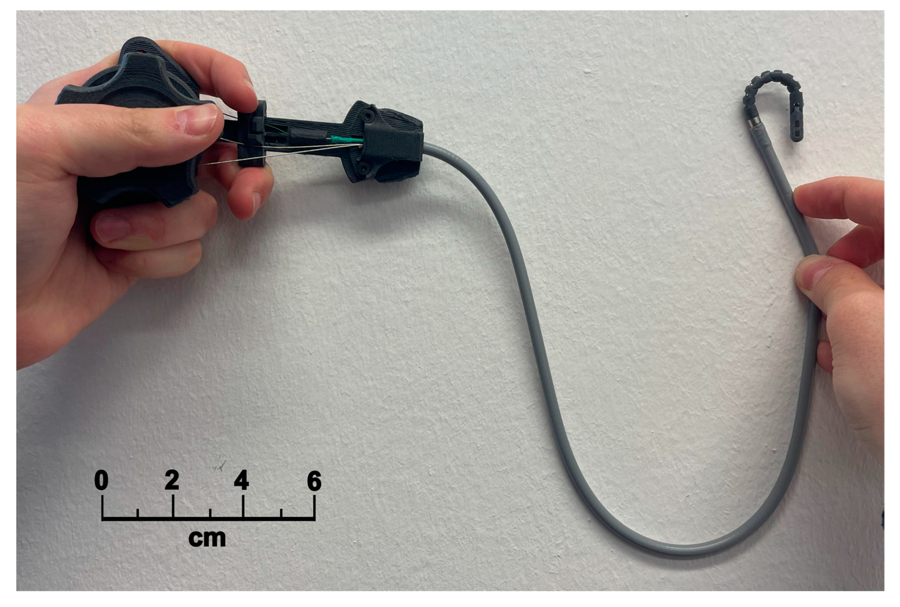
The adoption of 3D printing for prototype production is becoming increasingly popular due to its numerous advantages such as its low cost and minimum production time, and the wide range of materials available for 3D printing. The final prototype of the instrument, as depicted in Figure 3, displays the use of 3D printing technology to produce the distal tip and the control handle parts. The biocompatible BioMed Amber resin (Formlabs, Somerville, MA, USA) was used with the stereolithography 3D printer to fabricate the parts of the distal tip and grasper. The high accuracy of stereolithography 3D printers allows the production of highly optimized parts. Furthermore, the control handle parts were produced using a selective laser sintering process with Nylon PA 12 on Lisa Pro printer (Sinterit, Krakow, Poland), due to the adequate mechanical properties of this material. Approximately 2 h of labor was required for the complete assembly of the prototype.

2.3. Workspace of the Distal Tip

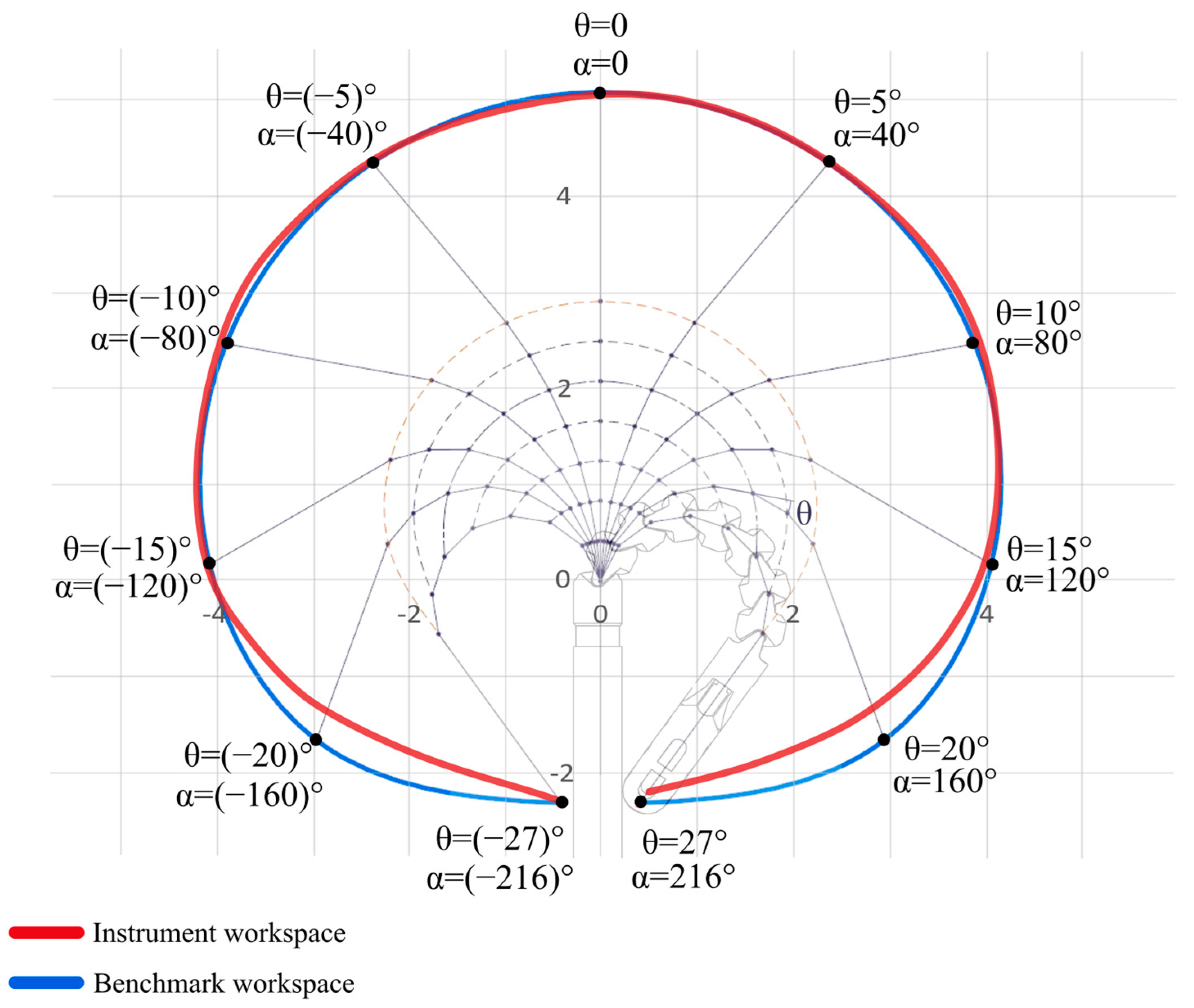
As a measure of maneuverability, the workspace of the instrument’s distal tip was mapped. The workspace is defined as the curve traced by the distal tip during bending, depicting its positions at different bending angles. To measure the workspace of the instrument, each segment of the distal tip was traced during bending, and points were marked on a millimeter paper. Based on the marked points for each segment, a set of polygons were generated, depicting the positioning of the distal tip at various bending angles.
Additionally, a benchmark workspace was modeled and used as the gold standard for comparison, as it represents the ideal range of motion of the designed distal tip. For modeling the benchmark workspace, several assumptions were considered. In particular, it was assumed that the bending is planar and gradual, and that the total length of the bending section does not change during bending.

2.4. Measurement of the Bending Force

In another bench test, the forces achieved by the instruments’ distal tip were measured and compared to those achieved by a combination of a gastroscope (Olympus GIF 190, Olympus Europa SE & Co. KG, Hamburg, Germany) and biopsy forceps (Radial Jaw 4—Standard Capacity, Boston Scientific, Marlborough, MA, USA). The combination of gastroscope and biopsy forceps is commonly used in endoscopic practice to perform different operational tasks in the upper GI tract, such as traction and tissue manipulation. A digital force gauge DFG-500 (PCE Instruments EURL, Soultz-sous-Forets, France) was used for measurements.
To perform the measurements of bending force, both the instrument and the gastroscope combined with biopsy forceps were fixed at the beginning of their bending sections with a table-clamping tool. The distal tips of both were left unrestrained to allow for full range of movement. To connect the grasping areas of both tools to the sensing shaft of the force gauge, a stiff steel wire was used. The wire was positioned perpendicular to the axes of the tools and then grasped. At last, the bending sections of the tools were maneuvered to pull the wire away from the force gauge, while the maximal achieved forces were recorded.

2.5. Performance Assessment in an Upper GI Porcine Model

To assess the instrument’s performance and properties inside the GI tract, an experiment was performed using an ex vivo porcine model, consisting of an esophagus and stomach. The porcine model was placed on a polystyrene (PS) board with a cutout shaped to resemble the upper GI tract. The PS board cutout was curved at its beginning to resemble a mouth and throat, with a cavity with a leak opening 35 cm away from the beginning. To provide access to the leak cavity on the PS board, a 1 cm hole was made in the esophagus of the porcine model.
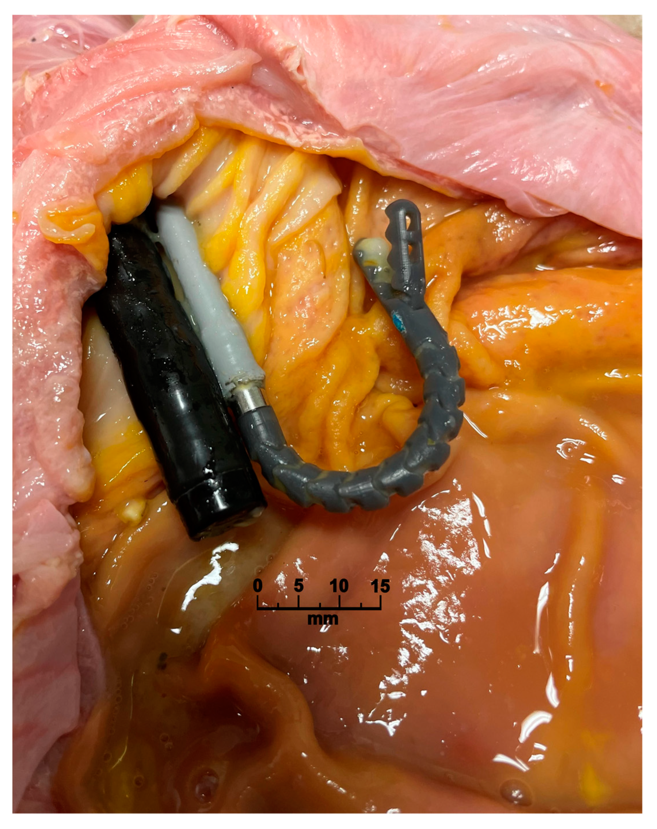
The instrument was inserted through the esophagus and advanced toward the leak opening. A 10.5 mm gastroscope was inserted alongside the instrument to provide the vision of the distal tip and operating area (Figure 4).
A series of tests were performed by a group composed of several physicians and engineers. A grading scale (0—insufficient, 1—limited, 2—sufficient, 3—good) was used to assess the performance of the prototype, as the properties of the instrument could not be accurately quantified inside the organs of the porcine model. Insufficient and limited properties were regarded as unacceptable for use and would require a significant improvement, while sufficient and good properties were regarded as acceptable for procedures in interventional endoscopy.
The following properties were assessed and graded:
  • Flexibility—the ability of the instrument to bend around the anatomical landmarks inside the porcine model.
  • Resistance to buckling—the ability to avoid bending (buckling) when pushed through the organ or when advancing an object to the leak-opening area.
  • Friction between instrument and organ surface.
  • The maneuverability of the distal tip inside the porcine model.
  • Torsional stiffness—the ability of the instrument to transfer the rotational movement from the handle to the distal tip.

2.6. Time Necessary to Perform Interventional Endoscopic Tasks in the Porcine Model

Additionally, we measured the time needed to perform different interventional tasks, including insertion, object manipulation, and object retrieval from the operating region. The same porcine model placed on the PS board was used and the instrument was inserted alongside the gastroscope. A sponge (1 cm³) was used as the object to be manipulated by the instrument. The following times were measured:
  • The time needed to insert and transport the object to the dedicated region. The sponge was grasped outside the porcine model and inserted together with the instrument and gastroscope until reaching the leak-opening region.
  • The time required to maneuver and place the object into the cavity space. When the dedicated region was reached, and leak opening was visible, the instrument would be bent into the cavity and used to fixate the sponge inside.
  • The time needed to grab and retract the object from the organ. The instrument was inserted into the organ without the sponge and then maneuvered into the cavity. Once the instrument was positioned, the already-placed sponge was grasped and completely removed from the organ.

2.7. Data Analysis

The analysis and visualization of recorded data were performed with MS Excel (Microsoft Corporation, Redmond, Washington, DC, USA) and Python (Python Software Foundation, Wilmington, NC, USA) using the NumPy, Pandas, and SciPy libraries. The normally distributed metrics are reported with mean values and their standard deviation (SD), while non-normally distributed metrics are reported as the median with corresponding interquartile range (IQR). The two-sample T-Test was used for normally distributed data sets to test if their distributions differed significantly. A p-value of 0.05 was adopted as the threshold of a significant difference.

2.8. Availability of the Instrument

The files necessary to reproduce the instrument (3D models, bill of materials and assembly instructions) are freely available to download at: https://www.ukw.de/research/inexen/code-data-and-3d-models/ (accessed on 2 May 2023).

3. Results

3.1. Workspace of the Distal Tip

The workspace curve of the instrument was mapped 10 times in total. The final curve was fitted through the mapped points, which is presented in red color in Figure 5. The instrument achieved the maximal bending angle of 216° in both directions, with a 6 mm bending radius. For absolute angles lower than 120°, the instrument workspace matched the benchmark one with deviations smaller than 1 mm. The first noticeable differences happened at absolute angles higher than 120°.
The highest deviation between the curves was recorded to be 3.4 mm at 170°. Furthermore, at the maximum bending angles, the two curves started to match each other again.

3.2. Measurement of the Bending Force

The maximum bending force was measured 30 times using both the instrument and the gastroscope–forceps combination. The mean bending force achieved by the instrument was 7.85 N (SD ± 0.53), while the gastroscope–forceps combination achieved a mean force of 7.68 N (SD ± 0.3). Although it was 3D-printed, the instrument demonstrated comparable bending force to its commonly used counterpart (p = 0.56).

3.3. Performance Assessment in the Upper GI Porcine Model

The instrument demonstrated the required flexibility, rigidity, maneuverability, and friction with the mucosal surface during the tests within the esophagus of the porcine model. A summary of the expert grading of the instrument’s performance is shown in Table 2.

3.4. Time Necessary to Perform Interventional Endoscopic Tasks in the Porcine Model

In total, 10 trials were performed with the instrument in the porcine model. The median time required to insert and transport an object from the organ entrance to the dedicated region was measured to be 120 (IQR: 90–140) seconds. No major obstructions or drawbacks were recorded during the experiment regarding the task. Once the object was carried to the dedicated region, about 70 (IQR: 60–110) seconds was needed to navigate through the leak opening in the organ and position the object inside the leak cavity. Finally, the median time necessary to insert, reach the cavity, grasp, and remove the object was measured to be 220 (IQR: 180–280) seconds.

4. Discussion

Interventional endoscopic procedures seem to have no limits regarding innovative procedures. The border between established surgical procedures and feasible endoscopic procedures is continually shifting [1,2,3,4,5,6,7,8,9,10,11,38]. An important result of this shift was the increased experience of physicians who perform complex endoscopic procedures. Recent advancements in interventional endoscopy have allowed for planned access to the abdominal cavity or the mediastinum, reducing fears of handling closure issues of the gut. Furthermore, clinical experience has shown that full-thickness lesions of the gut do not necessarily create problems if they undergo sufficient and competent closure. These developments have expanded the scope of endoscopic interventions, providing physicians with a wider range of options for the treatment of various conditions [11,29,30,31,38].
The latter development in interventional endoscopy has led to the need for special instruments that can solve rare clinical problems [39,40]. Ideally, one would need a stable grasper for the adequate endoscopic resection and improved maneuverability of the instrument distal tip for endoscopic dissection [5,9,22,24,39,40,41,42]. The commercially based development of endoscopic instruments can be very time consuming and expensive, if it ever happens, since the necessity for a specific use-case tailored instrument may not appear frequently enough to make a business out of it [25,28].
To address this challenge, the concept of creating affordable endoscopic instrument prototypes using 3D printing has emerged. This approach enables the early assessment of the instrument, highlighting its potential practical benefits before upscaling production. The initial results have been very promising, with several success stories reported [17,18,22,26,43].
Our work focuses on applying 3D-printing technology to develop a highly flexible instrument that would overcome current technological limitations in interventional therapeutic endoscopy. To our knowledge, there was no previous work that developed an instrument that achieves a comparable bending angle. Several previous works reported instruments that could achieve bending angles from 50 to 90° [27,44,45,46,47]. In the work by Culmone et al., an instrument that facilitates the insertion of additional endoscopic instruments was developed [15]. It has a hollow body that is 8 mm in diameter and is split into several compartments, creating a number of additional working channels. With unoccupied working channels, this instrument demonstrated bending angles of up to 160°. This is still lower than the bending angle of the instrument presented in our study, with measured angles of 216°. Moreover, the most-presented instruments, up until now, have a rigid shaft that cannot be inserted through the curved GI tract, while our instrument has a fully flexible shaft that can be expanded to lengths up to 200 cm.
Several works explored the concept of add-on devices on endoscopes, building additional working channels for the insertion of different resection tools, such as graspers [25,26,28]. Although they provide additional procedural capabilities, they are still firmly attached to the endoscope. This limits the number of options that a physician has during the procedure since the positioning of the endoscope can often be challenging. Our work presents an instrument that is completely independent from the endoscope, which is mainly used to provide vision of the operating region. This approach provides physicians with an additional level of freedom when performing technically complex procedures in the GI tract.
The measurement of bending force achieved by the distal tip showed a comparable performance (7.85 N) to the combination of gastroscope and biopsy forceps (7.68 N). For both cases, the limiting factor to achieving greater forces was that the bending was performed manually. A work by Brecht et al. demonstrates an instrument that achieves a maximal bending force of 5.6 N, which is still notably lower than in our case [27]. Additionally, instead of bending force, several previous works focused exclusively on rigid laparoscopic instruments and reported on a similar metric called payload capability. In their cases, this metric ranged from 0.26 to 10 N [15,47,48,49]. One noticeable difference is that payload capacity considers a range of tolerable deflection of the instrument under different loads that are applied to its tip. In our study, we did not systematically focus on such a metric. For flexible endoscopy, some deflection of the instrument could be regarded as an advantage since an instrument that does not deflect under outside forces could potentially perforate the delicate tissue of the GI tract during the process of insertion.
In the assessment performed in the porcine model, the medical experts involved graded the instrument’s properties as overall adequate for the intended use. The instrument’s smooth shape minimized friction with the organ surface, and no limitations were observed during the insertions, due to the instrument’s high degree of flexibility. However, it was noted that in some instances, more rigidity would be preferred, but without sacrificing flexibility. As rigidity and flexibility are two opposing properties, finding a balance is a challenging task that we tried to address. The need for improvement was observed mainly regarding the flexible shaft, as it encountered some degree of torsional deflection during the tests. This affected the responses of the distal tip to hand inputs when the instrument was highly bent. The instrument’s maneuverability within the esophagus alongside the endoscope and bending through the leak opening was unconstrained due to its slender shape, high bending angle, and small bending radius.
The latter performance of the instrument is supported by the measurements of the time necessary to perform different interventional tasks. As interventional endoscopic procedures are complex operations involving a wide range of tasks during their execution, we separately measured the time necessary to perform tasks that are commonly occurring. We demonstrated that the median time to insert a sponge in the region of the leak opening was 120 s, while a median of 70 s is necessary to properly enter and position the same sponge in the cavity. Additionally, the median time needed to remove an object from the cavity was measured to be 220 s, which is comparable to the time required for the removal of foreign objects from the esophageal region and stomach.
As a limitation of the presented instrument, it must be noted that during repetitive and extensive use of the instrument, some elements of the bending section experienced failure, which raises the question of the instrument’s durability in extended, repetitive use. A larger study is necessary to systematically record and analyze such occurrences. Regardless, due to its inherent affordability and a recently increasing adoption of single-use devices in endoscopy, this limitation might be readily surpassed [50,51,52]. Furthermore, it must be noted that an objective comparison with other works regarding the grading of the instrument and the time necessary to perform various interventional tasks is lacking, due to the specificity of the presented instrument and experiments in the porcine model.

5. Conclusions

In this study, we demonstrated that the features necessary for a flexible endoscopic grasper can be realized in a 3D-printed prototype, providing adequate bending strength, added steering capabilities, and an endoscope-independent approach to performing complex therapeutic interventions. The presented instrument demonstrated improved maneuverability and flexibility of the distal tip compared to state-of-the-art instruments, while sustaining strength comparable to the combination of a gastroscope and biopsy forceps. Furthermore, its narrow, smooth body allows for the rapid insertion and manipulation of objects and tissue inside the GI tract. Such properties keep the time necessary to perform different interventional tasks minimal. Regardless, further development and assessment of the instrument are necessary before its scalable adoption in daily clinical practice.

Author Contributions

Conceptualization: B.S., K.-H.F. and A.M.; methodology: B.S. and K.-H.F.; instrument design: B.S. and J.T.; formal analysis: B.S. and K.-H.F.; data acquisition: B.S. and K.-H.F.; writing—original draft preparation: B.S., J.T. and K.-H.F.; writing—review and editing: B.S., K.-H.F., A.M., J.T., A.H. and A.V. All authors have read and agreed to the published version of the manuscript.

Funding

This research received no external funding.

Institutional Review Board Statement

Not applicable.

Informed Consent Statement

Not applicable.

Data Availability Statement

The CAD files, bill of materials, and production instructions are available on the website: https://www.ukw.de/research/inexen/code-data-and-3d-models/ (accessed on 2 May 2023).

Conflicts of Interest

The authors declare no conflict of interest.

References

  1. Neuhaus, H.; Costamagna, G.; Devière, J.; Fockens, P.; Ponchon, T.; Rösch, T. Endoscopic Submucosal Dissection (ESD) of Early Neoplastic Gastric Lesions Using a New Double-Channel Endoscope (the “R-Scope”). Endoscopy 2006, 38, 1016–1023. [Google Scholar] [CrossRef] [PubMed]
  2. Ell, C.; May, A.; Pech, O.; Gossner, L.; Guenter, E.; Behrens, A.; Nachbar, L.; Huijsmans, J.; Vieth, M.; Stolte, M. Curative Endoscopic Resection of Early Esophageal Adenocarcinomas (Barrett’s Cancer). Gastrointest. Endosc. 2007, 65, 3–10. [Google Scholar] [CrossRef] [PubMed]
  3. Saito, Y.; Fukuzawa, M.; Matsuda, T.; Fukunaga, S.; Sakamoto, T.; Uraoka, T.; Nakajima, T.; Ikehara, H.; Fu, K.-I.; Itoi, T.; et al. Clinical Outcome of Endoscopic Submucosal Dissection versus Endoscopic Mucosal Resection of Large Colorectal Tumors as Determined by Curative Resection. Surg. Endosc. 2010, 24, 343–352. [Google Scholar] [CrossRef]
  4. Pimentel-Nunes, P.; Dinis-Ribeiro, M.; Ponchon, T.; Repici, A.; Vieth, M.; De Ceglie, A.; Amato, A.; Berr, F.; Bhandari, P.; Bialek, A.; et al. Endoscopic Submucosal Dissection: European Society of Gastrointestinal Endoscopy (ESGE) Guideline. Endoscopy 2015, 47, 829–854. [Google Scholar] [CrossRef]
  5. Stavropoulos, S.N.; Modayil, R.; Friedel, D. Current Applications of Endoscopic Suturing. WJGE 2015, 7, 777. [Google Scholar] [CrossRef]
  6. Schmidt, A.; Fuchs, K.-H.; Caca, K.; Küllmer, A.; Meining, A. The Endoscopic Treatment of Iatrogenic Gastrointestinal Perforation; Deutsches Ärzteblatt International: Cologne, Germany, 2016. [Google Scholar]
  7. Meier, B.; Schmidt, A.; Glaser, N.; Meining, A.; Walter, B.; Wannhoff, A.; Riecken, B.; Caca, K. Endoscopic Full-Thickness Resection of Gastric Subepithelial Tumors with the GFTRD-System: A Prospective Pilot Study (RESET Trial). Surg. Endosc. 2020, 34, 853–860. [Google Scholar] [CrossRef]
  8. Race, A.; Horgan, S. Overview of Current Robotic Technology. In Innovative Endoscopic and Surgical Technology in the GI Tract; Horgan, S., Fuchs, K.-H., Eds.; Springer International Publishing: Cham, Switzerland, 2021; pp. 1–17. [Google Scholar]
  9. Meining, A. Endoscopic Platforms. In Innovative Endoscopic and Surgical Technology in the GI Tract; Horgan, S., Fuchs, K.-H., Eds.; Springer International Publishing: Cham, Switzerland, 2021; pp. 313–316. [Google Scholar]
  10. Kaindlstorfer, A.; Koch, O.O.; Antoniou, S.A.; Asche, K.-U.; Granderath, F.A.; Pointner, R. A Randomized Trial on Endoscopic Full-Thickness Gastroplication Versus Laparoscopic Antireflux Surgery in GERD Patients Without Hiatal Hernias. Surg. Laparosc. Endosc. Percutaneous Tech. 2013, 23, 212–222. [Google Scholar] [CrossRef]
  11. Kato, M.; Takeuchi, Y.; Hoteya, S.; Oyama, T.; Nonaka, S.; Yoshimizu, S.; Kakushima, N.; Ohata, K.; Yamamoto, H.; Hara, Y.; et al. Outcomes of Endoscopic Resection for Superficial Duodenal Tumors: 10 Years’ Experience in 18 Japanese High Volume Centers. Endoscopy 2021, 54, 663–670. [Google Scholar] [CrossRef]
  12. Kermavnar, T.; Shannon, A.; O’Sullivan, K.J.; McCarthy, C.; Dunne, C.P.; O’Sullivan, L.W. Three-Dimensional Printing of Medical Devices Used Directly to Treat Patients: A Systematic Review. 3D Print. Addit. Manuf. 2021, 8, 366–408. [Google Scholar] [CrossRef]
  13. Yan, Q.; Dong, H.; Su, J.; Han, J.; Song, B.; Wei, Q.; Shi, Y. A Review of 3D Printing Technology for Medical Applications. Engineering 2018, 4, 729–742. [Google Scholar] [CrossRef]
  14. Culmone, C.; Lussenburg, K.; Alkemade, J.; Smit, G.; Sakes, A.; Breedveld, P. A Fully 3D-Printed Steerable Instrument for Minimally Invasive Surgery. Materials 2021, 14, 7910. [Google Scholar] [CrossRef] [PubMed]
  15. Culmone, C.; Henselmans PW, J.; van Starkenburg RI, B.; Breedveld, P. Exploring Non-Assembly 3D Printing for Novel Compliant Surgical Devices. PLoS ONE 2020, 15, e0232952. [Google Scholar] [CrossRef] [PubMed]
  16. George, M.; Aroom, K.R.; Hawes, H.G.; Gill, B.S.; Love, J. 3D Printed Surgical Instruments: The Design and Fabrication Process. World J. Surg. 2017, 41, 314–319. [Google Scholar] [CrossRef] [PubMed]
  17. Zizer, E.; Roppenecker, D.; Helmes, F.; Hafner, S.; Krieger, Y.; Lüth, T.; Meining, A. A New 3D-Printed Overtube System for Endoscopic Submucosal Dissection: First Results of a Randomized Study in a Porcine Model. Endoscopy 2016, 48, 765. [Google Scholar] [PubMed]
  18. Troya, J.; Krenzer, A.; Flisikowski, K.; Sudarevic, B.; Banck, M.; Hann, A.; Puppe, F.; Meining, A. New Concept for Colonoscopy Including Side Optics and Artificial Intelligence. Gastrointest. Endosc. 2022, 95, 794–798. [Google Scholar] [CrossRef] [PubMed]
  19. Meining, A. Endoneering: A New Perspective for Basic Research in Gastrointestinal Endoscopy. United Eur. Gastroenterol. J. 2020, 8, 241–245. [Google Scholar] [CrossRef]
  20. Meining, A. Setting the Stage for Research in Endoscopy. United Eur. Gastroenterol. J. 2019, 7, 177–178. [Google Scholar] [CrossRef]
  21. Walter, B.; Schmidbaur, S.; Rahman, I.; Albers, D.; Schumacher, B.; Meining, A. The BougieCap—A New Method for Endoscopic Treatment of Complex Benign Esophageal Stenosis: Results from a Multicenter Study. Endoscopy 2019, 51, 866–870. [Google Scholar] [CrossRef]
  22. Walter, B.; Schmidbaur, S.; Krieger, Y.; Meining, A. Improved Endoscopic Resection of Large Flat Lesions and Early Cancers Using an External Additional Working Channel (AWC): A Case Series. Endosc. Int. Open 2019, 7, E298–E301. [Google Scholar] [CrossRef]
  23. Hann, A.; Walter, B.M.; Epp, S.; Ayoub, Y.K.; Meining, A. The ‘Twist-Needle’—A New Concept for Endoscopic Ultrasound-Guided Fine Needle-Biopsy. Endosc. Int. Open 2019, 7, E1658–E1662. [Google Scholar] [CrossRef]
  24. Fuchs, K.-H.; Neki, K.; Lee, A.M.; Dominguez, R.; Broderick, R.; Sandler, B.; Horgan, S. New Suturing System for Flexible Endoscopy in The Gastrointestinal Tract. JJGH 2021, 6, 1–6. [Google Scholar]
  25. Fuchs, K.-H.; Breithaupt, W. Transgastric Small Bowel Resection with the New Multitasking Platform EndoSAMURAITM for Natural Orifice Transluminal Endoscopic Surgery. Surg. Endosc. 2012, 26, 2281–2287. [Google Scholar] [CrossRef]
  26. Bernhard, L.; Krumpholz, R.; Krieger, Y.; Czempiel, T.; Meining, A.; Navab, N.; Lüth, T.; Wilhelm, D. PLAFOKON: A New Concept for a Patient-Individual and Intervention-Specific Flexible Surgical Platform. Surg. Endosc. 2022, 36, 5303–5312. [Google Scholar] [CrossRef]
  27. Brecht, S.V.; Stock, M.; Stolzenburg, J.-U.; Lueth, T.C. 3D Printed Single Incision Laparoscopic Manipulator System Adapted to the Required Forces in Laparoscopic Surgery. In Proceedings of the 2019 IEEE/RSJ International Conference on Intelligent Robots and Systems (IROS), Macau, China, 3–8 November 2019; IEEE: Macau, China, 2019; pp. 6296–6301. [Google Scholar]
  28. Perretta, S.; Dallemagne, B.; Barry, B.; Marescaux, J. The ANUBISCOPE® Flexible Platform Ready for Prime Time: Description of the First Clinical Case. Surg. Endosc. 2013, 27, 2630. [Google Scholar] [CrossRef] [PubMed]
  29. Meining, A.; Feussner, H.; Swain, P.; Yang, G.; Lehmann, K.; Zorron, R.; Meisner, S.; Ponsky, J.; Martiny, H.; Reddy, N.; et al. Natural-Orifice Transluminal Endoscopic Surgery (NOTES) in Europe: Summary of the Working Group Reports of the Euro-NOTES Meeting 2010. Endoscopy 2011, 43, 140–143. [Google Scholar] [CrossRef]
  30. Rattner, D.W.; Hawes, R.; Schwaitzberg, S.; Kochman, M.; Swanstrom, L. The Second SAGES/ASGE White Paper on Natural Orifice Transluminal Endoscopic Surgery: 5 Years of Progress. Surg. Endosc. 2011, 25, 2441–2448. [Google Scholar] [CrossRef]
  31. Fuchs, K.H.; Meining, A.; von Renteln, D.; Fernandez-Esparrach, G.; Breithaupt, W.; Zornig, C.; Lacy, A. Euro-NOTES Status Paper: From the Concept to Clinical Practice. Surg. Endosc. 2013, 27, 1456–1467. [Google Scholar] [CrossRef]
  32. Fuchs, K.-H.; Broderick, R.C.; Lee, A.M. The Role of Endoscopic Technology in GastrointestinaI Surgery. In Innovative Endoscopic and Surgical Technology in the GI Tract; Horgan, S., Fuchs, K.-H., Eds.; Springer International Publishing: Cham, Switzerland, 2021; pp. 87–95. [Google Scholar]
  33. Broderick, R.C.; Tsai, C.; Lee, A.M.; Fuchs, K.-H. Endoscopic Surgical Platforms. In Innovative Endoscopic and Surgical Technology in the GI Tract; Horgan, S., Fuchs, K.-H., Eds.; Springer International Publishing: Cham, Switzerland, 2021; pp. 267–279. [Google Scholar]
  34. Golahmadi, A.K.; Khan, D.Z.; Mylonas, G.P.; Marcus, H.J. Tool-Tissue Forces in Surgery: A Systematic Review. Ann. Med. Surg. 2021, 65, 102268. [Google Scholar] [CrossRef]
  35. Hu, J.; Liu, T.; Zeng, H.; Chua, M.X.; Katupitiya, J.; Wu, L. Static Modeling of a Class of Stiffness-Adjustable Snake-like Robots with Gravity Compensation. Robotics 2022, 12, 2. [Google Scholar] [CrossRef]
  36. Jin, L.; Han, L. Kinematic Model and Real-Time Path Generator for a Wire-Driven Surgical Robot Arm with Articulated Joint Structure. Appl. Sci. 2019, 9, 4114. [Google Scholar] [CrossRef]
  37. Zolfagharian, A.; Bodaghi, M.; Winter, A.; Nahavandi, D. Non-Assembly Spherical Joints 3D-Printed for Soft Robotic Applications. In Proceedings of the 2021 IEEE International Conference on Recent Advances in Systems Science and Engineering (RASSE), Shanghai, China, 12–14 December 2021; IEEE: Shanghai, China, 2021; pp. 1–6. [Google Scholar]
  38. Werner, Y.B.; Hakanson, B.; Martinek, J.; Repici, A.; von Rahden BH, A.; Bredenoord, A.J.; Bisschops, R.; Messmann, H.; Vollberg, M.C.; Noder, T.; et al. Endoscopic or Surgical Myotomy in Patients with Idiopathic Achalasia. N. Engl. J. Med. 2019, 381, 2219–2229. [Google Scholar] [CrossRef] [PubMed]
  39. Tsuji, K.; Yoshida, N.; Nakanishi, H.; Takemura, K.; Yamada, S.; Doyama, H. Recent Traction Methods for Endoscopic Submucosal Dissection. WJG 2016, 22, 5917. [Google Scholar] [CrossRef] [PubMed]
  40. Urakawa, S.; Hirashita, T.; Hirashita, Y.; Lowenfeld, L.; Gurram, K.C.; Nishimura, M.; Milsom, J.W. Use of an Endoscopic Flexible Grasper as a Traction Tool for Excision of Polyps: Preclinical Trial. Sci. Rep. 2021, 11, 18674. [Google Scholar] [CrossRef] [PubMed]
  41. Zheng, S.; Ali, F.S.; Zhang, J.; Zhao, L.; Liu, B. Endoscopic Traction Techniques. Am. J. Gastroenterol. 2021, 116, 862–866. [Google Scholar] [CrossRef]
  42. Thompson, C.C.; Ryou, M.; Soper, N.J.; Hungess, E.S.; Rothstein, R.I.; Swanstrom, L.L. Evaluation of a Manually Driven, Multitasking Platform for Complex Endoluminal and Natural Orifice Transluminal Endoscopic Surgery Applications (with Video). Gastrointest. Endosc. 2009, 70, 121–125. [Google Scholar] [CrossRef] [PubMed]
  43. Steinemann, D.C.; Müller, P.C.; Apitz, M.; Nickel, F.; Kenngott, H.G.; Müller-Stich, B.P.; Linke, G.R. An Ad Hoc Three Dimensionally Printed Tool Facilitates Intraesophageal Suturing in Experimental Surgery. J. Surg. Res. 2018, 223, 87–93. [Google Scholar] [CrossRef]
  44. Swaney, P.J.; York, P.A.; Gilbert, H.B.; Burgner-Kahrs, J.; Webster, R.J. Design, Fabrication, and Testing of a Needle-Sized Wrist for Surgical Instruments. J. Med. Devices 2017, 11, 014501. [Google Scholar] [CrossRef]
  45. Krieger, Y.S.; Roppenecker, D.B.; Kuru, I.; Lueth, T.C. Multi-Arm Snake-like Robot. In Proceedings of the 2017 IEEE International Conference on Robotics and Automation (ICRA), Singapore, 29 May 2017–3 June 2017; IEEE: Singapore, 2017; pp. 2490–2495. [Google Scholar]
  46. Dearden, J.; Grames, C.; Jensen, B.D.; Magleby, S.P.; Howell, L.L. Inverted L-Arm Gripper Compliant Mechanism. J. Med. Devices 2017, 11, 034502. [Google Scholar] [CrossRef]
  47. Yang, Y.; Li, J.; Kong, K.; Wang, S. Design of a Dexterous Robotic Surgical Instrument with a Novel Bending Mechanism. Robot. Comput. Surg. 2022, 18, e2334. [Google Scholar] [CrossRef]
  48. Francis, P.; Eastwood, K.W.; Bodani, V.; Price, K.; Upadhyaya, K.; Podolsky, D.; Azimian, H.; Looi, T.; Drake, J. Miniaturized Instruments for the Da Vinci Research Kit: Design and Implementation of Custom Continuum Tools. IEEE Robot. Automat. Mag. 2017, 24, 24–33. [Google Scholar] [CrossRef]
  49. Haraguchi, D.; Kanno, T.; Tadano, K.; Kawashima, K. A Pneumatically Driven Surgical Manipulator With a Flexible Distal Joint Capable of Force Sensing. IEEE/ASME Trans. Mechatron. 2015, 20, 2950–2961. [Google Scholar] [CrossRef]
  50. Peter, S.; Bang, J.Y.; Varadarajulu, S. Single-Use Duodenoscopes: Where Are We and Where Are We Going? Curr. Opin. Gastroenterol. 2021, 37, 416–420. [Google Scholar] [CrossRef] [PubMed]
  51. Ellrichmann, M.; Eickhoff, A. Einmalprodukte in der Endoskopie—Vom Zubehör bis zu den “single-use scopes”: Die Zukunft? Gastroenterologe 2022, 17, 15–21. [Google Scholar] [CrossRef]
  52. Carlier, M.; Baboudjian, M.; Govidin, L.; Yahia, M.; Chiappini, J.; Lechevallier, E.; Boissier, R. Urétéroscope souple à usage unique versus réutilisable: Aspects techniques et médico-économiques. Progrès Urol. 2021, 31, 937–942. [Google Scholar] [CrossRef]
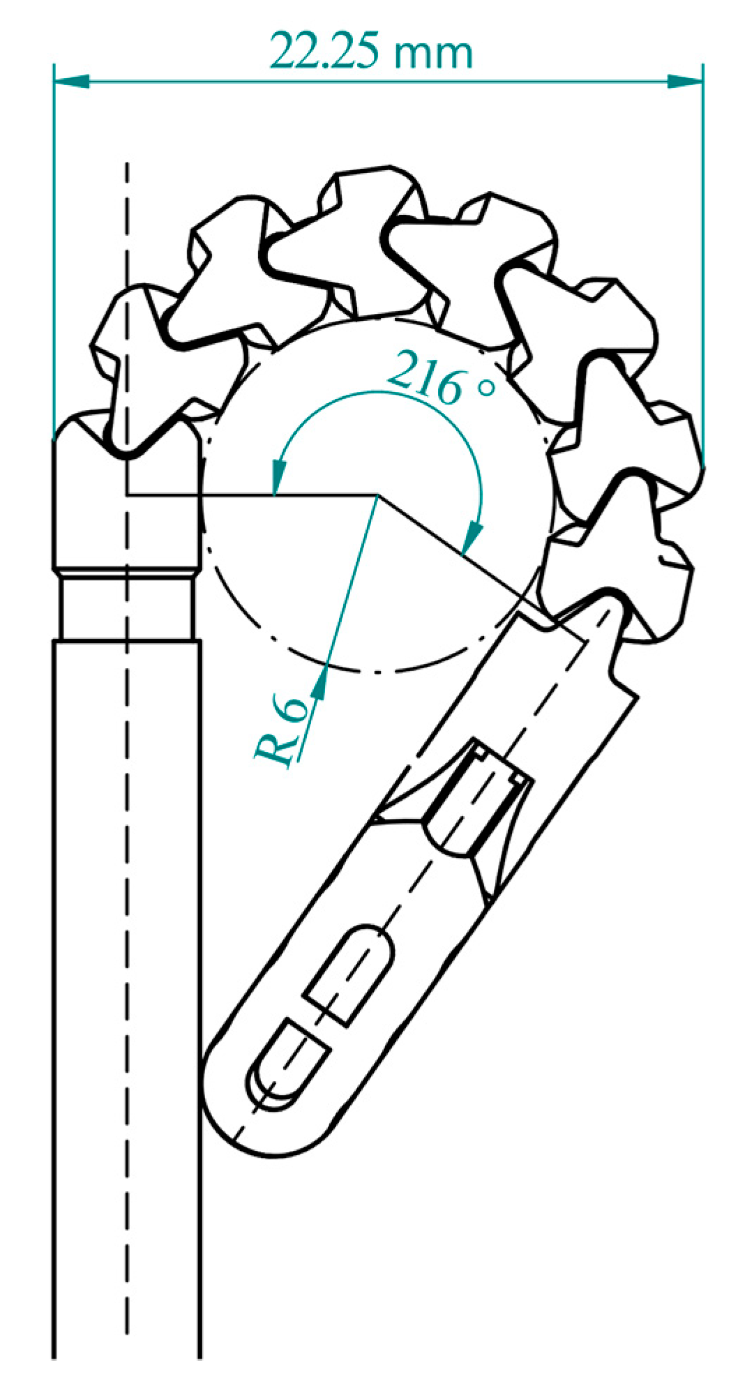
Figure 1. Image of the instrument’s distal tip at the maximal bending angle with annotated dimensions.
Applsci 13 05656 g001
Figure 2. Exploded view of the control handle with its main elements.
Applsci 13 05656 g002
Figure 3. Image of the prototype developed for the study. The flexible shaft and the distal tip are bent, showing the degree of the instrument’s flexibility. The depicted prototype is 50 cm long.
Applsci 13 05656 g003
Figure 4. Image of the instrument placed next to the gastroscope in the open stomach of the porcine model. The gastroscope was used to provide the vision for navigation of the instrument. The distal tip of the instrument is bent more than 180° and the grasping forceps are open.
Applsci 13 05656 g004
Figure 5. Visualization of the two workspaces, the instrument in red and the benchmark in blue color. Inside the curves, the bending section of the instrument is depicted at the maximal bending angle. The polygons that connect the black points with the origin represent different shapes that the distal tip forms during bending. The number outside the curves represents the angles achieved by bending elements and by the whole bending section, while the number inside the curves represents the lengths in centimeters.
Applsci 13 05656 g005
Table 1. The list of requirements for the 3D-pEI and its elements.
Instrument features
Adequate length to reach the dedicated region, at least 40 cm.
A diameter of 5 mm for intraluminal application alongside an endoscope.
Sufficient strength for movement independent from the endoscope.
Safe introduction and maneuvering inside the GI tract, with smooth parts and surfaces.
Biocompatibility of the parts with contact with organs.
Affordable manufacturing through 3D printing and simple assembly.
Grasping forceps
A smooth shape of the features that are in contact with the mucosal surfaces.
A minimum of 10 N grasping force to manipulate the tissue or a sponge.
Minimal length of non-grasping segments. The total length is less than 25 mm.
Jaw opening angle of at least 60°.
At least 8 mm-long grasping area with a gentle wave-like profile.
Bending section
Planar bending in both directions (left and right).
A high bending angle of at least 180° and a compact bending radius.
Adequate bending force for tissue and object manipulation of more than 5N.
Sufficient strength to withstand the loads introduced during maneuvering.
Flexible shaft
A high degree of flexibility for safe introduction into the dedicated region.
Sufficient torsional stiffness to precisely transfer the hand inputs to the distal tip.
Enough stiffness during pushing when transporting objects (no buckling).
Control handle and tendon wires
Convenient for single-hand use.
Star knob mechanism to control the bending of the distal tip, such as on endoscopes.
Push–pull motion for opening the grasping jaws, like on common biopsy forceps.
Table 2. The summary of expert grading from the assessment.
ParameterMedian GradeGrading RangeShort Conclusion
Flexibility22–3Adequate
Rigidity22–3Adequate
Torsional stiffness22Sufficient
Friction32–3Adequate
Maneuverability33Very good
Disclaimer/Publisher’s Note: The statements, opinions and data contained in all publications are solely those of the individual author(s) and contributor(s) and not of MDPI and/or the editor(s). MDPI and/or the editor(s) disclaim responsibility for any injury to people or property resulting from any ideas, methods, instructions or products referred to in the content.