Communication

Spontaneous Osteogenic Potential of Periosteum after Segmental Mandibulectomy in Patients with Medication-Related Osteonecrosis of the Jaw (MRONJ): A Retrospective Study of 14 Cases

1
Department of Conservative Dentistry, Wonju College of Medicine, Yonsei University, Wonju 26426, Republic of Korea
2
Department of Oral and Maxillofacial Surgery, Wonju College of Medicine, Yonsei University, Wonju 26426, Republic of Korea
*
Author to whom correspondence should be addressed.
Appl. Sci. 2023, 13(9), 5426; https://doi.org/10.3390/app13095426
Submission received: 20 March 2023 / Revised: 25 April 2023 / Accepted: 25 April 2023 / Published: 26 April 2023

Abstract

:
Medication-related osteonecrosis of the jaw (MRONJ) has chiefly involved patients receiving medications for the treatment of bone-related malignancies. The surgical methods are now cautiously recommended as the treatment option for MRONJ. The purpose of this retrospective study was to report new bone formation and examine possible factors influencing spontaneous bony bridge formation by using an R-plate for reconstruction without any graft material after segmental mandibulectomy in patients with MRONJ. A cohort of 54 patients who underwent mandibulectomy between 2017 and 2022 by a single operator were included. Of them, 14 MRONJ patients (12 females and 2 males) were selected based on the inclusion criteria. Data on new bone formation, bony bridge formation, R-plate fracture, patient age, and type and duration of medication were collected. The mean age of the patients was 74.8 ± 9.3 years. All patients showed new bone formation, and 71.4% showed bony bridge formation within a year after surgery. The older the patient, the lower was the tendency for bony bridge formation in the defect (p = 0.035). Statistical analysis was performed using Fisher’s exact test. Preservation of the periosteum might be one of the most important contributing factors to new bone formation, serving as an optimal treatment option for reconstruction after segmental mandibulectomy in patients with MRONJ.

1. Introduction

Bisphosphonate-related osteonecrosis of the jaw (BRONJ) was first reported in 2003 by Marx [1] and has chiefly involved patients receiving intravenous bisphosphonates for the treatment of bone-related malignancies [2]. It has primarily been reported in patients with bisphosphonate-related osteonecrosis of the jaw (BRONJ), but other antiresorptic drugs, such as denosumab, are also considered a risk factor for osteonecrosis of the jaw [3,4]. Further, ONJ is also associated with the intake of antiangiogenics for cancer treatment [5,6] and immunomodulators for Crohn’s disease or rheumatic arthritis [7,8]. Inhibitors of osteoclast differentiation and function have been introduced in the pharmacologic society for the treatment of diseases, causing a decreased bone turnover. However, the continued advent of new and complex therapies will eventually lead to an increase in the number of drugs inducing ONJ. The name of the disease was therefore revised from BRONJ to MRONJ and subsequently to drug-induced osteonecrosis of the jaw (DIONJ) [9].
After the first position statement on MRONJ in 2007, the American Association of Oral and Maxillofacial Surgeons (AAOMS) updated the position paper on strategies for the management of MRONJ in 2009 and 2014 [10], with the latest update in 2022. Although the most suitable treatment option for large bony defects in MRONJ remains debatable, AAOMS now cautiously recommends operative methods [11]. A nonoperative approach for MRONJ does not guarantee disease resolution. Early surgical intervention can provide good outcomes and should therefore be considered to inhibit disease progression [12]. Segmental or marginal resection of the mandible and partial maxillectomy are reliable methods to control MRONJ [13] and can be adopted during any stage of the disease, including stage 1 MRONJ [14]. After resecting the diseased part of the jaw, several reconstruction techniques can be used. A popular technique involves placement of a vascularized bone graft, which is the gold standard for mandibular reconstruction. The most common donor sites for osseous free-tissue grafts include the fibula, scapula, iliac bone, and radius [15]. Although the fibula free flap graft has been shown to be promising, it remains controversial due to requirements of advanced skill and technology and reports of postoperative progression of jaw bone necrosis, ONJ recurrence [16], and frequent failure caused by venous thrombosis [17]. In addition, this graft requires a donor site and is associated with a risk of donor site morbidity, which affects the quality of life of patients [18]. Other reconstruction techniques, such as distraction osteogenesis, which is very similar to the palatal distraction frequently used in orthodontic treatment, and the induced membrane technique (Masquelet technique), are other technical options [19,20]. The Masquelet technique can be summarized in two stages. The first stage consists of meticulous debridement at the lesion site, maintaining the stability of fracture ends, and implanting a polymethylmethacrylate cement spacer into the created bone defect. During the second stage with an interval of 6 to 8 weeks, the cement spacer is removed and the autologous bone graft is inserted. However, this method also requires a bone graft and a secondary surgery. Furthermore, these other options are also technical-sensitive and time-consuming [20].
Nonvascularized autogenous bone grafts or spontaneous regeneration of the bony defects without grafts constitute another reliable option for mandibular reconstruction [21,22]. These procedures utilize rigid plates made of titanium known as reconstruction plates (R-plates). R-plates are placed on the border of the mandible to bridge and stabilize the residual bone segments and defects. They can easily be used to maintain occlusion and the facial contour without modification. R-plates are used to affix nonvascularized autogenous bone grafts to the mandibular segments [23]. However, most reports on R-plates describe cases of reconstructive surgery for trauma or cancer-related defects (osteosarcoma) without ONJ. Graft materials are contraindicated in ONJ owing to a risk of inflammation. In addition, cases of spontaneous jaw bone reconstruction without bone graft materials are mostly limited to young patients or children.
There are no reports on the follow-up of MRONJ patients who have undergone jaw bone reconstruction with R-plates without bone grafts. Thus, the purpose of this retrospective study was to report new bone formation and examine possible factors influencing bony bridge formation after segmental mandibulectomy reconstruction using an R-plate without any graft material in patients with MRONJ.

2. Materials and Methods

Data on patients who received treatment between 2017 and 2022 were collected. All patients underwent surgery at the Department of Oral and Maxillofacial Surgery of the Wonju Severance Christian Hospital. Patients who underwent mandibular reconstruction without R-plates were excluded. Patients with MRONJ who underwent surgical intervention of the maxillary bones were also excluded as this study focused on the reconstruction of mandibular segmental defects. In addition, patients with ONJ due to the use of antineoplastic or antiangiogenic drugs were excluded to ensure that the ONJ was not associated with cancer or radiation therapy; therefore, we included only patients using antiresorptive drugs, such as bisphosphonates or denosumab. Thus, this retrospective observational study included patients who underwent the reconstruction of mandibular continuity defects resulting from surgical removal of the diseased bone in MRONJ using an R-plate without a bone graft. Prior to surgical intervention, these patients were diagnosed with MRONJ based on the following 2022 AAOMS criteria [11]:
  • Current or previous treatment with antiresorptive therapy alone or in combination with immune modulators or antiangiogenic medications.
  • Exposed bone or bone that can be probed through an intraoral or extraoral fistula(e) in the maxillofacial region persisting for more than 8 weeks.
  • No history of radiation therapy to the jaws or metastatic disease to the jaws.
Segmental mandibulectomy and mandibular reconstruction using an R-plate (Jeil Medical Corporation, Seoul, Republic of Korea) were performed. The bone-cutting margin was confirmed through clinical and radiographic assessments. The extent of the necrotic bone and defect was determined through panoramic radiography, cone-beam computed tomography (CBCT) (Carestream CS9300, Carestream Health, Rochester, NY, USA), and bone scanning. Intraoperatively, if a fresh blood supply and a sound bone margin could not be confirmed, the resection margin was extended. When removing the necrotic bone, the periosteum was gently handled and conserved to maximize its osteogenic potential. Following segmental mandibulectomy, the R-plate was placed. R-plates are rigid plates that are made with the intention of bridging a defect, stabilizing remaining segments, and maintaining occlusion and the facial contour. They are mainly used to fix bone blocks or vascularized bone grafts to the remaining mandible. R-plates were originally made of stainless steel, but the titanium plates are more preferable due to their biocompatibility [23]. There are several types of R-plates, but the main principle is to have one single plate of sufficient thickness and width of approximately 3 mm thick and 5 mm wide to hold the fragments in place. Every reconstruction case was held with a 2.4 mm thick titanium R-plate placed with 2.7 mm screws 5 mm above the inferior border of the mandible. Patient-specific R-plates were prepared preoperatively by pre-bending R-plates with the support of a rapid prototype model (RP model). After affixing the R-plates, they were completely covered with the conserved periosteum to utilize its osteogenic potential and prevent exposure of the surgical site.
Postoperatively, analgesics and antibiotics were routinely prescribed, and the patients were regularly monitored. In the first week postoperatively, we routinely used cephalosporin and acetaminophen or ketorolac intravenously. After 2–3 weeks, according to the symptoms of patients, antibiotics and analgesics were prescribed perorally. Intermaxillary fixation and radiographic examination, including panoramic radiography and CBCT, were performed a day after the operation. A Levin tube (L-tube) diet was indicated during the first postoperative week. The L-tube is a simple small-diameter tube that is inserted through the nares to pass through the posterior oropharynx, down the esophagus, and into the stomach. L-tubes can be placed to administer medications or nutrition in patients who have a functional gastrointestinal tract but are unable to tolerate oral intake [24]. Seven days postoperatively, the patients were instructed to take a sip of water. In the second postoperative week, the sutures and SAS screws were removed. The patients were then prescribed physical therapy, including mandibular extension for maximum mouth opening, and a soft diet. If the occlusion was unstable, intermaxillary fixation was performed again for another week. Panoramic and CBCT scans were obtained at 3, 6, 9, and 12 months postoperatively. The follow-up period was defined as the time from the placement of the plate to the last examination. During this period, complications related to the quality of the reconstruction (exposure of plates, loosening of screws, and fracture of plates) were recorded and appropriately managed.
Data on patient age and gender, medication type, and duration of intake were collected. New bone formation was evaluated on panoramic radiographs obtained periodically. On detecting new bone formation, the detection of bony bridge formation was also recorded. Statistical analysis of the correlation between the collected data and bony bridge formation was performed using Fisher’s exact test. The level of significance was set as a p-value of 5%. All statistical analyses were performed using SPSS 25.0 (IBM SPSS Statistics 25 software, IBM Corp., Armonk, NY, USA). The study protocol was reviewed and approved by the Institutional Review Board of Yonsei University Wonju Severance Christian Hospital, Wonju, Republic of Korea (CR322157).

3. Results

We evaluated the records of 54 patients who underwent mandibular surgery by a single operator in the Department of Oral and Maxillofacial Surgery between 2017 and 2022. Of them, eight diagnosed with ameloblastoma or squamous cell carcinoma were excluded. A total of 17 patients who developed osteonecrosis or osteomyelitis without intake of any MRONJ-inducing medication were also excluded. A total of 14 patients were excluded because their surgical management was marginal mandibulectomy or hemimandibulectomy. One case, which met the inclusion criteria but had a follow-up of <6 months, was also excluded. Finally, 14 cases were selected (Table 1).
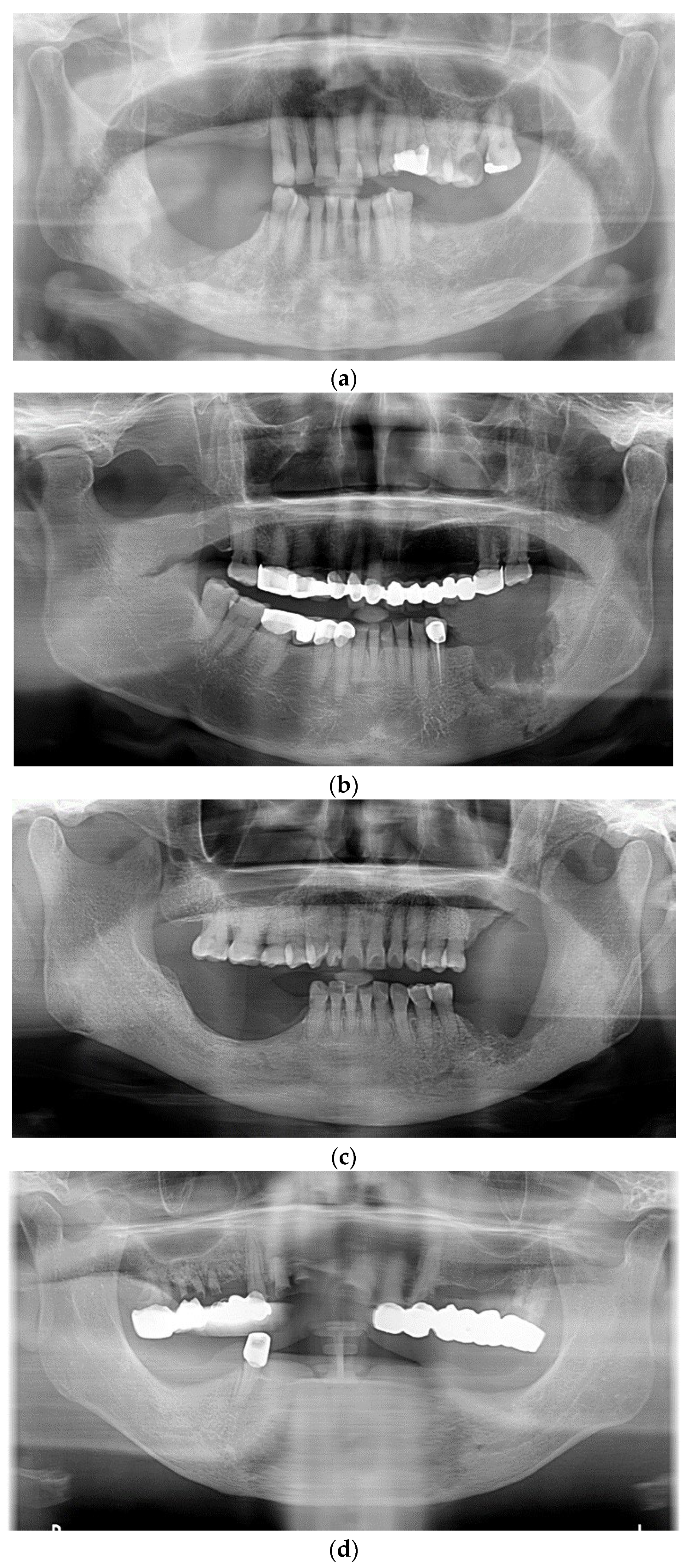
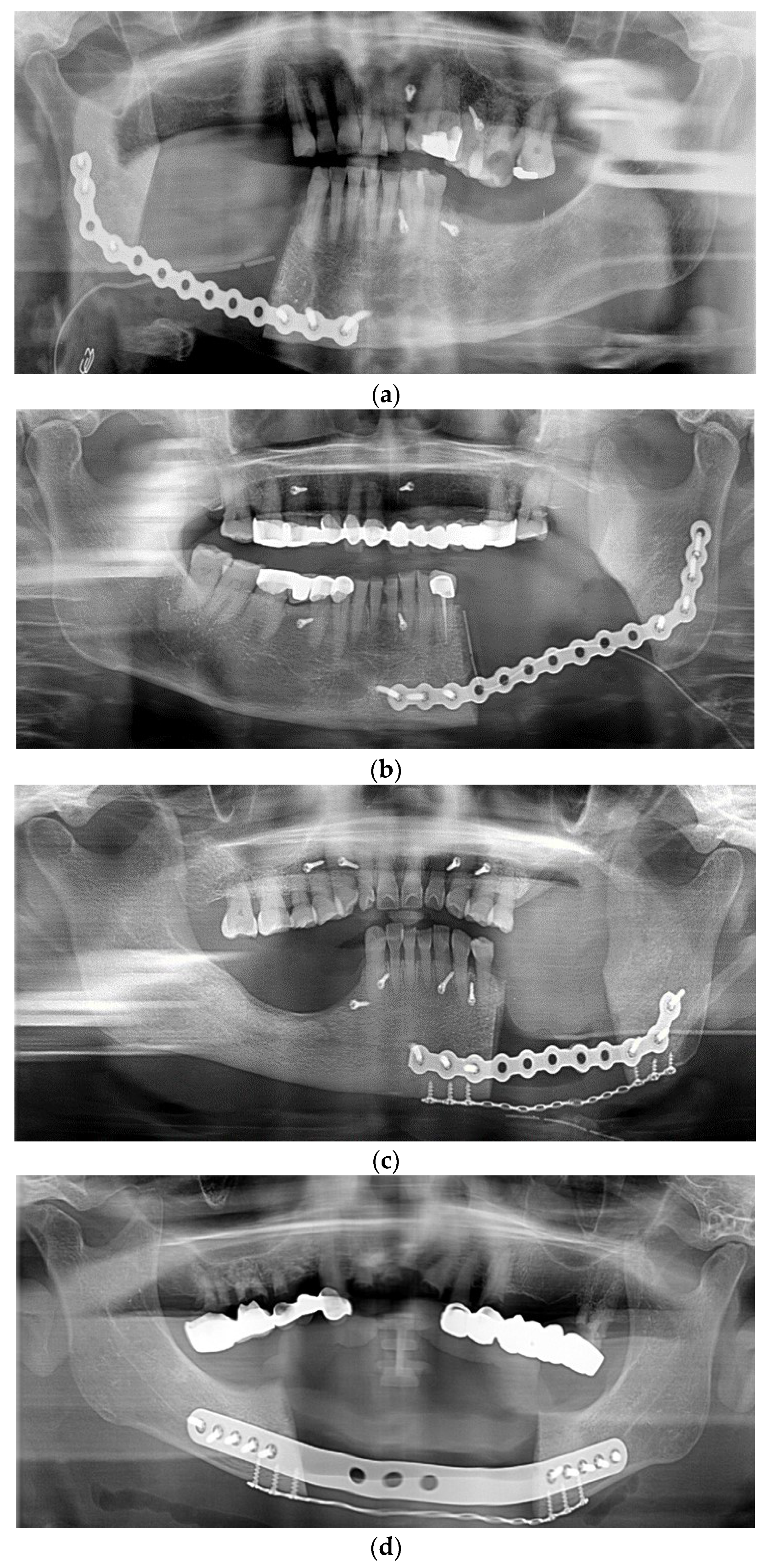
The distribution of cases is shown in Table 2. The included patients comprised two men (14.3%) and 12 women (85.7%) with a mean age of 74.8 ± 9.3 years (range, 50.3–87.2 years). The mean follow-up period after the surgery was 27.5 ± 11.6 months (range, 12.7–48.3 months). New bone formation was observed in all patients (100%), but bony bridge formation was detected in ten (71.4%). The bony bridge formation was detected within 1 year after surgery in all ten cases. The bony bridge formation was evaluated by comparing serial views in both the three-dimensional reconstruction CT view (Figure 1) and panoramic radiographs (Figure 2, Figure 3 and Figure 4, serial panoramic radiographs of each patient are shown in the Supplementary Materials). The older the patient, the lower was the tendency for bony bridge formation in the defect (p = 0.035). In 11 cases (84.6%), a sound state of the R-plate was maintained without any complication. In the other two (15.4%), a fracture of the R-plate was detected at the last follow-up. However, the R-plate fracture did not significantly affect bony bridge formation (p > 0.05).
Most cases were of MRONJ induced by bisphosphonate medication alone (78.6%). The duration of medication intake was >4 years in half of the cases (50%) and <4 years in another half (50%). The effects of medication type and duration on bone bridge formation were not statistically significant (p > 0.05).

4. Discussion

Several attempts have been made to achieve successful jaw reconstruction through spontaneous regeneration of the bone. However, this procedure is commonly recommended only for young patients, in whom regeneration can be achieved relatively easily [22,25]. Therefore, the literature on R-plate reconstruction with spontaneous new bone formation in elderly patients is limited. In this retrospective study, the average patient age was 74.4 ± 9.4 years. Nevertheless, all 14 patients showed new bone formation, indicating spontaneous regeneration, without use of graft materials.
Our study consists of only the osteonecrosis on mandible, where the vascular supply is limited and somewhat dynamic compared to the maxilla. Thus, it is essential to maintain absolute immobilization in the early period of healing and obtain sufficient vascular supply for spontaneous bone healing in the mandible.
Two cases with R-plate fracture occurrence might be mainly the fatigue fracture caused by the discrepancy between dynamic movement of the remaining mandibular structure and the static state of the R-plate. However, the plate fracture of both cases was found after the new bone formation was detected, indicating that the customized patient-specific R-plate reconstruction and postoperative intermaxillary fixation were sufficient to gain absolute immobilization in the early period of healing. Furthermore, the periosteum was preserved and repositioned to cover the inferior border of the mandible and R-plate in every case, which could also contribute to supplemental primary immobilization. Of the two cases of R-plate fractures, bony bridge formation was observed in one. The absence of bony bridge formation in the other may be attributed to an excessive occlusal force from the teeth opposing the surgery site [26]. It is thus critical to prevent distortion of the plate and unfavorable healing by alleviating the excessive occlusal load.
Another crucial factor of spontaneous bone healing is to gain vascular supply. The preserved periosteum is essential as it can serve as a direct source of vascular supply and osteogenic cells, and can promote bone regeneration [22]. An absence of periosteum has been associated with unfavorable healing outcomes in previous studies [22,27]. Preservation of the periosteum is impossible during mandibulectomy in cases of oral malignancy; thus, patients diagnosed with metastatic diseases were excluded. To eliminate the possible unfavorable effects of metastatic diseases on bone healing, patients taking antineoplastic or antiangiogenic drugs were also excluded. Preservation of the periosteum may have been the most important contributing factor for spontaneous new bone formation in our cases as all of them (100%) showed detectable new bone formation. Further, there were no cases of plate exposure in this study, which may also be attributed to the preservation of the periosteum and appropriate coverage by surrounding tissues such as platysma muscle, fibrous fatty tissue, and the skin.
The site of the bony bridge formation is the most suitable for the rehabilitation of occlusion and mastication. The bony bridge also provides a newly formed alveolar process, enhancing the volume and strength at the site of plate reconstruction and preventing pathologic fractures from occlusive loads [28]. Therefore, when assessing new bone formation from the perspective of functional rehabilitation, bony bridge formation is an important clinical outcome factor.
Most patients in the present study were female (84.6%), which is consistent with the findings of other studies on the incidence and prevalence of MRONJ. Previous studies have frequently cited patient gender (being female), age, and duration of medication intake (≥4 years) as prognostic factors for the prevalence of MRONJ [29,30]. Therefore, we used a cutoff of 4 years to categorize the duration of medication intake by patients. In our study, bony bridge formation was only statistically related to age, with no significant effect of R-plate fracture, medication type, or duration of medication intake.
There are several treatment options for the reconstruction of segmental mandibulectomy. The placement of a vascularized bone graft, which is the gold standard for mandibular reconstruction, remains controversial due to requirements of advanced skill and technology and several side-effects as mentioned above. Furthermore, it requires a donor site and is associated with a risk of donor site morbidity. Other reconstruction techniques, such as distraction osteogenesis and the induced membrane technique, are other technical options [19,20], but these treatment options require several visits or secondary surgery. The R-plate reconstruction could therefore be a promising and suitable treatment option for patients with MRONJ.
Recent studies have reported that some novel approaches and adjuvant therapies concomitant with the standard approach may offer advantages in terms of surgical techniques and bone healing [31]. Leukocyte and platelet-rich fibrin (LPRF) and leukocyte and platelet-rich plasma (LPRP) are used to promote bone healing and showed a significant improvement in quality of life [32]. A novel surgical technique called fluorescence-guided surgery was introduced for its enhancing effect of the transition from necrotic to vital bone during surgical debridement [33]. In addition, necrotic bone can be vaporized by laser ablation such as an Er:YAG laser, leading to a faster bone healing without risk of thermal damage [34]. Additionally, antimicrobial agents such as chlorhexidine gluconate mouthwash are prescribed in some cases to prevent oral infection on the surgery site with regard to intraoral microbial approaches. Some recently introduced compounds have been demonstrated as having a significant influence on the oral environment [35]. The use of lysates [36] and postbiotics [37] can modify clinical and microbiological parameters, so these products can also be considered an oral microbial approach in maxillofacial surgery. When these adjuvant techniques are combined with R-plate reconstruction, the healing outcome might be more favorable.
However, there are several limitations in this study. Along with the involvement with a single limited population, the retrospective nature of this study leads to a limited sample size of rare cases without sample size calculation. Furthermore, limitations within operator-related factors such as experience level or the learning curve exist as all of the cases were performed by a single operator. Additionally, comparisons on the healing outcome with other reconstruction are required, as this study investigated the healing outcome of only one surgical procedure.
A systematic review about the prognostic factors of MRONJ revealed that as prognostic factors of treatment outcomes, the type of medication, the stage of MRONJ, and the type of surgical intervention were significant independent factors in the multivariate analysis [38]. Thus, further studies might be needed to investigate the effects of medication type and MRONJ stage on the spontaneous osteogenesis in R-plate reconstruction after segmental mandibulectomy with larger case numbers of MRONJ patients. Furthermore, as only the presence of new bone formation and bony bridge formation was assessed in the present study, further research on the quantitative measurement of bony bridge formation using CBCT images and three-dimensional views is necessary.

5. Conclusions

Within the limitations of this study, all included cases (100%) showed new bone formation following segmental mandibulectomy and R-plate reconstruction without the use of bone graft materials in patients with MRONJ. In approximately 70% of the patients, bony bridge formation was observed within a year after surgery. The older the patient, the lower was the tendency for bony bridge formation in the defect (p = 0.035). Preservation of the periosteum may contribute significantly to new bone formation, offering an additional treatment option for jaw reconstruction after segmental mandibulectomy in patients with MRONJ.

Supplementary Materials

The following supporting information can be downloaded at: https://www.mdpi.com/article/10.3390/app13095426/s1. The Supplementary Materials contain serial panoramic radiography of each patient.

Author Contributions

Conceptualization, Y.K. and C.L.; methodology, Y.K. and C.L.; validation, C.L.; formal analysis, Y.K., Y.-Q.F. and S.L.; investigation, Y.K.; resources, C.L.; data curation, C.L.; writing—original draft preparation, Y.K. and C.L.; writing—review and editing, Y.K., Y.-Q.F., S.L. and C.L.; visualization, Y.K.; supervision, C.L. All authors have read and agreed to the published version of the manuscript.

Funding

This research was funded by the National Research Foundation of Korea (NRF) grant funded by the Korea government (MSIT) (No. RS-2022-00165819).

Institutional Review Board Statement

The study was conducted in accordance with the guidelines of the Declaration of Helsinki and was approved by the Institutional Review Board of Yonsei University of Wonju Severance Christian Hospital, Republic of Korea (CR322157).

Informed Consent Statement

Patient consent was waived due to the retrospective nature of this study.

Data Availability Statement

Data are available upon reasonable request.

Conflicts of Interest

The authors declare no conflict of interest.

References

  1. Marx, R.E. Pamidronate (Aredia) and zoledronate (Zometa) induced avascular necrosis of the jaws: A growing epidemic. J. Oral Maxillofac. Surg. 2003, 61, 1115–1117. [Google Scholar] [CrossRef]
  2. Ruggiero, S.L.; Mehrotra, B.; Rosenberg, T.J.; Engroff, S.L. Osteonecrosis of the jaws associated with the use of bisphosphonates: A review of 63 cases. J. Oral Maxillofac. Surg. 2004, 62, 527–534. [Google Scholar] [CrossRef] [PubMed]
  3. Aghaloo, T.L.; Dry, S.M.; Mallya, S.; Tetradis, S. Osteonecrosis of the jaw in a patient on denosumab. J. Oral Maxillofac. Surg. 2014, 72, 702–716. [Google Scholar] [CrossRef] [PubMed]
  4. Saag, K.G.; Petersen, J.; Brandi, M.L.; Karaplis, A.C.; Lorentzon, M.; Thomas, T.; Maddox, J.; Fan, M.; Meisner, P.D.; Grauer, A. Romosozumab or Alendronate for Fracture Prevention in Women with Osteoporosis. N. Engl. J. Med. 2017, 377, 1417–1427. [Google Scholar] [CrossRef]
  5. Estilo, C.L.; Fornier, M.; Farooki, A.; Carlson, D.; Bohle, G., 3rd; Huryn, J.M. Osteonecrosis of the jaw related to bevacizumab. J. Clin. Oncol. 2008, 26, 4037–4038. [Google Scholar] [CrossRef]
  6. Sivolella, S.; Lumachi, F.; Stellini, E.; Favero, L. Denosumab and anti-angiogenetic drug-related osteonecrosis of the jaw: An uncommon but potentially severe disease. Anticancer Res. 2013, 33, 1793–1797. [Google Scholar]
  7. Alsalleeh, F.; Keippel, J.; Adams, L.; Bavitz, B. Bisphosphonate-associated osteonecrosis of jaw reoccurrence after methotrexate therapy: A case report. J. Endod. 2014, 40, 1505–1507. [Google Scholar] [CrossRef]
  8. Henien, M.; Carey, B.; Hullah, E.; Sproat, C.; Patel, V. Methotrexate-associated osteonecrosis of the jaw: A report of two cases. Oral Surg. Oral Med. Oral Pathol. Oral Radiol. 2017, 124, e283–e287. [Google Scholar] [CrossRef]
  9. Hamadeh, I.S.; Ngwa, B.A.; Gong, Y. Drug induced osteonecrosis of the jaw. Cancer Treat. Rev. 2015, 41, 455–464. [Google Scholar] [CrossRef]
  10. Ruggiero, S.L.; Dodson, T.B.; Fantasia, J.; Goodday, R.; Aghaloo, T.; Mehrotra, B. American Association of Oral and Maxillofacial Surgeons position paper on medication-related osteonecrosis of the jaw—2014 update. J. Oral Maxillofac. Surg. 2014, 72, 1938–1956. [Google Scholar] [CrossRef]
  11. Ruggiero, S.L.; Dodson, T.B.; Aghaloo, T.; Carlson, E.R.; Ward, B.B.; Kademani, D. American Association of Oral and Maxillofacial Surgeons’ Position Paper on Medication-Related Osteonecrosis of the Jaws—2022 Update. J. Oral Maxillofac. Surg. 2022, 80, 920–943. [Google Scholar] [CrossRef] [PubMed]
  12. Ristow, O.; Rückschloß, T.; Müller, M.; Berger, M.; Kargus, S.; Pautke, C. Is the conservative non-surgical management of medication-related osteonecrosis of the jaw an appropriate treatment option for early stages? A long-term single-center cohort study. J. Craniomaxillofac. Surg. 2019, 47, 491–499. [Google Scholar] [CrossRef] [PubMed]
  13. Wilde, F.; Heufelder, M.; Winter, K.; Hendricks, J.; Frerich, B.; Schramm, A. The role of surgical therapy in the management of intravenous bisphosphonates-related osteonecrosis of the jaw. Oral Surg. Oral Med. Oral Pathol. Oral Radiol. Endodontol. 2011, 111, 153–163. [Google Scholar] [CrossRef]
  14. Kyrgidis, A.; Vahtsevanos, K.; Koloutsos, G.; Andreadis, C.; Boukovinas, I.; Teleioudis, Z. Bisphosphonate-related osteonecrosis of the jaws: A case-control study of risk factors in breast cancer patients. J. Clin. Oncol. 2008, 26, 4634–4638. [Google Scholar] [CrossRef] [PubMed]
  15. Rana, M.; Warraich, R.; Kokemüller, H.; Lemound, J.; Essig, H.; Tavassol, F. Reconstruction of mandibular defects-clinical retrospective research over a 10-year period. Head Neck Oncol. 2011, 3, 23. [Google Scholar] [CrossRef]
  16. Sacco, R.; Sacco, N.; Hamid, U.; Ali, S.H.; Singh, M.; Blythe, J.S.J. Microsurgical Reconstruction of the Jaws Using Vascularised Free Flap Technique in Patients with Medication-Related Osteonecrosis: A Systematic Review. BioMed Res. Int. 2018, 2018, 9858921. [Google Scholar] [CrossRef]
  17. Verhelst, P.J.; Dons, F.; Van Bever, P.J.; Schoenaers, J.; Nanhekhan, L.; Politis, C. Fibula Free Flap in Head and Neck Reconstruction: Identifying Risk Factors for Flap Failure and Analysis of Postoperative Complications in a Low Volume Setting. Craniomaxillofac. Trauma Reconstr. 2019, 12, 183–192. [Google Scholar] [CrossRef]
  18. Akinbami, B.O. Reconstruction of Continuity Defects of the Mandible with Non-vascularized Bone Grafts. Systematic literature review. Craniomaxillofac. Trauma Reconstr. 2016, 9, 195–205. [Google Scholar] [CrossRef]
  19. Sun, S.H.; Tsai, W.W.; Shiu, S.I.; Chen, C.H. Induced membrane technique for large bone defects: A systematic review and individual participant data meta-analysis. Medicine 2022, 101, e29292. [Google Scholar] [CrossRef]
  20. Hibi, H.; Ueda, M. Supraperiosteal transport distraction osteogenesis for reconstructing a segmental defect of the mandible. J. Oral Maxillofac. Surg. 2011, 69, 742–746. [Google Scholar] [CrossRef]
  21. Bede, S.Y.H.; Ismael, W.K.; Hashim, E.A. Reconstruction plate-related complications in mandibular continuity defects. Oral Maxillofac. Surg. 2019, 23, 193–199. [Google Scholar] [CrossRef]
  22. Anyanechi, C.E.; Saheeb, B.D.; Bassey, G.O. Spontaneous bone regeneration after segmental mandibular resection: A retrospective study of 13 cases. Int. J. Oral Maxillofac. Surg. 2016, 45, 1268–1272. [Google Scholar] [CrossRef] [PubMed]
  23. Goh, B.T.; Lee, S.; Tideman, H.; Stoelinga, P.J. Mandibular reconstruction in adults: A review. Int. J. Oral Maxillofac. Surg. 2008, 37, 597–605. [Google Scholar] [CrossRef] [PubMed]
  24. Rai, S.; Rattan, V.; Jolly, S.S.; Sharma, V.K.; Mubashir, M.M. Spontaneous Regeneration of Bone in Segmental Mandibular Defect. J. Maxillofac. Oral Surg. 2019, 18, 224–228. [Google Scholar] [CrossRef] [PubMed]
  25. Almansoori, A.A.; Choung, H.W.; Kim, B.; Park, J.Y.; Kim, S.M.; Lee, J.H. Fracture of Standard Titanium Mandibular Reconstruction Plates and Preliminary Study of Three-Dimensional Printed Reconstruction Plates. J. Oral Maxillofac. Surg. 2020, 78, 153–166. [Google Scholar] [CrossRef]
  26. Boyne, P.J. The restoration of resected mandibles in children without the use of bone grafts. Head Neck Surg. 1983, 6, 626–631. [Google Scholar] [CrossRef]
  27. Lemperle, S.M.; Calhoun, C.J.; Curran, R.W.; Holmes, R.E. Bony healing of large cranial and mandibular defects protected from soft-tissue interposition: A comparative study of spontaneous bone regeneration, osteoconduction, and cancellous autografting in dogs. Plast. Reconstr. Surg. 1998, 101, 660–672. [Google Scholar] [CrossRef]
  28. Sherris, D.A.; Murakami, C.S.; Larrabee, W.F., Jr.; Bruce, A.G. Mandibular reconstruction with transforming growth factor-beta1. Laryngoscope 1998, 108, 368–372. [Google Scholar] [CrossRef]
  29. Ishimaru, M.; Ono, S.; Morita, K.; Matsui, H.; Hagiwara, Y.; Yasunaga, H. Prevalence, Incidence Rate, and Risk Factors of Medication-Related Osteonecrosis of the Jaw in Patients with Osteoporosis and Cancer: A Nationwide Population-Based Study in Japan. J. Oral Maxillofac. Surg. 2022, 80, 714–727. [Google Scholar] [CrossRef]
  30. Aljohani, S.; Fliefel, R.; Ihbe, J.; Kühnisch, J.; Ehrenfeld, M.; Otto, S. What is the effect of anti-resorptive drugs (ARDs) on the development of medication-related osteonecrosis of the jaw (MRONJ) in osteoporosis patients: A systematic review. J. Craniomaxillofac. Surg. 2017, 45, 1493–1502. [Google Scholar] [CrossRef]
  31. Govaerts, D.; Piccart, F.; Ockerman, A.; Coropciuc, R.; Politis, C.; Jacobs, R. Adjuvant therapies for MRONJ: A systematic review. Bone 2020, 141, 115676. [Google Scholar] [CrossRef] [PubMed]
  32. Giudice, A.; Barone, S.; Giudice, C.; Bennardo, F.; Fortunato, L. Can platelet-rich fibrin improve healing after surgical treatment of medication-related osteonecrosis of the jaw? A pilot study. Oral Surg. Oral Med. Oral Pathol. Oral Radiol. 2018, 126, 390–403. [Google Scholar] [CrossRef] [PubMed]
  33. Ristow, O.; Otto, S.; Geiß, C.; Kehl, V.; Berger, M.; Troeltzsch, M.; Koerdt, S.; Hohlweg-Majert, B.; Freudlsperger, C.; Pautke, C. Comparison of auto-fluorescence and tetracycline fluorescence for guided bone surgery of medication-related osteonecrosis of the jaw: A randomized controlled feasibility study. Int. J. Oral Maxillofac. Surg. 2017, 46, 157–166. [Google Scholar] [CrossRef] [PubMed]
  34. Merigo, E.; Cella, L.; Oppici, A.; Cristina Arbasi, M.; Clini, F.; Fontana, M.; Fornaini, C. Combined Approach to Treat Medication-Related Osteonecrosis of the Jaws. J. Lasers Med. Sci. 2018, 9, 92–100. [Google Scholar] [CrossRef]
  35. Butera, A.; Pascadopoli, M.; Pellegrini, M.; Gallo, S.; Zampetti, P.; Scribante, A. Oral Microbiota in Patients with Peri-Implant Disease: A Narrative Review. Appl. Sci. 2022, 12, 3250. [Google Scholar] [CrossRef]
  36. Vale, G.C.; Mayer, M.P.A. Effect of probiotic Lactobacillus rhamnosus by-products on gingival epithelial cells challenged with Porphyromonas gingivalis. Arch. Oral. Biol. 2021, 128, 105174. [Google Scholar] [CrossRef]
  37. Butera, A.; Pascadopoli, M.; Pellegrini, M.; Gallo, S.; Zampetti, P.; Cuggia, G.; Scribante, A. Domiciliary Use of Chlorhexidine vs. Postbiotic Gels in Patients with Peri-Implant Mucositis: A Split-Mouth Randomized Clinical Trial. Appl. Sci. 2022, 12, 2800. [Google Scholar] [CrossRef]
  38. Yamada, S.I.; Kurita, H.; Kondo, E.; Suzuki, S.; Nishimaki, F.; Yoshimura, N.; Morioka, M.; Ishii, S.; Kamata, T. Treatment outcomes and prognostic factors of medication-related osteonecrosis of the jaw: A case- and literature-based review. Clin. Oral Investig. 2019, 23, 3203–3211. [Google Scholar] [CrossRef]
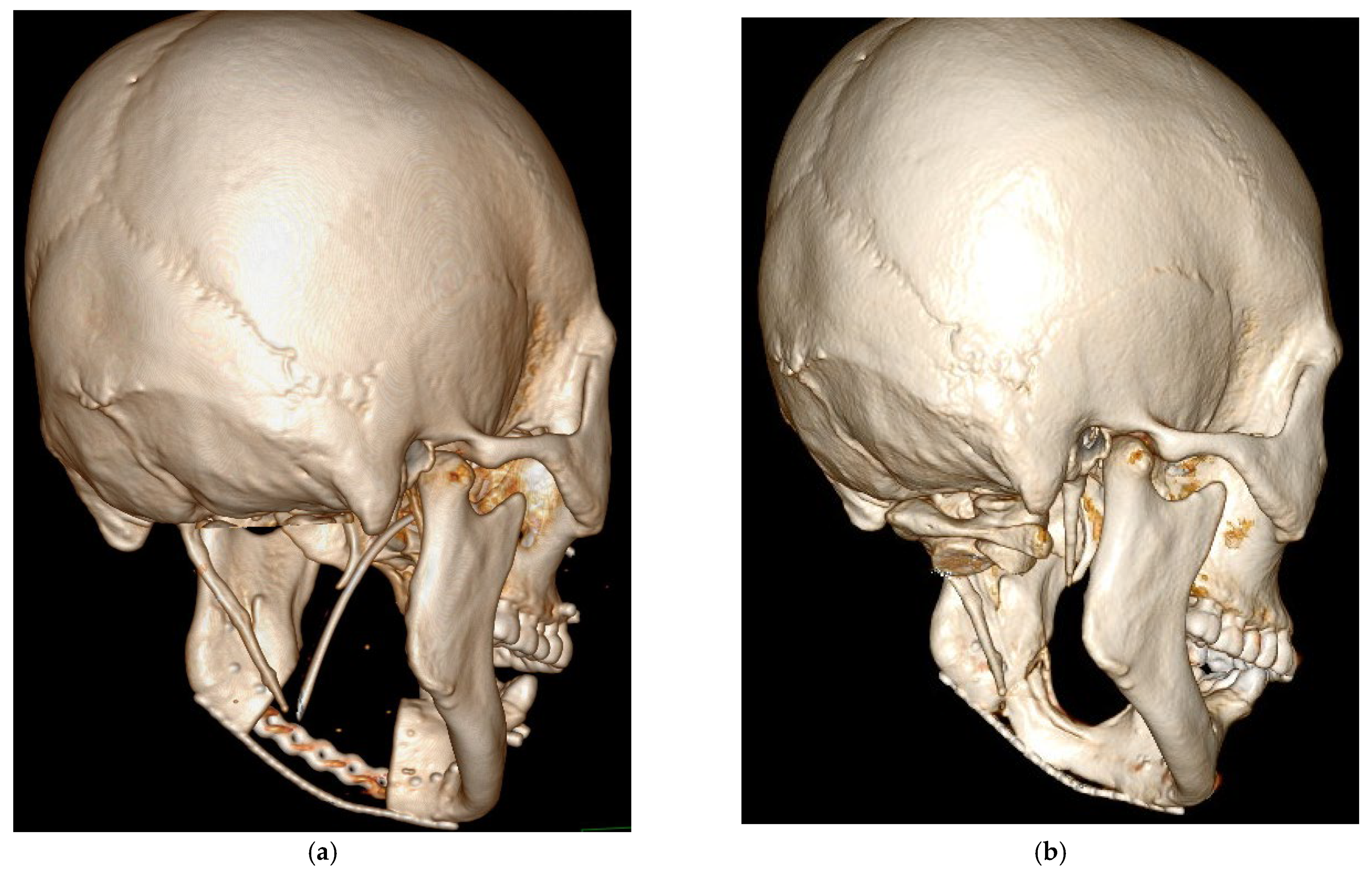
Figure 1. Three-dimensional Reconstruction CT View of Patient No. 4: (a) Postoperative; (b) Bony bridge formation within a year after the surgery.
Applsci 13 05426 g001
Figure 2. Preoperative Panoramic Radiographs. (a) Patient No. 1; (b) Patient No. 2; (c) Patient No. 4; (d) Patient No. 9.
Applsci 13 05426 g002
Figure 3. Postoperative Panoramic Radiographs. (a) Patient No. 1; (b) Patient No. 2; (c) Patient No. 4; (d) Patient No. 9.
Applsci 13 05426 g003
Figure 4. Panoramic Radiographs Showing Bony Bridge Formation. (a) Patient No. 1; (b) Patient No. 2; (c) Patient No. 4; (d) Patient No. 9.
Applsci 13 05426 g004
Table 1. Patient characteristics.
No.Age (Year)Follow-Up Period (Month)GenderNew Bone FormationBony Bridge FormationR-Plate FractureMedication TypeDuration
173.7650.08FemalePresentPresentNoneBoth>4 years
273.4845.42FemalePresentPresentNoneBisphosphonates>4 years
380.7241.25MalePresentAbsentPresentBisphosphonates2 years
470.7737.00FemalePresentPresentNoneBisphosphonates3 years
571.7032.42FemalePresentPresentNoneBisphosphonates3 years
685.0128.67FemalePresentAbsentNoneBisphosphonates>4 years
783.1225.17FemalePresentAbsentNoneBisphosphonates>4 years
878.3922.75FemalePresentAbsentNoneBisphosphonates2 years
971.7222.67FemalePresentPresentNoneBisphosphonates>4 years
1050.2719.08FemalePresentPresentNoneBisphosphonates3 years
1162.5016.92MalePresentPresentPresentDenosumab>4 years
1278.9816.25FemalePresentPresentNoneBisphosphonates1 year
1387.1914.58FemalePresentPresentNoneBisphosphonates1 year
1479.2212.67FemalePresentPresentNoneDenosumab2 years
Table 2. Distribution of Cases by Characteristics, N = 14.
Characteristic
Age, y, mean ± SD74.8 ± 9.3
 Range, y50.3–87.2
Follow-up period, m, mean ± SD27.5 ± 11.6
 Range, m12.7–48.3
Gender, n (%) 
 Male2 (14.3%)
 Female12 (85.7%)
New bone formation, n (%) 
 Present14 (100%)
 Absent0 (0%)
Bony bridge formation, n (%) 
 Present10 (71.4%)
 Absent4 (28.6%)
R-plate fracture, n (%) 
 None12 (85.7%)
 Present2 (14.3%)
Medication type 
 Bisphosphonates11 (78.6%)
 Denosumab2 (14.3%)
 Both1 (7.1%)
Duration of medication 
 >4 years7 (50%)
 <4 years7 (50%)
y = years, m = months.
Disclaimer/Publisher’s Note: The statements, opinions and data contained in all publications are solely those of the individual author(s) and contributor(s) and not of MDPI and/or the editor(s). MDPI and/or the editor(s) disclaim responsibility for any injury to people or property resulting from any ideas, methods, instructions or products referred to in the content.