

Quadriceps Muscle and Medial Retinaculum Combinate Effects on Patellar Instability during Knee Flexion
Abstract
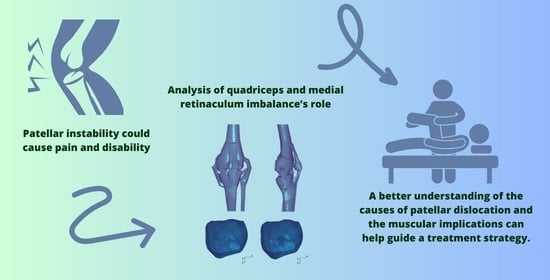
1. Introduction
2. Material and Methods
2.1. Computational Modelling
2.2. Material Properties
2.3. Model Loading
3. Results
4. Discussion
5. Conclusions
Author Contributions
Funding
Institutional Review Board Statement
Informed Consent Statement
Data Availability Statement
Conflicts of Interest
References
- Erdemir, A. Open Knee: Open Source Modeling and Simulation in Knee Biomechanics. J. Knee Surg. 2016, 29, 107–116. [Google Scholar] [CrossRef] [PubMed]
- Shao, B.; Xing, J.; Zhao, B.; Wang, T.; Mu, W. Role of the proximal tibiofibular joint on the biomechanics of the knee joint: A three-dimensional finite element analysis. Injury 2022, 53, 2446–2453. [Google Scholar] [CrossRef] [PubMed]
- Biko, D.M.; Miller, A.L.; Ho-Fung, V.; Jaramillo, D. MRI of congenital and developmental abnormalities of the knee. Clin. Radiol. 2012, 67, 1198–1206. [Google Scholar] [CrossRef] [PubMed]
- Thompson, P.; Metcalfe, A.J. Current concepts in the surgical management of patellar instability. Knee 2019, 26, 1171–1181. [Google Scholar] [CrossRef]
- Bowd, J.; Van Rossom, S.; Williams, D.; Elson, D.; Wilson, C.; Whatling, G.; Holt, C.; Jonkers, I. Using musculoskeletal modelling to estimate knee joint loading pre and post high tibial osteotomy. Clin. Biomech. 2023, 101, 105855. [Google Scholar] [CrossRef]
- Zibis, A.H.; Vassalou, E.E.; Raoulis, V.A.; Lampridis, V.; Klontzas, M.E.; Fyllos, A.; Stavlas, P.; Karantanas, A.H. Knee Capsule Anatomy: An MR Imaging and Cadaveric Study. Diagnostics 2021, 11, 1965. [Google Scholar] [CrossRef] [PubMed]
- Migliorini, F.; Maffulli, N.; Vaishya, R. Patellofemoral instability: Current status and future perspectives. J. Orthop. 2023, 36, 49–50. [Google Scholar] [CrossRef]
- Sato, A.; Takagi, H.; Koya, T.; Espinoza Orias, A.A.; Kanzaki, K.; Inoue, N. Clinical three-dimensional anatomy of the femur considering navigation-aided surgery of total knee arthroplasty in Japanese patients. Knee 2023, 41, 214–220. [Google Scholar] [CrossRef] [PubMed]
- Patterson, B.E.; Girdwood, M.A.; West, T.J.; Bruder, A.M.; Oiestad, B.E.; Juhl, C.; Culvenor, A.G. Muscle strength and osteoarthritis of the knee: A systematic review and meta-analysis of longitudinal studies. Skeletal. Radiol. 2022. [Google Scholar] [CrossRef] [PubMed]
- Loudon, J.K. Biomechanics and Pathomechanics of the Patellofemoral Joint. Int. J. Sport. Phys. Ther. 2016, 11, 820–830. [Google Scholar]
- Lack, S.; Anthony, L.; Noake, J.; Brennan, K.; Zhang, B.; Morrissey, D. Medial and Lateral Patellofemoral Joint Retinaculum Thickness in People with Patellofemoral Pain: A Case-Control Study. J. Ultrasound Med. 2019, 38, 1483–1490. [Google Scholar] [CrossRef] [PubMed]
- Teichtahl, A.J.; Wluka, A.E.; Cicuttini, F.M. Frontal plane knee alignment is associated with a longitudinal reduction in patella cartilage volume in people with knee osteoarthritis. Osteoarthr. Cartil. 2008, 16, 851–854. [Google Scholar] [CrossRef] [PubMed]
- Seyyed Hossein, H.; Sara, A.; Hasan, D.; Navid, K. The effect of three types of exercises programs on the patella location in athletes with patellofemoral pain. Knee 2023, 41, 97–105. [Google Scholar] [CrossRef] [PubMed]
- Cascio, F.; Cacciola, A.; Portaro, S.; Basile, G.A.; Rizzo, G.; Felippu, A.W.D.; Felippu, A.W.D.; Bruschetta, A.; Anfuso, C.; Cascio, F.; et al. In Vivo Computed Tomography Direct Volume Rendering of the Anterior Ethmoidal Artery: A Descriptive Anatomical Study. Int. Arch. Otorhinolaryngol. 2020, 24, e38–e46. [Google Scholar] [CrossRef] [PubMed]
- Schache, A.G.; Sritharan, P.; Culvenor, A.G.; Patterson, B.E.; Perraton, L.G.; Bryant, A.L.; Guermazi, A.; Morris, H.G.; Whitehead, T.S.; Crossley, K.M. Patellofemoral joint loading and early osteoarthritis after ACL reconstruction. J. Orthop. Res. 2023. [Google Scholar] [CrossRef] [PubMed]
- Krebs, C.; Tranovich, M.; Andrews, K.; Ebraheim, N. The medial patellofemoral ligament: Review of the literature. J. Orthop. 2018, 15, 596–599. [Google Scholar] [CrossRef]
- Tisano, A.; Alito, A.; Milardi, D.; Fazio, R.; Virelli, L.; Zanella, C.; Ruggeri, C.; Filardi, V.; Bruschetta, D. Statistical investigation about spinal clinical asymmetry in a school population. J. Orthop. 2020, 22, 336–340. [Google Scholar] [CrossRef] [PubMed]
- Simonaityte, R.; Rutkauskas, S.; Cekanauskas, E.; Labanauskas, L.; Barauskas, V. First-Time Acute Lateral Patellar Dislocation in Children and Adolescents: What about Unaffected Knee Patellofemoral Joint Anatomic Abnormalities? Medicina 2021, 57, 206. [Google Scholar] [CrossRef]
- Sinikumpu, J.; Nicolaou, N. Current concepts in the treatment of first-time patella dislocation in children and adolescents. J. Child. Orthop. 2023, 17, 28–33. [Google Scholar] [CrossRef] [PubMed]
- Palco, M.; Rizzo, P.; Basile, G.C.; Alito, A.; Bruschetta, D.; Accorinti, M.; Restuccia, R.; Leonetti, D. Short- and Midterm Comparison of Platelet-Rich Plasma with Hyaluronic Acid versus Leucocyte and Platelet-Rich Plasma on Pain and Function to Treat Hip Osteoarthritis. A Retrospective Study. Gels 2021, 7, 222. [Google Scholar] [CrossRef]
- Trisolino, G.; Depaoli, A.; Gallone, G.; Ramella, M.; Olivotto, E.; Zarantonello, P.; Stallone, S.; Persiani, V.; Casadei, G.; Rocca, G. A 20-Year Retrospective Study of Children and Adolescents Treated by the Three-in-One Procedure for Patellar Realignment. J. Clin. Med. 2023, 12, 702. [Google Scholar] [CrossRef]
- Palco, M.; Rizzo, P.; Sanzarello, I.; Nanni, M.; Leonetti, D. Congenital and Bilateral Dislocation of the Knee: Case Report and Review of Literature. Orthop. Rev. 2022, 14, 33926. [Google Scholar] [CrossRef] [PubMed]
- Schubert, I.; Morris, P.; Dickschas, J.; Strohm, P.C. The Impact of Anatomical Predisposition and Mechanism of Trauma on Dislocation of the Patella: A Retrospective Analysis of 104 Cases. J. Pers. Med. 2022, 13, 84. [Google Scholar] [CrossRef]
- Chen, J.; Li, Q.; Liu, S.; Fan, L.; Yin, B.; Yang, X.; Wang, L.; Xu, Z.; Zhang, J.; Quan, Z.; et al. Prediction of Subsequent Contralateral Patellar Dislocation after First-Time Dislocation Based on Patellofemoral Morphologies. J. Clin. Med. 2022, 12, 180. [Google Scholar] [CrossRef] [PubMed]
- Kuwabara, A.; Cinque, M.; Ray, T.; Sherman, S.L. Treatment Options for Patellofemoral Arthritis. Curr. Rev. Musculoskelet. Med. 2022, 15, 90–106. [Google Scholar] [CrossRef] [PubMed]
- Jain, N.P.; Khan, N.; Fithian, D.C. A treatment algorithm for primary patellar dislocations. Sport. Health 2011, 3, 170–174. [Google Scholar] [CrossRef]
- Atkin, D.M.; Fithian, D.C.; Marangi, K.S.; Stone, M.L.; Dobson, B.E.; Mendelsohn, C. Characteristics of patients with primary acute lateral patellar dislocation and their recovery within the first 6 months of injury. Am. J. Sport. Med. 2000, 28, 472–479. [Google Scholar] [CrossRef]
- Pratelli, E.; Alito, A.; Zanella, C.; Busi, L.; Mangone, G.; Scarselli, M.; Pasquetti, P. Lower limbs heterometry correction in patients with osteoporosis and increased risk of falls. Clin. Cases Min. Bone Metab. 2017, 14, 294–297. [Google Scholar] [CrossRef] [PubMed]
- Biyani, R.; Elias, J.J.; Saranathan, A.; Feng, H.; Guseila, L.M.; Morscher, M.A.; Jones, K.C. Anatomical factors influencing patellar tracking in the unstable patellofemoral joint. Knee Surg. Sport. Traumatol. Arthrosc. 2014, 22, 2334–2341. [Google Scholar] [CrossRef] [PubMed]
- Sakai, N.; Luo, Z.P.; Rand, J.A.; An, K.N. The influence of weakness in the vastus medialis oblique muscle on the patellofemoral joint: An in vitro biomechanical study. Clin. Biomech. 2000, 15, 335–339. [Google Scholar] [CrossRef] [PubMed]
- Stephen, J.; Alva, A.; Lumpaopong, P.; Williams, A.; Amis, A.A. A cadaveric model to evaluate the effect of unloading the medial quadriceps on patellar tracking and patellofemoral joint pressure and stability. J. Exp. Orthop. 2018, 5, 34. [Google Scholar] [CrossRef] [PubMed]
- Siljander, B.; Tompkins, M.; Martinez-Cano, J.P. A Review of the Lateral Patellofemoral Joint: Anatomy, Biomechanics, and Surgical Procedures. J. Am. Acad. Orthop. Surg. Glob. Res. Rev. 2022, 6, e21.00255. [Google Scholar] [CrossRef]
- Yoo, Y.H.; Lee, S.J.; Jeong, S.W. Effects of quadriceps angle on patellofemoral contact pressure. J. Vet. Sci. 2020, 21, e69. [Google Scholar] [CrossRef]
- Choi, W.; Lee, S.J.; Oh, J.; Baek, H.; Yang, J.; Shin, J.; Jung, B.; Lee, S. Magnetic Resonance Imaging of Patellofemoral Morphometry Reveals Age and Gender Variations in the Knees of Children and Adolescents. Diagnostics 2021, 11, 1985. [Google Scholar] [CrossRef] [PubMed]
- Wolfe, S.; Varacallo, M.; Thomas, J.D.; Carroll, J.J.; Kahwaji, C.I. Patellar Instability; StatPearls: Treasure Island, FL, USA, 2022. [Google Scholar]
- Huddleston, H.P.; Shewman, E.F.; Knapik, D.; Yanke, A.B. Lateral Patellofemoral Ligament Reconstruction: A Biomechanical Comparison of 2 Techniques. Am. J. Sport. Med. 2023, 51, 446–452. [Google Scholar] [CrossRef] [PubMed]
- Zarins, B. Tibial External Rotation Test for Patellar Instability. J. Bone Jt. Surg. Am. 2022, 104, e16. [Google Scholar] [CrossRef]
- White, A.E.; Otlans, P.T.; Horan, D.P.; Calem, D.B.; Emper, W.D.; Freedman, K.B.; Tjoumakaris, F.P. Radiologic Measurements in the Assessment of Patellar Instability: A Systematic Review and Meta-analysis. Orthop. J. Sport. Med. 2021, 9, 2325967121993179. [Google Scholar] [CrossRef]
- Alito, A.; Filardi, V.; Fama, F.; Bruschetta, D.; Ruggeri, C.; Basile, G.; Stancanelli, L.; D’Amico, C.; Bianconi, S.; Tisano, A. Traumatic and non-traumatic spinal cord injury: Demographic characteristics, neurological and functional outcomes. A 7-year single centre experience. J. Orthop. 2021, 28, 62–66. [Google Scholar] [CrossRef]
- Jibri, Z.; Jamieson, P.; Rakhra, K.S.; Sampaio, M.L.; Dervin, G. Patellar maltracking: An update on the diagnosis and treatment strategies. Insights Imaging 2019, 10, 65. [Google Scholar] [CrossRef]
- Sanchis-Alfonso, V.; Teitge, R.A. Torsional Abnormality: The Forgotten Issue in the Diagnosis and Treatment of the Anterior Knee Pain Patient. J. Clin. Med. 2022, 11, 3530. [Google Scholar] [CrossRef]
- Colvin, A.C.; West, R.V. Patellar instability. J. Bone Jt. Surg. 2008, 90, 2751–2762. [Google Scholar] [CrossRef] [PubMed]
- Lin, Y.F.; Lin, J.J.; Jan, M.H.; Wei, T.C.; Shih, H.Y.; Cheng, C.K. Role of the vastus medialis obliquus in repositioning the patella: A dynamic computed tomography study. Am. J. Sport. Med. 2008, 36, 741–746. [Google Scholar] [CrossRef]
- Siegel, M.; Maier, P.; Taghizadeh, E.; Fuchs, A.; Yilmaz, T.; Meine, H.; Schmal, H.; Lange, T.; Izadpanah, K. Change in Descriptive Kinematic Parameters of Patients with Patellofemoral Instability When Compared to Individuals with Healthy Knees—A 3D MRI In Vivo Analysis. J. Clin. Med. 2023, 12, 1917. [Google Scholar] [CrossRef]
- Mascal, C.L.; Landel, R.; Powers, C. Management of patellofemoral pain targeting hip, pelvis, and trunk muscle function: 2 case reports. J. Orthop. Sports Phys. Ther. 2003, 33, 647–660. [Google Scholar] [CrossRef]
- Ng, G.Y.; Cheng, J.M. The effects of patellar taping on pain and neuromuscular performance in subjects with patellofemoral pain syndrome. Clin. Rehabil. 2002, 16, 821–827. [Google Scholar] [CrossRef]
- Cancienne, J.M.; Christian, D.R.; Redondo, M.L.; Huddleston, H.P.; Shewman, E.F.; Farr, J.; Cole, B.J.; Yanke, A.B. The Biomechanical Effects of Limited Lateral Retinacular and Capsular Release on Lateral Patellar Translation at Various Flexion Angles in Cadaveric Specimens. Arthrosc. Sport. Med. Rehabil. 2019, 1, e137–e144. [Google Scholar] [CrossRef]
- Filardi, V.; Milardi, D. Experimental strain analysis on the entire bony leg compared with FE analysis. J. Orthop. 2017, 14, 115–122. [Google Scholar] [CrossRef]
- Filardi, V. FE analysis of stress and displacements occurring in the bony chain of leg. J. Orthop. 2014, 11, 157–165. [Google Scholar] [CrossRef] [PubMed]
- Filardi, V. Stress shielding in the bony chain of leg in presence of varus or valgus knee. J. Orthop. 2015, 12, 102–110. [Google Scholar] [CrossRef] [PubMed]
- Filardi, V.; Risitano, G.; Vaishya, R. Numerical investigation of patellar instability during knee flexion due to an unbalanced medial retinaculum loading effect. J. Orthop. 2023, 36, 57–64. [Google Scholar] [CrossRef] [PubMed]
- Filardi, V. Hallux valgus (HV): A multi-approach investigation analysis. J. Orthop. 2020, 18, 166–170. [Google Scholar] [CrossRef] [PubMed]
- Filardi, V. Flatfoot and normal foot a comparative analysis of the stress shielding. J. Orthop. 2018, 15, 820–825. [Google Scholar] [CrossRef]
- Petri, M.; Ettinger, M.; Stuebig, T.; Brand, S.; Krettek, C.; Jagodzinski, M.; Omar, M. Current Concepts for Patellar Dislocation. Arch. Trauma Res. 2015, 4, e29301. [Google Scholar] [CrossRef] [PubMed]
- Jamari, J.; Ammarullah, M.I.; Santoso, G.; Sugiharto, S.; Supriyono, T.; Permana, M.S.; Winarni, T.I.; van der Heide, E. Adopted walking condition for computational simulation approach on bearing of hip joint prosthesis: Review over the past 30 years. Heliyon 2022, 8, e12050. [Google Scholar] [CrossRef] [PubMed]
- Ahmed, A.M.; Duncan, N.A. Correlation of patellar tracking pattern with trochlear and retropatellar surface topographies. J. Biomech. Eng. 2000, 122, 652–660. [Google Scholar] [CrossRef]
- Goh, J.C.; Lee, P.Y.; Bose, K. A cadaver study of the function of the oblique part of vastus medialis. J. Bone Jt. Surg. Br. 1995, 77, 225–231. [Google Scholar] [CrossRef]
- Ruffilli, A.; Traina, F.; Pilla, F.; Fenga, D.; Faldini, C. Marchetti Vicenzi elastic retrograde nail in the treatment of humeral shaft fractures: Review of the current literature. Musculoskelet. Surg. 2015, 99, 201–209. [Google Scholar] [CrossRef] [PubMed]
- Ammarullah, M.I.; Hartono, R.; Supriyono, T.; Santoso, G.; Sugiharto, S.; Permana, M.S. Polycrystalline Diamond as a Potential Material for the Hard-on-Hard Bearing of Total Hip Prosthesis: Von Mises Stress Analysis. Biomedicines 2023, 11, 951. [Google Scholar] [CrossRef]
- Farahmand, F.; Naghi Tahmasbi, M.; Amis, A. The contribution of the medial retinaculum and quadriceps muscles to patellar lateral stability—An in-vitro study. Knee 2004, 11, 89–94. [Google Scholar] [CrossRef]
- Carson, W.G., Jr.; James, S.L.; Larson, R.L.; Singer, K.M.; Winternitz, W.W. Patellofemoral disorders: Physical and radiographic evaluation. Part II: Radiographic examination. Clin. Orthop. Relat. Res. 1984, 185, 178–186. [Google Scholar] [CrossRef]
- Zimmermann, F.; Privalov, M.; Franke, J.; Grutzner, P.A.; Balcarek, P.; Vetter, S.Y. Reconstruction of the medial patellofemoral ligament with nonresorbable suture tape normalizes patellar maltracking independent of patella-side fixation technique. Knee Surg. Sport. Traumatol. Arthrosc. 2022. [Google Scholar] [CrossRef]
- Koh, J.L.; Stewart, C. Patellar instability. Orthop. Clin. N. Am. 2015, 46, 147–157. [Google Scholar] [CrossRef] [PubMed]
- Abelleyra Lastoria, D.A.; Benny, C.K.; Hing, C.B. The effect of quadriceps anatomical factors on patellar stability: A systematic review. Knee 2023, 41, 29–37. [Google Scholar] [CrossRef]
- Maine, S.T.; O’Gorman, P.; Barzan, M.; Stockton, C.A.; Lloyd, D.; Carty, C.P. Rotational Malalignment of the Knee Extensor Mechanism: Defining Rotation of the Quadriceps and Its Role in the Spectrum of Patellofemoral Joint Instability. JBJS Open Access 2019, 4, e0020. [Google Scholar] [CrossRef]
- Saad, A.; Iqbal, A.; Ehiogu, U.; James, S.; Botchu, R. Do the quadriceps and hamstring muscles have an effect on patella stability in trochlear dysplasia? Pol. J. Radiol. 2021, 86, e232–e238. [Google Scholar] [CrossRef]
- Woods, G.W.; Elkousy, H.A.; O’Connor, D.P. Arthroscopic release of the vastus lateralis tendon for recurrent patellar dislocation. Am. J. Sport. Med. 2006, 34, 824–831. [Google Scholar] [CrossRef]
- Miller, J.R.; Adamson, G.J.; Pink, M.M.; Fraipont, M.J.; Durand, P., Jr. Arthroscopically assisted medial reefing without routine lateral release for patellar instability. Am. J. Sport. Med. 2007, 35, 622–629. [Google Scholar] [CrossRef] [PubMed]
- Ricchetti, E.T.; Mehta, S.; Sennett, B.J.; Huffman, G.R. Comparison of lateral release versus lateral release with medial soft-tissue realignment for the treatment of recurrent patellar instability: A systematic review. Arthroscopy 2007, 23, 463–468. [Google Scholar] [CrossRef] [PubMed]
- Drez, D., Jr.; Edwards, T.B.; Williams, C.S. Results of medial patellofemoral ligament reconstruction in the treatment of patellar dislocation. Arthroscopy 2001, 17, 298–306. [Google Scholar] [CrossRef]
- Filardi, V.; Simona, P.; Cacciola, G.; Bertino, S.; Soliera, L.; Barbanera, A.; Pisani, A.; Milardi, D.; Alessia, B. Finite element analysis of sagittal balance in different morphotype: Forces and resulting strain in pelvis and spine. J. Orthop. 2017, 14, 268–275. [Google Scholar] [CrossRef]
- Filardi, V. The healing stages of an intramedullary implanted tibia: A stress strain comparative analysis of the calcification process. J. Orthop. 2015, 12, S51–S61. [Google Scholar] [CrossRef]
- Sanchis-Alfonso, V.; Ginovart, G.; Alastruey-Lopez, D.; Montesinos-Berry, E.; Monllau, J.C.; Alberich-Bayarri, A.; Perez, M.A. Evaluation of Patellar Contact Pressure Changes after Static versus Dynamic Medial Patellofemoral Ligament Reconstructions Using a Finite Element Model. J. Clin. Med. 2019, 8, 2093. [Google Scholar] [CrossRef] [PubMed]
- Sayed-Noor, A.S.; Mukka, S.; Mohaddes, M.; Karrholm, J.; Rolfson, O. Body mass index is associated with risk of reoperation and revision after primary total hip arthroplasty: A study of the Swedish Hip Arthroplasty Register including 83,146 patients. Acta Orthop. 2019, 90, 220–225. [Google Scholar] [CrossRef]
- Ammarullah, M.I.; Santoso, G.; Sugiharto, S.; Supriyono, T.; Tauviqirrahman, M.; Winarni, T.I.; Jamari, J. Tresca stress study of CoCrMo-on-CoCrMo bearings based on body mass index using 2D computational model. J. Tribol. 2022, 33, 31–38. [Google Scholar]
- Kaiser, D.; Trummler, L.; Gotschi, T.; Waibel, F.W.A.; Snedeker, J.G.; Fucentese, S.F. The quantitative influence of current treatment options on patellofemoral stability in patients with trochlear dysplasia and symptomatic patellofemoral instability—A finite element simulation. Clin. Biomech. 2021, 84, 105340. [Google Scholar] [CrossRef] [PubMed]
- Sanchis-Alfonso, V.; Alastruey-Lopez, D.; Ginovart, G.; Montesinos-Berry, E.; Garcia-Castro, F.; Ramirez-Fuentes, C.; Monllau, J.C.; Alberich-Bayarri, A.; Perez, M.A. Parametric finite element model of medial patellofemoral ligament reconstruction model development and clinical validation. J. Exp. Orthop. 2019, 6, 32. [Google Scholar] [CrossRef] [PubMed]
- Watanabe, K.; Mutsuzaki, H.; Fukaya, T.; Aoyama, T.; Nakajima, S.; Sekine, N.; Mori, K. Simulating Knee-Stress Distribution Using a Computed Tomography-Based Finite Element Model: A Case Study. J. Funct. Morphol. Kinesiol. 2023, 8, 15. [Google Scholar] [CrossRef]
- Watson, N.A.; Duchman, K.R.; Grosland, N.M.; Bollier, M.J. Finite Element Analysis of Patella Alta: A Patellofemoral Instability Model. Iowa Orthop. J. 2017, 37, 101–108. [Google Scholar] [PubMed]
- Nomura, E.; Horiuchi, Y.; Kihara, M. Medial patellofemoral ligament restraint in lateral patellar translation and reconstruction. Knee 2000, 7, 121–127. [Google Scholar] [CrossRef]
- Abdelrahman, T.; Moatshe, G.; Arendt, E.; Feller, J.; Getgood, A. Combined Medial Patellofemoral Ligament and Medial Patellotibial Ligament Reconstruction for Recurrent Lateral Patellar Dislocation in Flexion. Arthrosc. Tech. 2021, 10, e385–e395. [Google Scholar] [CrossRef]


| Eq. Von Mises Stress | |||
|---|---|---|---|
| Bony Parts | Healthy [MPa] | Unhealthy [MPa] | Difference [%] |
| Femur | 53 | 79 | 49% |
| Tibia | 37 | 47 | 27% |
| Fibula | 22 | 26 | 18% |
| Patella | 30 | 39 | 30% |
| Eq. Von Mises Stress | |||
|---|---|---|---|
| Ligaments | Healthy [MPa] | Unhealthy [MPa] | Difference [%] |
| Patellar Tendon | 30 | 43 | 43% |
| Lateral collateral lig. | 26 | 31 | 19% |
| Medial collateral lig. | 53 | 65 | 23% |
| Anterior cruciate lig. | 55 | 62 | 13% |
| Posterior cruciate lig. | 56 | 75 | 34% |
| Cartilages | Healthy [MPa] | Unhealthy [MPa] | Difference [%] |
| Femoral | 20 | 25 | 25% |
| Tibial | 19 | 23 | 21% |
Disclaimer/Publisher’s Note: The statements, opinions and data contained in all publications are solely those of the individual author(s) and contributor(s) and not of MDPI and/or the editor(s). MDPI and/or the editor(s) disclaim responsibility for any injury to people or property resulting from any ideas, methods, instructions or products referred to in the content. |
© 2023 by the authors. Licensee MDPI, Basel, Switzerland. This article is an open access article distributed under the terms and conditions of the Creative Commons Attribution (CC BY) license (https://creativecommons.org/licenses/by/4.0/).
Share and Cite
Alito, A.; Filardi, V.; Milardi, D. Quadriceps Muscle and Medial Retinaculum Combinate Effects on Patellar Instability during Knee Flexion. Appl. Sci. 2023, 13, 5420. https://doi.org/10.3390/app13095420
Alito A, Filardi V, Milardi D. Quadriceps Muscle and Medial Retinaculum Combinate Effects on Patellar Instability during Knee Flexion. Applied Sciences. 2023; 13(9):5420. https://doi.org/10.3390/app13095420
Chicago/Turabian StyleAlito, Angelo, Vincenzo Filardi, and Demetrio Milardi. 2023. "Quadriceps Muscle and Medial Retinaculum Combinate Effects on Patellar Instability during Knee Flexion" Applied Sciences 13, no. 9: 5420. https://doi.org/10.3390/app13095420
APA StyleAlito, A., Filardi, V., & Milardi, D. (2023). Quadriceps Muscle and Medial Retinaculum Combinate Effects on Patellar Instability during Knee Flexion. Applied Sciences, 13(9), 5420. https://doi.org/10.3390/app13095420