Article

A Dynamic In Vitro Model for Testing Intestinal Absorption of Different Vegetable Food Secondary Metabolites

1
Department of Drug Sciences, University of Pavia, V. le Taramelli 12, 27100 Pavia, Italy
2
C.S.G.I., Center for Colloid and Surface Science, 16, 27100 Pavia, Italy
*
Author to whom correspondence should be addressed.
Appl. Sci. 2023, 13(8), 5033; https://doi.org/10.3390/app13085033
Submission received: 2 April 2023 / Revised: 13 April 2023 / Accepted: 14 April 2023 / Published: 17 April 2023
(This article belongs to the Special Issue State-of-the-Art of Food Science and Technology in Italy)

Abstract

:
Cell-based bioreactors are important tools for evaluating molecule absorption in dynamic conditions, simulating simil-physiological flow, transport, and biological barriers. They allow for absorption and metabolization studies to be performed, obtaining very predictive data of in vivo conditions. In this paper, a new dynamic model is proposed to evaluate the intestinal absorption and toxicity of different vegetable food secondary metabolites, by using a LiveFlow® bioreactor. Different food secondary metabolites, such as caffeic, quinic, and rosmarinic acids, quercetin, and rutin, belonging to the polyphenols class, were selected. The aim was to study their different intestinal absorptions in order to validate this new system as an alternative strategy or a more advanced method compared to conventional culture systems for absorption screening and testing. The molecule absorption and the potential generation of metabolites were evaluated by RP-HPLC-DAD. This new dynamic platform represents a promising in vitro methodology which can provide more information than the traditional static in vitro approaches, and an efficient alternative to animal models, at least in preliminary experiments.

1. Introduction

The digestion process can modify the bioactivity of a compound, affecting its bioaccessibility and bioavailability. Therefore, the simulation of the gastrointestinal process is fundamental to predict its potential effect in vivo [1]. To mimic the physiological absorption processes, in vitro digestion models represent very promising approaches as they can be very predictive of in vivo conditions in the study of different food constituents [2,3].
The static approaches (non-cell-based or cell-based platforms) are widely used to monitor digestibility, bioaccessibility, bioavailability, and bioactivities of different compounds as they can provide fast and cheap results. While they are able to reproduce some digestion conditions, simulating the gastrointestinal fluids (pH values, salt, and enzyme concentrations) and digestive times, they lack mechanical aspects (motility, dynamic circulation, and passive diffusion) and dynamics that cannot provide information concerning biochemical parameters. Another important issue is related to the very wide number of static digestion protocols proposed in the literature, which makes the comparison of results obtained in different laboratories difficult [4,5,6]. An international consensus was achieved in the framework of the COST Action InfoGest, which involved more than 40 countries (European countries, USA, Canada, and others) and almost 500 researchers, with a static digestion protocol (COST action InfoGest, 2011–2015). This approach and its advances (InfoGest 2.0, 2019) were projected to set-up an international network working to promote the harmonization of a validated food digestive non-cell-based method consisting of three standardized phases (oral, gastric, and small intestinal duodenum) predicting the in vivo gastrointestinal digestion [7,8,9,10]. Some authors completed this static model by adding the colon step, thus highlighting the importance of colon enzymes and microbial fermentation in the bioavailability and bioactivation of some molecules, as well as in the study of microbiota [11]. Currently, the possibility of an internationally recognized digestion protocol, and the numerous modifications and advancements performed, have also allowed the set-up of models for specific human populations (from infant to elderly) or pathological gastrointestinal conditions [6,12,13,14].
For a more rapid prediction of the intestinal absorption, the digestion set-up can be simplified by using only an intestinal cell-based step, which can also potentially be combined with the abovementioned non-cell-based static approaches to have a more complete digestion process [2,6]. Over the years, human colon carcinoma cells, such as Caco-2 and HT-29, have been widely used as well-known conventional models to reproduce the intestinal cell transport, uptake, and mucus layer secretion. In fact, they form a monolayer of epithelial cells able to simulate the intestinal structure and functions, ensuring a spontaneous enterocyte-like differentiation and a brush border hydrolytic ability [2,15,16,17]. An important limit of these cell lines is their lower permeability than that registered in in vivo conditions, even if they remain a real promising approach to study molecule absorption.
The set-up of dynamic conditions represents an interesting advancement in comparison to static non-cell-based models and static culture systems in the simulation of more simil-physiological conditions. In addition, dynamic models can be promising approaches alternative to animal models, at least in preliminary experiments. However, the important issue related to the complexity of dynamic model validation currently remains unsolved. Over the years, some dynamic models, such as the TNO Gastro-Intestinal Model (TIM), the Human Gastric Simulator (HGS, also called the Riddet model), and the Dynamic Gastric Model (DGM), have been proposed to study gastrointestinal digestion. They are complex computer-controlled systems, which are equipped with different chambers, pumps, connections, valves, and filtration systems to reproduce peristaltic movements, pH changes and gradual secretions [2,18,19,20].
Recently, the miniaturization trend has led to growing interest in microfluidic devices (chips) and millifluidic platforms. These are mainly used as reactors for extraction or synthesis, but they can also represent very promising solutions to rapidly test compound absorption and bioactivities (therapeutic effect and toxicity), when they are used as platforms for cell cultures [21,22,23]. They can reproduce simil-physiological flows (as for example, the tangential one), improving the cellular permeability of traditional static Caco-2 cell models, and consequently, the studies concerning intestinal absorption and bioactivities [24,25,26,27].
In the last few years, interest in millifluidic systems has grown, as they are often easier and cheaper to manufacture in comparison to chips, and can reproduce biological barriers and fluid volume/mixing similarly to what happens in in vivo compartments. In addition, these systems are very useful to study a compound behavior when interacting with cells [28,29].
In our previous investigations, a multi-organ cell-based dynamic platform was set up by using a commercial millifluidic bioreactor, namely LiveFlow® (IVTech Srl., Massarosa, LU, Italy), to study gastrointestinal digestion of food molecules/by-products [30,31].
The aim of the present research was to focus on the intestinal step using Caco-2 cells under dynamic conditions, searching for a screening platform able to clarify the absorption mechanism of different secondary metabolites. We tested different phenolic acids (caffeic, quinic, and rosmarinic acids) and flavonols (quercetin and rutin) in order to compare their different intestinal absorptions and assess the potential advantages deriving from the use of this dynamic simulation. The results were further compared to the use of Caco-2 cells under static conditions.

2. Materials and Methods

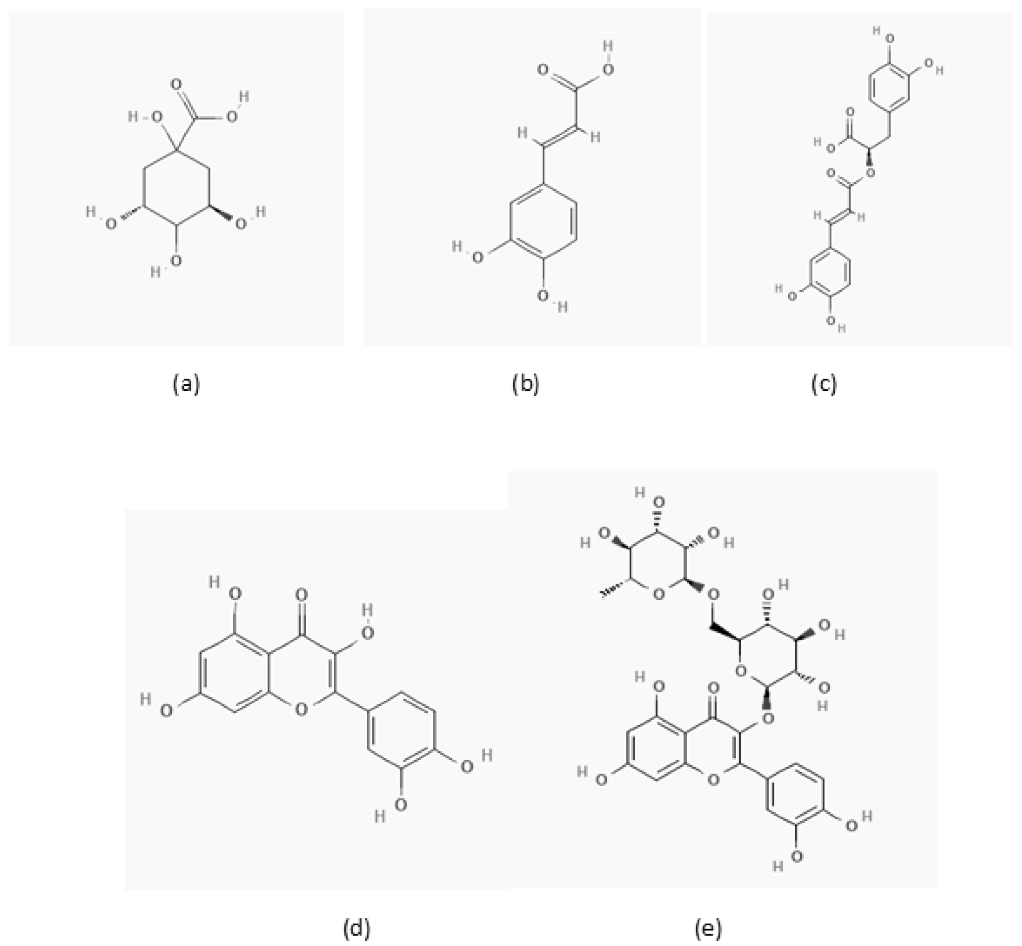
2.1. Reagents and Chemicals

Rosmarinic acid (RA) (MW 360.3 g/mol) was purchased by Extrasynthese (Genay, France), while caffeic acid (CA) (MW 180.16 g/mol), quinic acid (QC) (MW 192.17 g/mol), quercetin (QCT) (MW 302.23 g/mol), rutin (quercetin-3-rutinoside) (RU) (MW 610.5 g/mol) (Figure 1), and HPLC-MS-grade organic solvents were provided by Sigma-Aldrich (St. Louis, Mo, USA).
RPMI-1640 medium, fetal bovine serum (FBS), L-glutamine (200 mM)–penicillin (10,000 U)–streptomycin (10 mg/mL) solution, poly-L-lysine solution, and an in vitro resazurin-based toxicology assay kit were bought from Merck (Darmstadt, Germany). Caco-2 cells were purchased from the European Collection of Authenticated Cell Cultures (Salisbury, UK). MTS-based Cell Titer 96 AQueous One Solution Cell Proliferation Assay was provided by Promega (Madison, WI, USA).
LC-Grade water was obtained from a Millipore Direct-Q® System (Merck Millipore, Milan, Italy).

2.2. Sample Preparation

Standard solutions (1 or 2 mg/mL) were prepared in LC-grade water (for QC) or ethanol (for CA, RA, RU, and QCT), and then diluted to 0.1 mg/mL in the medium before starting with the experiments.

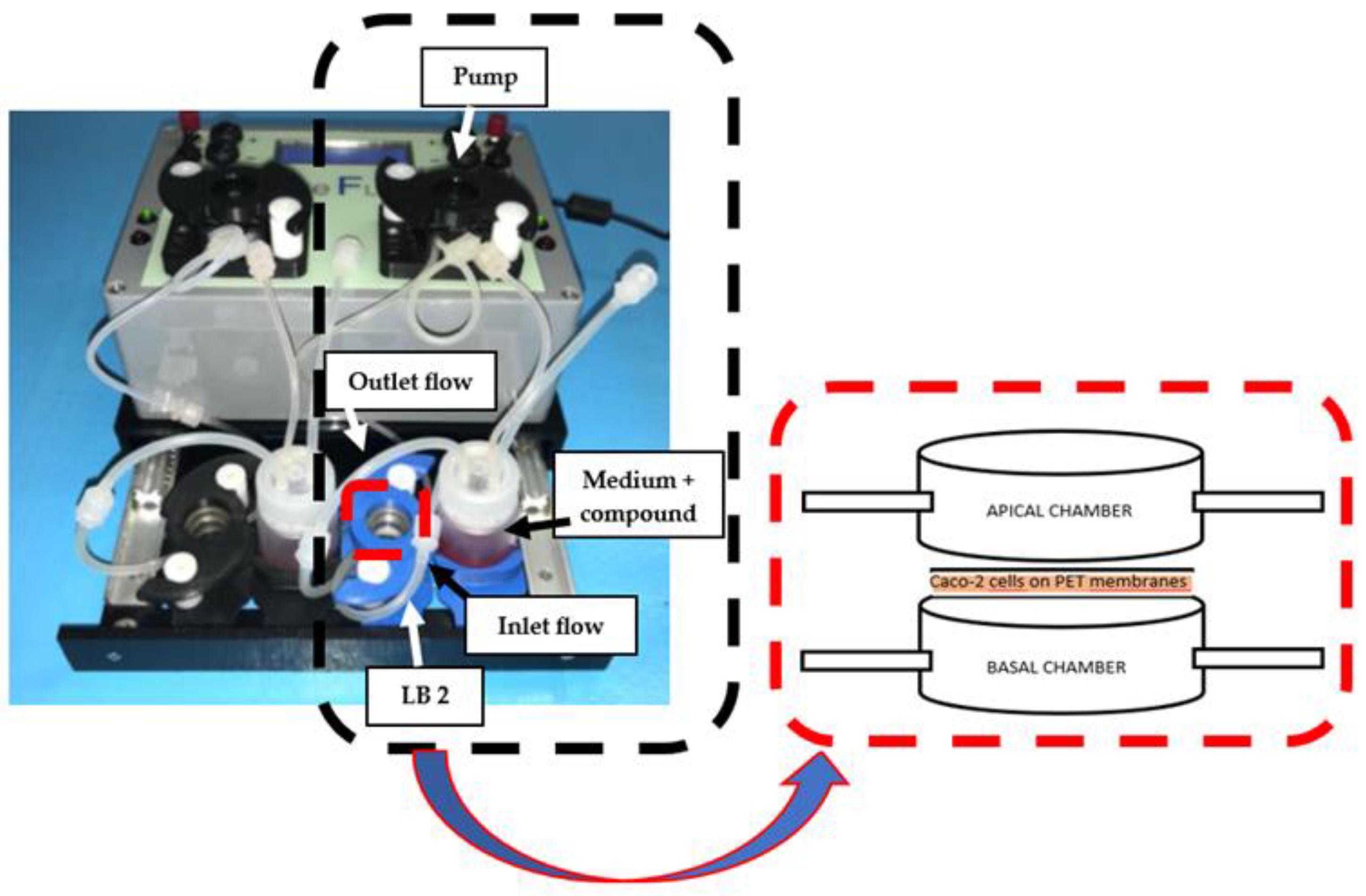
2.3. Dynamic Bioreactor Set-Up

A LiveFlow® system was purchased from IVTech Srl (IVTech Srl., Massarosa, LU, Italy) together with a multi-compartmental modular chamber, namely LB2, consisting of two flow inlets and outlets and two compartments (apical and basal). This chamber was projected to simulate physiological barriers, as intestinal membranes, and to interconnect dynamic cell cultures.
LB2 was set-up in a cell-culture incubator with Caco-2 cells, which were seeded on a permeable PET membrane (0.45 μm diameter, Delchimica, Naples, Italy) and the chamber was configurated to reproduce a tangential flow (see Figure 2). The flow rate was set at 150 μL/min and the RPMI-1640 medium flowed through LB2 for 48 h before the experiments. Then, each polyphenol solution (0.1 mg/mL) was added to the medium, circulating for 24 h. Time 0 (t0) was considered as the concentration of each molecule before the contact with Caco-2 cells; two withdrawals (extracellular concentrations) from LB2 were then collected and monitored by RP-HPLC-DAD over time (1, 2, 4, 6, and 24 h). All samples were filtered on regenerated cellulose (RC) membrane filters (pore size: 0.2 μm) supplied by Phenomenex® (Torrance, CA, USA) before the HPLC analysis. The experiments were replicated six times.

2.4. Cell Cultures and Cell Viability Assays

To set up the experimental conditions and find out the compounds’ concentration to be used in dynamic experiments, three concentrations of each selected compound were used to test cell viability by MTS assay (Promega). In brief, Caco-2 cells were grown as reported in [30] and plated in 96 multiwells at a density of 5000 cells/cm2 in the presence of 5% fetal bovine. Three concentrations for each compound were tested (0.02, 0.1, and 0.2 mg/mL). After incubation time (6 and 24 h), the MTS Cell Titer 96 AQueous One Solution (20 µL) was added to the wells, and the cells were incubated for 2 h at 37 °C. The absorbance was read at 450 nm by a multiwell plate reader (HT Synergy). Eight wells were used for each experimental point and each independent experiment was repeated six times. Cell viability was measured as percentage of viable cells in presence of each compound compared to controls (untreated cells). For all the experiments, the controls exhibited cell viability % in the range of 100.00 ± 2.87–100.00 ± 8.48.
For experiments under dynamic conditions, Caco-2 cells were plated in the millifluidic chambers at a density of 5000 cells/cm2 under the same growth conditions and treated with the indicated concentrations of each compound for 6 and 24 h.

2.5. RP-HPLC-DAD Analyses and Methods Validation

Chromatographic analyses of the different withdrawals were carried out on a 1200 series HPLC system (Agilent Technologies, Santa Clara, CA, USA), equipped with a quaternary gradient pump, a degasser, an autosampler, a thermostatted column oven set at 30.0 ± 0.5 °C, and a diode-array detector (DAD). Data acquisition was performed using ChemStation software (B.04.01). Separations were carried out on a Gemini C-18 column (150 × 2.1 mm, i.d.; 5 μm; 110 Å, Phenomenex, Torrance, CA, USA) with an XSelect HSS T3 Vanguard column (5 × 2.1 mm i.d.; 3.5 um; 100 Å, Waters Milford, MA, USA) at a constant flow rate of 0.3 mL/min (injection volume: 10 μL). A binary mobile phase consisting of 0.1% formic acid aqueous solution and methanol was used at different volume ratios according to the tested molecule: caffeic and quinic acids 65/35, v/v; rosmarinic acid 55/45, v/v; quercetin and rutin 35/65, v/v. All the elutions were carried out in isocratic mode. Chromatograms were recorded at 325 nm for caffeic acid, quinic acid, and rosmarinic acid and at 360 nm for quercetin and rutin. Each withdrawal at each monitoring time was analyzed in triplicate.
According to ICH guidelines [32], different parameters were tested. To confirm specificity, the chromatographic profiles of the medium and of each compound in the medium were compared and no peak was observed in the medium at the retention time of each compound. Calibration curves produced on the same day and over three consecutive days by plotting the peak area of the medium spiked with each compound at five different experimental concentrations (range 0.02–0.2 mg/mL) injected three times vs. the theoretical concentration were used to evaluate the linearity. All R2 values were higher than 0.9900. Limit of detection (LOD) and limit of quantification (LOQ) for all the compounds were in the range of 0.002–0.004 mg/mL and 0.01–0.02 mg/mL, respectively.
The method’s accuracy (intra- and inter-day) was calculated for each analyte by analyzing in triplicate the three different concentration levels (0.02, 0.1, and 0.2 mg/mL) over three consecutive days; it ranged from 92.10% to 103.85% for all the compounds. To determine the method’s precision, the same three concentration levels for each analyte were analyzed six times within a single day (intra-day assay) and in triplicate each day for three consecutive days (inter-day assay). Precision values were lower than 2.0%.

2.6. Statistical Analysis

Mean and standard deviation were always calculated for six replicated experiments. Differences were considered significant at p ≤ 0.05 (analysis of variance, ANOVA). All statistical analyses were carried out using Microsoft Excel 2010.

3. Results and Discussion

Caffeic acid, quinic acid, rosmarinic acid, quercetin, and rutin (Figure 1) were selected because of the recent great interest in them for dietary polyphenols and their supplementation, as they are known to exhibit several biological activities, mainly antioxidant and anti-inflammatory properties, and for their direct effect on the intestinal environment modulation. These compounds are widely distributed in vegetable foods and beverages. In particular caffeic and quinic acids are the most abundant phenolic acids in coffee beans, while rosmarinic acid is present in different aromatic herbs (Lamiaceae family), and many fruits (e.g., grapes, apricots, apples, cherries, and blackberries) are rich in quercetin and rutin [33,34,35,36]. Despite the abundance in many different foods, their intestinal absorption is low with important implications for their bioactivities.
Therefore, Caco-2 cells seeded on a permeable PET membrane in the dynamic bioreactor were used to better investigate the absorption mechanism of these compounds possessing similar chemical features, and in order to verify the results reported in the literature on static Caco-2 cell models. Cells were plated in millifluidic chambers and treated with the compounds described above at a concentration of 0.1 mg/mL for 6 and 24 h, under a medium flux of 150 μL/min. During the incubation, withdrawals were made at increasing contact times (1, 2, 4, 6 and 24 h) and the samples were analyzed by an RP-HPLC-DAD method, in which mobile phase composition was modified for each compound, to test the quantity and trend of absorption. In addition, cell viability under dynamic conditions was evaluated after 6 and 24 h contact by the Alamar Blue method and no toxicity was observed.
The tested concentration (0.1 mg/mL) was fixed after cell viability experiments, considering the potential daily intake of each compound.

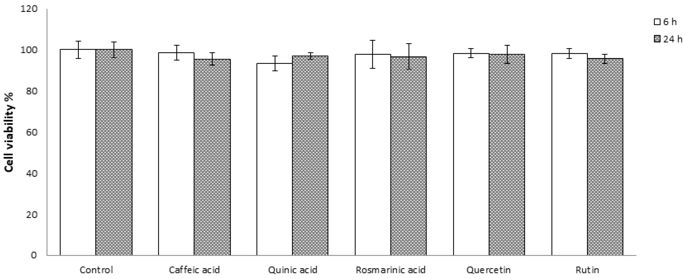
3.1. Effect of the Tested Molecules on Caco-2 Cells Viability

Preliminary cytotoxicity tests on the selected molecules were performed to define the concentrations to be used in the absorption studies. Therefore, three different concentrations of compounds (0.02, 0.1, and 0.2 mg/mL) were evaluated by an MTS-based assay. No cytotoxic effect was observed for any of the tested molecules at any concentration, as evident by cell viability % registered in comparison to the control after 6 and 24 h of incubation. The intermediate concentration (0.1 mg/mL) (Figure 3) was thus selected for the absorption test, as it was considered safe on Caco-2 cells to avoid any cell viability impairment on the absorption data.

3.2. Absorption Evaluation Using the In Vitro Dynamic Intestinal Model

All the dietary polyphenols are characterized by an intestinal metabolization and a low intestinal absorption, which represent the main reason for the decrease in their bioactivities registered in the literature. Conversely, they seem to be stable at the gastric level in the presence of acidic conditions. In addition, polyphenol stability and bioaccessibility are strictly dependent on their chemical class and the composition and structure of the food matrix. Another important issue affecting the absorption data reported in the literature is the applied digestion method; in fact, the bioaccessibility values can vary widely in relation to the approach used to measure them [37,38,39].
In this study, a dynamic system was used to set-up the digestion key-step (i.e., the intestinal chamber (Figure 2)) by using Caco-2 cells, which are a well-known traditional model to study epithelial barrier function and transepithelial transporters expressed in these cells, such as P-glycoprotein (Pgp) and multidrug-resistant proteins (MRPs); these proteins clearly participate in the intestinal transport and absorption mechanisms of compounds [2]. To assess the functionality of the Caco-2 epithelial barrier plated in the millifluidic chambers, Transepithelial Electrical Resistance (TEER) [40] was measured at the starting (t0) and end (24 h) points of the experiment to monitor the intestinal barrier intercellular junctional integrity and the possible damage of the cellular monolayer over time. TEER values were measured by a voltmeter Millicell® ERS-2 Voltohmmeter (Merck Millipore). The Caco-2 monolayer generates a stable TEER measurement of 250 Ω.cm2, which is a value commonly reported in the literature for Caco-2 cells to confirm the corrected tight junction (TJ) integrity and monolayer permeability [40].
The effect of in vitro dynamic conditions on the tested molecule absorption was then investigated by collecting and analyzing the medium circulating in the system after the addition of the tested compounds, at a prefixed time. The analyses were performed by an RP-HPLC-DAD method, adapting the binary mobile phase to the different compounds with slight modifications to the existent literature [41,42,43,44].
The percentage of each molecule in the extracellular fluid was considered as the non-absorbed fraction by Caco-2 cells and it was calculated at different times (from 1 to 24 h), considering the initial concentration (t0 = 0.1 mg/mL) as 100% of not-absorbed compound.
The results obtained for each tested compound at different monitoring times are reported in Table 1.
The chromatographic profiles registered during the monitoring period indicated that no metabolites were present, and therefore, the quantification was performed considering only the peak related to the tested molecule (Figures S1–S4).
The most important monitoring time was 6 h, as it represents the time required for food to pass from the small intestine to the colon (normal range: 5–6 h), while 24 h can be considered the time required for a complete digestion [45].
Quinic acid (QA) (Figure 1a) was completely absorbed after 1 h as evident from the chromatographic profile where no peak was detected. In fact, 0.1 mg/mL QA was not toxic (Figure 3), and therefore, no loss of Caco-2 cell membrane integrity that could affect the absorption mechanism can be speculated, and apparently the obtained result could be attributed to a total absorption. However, the analysis of t0 sample also revealed no peak, and therefore, QA behavior does not seem to depend on the contact between the compound and cell culture. In the literature, a high permeability has been attributed to QA, but with a resulting low intestinal absorption. Its absorption mechanism is not yet well known, and only recently some authors have suggested that QA controversial data on permeability and absorption could be explained by an active efflux mechanism, acting on QA with a potential saturation. This mechanism seems to be concentration dependent, as demonstrated by experiments on Caco-2 cells testing low concentrations (≥10 µM) [46].
Our results could be due to QA potential interaction with the RPMI-1640 medium, giving the formation of stable complexes, often polymeric structures, which could involve a QA α-hydroxycarboxylic group in which its electron-attractive nature increases the strength of the carboxylic acid [47,48]. This does not affect QA solubility, but compromises its detection by RP-HPLC-DAD using our method.
Caffeic acid (CA) (Figure 1b) is well known to have a very low permeability across Caco-2 cells and simulated intestinal digestion studies present in the literature have shown that CA is one of the least bioaccessible polyphenols. Two different absorption mechanisms have been proposed: one is an active transport by the monocarboxylic acid transporter (MCT), and the other via paracellular diffusion (as chlorogenic acid) [39,49,50,51]. In particular, in vitro experiments on Caco-2 cells performed in static conditions and in vivo experiments on animal models have shown that CA can be classified as a poorly absorbed compound (intestinal absorption: 0–20%) in the first hour of the intestinal digestion phase [49], as our results confirmed. In fact, 91.48% ± 4.92 and 86.14% ± 7.15 of CA was present in the medium after 4 and 6 h of contact with Caco-2 cells, respectively. Therefore, the absorbed CA fraction by Caco-2 cells was approximately 10–15% (Table 1).
Rosmarinic acid (RA) (Figure 1c) has been classified as having a low intestinal permeability as well, but it can be considered one of the most bioaccessible polyphenols (50% absorbed compound). The paracellular diffusion seemed to be the main absorption process, but an active transport has also been supposed [39,52,53,54]. Efflux transporters and TJ proteins seemed to have an important role in the RA absorption [53]. In vivo studies have confirmed that RA is quite rapidly metabolized during digestion [54]. In particular, literature data have shown that RA has a very high resistance to digestion at the intestinal level, thanks to its stability at neutral/alkaline pH. According to Zorić et al., RA stability rate, corresponding to its concentration before and after in vitro intestinal digestion ratio, was approximately 78% after 2 h, as also confirmed by our data indicating 86.23% ± 4.05 non-absorbed RA at 2 h (Table 1) [55]. Our results indicated that the absorption increases over time reaching approximately 40% after 6 h and it exhibited a complete metabolization after 24 h in accordance with data obtained on animal models [56].
In particular, quercetin (QRC) and rutin (RU) have a great and important impact on the gastrointestinal tract, and QRC was especially reported to protect and stabilize TJ proteins of the Caco-2 cell monolayer [36]. Literature data have shown that QCT (Figure 1d) uptake by Caco-2 cells should be approximately 50%, as confirmed by our results obtained using the dynamic system in which 50–60% QRC was absorbed after 4–6 h. These results highlighted its quick intestinal absorption starting from 4 h of contact with cells (Table 1) [37,57], which reaches a complete metabolization after 24 h and could be ascribed to its strong metabolization into quercetin glucuronide [37,58].
Nowadays, RU (Figure 1e) is considered the least absorbed flavonoid, notwithstanding the existence of very different bioaccessibility results in the literature, because it particularly depends on the analyzed concentration [37,59]. Our dynamic set-up confirmed that RU is not absorbed until 6 h when non-absorbed RU% is 90.74 ± 4.70, and that its absorption remained constant over time till 24 h when non-absorbed RU% was 86.85 ± 8.63. This indicated that RU permeation across the Caco-2 cell monolayer was almost not detected, which be due not only to the lack of Caco-2 cell permeability, but also to an efflux permeability mechanism. In fact, the measurement of apparent RU permeability coefficients in the static Caco-2 cell model has been a well-known indication of non-existent or low permeation across the intestinal barrier with the potential role of PgP, which is an efflux transporter [37,59].

4. Conclusions

Our data show that cell cultures in a millifluidic system can be easily used for absorption studies under dynamic conditions, which can be predictive of the in vivo environment. The use of Caco-2 cells in bioreactors can clarify bioaccessibility results obtained under static conditions. In fact, this platform can be useful for screening and testing nutraceuticals and nutrients instead of or for a comparison with traditional absorption tests. The versatility of this millifluidic system and its modular chambers also opens the possibility to set-up more sophisticated tools, which could be validated as real-time platforms for monitoring absorption and for testing bioactivities of different molecules.

Supplementary Materials

The following supporting information can be downloaded at: https://www.mdpi.com/article/10.3390/app13085033/s1, Figure S1: Chromatographic profiles obtained by RP-HPLC-DAD of caffeic acid using the dynamic intestinal model. Samples were injected at different digestion times. From top to bottom: undigested sample at (t0), 2, 4, 6 and 24 h. Figure S2. Chromatographic profiles obtained by RP-HPLC-DAD of rosmarinic acid using the dynamic intestinal model. Samples were injected at different digestion times. From top to bottom: undigested sample at (t0), 2, 4, 6 and 24 h. Figure S3. Chromatographic profiles obtained by RP-HPLC-DAD of quercetin using the dynamic intestinal model. Samples were injected at different digestion times. From top to bottom: undigested sample at (t0), 2, 4, 6 and 24 h. Figure S4. Chromatographic profiles obtained by RP-HPLC-DAD of rutin using the dynamic intestinal model. Samples were injected at different digestion times. From top to bottom: undigested sample at (t0), 2, 4, 6 and 24 h.

Author Contributions

Conceptualization, R.C. and A.P.; methodology, R.C. and M.P.; formal analysis: R.C. and M.P.; investigation, R.C. and M.P.; data curation, R.C. and M.P.; writing—original draft preparation, R.C.; writing—review and editing, R.C. and A.P.; supervision, A.P.; funding acquisition, R.C. and A.P. All authors have read and agreed to the published version of the manuscript.

Funding

This research received no external funding.

Institutional Review Board Statement

Not applicable.

Informed Consent Statement

Not applicable.

Data Availability Statement

The data presented in this study are available on request from the corresponding author.

Acknowledgments

The authors thank “Fondo Ricerca e Giovani (FRG 2021)” (Department of Drug Sciences, University of Pavia) for the contribution awarded to Raffaella Colombo as a partial financial support in the purchasing of materials used for the experiments.

Conflicts of Interest

The authors declare no conflict of interest.

References

  1. Sunantha, K.; Reginio, F.C., Jr.; Thuengtung, S.; Ogawa, Y. Changes in bioactive compounds and antioxidant activity of plant-based foods by gastrointestinal digestion: A review. Crit. Rev. Food Sci. Nutr. 2022, 62, 4684–4705. [Google Scholar]
  2. Verhoeckx, K.; Cotter, P.; López-Expósito, I.; Kleiveland, C.; Lea, T.; Mackie, A.; Requena, T.; Swiatecka, D.; Wichers, H. The Impact of Food Bio-Actives on Gut Health: In Vitro and Ex Vivo Models; Springer: Berlin/Heidelberg, Germany, 2015. [Google Scholar]
  3. Lucas-González, R.; Viuda-Martos, M.; Pèrez-Alvarez, J.A.; Fernàndez-Lòpez, J. In vitro digestion models suitable for foods: Opportunities for new fields of application and challenges. Food Res. Int. 2018, 107, 423–436. [Google Scholar] [CrossRef] [PubMed]
  4. Van De Wiele, T.R.; Oomen, A.G.; Wragg, J.; Cave, M.; Minekus, M.; Hack, A.; Cornelis, C.; Rompelberg, C.J.; De Zwart, L.L.; Klinch, B.; et al. Comparison of five in vitro digestion models to in vivo experimental results: Lead bioaccessibility in the human gastrointestinal tract. J. Environ. Sci. Health 2007, 42, 1203–1211. [Google Scholar] [CrossRef] [PubMed]
  5. Hur, S.J.; Lim, B.O.; Decker, E.A.; McClements, D.J. In vitro human digestion models for food applications. Food Chem. 2011, 125, 1–12. [Google Scholar] [CrossRef]
  6. Colombo, R.; Ferron, L.; Frosi, I.; Papetti, A. Advances in static in vitro digestion models after the COST action Infogest consensus protocol. Food Funct. 2021, 12, 7619–7636. [Google Scholar] [CrossRef]
  7. Minekus, M.; Alminger, M.; Alvito, P.; Ballance, S.; Bohn, T.; Bourlieu, C.; Carrière, F.; Boutrou, R.; Corredig, M.; Dupont, D.; et al. A standardized static in vitro digestion method suitable for food—An international consensus. Food Funct. 2014, 5, 1113–1124. [Google Scholar] [CrossRef]
  8. Brodkorb, A.; Egger, L.; Alminger, M.; Alvito, P.; Assunção, R.; Ballance, S.; Bohn, T.; Bourlieu-Lacanal, C.; Boutrou, R.; Carrière, F.; et al. Infogest static in vitro simulation of gastrointestinal food digestion. Nat. Protoc. 2019, 14, 991–1040. [Google Scholar] [CrossRef]
  9. Cost-Infogest.eu. Available online: http://www.cost-infogest.eu (accessed on 27 March 2023).
  10. Egger, L.; Schlegel, P.; Baumann, C.; Stoffers, H.; Guggisberg, D.; Brügger, C.; Dürr, D.; Stoll, P.; Vergères, G.; Portmann, R. Physiological comparability of the harmonized INFOGEST in vitro digestion method to in vivo pig digestion. Food Res. Int. 2017, 102, 567–574. [Google Scholar] [CrossRef]
  11. Hamzalıoğlu, A.; Gökmen, V. Investigations on the reactions of α-dicarbonyl compounds with amino acids and proteins during in vitro digestion of biscuits. Food Funct. 2016, 6, 109–114. [Google Scholar] [CrossRef]
  12. Egger, L.; Ménard, O.; Delgado-Andrade, C.; Alvito, P.; Assunção, R.; Balance, S.; Bradkorb, A.; Cattenoz, T.; Clemente, A.; Comi, I.; et al. The harmonized INFOGEST in vitro digestion method: From knowledge to action. Food Res. Int. 2016, 88, 217–225. [Google Scholar] [CrossRef]
  13. Ménard, O.; Bourlieu, C.; De Oliveira, S.C.; Dellarosa, N.; Laghi, L.; Carrière, F.; Capozzi, F.; Dupont, D.; Deglaire, A. A first step towards a consensus static in vitro model for simulating full-term infant digestion. Food Chem. 2018, 240, 338–345. [Google Scholar] [CrossRef] [PubMed]
  14. Calvo-Lerma, J.; Fornés-Ferrer, V.; Heredia, A.; Andrés, A. In vitro digestion models to assess lipolysis: The impact of the simulated conditions of gastric and intestinal pH, bile salts and digestive fluids. Food Res. Int. 2019, 125, 108511. [Google Scholar] [CrossRef] [PubMed]
  15. Wikman-Larhed, A.; Artursson, P. Co-cultures of human intestinal goblet (HT29-H) and absorptive (Caco-2) cells for studies of drug and peptide abs orption. Eur. J. Pharm. Sci. 1995, 3, 171–183. [Google Scholar] [CrossRef]
  16. Lennernäss, H. Intestinal permeability and its relevance for absorption and elimination. Xenobiotica 2007, 37, 1015–1051. [Google Scholar] [CrossRef] [PubMed]
  17. Cai, X.; Patel, S.; Huang, C.; Paiva, A.; Sun, Y.; Barker, G.; Weller, H.; Shou, W. Comprehensive characterization and optimization of Caco-2 cells enabled the development of a miniaturized 96-well permeability assay. Xenobiotica 2022, 52, 742–750. [Google Scholar] [CrossRef]
  18. Minekus, M.; Marteau, P.; Havenaar, R.; Huis in’t Veld, J.H. A multicompartmental dynamic computer-controlled model simulating the stomach and small intestine. Altern. Lab. Anim. 1995, 23, 197–209. [Google Scholar] [CrossRef]
  19. Kong, F.; Singh, R.P.A. A Human Gastric Simulator (HGS) to study food digestion in human stomach. J. Food Sci. 2010, 75, 625–635. [Google Scholar] [CrossRef]
  20. Wickham, M.J.S.; Faulks, R.M.; Mann, J.; Mandalari, G. The design, operation, and application of a dynamic gastric model. Dissolution Technol. 2012, 19, 15–22. [Google Scholar] [CrossRef]
  21. Huh, D.; Torisawa, Y.S.; Hamilton, G.A.; Kim, H.J.; Ingber, D.E. Microengineered physiological biomimicry: Organs-on-chips. Lab Chip 2012, 12, 2156–2164. [Google Scholar] [CrossRef]
  22. Langhans, S.A. Three-dimensional in vitro cell culture models in drug discovery and drug repositioning. Front. Pharmacol. 2018, 23, 6. [Google Scholar] [CrossRef]
  23. Mazzei, D.; Giusti, S.; Sbrana, T.; Ahluwalia, A. Multicompartmental modular bioreactor as innovative system for dynamic cell cultures and co-cultures. In Bioreactors: Design, Properties and Applications; Antolli, P.G., Liu, Z., Eds.; Nova Science Publishers: Hauppauge, NY, USA, 2011; pp. 159–178. [Google Scholar]
  24. Giusti, S.; Sbrana, T.; La Marca, M.; Di Patria, V.; Martinucci, V.; Tirella, A.; Domenici, C.; Ahluwalia, A. A novel dual-flow bioreactor simulates increased fluorescein permeability in epithelial tissue barriers. Biotechnol. J. 2014, 9, 1175–1184. [Google Scholar] [CrossRef] [PubMed]
  25. Jung, S.-M.; Kim, S. In vitro models of the small intestine for studying intestinal diseases. Front. Microbiol. 2022, 12, 767038. [Google Scholar] [CrossRef]
  26. Carton, F.; Malatesta, M. In vitro models of biological barriers for nanomedical research. Int. J. Mol. Sci. 2022, 23, 8910. [Google Scholar] [CrossRef] [PubMed]
  27. Fedi, A.; Viatle, C.; Ponschin, G.; Ayehunie, S.; Fato, M.; Scaglione, S. In vitro models replicating the human intestinal epithelium for absorption and metabolism studies: A systematic review. J. Control. Release 2021, 335, 247–268. [Google Scholar] [CrossRef]
  28. Cacopardo, L.; Costa, J.; Giusti, S.; Buoncompagni, L.; Meucci, S.; Corti, A.; Mattei, G.; Ahluwalia, A. Real-time cellular impedance monitoring and imaging of biological barriers in a dual-flow membrane bioreactor. Biosens. Bioelectron. 2019, 140, 111340. [Google Scholar] [CrossRef]
  29. Costa, J.; Almonti, V.; Cacopardo, L.; Poli, D.; Rapposelli, S.; Ahluwalia, A. Investigating curcumin/intestinal epithelium interaction in a millifluidic bioreactor. Bioengineering 2020, 7, 100. [Google Scholar] [CrossRef]
  30. Colombo, R.; Paolillo, M.; Papetti, A. A new millifluidic-based gastrointestinal platform to evaluate the effect of simulated dietary methylglyoxal intakes. Food Funct. 2019, 10, 4330–4338. [Google Scholar] [CrossRef]
  31. Colombo, R.; Paolillo, M.; Frosi, I.; Ferron, L.; Papetti, A. Effect of the simulated digestion process on the chlorogenic acid trapping activity against methylglyoxal. Food Funct. 2023, 14, 541–549. [Google Scholar] [CrossRef]
  32. ICH Guideline Q2(R1), Validation of Analytical Procedures: Text and Methodology Guidance for Industry; 2021. Available online: https://www.fda.gov/regulatory-information/search-fda-guidance-documents/q2r1-validation-analytical-procedures-text-and-methodology-guidance-industry (accessed on 15 January 2022).
  33. Rudrapal, M.; Khairnar, S.J.; Khan, J.; Dukhyil, A.B.; Ansari, M.A.; Alomary, M.N.; Alshabrmi, F.M.; Palai, S.; Deb, P.K.; Devi, R. Dietary polyphenols and their role in oxidative stress-induced human diseases: Insights into protective effects, antioxidant potentials and mechanism(s) of action. Front. Pharmacol. 2022, 13, 806470. [Google Scholar] [CrossRef]
  34. Alam, M.; Ashraf, G.M.; Sheikh, K.; Khan, A.; Ali, S.; Ansari, M.M.; Adnan, M.; Pasupuleti, V.R.; Hassan, M.I. Potential therapeutic implications of caffeic acid in cancer signaling: Past, present, and future. Front. Pharmacol. 2022, 13, 845871. [Google Scholar] [CrossRef] [PubMed]
  35. Cheng, Z.; Wang, Y.; Li, B. Dietary polyphenols alleviate autoimmune liver disease by mediating the intestinal microenvironment: Challenges and hopes. J. Agric. Food Chem. 2022, 70, 10708–10737. [Google Scholar] [CrossRef]
  36. Oteiza, P.I.; Fraga, C.G.; Mills, D.A.; Taft, D.H. Flavonoids and the gastrointestinal tract: Local and systemic effects. Mol. Aspects Med. 2018, 61, 41–49. [Google Scholar] [CrossRef] [PubMed]
  37. Tian, X.J.; Yang, X.W.; Yang, X.; Wang, K. Studies of intestinal permeability of 36 flavonoids using Caco-2 cell monolayer model. Int. J. Pharm. 2009, 367, 58–64. [Google Scholar] [CrossRef]
  38. Pineda-Vadillo, C.; Nau, F.; Dubiard, C.G.; Cheynier, V.; Meudec, E.; Sanz-Buenhombre, M.; Guadarrama, A.; Tóth, T.; Csavajda, É.; Hingyi, H.; et al. In vitro digestion of dairy and egg products enriched with grape extracts: Effect of the food matrix on polyphenol bioaccessibility and antioxidant activity. Food Res. Int. 2016, 88, 284–292. [Google Scholar] [CrossRef]
  39. Gayoso, L.; Claerbout, A.S.; Calvo, M.I.; Cavero, R.Y.; Astiasarán, I.; Ansorena-Artieda, D. Bioaccessibility of Rutin, Caffeic Acid and Rosmarinic Acid: Influence of the in Vitro Gastrointestinal Digestion Models. J. Funct. Foods 2016, 26, 428–438. [Google Scholar] [CrossRef]
  40. Srinivasan, B.; Kolli, A.R.; Esch, M.B.; Abaci, H.E.; Shuler, M.L.; Hickman, J.J. TEER measurement techniques for in vitro barrier model systems. J. Lab. Autom. 2015, 20, 107–126. [Google Scholar] [CrossRef] [PubMed]
  41. Magnani Spagnol, C.; Issac, V.L.; Corrêa, M.A.; Salgado, H.R. Validation of HPLC–UV assay of caffeic acid in emulsions. J. Chromatogr. Sci. 2016, 54, 305–311. [Google Scholar]
  42. Chaowuttikul, C.; Palanuvej, C.; Ruangrungs, N. Quantification of chlorogenic acid, rosmarinic acid, and caffeic acid contents in selected Thai medicinal plants using RP-HPLC-DAD. Braz. J. Pharm. Sci. 2020, 56, e17547. [Google Scholar] [CrossRef]
  43. Papetti, A.; Mascherpa, D.; Carazzone, C.; Stauder, M.; Spratt, D.A.; Wilson, M.; Pratten, J.; Ciric, L.; Lingström, P.; Zaura, E.; et al. Identification of organic acids in Cichorium intybus inhibiting virulence-related properties of oral pathogenic bacteria. Food Chem. 2013, 138, 1706–1712. [Google Scholar] [CrossRef] [PubMed]
  44. Liu, H.P.; Shi, X.F.; Zhang, Y.C.; Li, Z.X.; Zhang, L.; Wang, Z.Y. Quantitative analysis of quercetin in euphorbia helioscopia L by RP-HPLC. Cell Biochem. Biophys. 2011, 61, 59–64. [Google Scholar] [CrossRef]
  45. Camilleri, M.; Colemont, L.J.; Phillips, S.F.; Brown, M.L.; Thormforde, G.M.; Chapman, N.; Zinsmeister, A.N. Human gastric emptying and colonic filling of solids characterized by a new method. Am. J. Physiol. 1989, 257, G284–G290. [Google Scholar] [CrossRef]
  46. Mortelè, O.; Jörissen, J.; Spacova, I.; Lebeer, S.; van Nuijs, A.L.N.; Hermans, N. Demonstrating the involvement of an active efflux mechanism in the intestinal absorption of chlorogenic acid and quinic acid using a Caco-2 bidirectional permeability assay. Food Funct. 2021, 12, 417–425. [Google Scholar] [CrossRef]
  47. Bermejo, E.; Carballo, R.; Castiñeiras, A.; Lago, A.B. Coordination of α-hydroxycarboxylic acids with first-row transition ions. Coord. Chem. Rev. 2013, 257, 2639–2651. [Google Scholar] [CrossRef]
  48. Barba-Behrens, N.; Salazar-García, F.; Mara Bello-Ramírez, A.; García-Báez, E.; de Jesús Rosales-Hoz, M.; Contreras, R.; Flores-Parra, A. Novel coordination compounds of quinic acid. X-ray diffraction study of copper (II) complexes where the metal ion was a chiral centre. Trans. Metal Chem. 1994, 19, 575–581. [Google Scholar] [CrossRef]
  49. Wang, S.J.; Zeng, J.; Yang, B.K.; Zhing, Y.M. Bioavailability of caffeic acid in rats and its absorption properties in the Caco-2 cell model. Pharm. Biol. 2014, 52, 1150–1157. [Google Scholar] [CrossRef] [PubMed]
  50. Birková, A.; Hubková, B.; Bolerázska, B.; Mareková, M.; Čižmárová, B. Caffeic acid: A brief overview of its presence, metabolism, and bioactivity. Bioact. Compd. Health Dis. 2020, 3, 74–81. [Google Scholar] [CrossRef]
  51. Veras, K.S.; Fachel, F.N.S.; de Araújo, B.V.; Teixeira, H.F.; Koester, L.S. Oral pharmacokinetics of hydroxycinnamic acids: An updated erview. Pharmaceutics 2022, 14, 2663. [Google Scholar] [CrossRef] [PubMed]
  52. Konishi, Y.; Kobayashi, S. Transepithelial transport of rosmarinic acid in intestinal Caco-2 cell monolayers. Biosci. Biotechnol. Biochem. 2005, 69, 583–591. [Google Scholar] [CrossRef] [PubMed]
  53. Qiang, Z.; Ye, Z.; Hauck, C.; Murphy, P.A.; McCoy, J.A.; Widrlechner, M.P.; Reddy, M.B.; Hendrich, S. Permeability of rosmarinic acid in Prunella vulgaris and ursolic acid in Salvia officinalis extracts across Caco-2 cell monolayers. J. Ethnopharmacol. 2011, 137, 1107–1112. [Google Scholar] [CrossRef]
  54. Amoah, S.K.; Sandjo, L.P.; Kratz, J.M.; Biavatti, M.W. Rosmarinic Acid—Pharmaceutical and Clinical Aspects. Planta Med. 2016, 82, 388–406. [Google Scholar] [CrossRef]
  55. Zorić, Z.; Markić, J.; Pedisić, S.; Bučević-Popović, V.; Generalić-Mekinić, I.; Grebenar, K.; Kulišić-Bilušić, T. Stability of rosmarinic acid in aqueous extracts from different lamiaceae species after in vitro digestion with human gastrointestinal enzymes. Food Technol. Biotechnol. 2016, 54, 97–102. [Google Scholar] [CrossRef] [PubMed]
  56. Liu, Y.Q.; Cai, Q.; Liu, C.; Bao, F.W.; Zhang, Z.Q. Simultaneous determination and pharmacokinetic comparisons of multi-ingredients after oral administration of radix salviae miltiorrhizae extract, hawthorn extract, and a combination of both extracts to rats. J. Anal. Methods Chem. 2014, 2014, 617367. [Google Scholar] [CrossRef]
  57. Boyer, J.; Brown, D.; Liu, R.H. In vitro digestion and lactase treatment influence uptake of quercetin and quercetin glucoside by the Caco-2 cell monolayer. Nutr. J. 2005, 4, 1–15. [Google Scholar] [CrossRef] [PubMed]
  58. Walgren, R.A.; Walle, U.K.; Walle, T. Transport of quercetin and its glucosides across human intestinal epithelial Caco-2 cells. Biochem. Pharmacol. 1998, 55, 1721–1727. [Google Scholar] [CrossRef]
  59. Zhang, X.; Song, J.; Shi, X.; Miao, S.; Li, Y.; Wen, A. Absorption and metabolism characteristics of rutin in Caco-2 cells. Sci. World J. 2013, 2013, 382350. [Google Scholar] [CrossRef] [PubMed]
Figure 1. Molecular structures of the compounds tested: caffeic acid (a), quinic acid (b), rosmarinic acid (c), quercetin (d), and rutin (e).
Applsci 13 05033 g001
Figure 2. LiveFlow® bioreactor in our set-up.
Applsci 13 05033 g002
Figure 3. Caco-2 cell viability (%) when exposed to 0.1 mg/mL of tested compounds for 6 and 24 h.
Applsci 13 05033 g003
Table 1. Percentage of the non-absorbed fraction of each compound at different monitoring times.
Time (h)
Compound (0.1 mg/mL)124624
Quinic acid00000
Caffeic acid98.48 ± 1.6296.67 ± 2.1491.48 ± 4.9286.14 ± 7.1552.52 ± 2.31
Rosmarinic acid93.15 ± 3.5886.23 ± 4.0568.20 ± 3.3859.53 ± 0.980
Quercetin99.55 ± 4.15 94.10 ± 5.3649.16 ± 3.7337.15 ± 2.680
Rutin96.67 ± 1.6696.58 ± 1.0090.98 ± 7.0890.74 ± 4.7086.85 ± 8.63
Disclaimer/Publisher’s Note: The statements, opinions and data contained in all publications are solely those of the individual author(s) and contributor(s) and not of MDPI and/or the editor(s). MDPI and/or the editor(s) disclaim responsibility for any injury to people or property resulting from any ideas, methods, instructions or products referred to in the content.

