Article

Rare Variants Residing in Novel Cis-Acting Element in Visual System Homeobox 1 and Their Contribution in the Pathogenesis of Keratoconus

by
Farhan Khashim Alswailmi
1,
Rida Khursheed Malik
2,
Mujeeb Ur Rehman Parrey
3,
Abdul Rauf Siddiqi
2,
Shaik Karimulla
1,
Abdulkareem A. Alanezi
4,
Raheel Qamar
5,6,
Maleeha Azam
2,* and
Ashfaq Ahmad
1,*
1
Department of Pharmacy Practice, College of Pharmacy, University of Hafr Al Batin, Hafr Al-Batin 39911, Saudi Arabia
2
Translational Genomics Laboratory, Department of Biosciences, COMSATS University Islamabad, Islamabad 45600, Pakistan
3
Faculty of Medicine, Northern Border University, Arar 91431, Saudi Arabia
4
Department of Pharmaceutics, College of Pharmacy, University of Hafr Al Batin, Hafr Al-Batin 39911, Saudi Arabia
5
Pakistan Academy of Sciences, Islamabad 45600, Pakistan
6
Science and Technology Sector, ICESCO, Rabat 10104, Morocco
*
Authors to whom correspondence should be addressed.
Appl. Sci. 2023, 13(8), 4888; https://doi.org/10.3390/app13084888
Submission received: 4 March 2023 / Revised: 5 April 2023 / Accepted: 11 April 2023 / Published: 13 April 2023
(This article belongs to the Special Issue Recent Advances in Pathogenesis and Management of Eye Diseases)

Abstract

:
(1) Background: The visual system homeobox 1 (VSX1) may contribute to the incidence of keratoconus (KC) in different populations. The present study investigated the role of VSX1 in autosomal recessive Pakistani families and sporadic KC patients using in silico analysis of the rare variants for the identification of the cis-acting elements in VSX1; (2) Methods: Mutation analysis of VSX1 was undertaken using Sanger sequencing of samples from seven KC families and 100 sporadic patients. In silico analysis of the rare variants and identification of cis-acting elements was determined using Human Splicing Finder (HSF), ESE finder, RESCUE-ESE and through Exon- Identity Element (EIEs) prediction software suits, combined with various algorithms to identify the effect of variations in splicing motifs; (3) Results: Screening of VSX1 did not reveal any novel mutation in the KC panel, but a synonymous polymorphism rs12480307 (c.546A>G; p.Ala182Ala) in exon three and 3′UTR rs76499395 (c.*496A>G) were observed in two separate probands. These polymorphisms were not found in any of the sporadic KC cases or 100 ethnically matched control samples. The analysis of these rare variants revealed a plausible role for these two single nucleotide polymorphisms (SNPs) in KC development through the identification of novel cis-acting elements, an exonic splicing enhancer element (ESE) and binding motifs for two micro RNAs, miRNA-203 binding and hsa-miR-3938, in the VSX1 gene structure; (4) Conclusions: Rare genetic variations in the VSX1 were found to have a potential contribution to KC development.

1. Introduction

Keratoconus is cone shaped and occurs as bilateral corneal dystrophy, which is expressed as a progressive thinning of the corneal layers of stroma [1]. The condition has a multifactorial origin and is progressive but non-inflammatory. The initial corneal thinning leads to irregular astigmatism, a prominent level of myopia and a severe refractive error in the patient [2,3], resulting in decreased visual acuity. At the initial stages of the disease, there are no obvious symptoms other than a mild loss of visual acuity. As the disease progresses, severe signs start to appear, such as Fleischer’s ring, corneal protrusion, Vogt’s striae and Munson [4]. Both eyes are affected in an asymmetric manner [5]. Globally the prevalence of keratoconus is 1 out of 2000 individuals, which varies with ethnicity having a prevalence rate in Caucasian populations of between 8.8–54.5 per 100,000 individuals [1], in a Malaysian population of 1 per 100 persons [6] and in an Iranian population 760 patients out of 100,000 individuals which is much higher than the prevalence of KC in the Western world population [7]. The same disease was studied in the United States for 48 years, and the data collected over this time reported the disease prevalence to be 54.5 patients per 100,000 individuals [8]. A study reported in 2017 showed the prevalence of disease in children in Saudi Arabia to be 4.8% [9].
Manifestation of KC usually starts in the second decade of life and progresses in severity over the third and fourth [10], while the prevalence of this disease is higher in males compared to females in certain populations [11,12]. The pattern of the disease prevalence is more sporadically oriented than familial linked, but a positive family history does contribute to the risk of the disease in the offspring. In familial linked cases, a history of the presence of inherited autosomal dominant mode is frequent when compared to X-linked or recessive inheritance [13,14]. The exact etiological cause of KC is unclear due to the varying prevalence of the disease in different races and families, and it occurs sporadically. Nonetheless, bearing in mind KC is a multifactorial disease, it can be speculated that both the environmental as well as genetic factors play their individual and specific roles in the pathogenesis of the disease [15].
Linkage analysis of the Keratoconus patient population of different ethnicities and Genome-wide association studies (GWAS) have indicated the involvement of various single nucleotide polymorphisms (SNPs) and loci in the pathogenesis of the disease [16], which includes matrix metalloproteinase (MMP-9), inhibitors of metalloproteinase 3 (TIMP-3), lysyl oxidase (LOX), collagen type VI alpha-1 chain (COL6A1), hepatocyte growth factor (HGF), superoxide dismutase (SOD), crumbs homolog 1 (CRB1), zinc finger e-box binding homeobox 1 (ZEB1) and visual system homeobox 1 (VSX1). Among all these genes, proteins and factors, VSX1 is involved in human ocular development [17] but it is also expressed in embryonic craniofacial tissue [18], the inner layer of the retina, as well as corneal tissue in the adult [19].
Despite the number of associated genes, the role of genetic deficits in the pathogenesis of keratoconus has not been explored in Pakistani families or the sporadic population. Moreover, bioinformatic information regarding the role of genetic variants and their impact on gene/protein function is severely lacking. The present study is designed to explore the contribution of the VSX1 genes in the recessive form of the disease in Pakistani KC patients and to perform bioinformatic analysis on identified variants to explore their potential contributions to KC development.

2. Materials and Methods

2.1. Sample Collection and DNA Isolation

Seven (07) autosomal recessive KC families (Figure 1) and one hundred (100) sporadic KC cases were recruited locally from hospitals in Islamabad, Pakistan. Blood sampling of the Keratoconus patients was performed irrespective of age, sex or occupation (males = 64, females = 36). The average age of onset of KC in the current cohort was 20 years. Patients with a positive family history of KC and those who experienced eye rubbing due to allergies, together with a family history of KC, were recruited into the study. Exclusion criteria from the study consisted of eye trauma and other corneal pathologies in patients where keratoconus was secondary to another primary disease, infection or those who had excessive eye rubbing.
All the recruited KC patients underwent corneal examination. This comprised an examination to determine the average Orb scan measurements, which evaluates the curvature of both anterior and posterior corneal layer. In the KC patients, it was determined to be 438.6 ± 47.7 μm (normal range~550 μm), which suggested a thinning of the cornea. Based on the measurement of corneal thinning, corneal pachymetry showed that the patients had an average thickness of 462.5 μm ± 8.0 (normal range~554.9 μm ± 7.4). The curvature of the anterior surface was measured using Keratometry, and the patients had a curvature of 52.5 ± 8.8 D (normal range 7.2 ± 0.8 D). Age, sex and occupation matched 100 healthy control individuals (males = 62, females = 38) who were also randomly recruited into the study as control subjects for the KC cohort, and these controls were naïve for any eye disease or inherited disease and had no major health issues. Once all the legal requirements and written consent had been obtained, blood samples were taken from all KC patients and control individuals. The present study complied with the Helsinki declaration, while ethical and scientific approval was obtained from the Ethics Committee/Institutional Review Board Department of Biosciences, COMSATS University Islamabad, Pakistan.
Genomic DNA was extracted from the plasma lymphocytes of the patient using the salting-out method as described previously [20]. Briefly, DNA extraction consisted of lysis of red blood cells (RBC) with erythrocyte lysis buffer (ELB) followed by the lysis of white blood cells (WBC) with Tris-Natrium chloride-ethylenediamine tetra acetate buffer (TNE), SDS and Pronase E. These samples were incubated overnight at room temperature, and DNA was salted out using NaCl salt and ethanol solution, which later then precipitated and re-suspended in TE buffer. This DNA was stored at −20 °C for further study.

2.2. VSX1 Sequencing

The exons and exon-intron boundaries of VSX1 in 9 KC family probands (Figure 1) and 100 sporadic cases were sequenced. Amplification of the probands from the KC sporadic cases was performed using the primers and conditions given in Table 1. The reaction consisted of mixing 50–70 ng of genomic DNA, 0.5 mM dNTPs, 1X ammonium sulfate Taq buffer, 2.5 U Taq DNA polymerase and GIBCO® water. Thermocycling was performed at an initial denaturation at 95 °C for 5 min, immediately followed by 35 cycles of denaturation at the same temperature (95 °C) for 30 s; primer annealing was conducted for 30 s at the respective temperatures given in Table 1 and chain extension at 72 °C for 30 s. The final extension was conducted at temperature 72 °C for 7 min.

2.3. In Silico Analysis

The exon three sequence of VSX1 was analyzed for the presence of potential cis-acting splicing-regulatory elements (SREs) using Human Splicing Finder (HSF), Exonic splice enhancer (ESE) element finder and through Exon-Identity Element (EIEs) prediction software. All three software have been widely used and cited to predict SREs. HSF predicts the effect of a mutation in an exonic region on splicing by combining twelve different algorithms, which include position weight matrices, maximum entropy and motif comparison algorithms [21]. The ESE finder identifies and predicts the ESE elements by scanning the subject’s exonic sequence for the presence of binding motifs of four SR proteins; the binding motifs are searched on the basis of position-specific scoring matrices based on frequency weighting values derived from the alignment of winner sequences [22]. Zhang et al. [23] proposed a model for the identification of cis-acting SREs which scans the subject’s sequence for exon-identity elements (EIEs) and intron-identity elements (IIEs). The program identifies EIEs and IIEs based on asymmetry in the nucleotide frequency and distribution compared to the rest of genomic DNA. The asymmetry exhibited by the SREs would have developed due to evolutionary selection pressure over the SREs, but the remainder of the nucleic acid shows a symmetric frequency of distribution of bases. Another program used was RESCUE-ESE, which uses a position weight matrix-based strategy validated experimentally and known as ESE sequences [24].
The 3′UTR sequence of VSX1 was checked using PITA (Probability of Interaction by Target Accessibility) software. PITA is a widely used algorithm for screening miRNA binding regions in the 3′UTR [25]. PITA has devised a model which successfully predicts miRNA binding sequence motifs based on the difference between free energies of unbound duplexed target mRNA with a hidden miRNA site and those of an open mRNA with a binding site accessible to the subject miRNA. The difference between two free energies is termed ΔΔG; the lower the ΔΔG value, the better will be the binding of miRNA with the target.
The 3′UTR of VSX1 was also analyzed through the miRanda algorithm. The miRanda algorithm functions, such as the Smith-Waterman alignment algorithm, to search for potential miRNA binding sequences, but instead of aligning the matching nucleotides (A-A and U-U), it searches for the target sequence based on the complementarity of base pairs (A=U or G≡C). miRanda is a widely cited algorithm, it searches for complementarity matches between miRNAs and 3′UTR targets through alignment generated by dynamic programming [26,27].

3. Results

3.1. Pedigree Analysis

The detailed interview of family members and review of previous medical records of keratoconus patients revealed the families as having a positive family history of the disease in different branches of the families. Sample collection from available affected members of the families along with their parents, followed by a pedigree analysis of the phenotype, showed the mode of inheritance to be autosomal recessive in all the seven families sampled.

3.2. Screening of VSX1

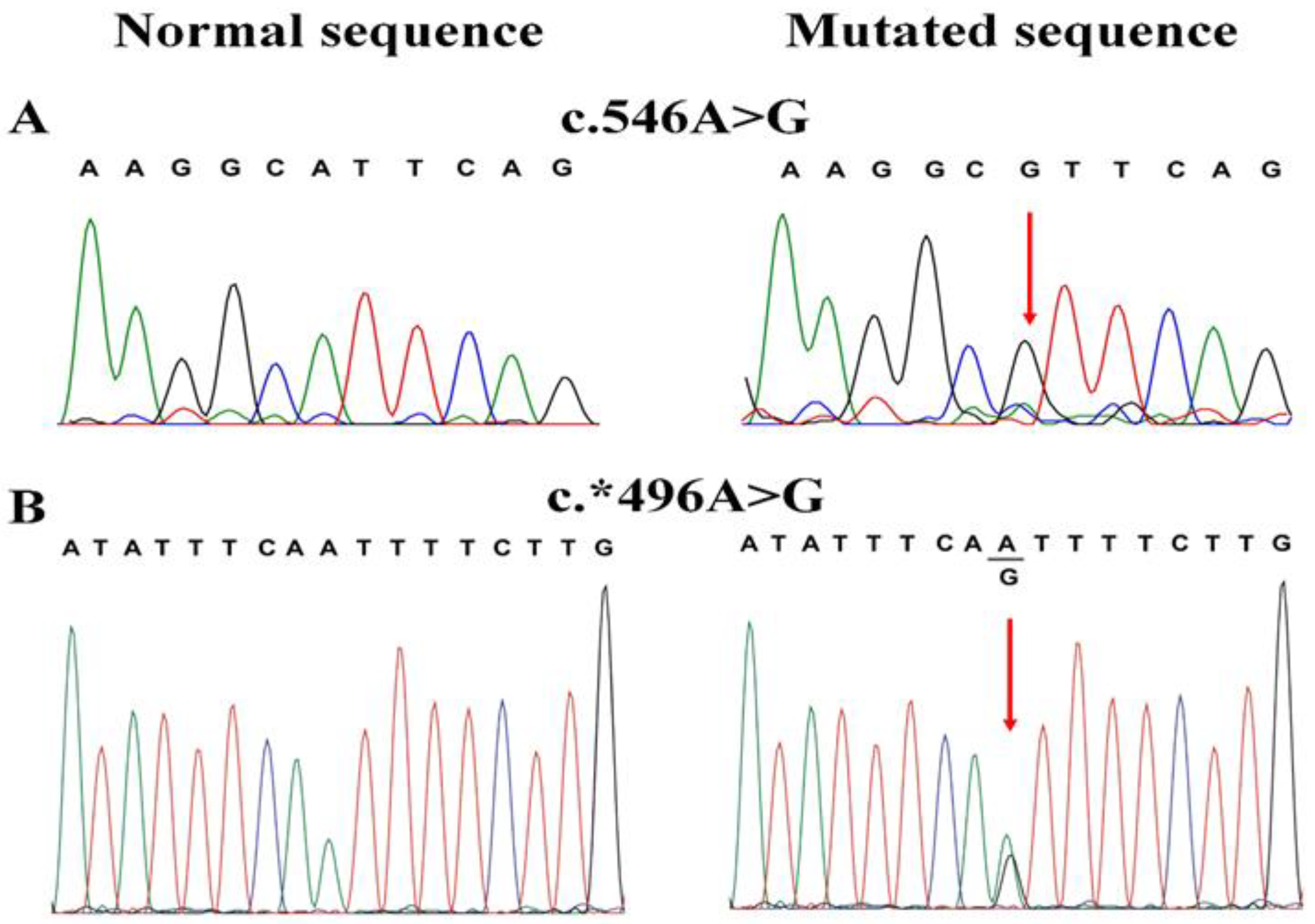
Sequencing of the VSX1 gene did not show any mutations in the seven keratoconus families. However, a single nucleotide polymorphism (SNP), rs12480307 (c.546A>G; p.Ala182Ala) in exon three with minor allele frequency (MAF) of G = 0.252, was present homozygously in the proband of a family KC2 (Figure 1 and Figure 2A) and another 3′UTR rs76499395 (c.*496A>G) with MAF = 0.02 SNP, was present heterozygously in the proband of family KC7 (Figure 1 and Figure 2B). Sequencing of VSX1 did not show any mutations in the one hundred sporadic keratoconus cases.

3.3. In Silico Analysis of c.546A>G

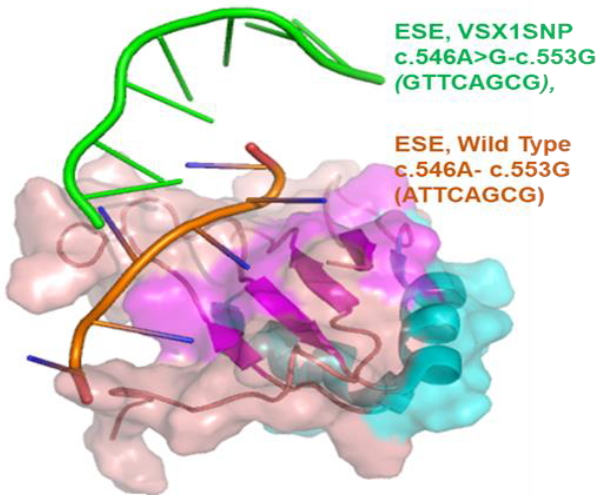
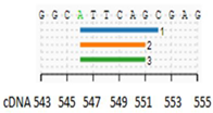
The rs12480307 (c.546A>G; p. Ala182Ala) sequence lies in the third exon of the VSX1, which is close to a splice site; therefore, the SNP, along with flanking sequences, was analyzed to determine whether these contained any ESE or ESS sequences. It was observed that the VSX1 SNP c.546A>G was altered and broke an ESE element lying in the third exon of the VSX1. This has been observed using three different splicing analytical tools used in this study. HSF predicted an ESE element starting from position VSX1 c.546, spanning over the next seven 3′ nucleotides with the sequence ATTCAGC. HSF predicted that in terms of SNP c.546A>G, the ESE element would be altered and broken. RESCUE-ESE also predicted the same ESE at position c546-552 of VSX1. The EIE identifier also predicted an exonic SRE at the same site (Table 2). The ESE finder identified a strong SRSF5 binding motif ATTCAGC (VSX1 c546-552) with a high binding score of 3.07, well above the threshold score of 2.67. In the case of SNP c.546A>G SRSF5, binding was lost for SRSF 5, but the mutant motif GTTCAGC showed a binding affinity with the SRSF2 protein (Table 3). Figure 3 illustrates the pre-mRNA analogs of the ESE elements, VSX1 Wild Type c.546A-c.553G (ATTCAGCG) and VSX1 SNP c.546A>G-c.553G (GTTCAGCG), bound to SRSF5 as revealed by docking. The wild type of ESE segment, c.546A-c.553G (ATTCAGCG), binds right in the binding pocket of the SRSF 5 protein, whereas the SNP segment, c.546A>G-c.553G (GTTCAGCG), did not bind well except for its initial 5′ region, docking nearly 3–4 Å away from the binding pocket of the SRSF 5 molecule. This would affect the splicing of the corresponding exonic region.
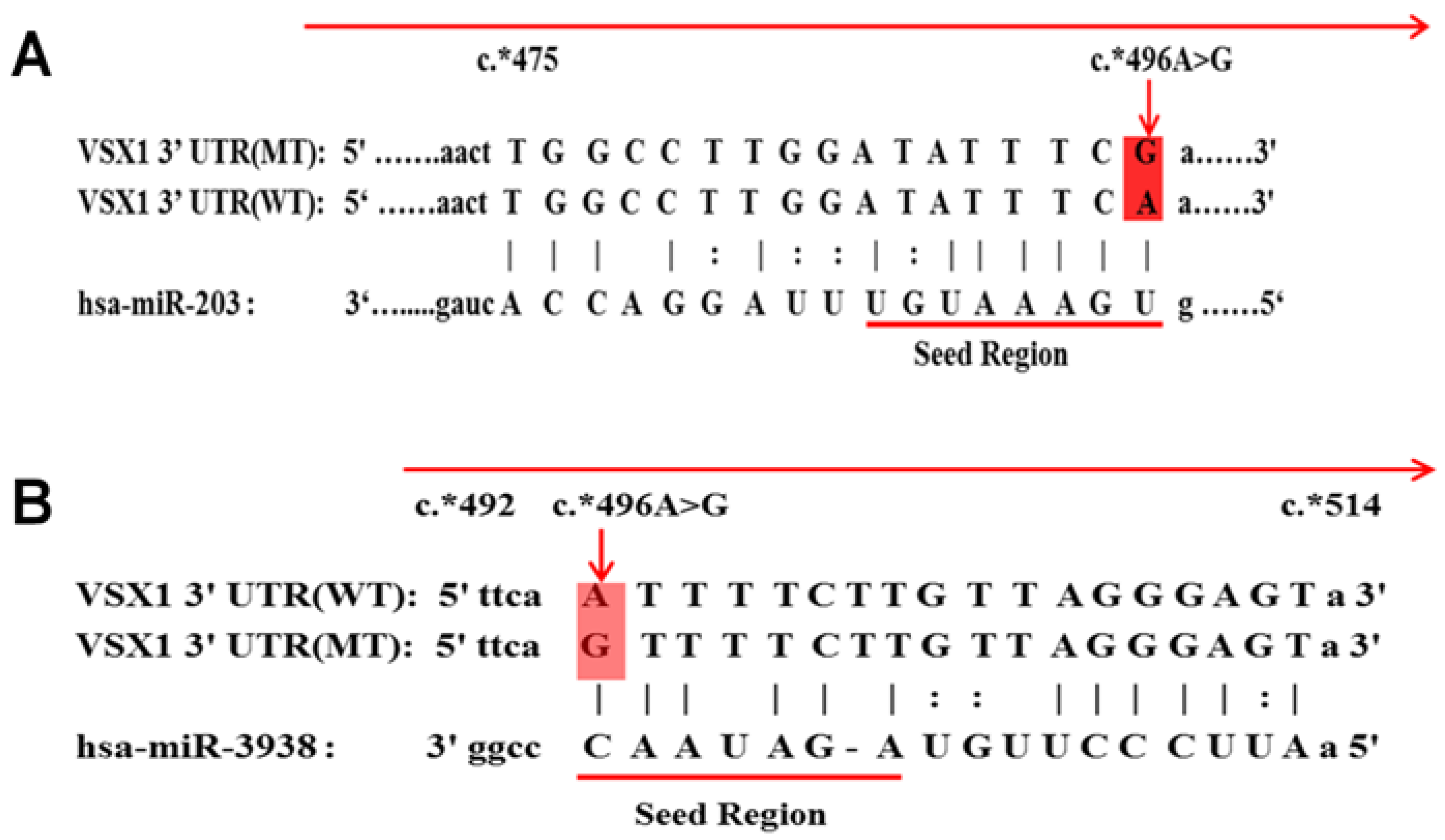
Both PITA and miRANDA predicted a miRNA binding site at VSX1 3′UTR c.*496 position (Table 4). The wildtype (WT) sequence was found to show strong binding with miRNA hsa-miR-203, and the binding site spanned across c*475 to c.*496 in VSX1 3′UTR wildtype (VSX1 3′UTR (WT) (Figure 4A). PITA estimated ΔΔG values for miRNA-203 binding with VSX1 3′UTR (WT) as −9.34, the seed region spanned over eight base pairs with all bases pairing up with target mRNA as one wobble pair. The miRNA-203 showed a minimum free energy of −15–50 kcal/mol with a binding score of 150, as determined by miRANDA. Conversely, the VSX1 3′UTR mutant type (MT), c.*496 A>G lost the binding site for miRNA-203, but instead, c.*496G was revealed as the binding site for hsa-miR-3938 from c.*496-c.*514 with a binding affinity of −12 kcal/mol and a binding score of 142 (Table 4; Figure 4A,B).

4. Discussion

This study was designed to investigate the contribution of VSX1 in autosomal recessive genes in Pakistani keratoconus families compared with that in subjects appearing as sporadic cases. This study was also set to perform an in silico analysis of the rare variants to allow for the identification of cis-acting elements in VSX1. In the present study, screening of the previously reported KC-associated gene VSX1 revealed a potential role for rare variants which might contribute to KC manifestation. This was achieved through the identification of novel cis-acting elements in VSX1 based on bioinformatic analysis of the gene.
The results of the VSX1 screening in the current panel were not in agreement with those reported earlier, where most of the cases carried mutations in the coding part of the gene and were reported for the dominant form of the disease. However, in the present cohort of autosomal recessive KC families, screening of VSX1 found no novel or recurrent mutation except for two SNPs rs12480307, a synonymous variation, and a 3′UTR rs76499395. A reasonable number of studies using VSX1 mutational screening reported VSX1 exon two as a mutation hot spot in KC cases, but the SNPs observed in two patients in the present study lie in the third exon and 3′UTR sequences. The results of the sporadic cases were also like that of the familial probands, exhibiting no VSX1 mutations. Regarding the mutation identification, the findings of the present study were consistent with those undertaken in Caucasian [28], Czech [29], Italian [30], Slovenian [31], South Indian [32] and North Indian [33] populations, in which no coding sequence VSX1 mutations were found in KC cases while ignoring very rare variants and their possible role in disease manifestation. Moreover, the expression analysis of VSX1 in human and mouse corneas also indicated contradictory findings. In humans [19], VSX1 expression in the adult cornea was not detected, but it was found to be higher in retinal tissues [34].
Although polymorphisms in different genes have been reported to be one of the risk factors in the pathogenesis of KC worldwide, a number of studies have reported that the association of SNPs with KC is low but did not investigate the underlying gene regulatory mechanisms, SREs and miRNAs which might be implicated in such associations. The identification of the ESE element (containing c.546A>G SNP) and a miRNA binding site at VSX1 3′UTR c.*496 position has highlighted the functional aspects of SNPs on cis-acting elements and their consequent effect on gene expression. Among the regulatory sequences, splicing is determined by a regulatory complex comprising a number of snRNP (small nuclear ribonucleo proteins) often named after their small nuclear RNA (snRNA) segments U1, U2, U4, U5 and U6 [35]. The role of snRNPs is to identify and bind at specific splice donor, branch and acceptor sites characterized by GU, polypyrimidine and AG nucleotides lying in the intronic region. The recruitment of these proteins is initiated by specific serine/arginine-rich (SR) proteins, the SRSF or SR (specific serine/arginine-rich) proteins. These start the recruitment process of snRNPs only after binding at specific exonic splicing enhancer (ESE) motifs lying in the flanking exonic regions of the subject introns. Twelve different SRSF proteins are known to bind at their specific ESE elements in pre-mRNA to initiate the splicing regulatory complex. Once the complex is formed, the spliceosome splices out the intronic region joining the two neighboring exons to form the mature mRNA for translation [36,37]. The current findings, therefore, clearly indicated that the VSX1 c546-552 was occupied by a highly putative ESE site, and in the case of the SNP c.546A>G, the homozygously present G allele, the ESE loses its affinity for a specific SR protein. This alteration could have consequences, such as exon skipping, for example, leading to the exclusion of an entire exon from VSX1 pre-mRNA transcript and consequently affecting gene expression. However, these findings need further investigation and validation through in vitro functional analysis.
MiR-203 is a widely studied miRNA for mediation in repressing several types of disorders, including multiple types of malignancies and cancer [38,39]. miRNA-203 has been found to suppress cell proliferation and the metastasis of several types of colorectal cancers by targeting Eukaryotic initiation factor 5A2 (EIF5A2) [40]. On the other hand, miRNA-3938 not only plays a role in cancers but has also been reported to affect macrophage cell development [41]. The loss of binding of miRNA-203 with VSX1 3′UTR c*496A>G (MT) and the binding of miRNA-3938 could be implicated in KC; however, the presence of variant c*496A>G heterozygously in KC patients could indicate a penetrance/dosage effect because one of the parents that carried the variation did not exhibit the phenotype, whereas disease manifestation was seen in offspring. The dosage effect of VSX1 variants in KC families has been observed in one previous study [42], which reported the Q175H VSX1 variant as being pathogenic with incomplete penetrance in the patient but not in the carrier mother. The genetic variants demonstrating such reduced penetrance have been reported to involve some modifier genes or environmental factors for disease presentation. It is also possible that the variant c*496A>G requires some genetic modifier or environmental factor to be pathogenic.
Like the previous studies by Aldave et al. [28], Liskova et al. [29], Tang et al. [30], Stabuc-Silih et al. [31], Verma et al. [32] and Tanwar et al. [33] on VSX1 screening in different ethnicities worldwide (Table 5), there seems a similarity in previous studies regarding the cohort selection and sample size. However, none of the studies were able to solve a complete cohort involving sporadic as well as familial cases. Most of the studies reported polymorphic variations, including synonymous as well as non-synonymous and intronic variations; however, due to the absence of in silico analysis, the exact role of the non-coding variations and microRNA was not investigated in previous studies. These contradictory results, therefore, highlight and suggest that KC cohorts, including sporadic and familial cases belonging to different ethnicities, to be genetically screened using state-of-the-art techniques, such as genome and exome sequencing to identify the exact genetic cause. These limitations in the current and previous studies, therefore, suggest thorough investigations involving larger KC cohorts and genome sequencing of KC patients. Another limitation of the current and previous findings is the lack of in vitro and in vivo functional characterization of identified genetic variants in KC patients. Regarding the cis-acting element and their predicted association with microRNA in the current study, though functional analysis could not be performed, however number of studies have reported the role of microRNA-203 in the increased inflammatory response [43]. The role of inflammation in KC pathogenesis is well defined [44,45]; therefore, the functional validation of the role of microRNAs in KC manifestation is suggested in future KC investigations.

5. Conclusions

The present study is one of the first to report the pathophysiological involvement of genetics role of cis-acting elements and microRNA in KC, where the role of rare variants of VSX1 was highlighted through the identification of novel cis-acting elements and their contribution to KC development as guided by in silico analysis. The identification of miRNA binding sites in the case of wild type and variant sequences has given new insight into disease causation. In vitro experimentation is required further to validate the current findings.

Author Contributions

Concept, F.K.A., M.A. and A.A.A.; methodology, R.K.M.; validation, F.K.A. and M.U.R.P.; investigation, F.K.A. and R.Q.; formal analysis, M.U.R.P., A.R.S. and R.K.M.; software, A.R.S., M.A. and A.A.; data curation, S.K. and F.K.A. resources, M.A., F.K.A. and A.A.A.; writing—original draft preparation, M.A., R.K.M. and F.K.A.; writing—review and editing, A.R.S., A.A.A. and S.K. project administration, F.K.A. and A.A.A.; supervision, A.A.A. and M.A.; funding acquisition, F.K.A. All authors have read and agreed to the published version of the manuscript.

Funding

The current study acknowledges the Deanship of Scientific Research, University of Hafr Al batin for research grant project No. 0015-1443-S.

Institutional Review Board Statement

The project was approved by the Ethics Review Board, Department of Biosciences (CUI-Reg/Notif-452/20/526), COMSATS University Islamabad. The study was conducted in accordance with the Declaration of Helsinki.

Informed Consent Statement

Informed consent was obtained from all subjects involved in the study.

Data Availability Statement

All data generated in this study is included as a supplementary file.

Acknowledgments

The authors thank all the participants for their contributions to the study.

Conflicts of Interest

The authors declare no conflict of interest.

References

  1. Rabinowitz, Y.S. Keratoconus. Surv. Ophthalmol. 1998, 42, 297–319. [Google Scholar] [CrossRef] [PubMed]
  2. Atilano, S.R.; Coskun, P.; Chwa, M.; Jordan, N.; Reddy, V.; Le, K.; Wallace, D.C.; Kenney, M.C. Accumulation of Mitochondrial DNA Damage in Keratoconus Corneas. Investig. Opthalmology Vis. Sci. 2005, 46, 1256–1263. [Google Scholar] [CrossRef] [PubMed] [Green Version]
  3. Avetisov, S.; Mamikonian, V.; Novikov, I. The role of tear pH values and Cu-cofactor of lysyl oxidase activity in the pathogenesis of keratoconus. Vestn. Oftalmol. 2011, 2, 3–8. [Google Scholar]
  4. Romero-Jiménez, M.; Santodomingo-Rubido, J.; Wolffsohn, J.S. Keratoconus: A review. Contact Lens Anterior Eye 2010, 33, 157–166. [Google Scholar] [CrossRef]
  5. Li, X.; Yang, H.; Rabinowitz, Y.S. Longitudinal study of keratoconus progression. Exp. Eye Res. 2007, 85, 502–507. [Google Scholar] [CrossRef] [Green Version]
  6. Mohd-Ali, B.; Liew, L.Y.; Tai, H.J.; Wong, Y.Y. Tears evaluation of one sample of keratoconus patients in Kuala Lumpur. Med. J. Malays. 2011, 66, 53. [Google Scholar]
  7. Hashemi, H.; Beiranvand, A.; Khabazkhoob, M.; Asgari, S.; Emamian, M.H.; Shariati, M.; Fotouhi, A. Prevalence of Keratoconus in a Population-based Study in Shahroud. Cornea 2013, 32, 1441–1445. [Google Scholar] [CrossRef]
  8. Kennedy, R.H.; Bourne, W.M.; Dyer, J.A. A 48-Year Clinical and Epidemiologic Study of Keratoconus. Am. J. Ophthalmol. 1986, 101, 267–273. [Google Scholar] [CrossRef]
  9. Netto EA, T.; Al-Otaibi, W.M.; Hafezi, N.L.; Kling, S.; Al-Farhan, H.M.; Randleman, J.B.; Hafezi, F. Prevalence of keratoconus in paediatric patients in Riyadh, Saudi Arabia. Br. J. Ophthalmol. 2018, 102, 1436. [Google Scholar] [CrossRef]
  10. Krachmer, J.H.; Feder, R.S.; Belin, M.W. Keratoconus and related noninflammatory corneal thinning disorders. Surv. Ophthalmol. 1984, 28, 293–322. [Google Scholar] [CrossRef]
  11. Lim, N.; Vogt, U. Characteristics and functional outcomes of 130 patients with keratoconus attending a specialist contact lens clinic. Eye 2002, 16, 54–59. [Google Scholar] [CrossRef]
  12. Owens, H.; Gamble, G. A Profile of Keratoconus in New Zealand. Cornea 2003, 22, 122–125. [Google Scholar] [CrossRef]
  13. Tyynismaa, H.; Sistonen, P.; Tuupanen, S.; Tervo, T.; Dammert, A.; Latvala, T.; Alitalo, T. A locus for autosomal dominant keratoconus: Linkage to 16q22.3-q23.1 in Finnish families. Investig. Opthalmology Vis. Sci. 2002, 43, 3160–3164. [Google Scholar]
  14. Wang, Y.; Rabinowitz, Y.S.; Rotter, J.I.; Yang, H. Genetic epidemiological study of keratoconus: Evidence for major gene determination. Am. J. Med. Genet. 2000, 93, 403–409. [Google Scholar] [CrossRef]
  15. Tuft, S.J.; Hassan, H.; George, S.; Frazer, D.G.; Willoughby, C.; Liskova, P. Keratoconus in 18 pairs of twins. Acta Ophthalmol. 2012, 90, e482–e486. [Google Scholar] [CrossRef]
  16. Nowak, D.M.; Gajecka, M. The genetics of keratoconus. Middle East Afr. J. Ophthalmol. 2011, 18, 2. [Google Scholar]
  17. Chow, R.L.; Snow, B.; Novak, J.; Looser, J.; Freund, C.; Vidgen, D.; Ploder, L.; McInnes, R.R. Vsx1, a rapidly evolving paired -like homeobox gene expressed in cone bipolar cells. Mech. Dev. 2001, 109, 315–322. [Google Scholar] [CrossRef]
  18. Hayashi, T.; Huang, J.; Deeb, S.S. RINX(VSX1), a Novel Homeobox Gene Expressed in the Inner Nuclear Layer of the Adult Retina. Genomics 2000, 67, 128–139. [Google Scholar] [CrossRef]
  19. Semina, E.; Mintz-Hittner, H.; Murray, J. Isolation and Characterization of a Novel Human paired-like Homeodomain-Containing Transcription Factor Gene, VSX1, Expressed in Ocular Tissues. Genomics 2000, 63, 289–293. [Google Scholar] [CrossRef]
  20. Arbab, M.; Tahir, S.; Niazi, M.K.; Ishaq, M.; Hussain, A.; Siddique, P.M.; Saeed, S.; Khan, W.A.; Qamar, R.; Butt, A.M.; et al. TNF-α Genetic Predisposition and Higher Expression of Inflammatory Pathway Components in Keratoconus. Investig. Opthalmology Vis. Sci. 2017, 58, 3481–3487. [Google Scholar] [CrossRef] [Green Version]
  21. Desmet, F.-O.; Hamroun, D.; Lalande, M.; Collod-Béroud, G.; Claustres, M.; Béroud, C. Human Splicing Finder: An online bioinformatics tool to predict splicing signals. Nucleic Acids Res. 2009, 37, e67. [Google Scholar] [CrossRef] [PubMed] [Green Version]
  22. Cartegni, L.; Wang, J.; Zhu, Z.; Zhang, M.Q.; Krainer, A.R. ESEfinder: A web resource to identify exonic splicing enhancers. Nucleic Acids Res. 2003, 31, 3568–3571. [Google Scholar] [CrossRef] [PubMed] [Green Version]
  23. Zhang, C.; Li, W.-H.; Krainer, A.R.; Zhang, M.Q. RNA landscape of evolution for optimal exon and intron discrimination. Proc. Natl. Acad. Sci. USA 2008, 105, 5797–5802. [Google Scholar] [CrossRef] [PubMed] [Green Version]
  24. Fairbrother, W.; Yeo, E.; Yeh, R.; Goldstein, P.; Mawson, M.; Sharp, P.A.; Burge, C.B. RESCUE-ESE identifies candidate exonic splicing enhancers in vertebrate exons. Nucleic Acids Res. 2004, 32, W187–W190. [Google Scholar] [CrossRef] [PubMed] [Green Version]
  25. Kertesz, M.; Iovino, N.; Unnerstall, U.; Gaul, U.; Segal, E. The role of site accessibility in microRNA target recognition. Nat. Genet. 2007, 39, 1278–1284. [Google Scholar] [CrossRef]
  26. Enright, A.J.; John, B.; Gaul, U.; Tuschl, T.; Sander, C.; Marks, D.S. MicroRNA targets in Drosophila. Genome Biol. 2003, 5, R1. [Google Scholar] [CrossRef] [Green Version]
  27. Huang, H.-Y.; Chien, C.-H.; Jen, K.-H. RegRNA: An integrated web server for identifying regulatory RNA motifs and elements. Nucleic Acids Res. 2006, 34, W429–W434. [Google Scholar] [CrossRef] [Green Version]
  28. Aldave, A.J.; Yellore, V.S.; Salem, A.K.; Yoo, G.L.; Rayner, S.A.; Yang, H.; Tang, G.Y.; Piconell, Y.; Rabinowitz, Y.S. NoVSX1Gene Mutations Associated with Keratoconus. Investig. Opthalmology Vis. Sci. 2006, 47, 2820–2822. [Google Scholar] [CrossRef] [Green Version]
  29. Liskova, P.; Ebenezer, N.D.; Hysi, P.G.; Gwilliam, R.; El-Ashry, M.F.; Moodaley, L.C.; Hau, S.; Twa, M.; Tuft, S.J.; Bhatacharya, S.S. Molecular analysis of the VSX1 gene in familial keratoconus. Mol. Vis. 2007, 13, 1887–1891. [Google Scholar]
  30. Tang, Y.G.; Picornell, Y.; Su, X.; Li, X.; Yang, H.; Rabinowitz, Y.S. Three VSX1 Gene Mutations, L159M, R166W, and H244R, Are Not Associated with Keratoconus. Cornea 2008, 27, 189–192. [Google Scholar] [CrossRef]
  31. Štabuc-Šilih, M.; Stražišar, M.; Hawlina, M.; Glavac, D. Absence of pathogenic mutations in VSX1 and SOD1 genes in patients with keratoconus. Cornea 2010, 29, 172–176. [Google Scholar] [CrossRef]
  32. Verma, A.; Das, M.; Srinivasan, M.; Prajna, N.V.; Sundaresan, P. Investigation of VSX1 sequence variants in South Indian patients with sporadic cases of keratoconus. BMC Res. Notes 2013, 6, 103–105. [Google Scholar] [CrossRef] [Green Version]
  33. Tanwar, M.; Kumar, M.; Nayak, B.; Pathak, D.; Sharma, N.; Titiyal, J.S.; Dada, R. VSX1 gene analysis in keratoconus. Mol. Vis. 2010, 16, 2395–2401. [Google Scholar]
  34. Héon, E.; Greenberg, A.; Kopp, K.K.; Rootman, D.; Vincent, A.L.; Billingsley, G.; Priston, M.; Dorval, K.M.; Chow, R.L.; McInnes, R.R.; et al. VSX1: A gene for posterior polymorphous dystrophy and keratoconus. Hum. Mol. Genet. 2002, 11, 1029–1036. [Google Scholar] [CrossRef] [Green Version]
  35. Cartegni, L.; Chew, S.L.; Krainer, A. Listening to silence and understanding nonsense: Exonic mutations that affect splicing. Nat. Rev. Genet. 2002, 3, 285–298. [Google Scholar] [CrossRef]
  36. Blencowe, B.J. Exonic splicing enhancers: Mechanism of action, diversity and role in human genetic diseases. Trends Biochem. Sci. 2000, 25, 106–110. [Google Scholar] [CrossRef]
  37. Cáceres, J.F.; Kornblihtt, A.R. Alternative splicing: Multiple control mechanisms and involvement in human disease. Trends Genet. 2002, 18, 186–193. [Google Scholar] [CrossRef]
  38. Furuta, M.; Kozaki, K.-I.; Tanaka, S.; Arii, S.; Imoto, I.; Inazawa, J. miR-124 and miR-203 are epigenetically silenced tumor-suppressive microRNAs in hepatocellular carcinoma. Carcinogenesis 2010, 31, 766–776. [Google Scholar] [CrossRef] [Green Version]
  39. Saini, S.; Majid, S.; Yamamura, S.; Tabatabai, L.; Suh, S.O.; Shahryari, V.; Chen, Y.; Deng, G.; Tanaka, Y.; Dahiya, R. Regulatory Role of mir-203 in Prostate Cancer Progression and MetastasismiR-203 in Metastatic Prostate Cancer. Clin. Cancer Res. 2011, 17, 5287–5298. [Google Scholar] [CrossRef] [Green Version]
  40. Viticchiè, G.; Lena, A.M.; Latina, A.; Formosa, A.; Gregersen, L.H.; Lund, A.H.; Bernardini, S.; Mauriello, A.; Miano, R.; Spagnoli, L.G.; et al. MiR-203 controls proliferation, migration and invasive potential of prostate cancer cell lines. Cell Cycle 2011, 10, 1121–1131. [Google Scholar] [CrossRef] [Green Version]
  41. Meng, Q.-L.; Liu, F.; Yang, X.-Y.; Liu, X.-M.; Zhang, X.; Zhang, C.; Zhang, Z.-D. Identification of latent tuberculosis infection-related microRNAs in human U937 macrophages expressing Mycobacterium tuberculosis Hsp16.3. BMC Microbiol. 2014, 14, 37. [Google Scholar] [CrossRef] [PubMed] [Green Version]
  42. Paliwal, P.; Tandon, R.; Dube, D.; Kaur, P.; Sharma, A. Familial segregation of a VSX1 mutation adds a new dimension to its role in the causation of keratoconus. Mol. Vis. 2011, 17, 481–485. [Google Scholar] [PubMed]
  43. Dong, Q.; Gu, Y.; Groome, L.J.; Wang, Y. OS073. Over-expression of MIRNA-203 results in increased inflammatory response in endothelial cells: A mechanism of increased endothelial inflammatory response in preeclampsia. Pregnancy Hypertens. 2012, 2, 217. [Google Scholar] [CrossRef] [PubMed]
  44. Reyhan, A.; Karadağ, A.; Çınar, Ş.Ş. Assessing the role of systemic inflammation in the etiopathogenesis of advanced stage keratoconus. Indian J. Ophthalmol. 2021, 69, 2658. [Google Scholar] [CrossRef]
  45. Nichani, P.A.H.; Solomon, B.; Trinh, T.; Mimouni, M.; Rootman, D.; Singal, N.; Chan, C.C. Investigating the role of inflammation in keratoconus: A retrospective analysis of 551 eyes. Eur. J. Ophthalmol. 2022, 33, 35–43. [Google Scholar] [CrossRef]
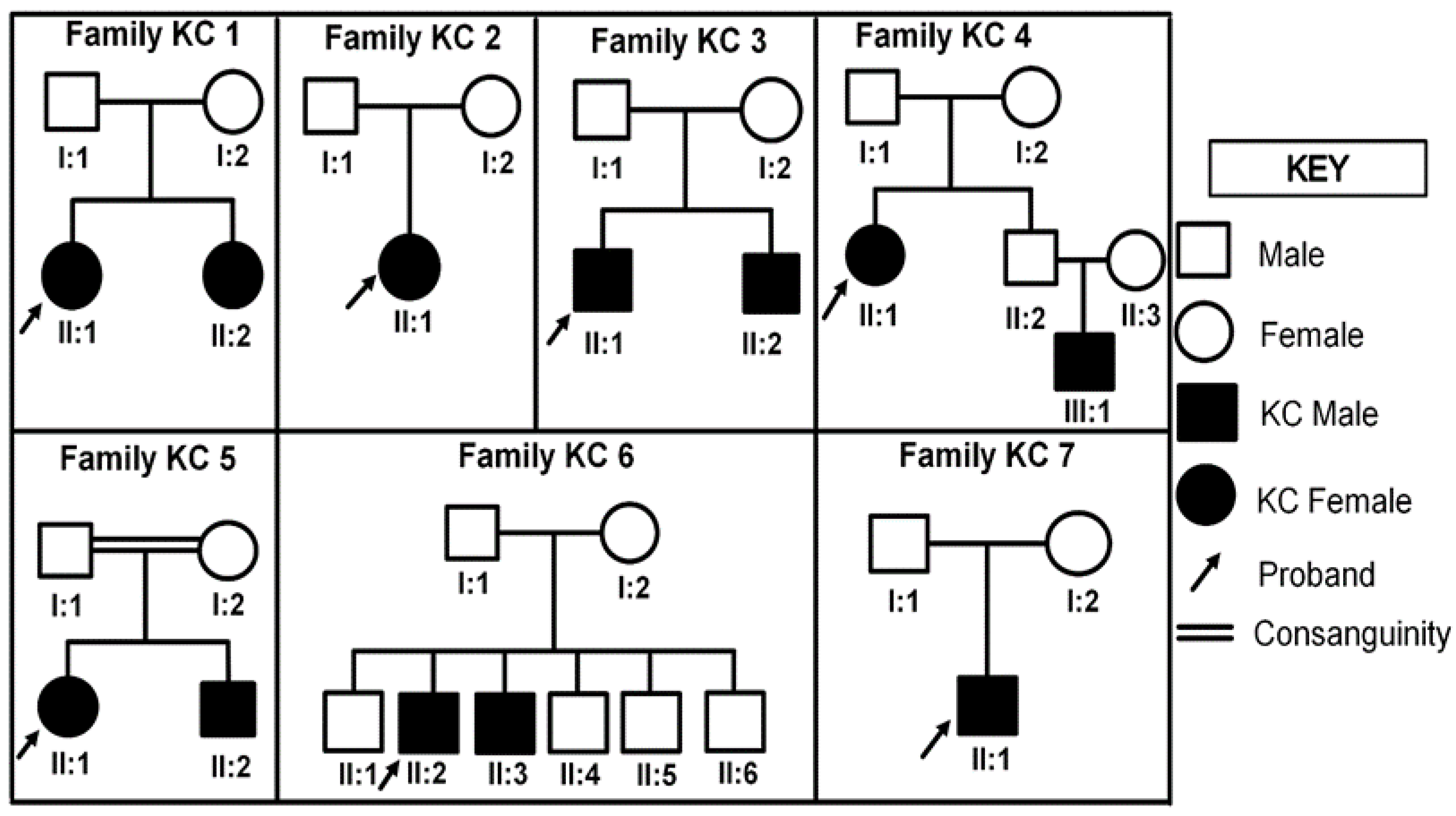
Figure 1. Pedigrees for seven autosomal recessive KC families. In the pedigrees white circles represent unaffected females whereas white squares represent unaffected males. Filled symbols represent affected individuals. Double lines indicate consanguineous marriage between parents.
Applsci 13 04888 g001
Figure 2. Sequence chromatogram of observed variants. Upper panel (A) represents normal and mutated sequence for rs12480307 (c.546A>G) while lower panel (B) is of rs76499395 (c.*496A>G) polymorphism.
Applsci 13 04888 g002
Figure 3. ESE elements, VSX1 Wild Type c.546A-c.553G (ATTCAGCG) shown in brown and VSX1 SNP c.546A>G-c.553G (GTTCAGCG) showing green docked to the SRSF 5 binding pocket. Notice the Wild Type ESE element binds well into the SRSF 5 binding pocket, while the green one binds away, excepting its 5′ region.
Applsci 13 04888 g003
Figure 4. hsa-miRNA-203 binding site on VSX1 3′UTR(WT). (A) c*475-c*496 as predicted by PITA and miRANDA. (B) In the case of VSX1 3′UTR(MT) c*496 A>G, hsa-miRNA-203 there is no binding.
Applsci 13 04888 g004
Table 1. Sequencing primers for VSX1 exons with amplification conditions.
ExonsPrimer SequenceAnnealing
(°C)
Product Size
(bp)
MgCl2
(mM)
Primer
(µM)
Exon 1_1F: 5′-TTTCGAGGGACAGGCAGAC-3′
R: 5′-AGGTCCGTGATGGCGAAG-3′
6044920.4
Exon 1_2F: 5′-TGCTTGCTAAGGAACCATGAC-3′
R: 5′-TCAGAGCCTAGGGGACAGG-3′
614892.50.32
Exon 2F: 5′-AATGCTGGCTCATACTGTAAAC-3′
R: 5′-AACCAGGAAACCACTGGG-3′
583272.50.4
Exon 3F: 5′-AGCAGAGGAAGCAGGCAC-3′
R: 5′-CTATGCAAAGGGAGCGTG-3′
583322.50.4
Exon 4F: 5′-ATCATGCTCGGGAGAGAAG-3′
R: 5′-TTGCTTTGCTTTGGAAATG-3′
5839130.4
Exon 5_1F: 5′-CCCCAGAGATAGGCACTGAC-3′
R: 5′-TGCCAGTGAGGAATATGCAC-3′
5847030.4
Exon 5_2F: 5′-GCAGGAGACCAAGAAAGTGC-3′
R: 5′-CTCAAATGATGCCCAGCAG-3′
584162.50.4
Exon 5_3F: 5′-ATGCCACTTGCTTTAAGAGG-3′
R: 5′-TGCAGAAACGACTAGAGTATGG-3′
5846430.4
Exon 5_4F: 5′-TACCTTGAACTTGGCCTTGG-3′
R: 5′-TGGCTGGGATCAGAGATAGTG-3′
583912.50.4
VSX1, Visual system homeobox gene; F, forward primer; R, reverse primer.
Table 2. Splicing analysis of the exon 3 sequence of the VSX1 and its SNP c.546A>G, effect on the ESE site the formation of splicing regulatory complex.
Predicted SignalPrediction AlgorithmcDNA PositionInterpretation
ESE Site Broken1. ESE-Finder-SRp40Applsci 13 04888 i001Alteration of an exonic ESE site.
Potential alteration of spllicing
2. RESCUE ESE Hexamers
3. EIEs from Zhang et al. [23]
Table 3. Variation in ESE-SRSF protein binding properties between VSX1 Wild Type c.546A as compared to VSX1 SNP c.546A>G analyzed using the ESE finder.
TypeSRSF Binding Properties
Biding MotifSRSF ProteinBinding Score BS Threshold
VSX1 Wild Type c.546A-c.553G ATTCAGCGSRSF53.072.67
VSX1 SNP c.546A>G-c.553G GTTCAGCGSRSF23.202.38
Table 4. Variations in binding properties of miRNA-203 and miRNA-3938 with VSX1 3′UTR (WT) as predicted by miRANDA and PITA.
miRNAWild and Mutant Type SequencesmiRANDAPITA
BindsBinding Free Energy (kcal/mol)ScoreBindsΔΔG
miRNA-203VSX1 3′UTR(WT) c* 496 A−15.50150−9.35
VSX1 3′UTR(MT) c* 496 A>GX00X0
miRNA-3938VSX1 3′UTR(WT) c* 496 AX00X0
VSX1 3′UTR(MT) c* 496 A>G−12.00140−7.65
Table 5. Comparison of the studies conducted on same pattern on different ethnicity, study type and cohort size, techniques, identified variants and number of patients.
EthnicityStudy Type and Cohort SizeIsolated/Familial CohortTechniqueIdentified Variant(s)No.of Patient(s)Variant TypeReference
CaucasianAssociation study. 100 unrelated KC PatientsIsolated cohortSanger Sequencingp.Asp144Glu
p.Ser6Ser
p.Pro58Pro
p.Arg131Ser
p.Ala182Ala
1
4
2
1
51
Non-synonymous
Synonymous
Synonymous
Non-synonymous
Synonymous
[28]
Caucasian+Asian
+African
85 ProbandsFamilialSanger Sequencingp.D144E
c.504-10G>A
504-24C>T
1
1
3
Non-synonymous
Intronic
Intronic
[29]
Whites+ Hispanics+OthersAssociation study with 77 unrelated KC Patients and 75 familiesIsolated cohort+familial cohortARMS-PCR and RFLPp.H244R
p.L159M
3
5
Non-synonymous
Non-synonymous
[30]
Slovenian113 patients with sporadic and familial KCIsolated cohort+familial cohortSanger Sequencingp.S6S
p.A128A
p.D144E
504-24C.T
627+23G.A
21
35
1
0
44
Synonymous
Synonymous
Non-synonymous
Intronic
Intronic
[31]
Indian117 sporadic cases of keratoconusIsolated cohortSanger Sequencingp.A182A
c.627+23G>A
c.627+84T>A
c.504-24C>T
7
3
9
7
Non-synonymous
Intronic
Intronic
Intronic
[32]
Indian50 sporadic casesIsolated cohortSanger Sequencingp.R217H
p.P237P
1
3
Non-synonymous
Synonymous
[33]
Disclaimer/Publisher’s Note: The statements, opinions and data contained in all publications are solely those of the individual author(s) and contributor(s) and not of MDPI and/or the editor(s). MDPI and/or the editor(s) disclaim responsibility for any injury to people or property resulting from any ideas, methods, instructions or products referred to in the content.