Article

The Purification and Characterization of a Cutinase-like Enzyme with Activity on Polyethylene Terephthalate (PET) from a Newly Isolated Bacterium Stenotrophomonas maltophilia PRS8 at a Mesophilic Temperature

1
Department of Microbiology, Faculty of Biological Sciences, Quaid-i-Azam University, Islamabad 45320, Pakistan
2
Department of Biology, Karadeniz Technical University, Trabzon 61080, Turkey
3
Division of Biotechnology, Department of Chemistry, Faculty of Engineering (LTH), Lund University, P.O. Box 124, SE-22100 Lund, Sweden
4
Department of Microbiology, Kohsar University Murree, Murree 47150, Pakistan
5
Centre for Analysis and Synthesis, Department of Chemistry, Faculty of Engineering (LTH), Lund University, SE-22100 Lund, Sweden
*
Authors to whom correspondence should be addressed.
Appl. Sci. 2023, 13(6), 3686; https://doi.org/10.3390/app13063686
Submission received: 7 December 2022 / Revised: 4 March 2023 / Accepted: 9 March 2023 / Published: 14 March 2023
(This article belongs to the Section Applied Biosciences and Bioengineering)

Abstract

:

Featured Application

The potential use of bacteria and their enzymes for the degradation of plastic waste and the recovery of value-added products considering bio-up recycling for the circular economy.

Abstract

A polyethylene terephthalate (PET)-degrading bacterium identified as Stenotrophomonas maltophilia PRS8 was isolated from the soil of a landfill. The degradation of the PET bottle flakes and the PET prepared as a powder were assessed using live cells, an extracellular medium, or a purified cutinase-like enzyme. These treated polymers were analyzed using Fourier transform infrared spectroscopy (FTIR) and scanning electron microscopy (SEM). The depolymerization products, identified using HPLC and LC-MS, were terephthalic acid (TPA), mono(2-hydroxyethyl)-TPA (MHET), and bis(2-hydroxyethyl)-TPA (BHET). Several physicochemical factors were optimized for a better cutinase-like enzyme production by using unique single-factor and multi-factor statistical models (the Plackett–Burman design and the central composite design software). The enzyme was purified for homogeneity through column chromatography using Sephadex G-100 resin. The molecular weight of the enzyme was approximately 58 kDa. The specific activity on para nitrophenyl butyrate was estimated at 450.58 U/mg, with a purification of 6.39 times and a yield of 48.64%. The enzyme was stable at various temperatures (30–40 °C) and pH levels (8.0–10.0). The enzyme activity was significantly improved by the surfactants (Triton X-100 and Tween-40), organic solvent (formaldehyde), and metals (NiCl2 and Na2SO4). The extracellular medium containing the cutinase-type enzyme showed a depolymerization yield of the PET powder comparable to that of Idonella skaiensis IsPETase and significantly higher than that of Humicola insolens thermostable HiCut (HiC) cutinase. This study suggests that S. maltophilia PRS8 is able to degrade PET at a mesophilic temperature and could be further explored for the sustainable management of plastic waste.

1. Introduction

Cutinase-like enzymes are gaining interest because some exhibit degradation activity on polyethylene terephthalate (PET). Since the first report on a PET-active cutinase was published in 2005 [1], reports on PET-depolymerizing enzymes have significantly increased in recent years, after the publication of the Ideonella sakaiensis bacterium that was able to grow using PET as the sole carbon source [2]. It is worth emphasizing that the amorphous domain of PET can be more accessible to an enzyme attack than crystalline domains. Indeed, thermostabilized PETase, acting at temperatures closer to the glass transition temperature of PET, has shown a better depolymerizing performance [3]. However, mesophilic enzymes and microorganisms could play an important role in the systems where high temperatures are not suitable, such as in wastewater plants.
The annual production of plastics was estimated to be 348 million tons in 2017, of which ~90% was derived from fossil fuels [4,5]. By 2030, it is expected to increase to 590 million tons [6]. Owing to their expanding use and versatility, plastic products have become a part of everyday life [7]. Polyethylene terephthalate (PET) consists of ethylene glycol (EG) and terephthalic acid (TPA) monomers linked by ester bonds. It is considered one of the most widely used synthetic polymers and has many applications in drinking bottles, food packaging, and textile fiber materials [8,9]. In 2017, PET production exceeded 30 million tons. Packaging materials always rank high, accounting for ~40% of the total plastics produced [4]. They accumulate in urban solid waste, landfills, and oceans, causing global environmental pollution [10]. This situation has caused severe damage to marine life and threatened some species as most plastic waste on land ends up in water bodies [11].
Plastics are recalcitrant materials for biological degradation. However, 30,000 non-redundant enzyme homologs with putative activity in 10 different plastic types were recently found using bioinformatics methods [12], supporting the current trend of developing enzymatic processes for plastic recycling. Nevertheless, only a small number of fungi and bacteria capable of partially depolymerizing PET have been experimentally studied [13]. The crystallinity of PET is one of the main obstacles to its biodegradation. The enzymes that hydrolyze PET are serine hydrolases, such as cutinases, lipases, and carboxylesterases. The I. sakaiensis enzyme, PETase (PET-active enzyme), mainly produces mono(2-hydroxyethyl) terephthalic acid (MHET) and, to a lesser extent, terephthalic acid (TPA) and bis(2-hydroxyethyl)-TPA [2]. The cutinases are part of the α/β family of serine hydrolases that perform ester bond hydrolysis using the canonical Ser-His-Asp catalytic triad. PET and cutin generally share small structural features; they have ester bonds that can be cleaved by cutinases [14,15].
Plastic pretreatment is an important process before enzymatic depolymerization. Plastic crystallinity is one of the main obstacles to an enzymatic attack [16]. Therefore, several pretreatment strategies focus on increasing the amorphization. For example, extrusion and micronization are processes widely used in the plastic industries that amorphize and increase the exchange surface of plastics, facilitating an enzymatic attack. These technologies have maximized PET depolymerization by using an outperforming engineered leaf-branch compost cutinase (LCC), resulting in an outstanding depolymerization yield in a few hours [3]. At the laboratory level and for research purposes, as in this work, PET and other polyesters can also be dissolved in organic solvents and then precipitated as powders [17]. These polyester powders have a lower crystallinity and are suitable for assessing enzymatic activities. On the other hand, green plastic pretreatments should be developed for practical purposes and industrial processes.
In the present study, a bacterial strain capable of growing in a saline medium containing PET as the sole carbon source was isolated from landfill soil. This strain was identified as Stenotrophomonas maltophilia. S. melophilia is a gram-negative bacterium and belongs to the γ-proteobacteria. It is present in water, soil, and plant rhizospheres and is considered an opportunistic pathogen [18]. This species has previously been reported to play a role in the bioremediation of various contaminants, including xenobiotics, oils, dyes, and polyaromatic hydrocarbons (PAHs) [19,20,21]. However, to our knowledge, this is the first report of the PET-depolymerizing activity of an S. melophilia strain, and the purification of an enzyme associated with this activity.
The S. melophilia PRS8-degrading activity of the plastic was assessed using plastic weight loss, Fourier transform infrared spectroscopy (FTIR), and scanning electron microscopy (SEM). An enzyme with a cutinase-like activity was purified from a bacterial enzyme production culture optimized for nutrients and physical parameters using the Plackett–Burman and central composite design. The enzyme was purified for homogeneity and characterized to determine its stability and activity, using p-nitrophenyl butyrate as the substrate. The enzymatic depolymerization of the PET flakes and powders was assessed by detecting and quantifying the PET hydrolysis products using high-performance liquid chromatography (HPLC) and liquid chromatography-mass spectrometry (LC-MS).

2. Materials and Methods

2.1. Chemicals

The PET flakes from mineral water bottles (Nestle) were used in this study. The PET beads were obtained from Indorama. The nutrient broth (NB) and nutrient agar (NA) were obtained from Merk (GmBH, Darmstadt Germany). A p-nitrophenyl butyrate (p-NPB) standard solution (12 mM) in isopropanol was utilized. Methanol (MeOH), 1,1,1,3,3,3-Hexafluoropropan-2-ol (HFIP) (≥99%), and chloroform (CHCl3) (stabilized with 0.6% ethanol) were purchased from VWR Chemicals (Radnor, PA, USA). All the other media and chemicals utilized in this study were of the highest commercial quality.

2.2. Collection of the Samples and the Isolation of the Bacteria

The soil samples were collected from a garbage dump. The bacterial strains were isolated using serial dilutions and cultured on NA plates at 37 °C for 24 h. Then, the isolated colonies were transferred to the NA broth for propagation and further analysis.

2.3. Screening of the Bacterial Isolates for PET Degradation

A total of 24 isolated bacterial strains were subjected to the PET biodegradation screening. The assays consisted of assessing the ability of the bacteria to grow in a salt media MSM [21] (composition in g/L: 1.0 K2HPO4, 0.2 KH2PO4, 0.1 NaCl, 0.002 CaCl2.2H2O, 0.1 (NH4)2SO4, 0.5 MgSO4.7H2O, and 0.01 FeSO4.7H2O) with a PET flake as the sole carbon source. The PET flakes were washed with autoclaved water and sterilized with 70% ethanol, followed by exposure to UV light for 5 min. No differences between the PET exposed and not exposed to UV light were detected through the FTIR spectroscopy analysis (Supplementary Materials S1 and Figure S1). The cultures were prepared in 100 mL of a culture medium in a 250 mL shake flask and incubated under shaking (150 rpm) at 35 °C for one week. To enrich the potential degrading bacteria, 1 mL of the previous cultivation was inoculated in the same medium with PET and incubated under the same conditions for four consecutive times. A strain designated PRS8 was selected for further studies based on its maximum growth.

2.4. Biofilm Assay for Bacterial Strain

The capacity of S. maltophilia PRS8 for the biofilm formation on PET was assessed using a crystal violet staining method [22,23] adapted to the PET sheets. S. maltophilia PRS8 was inoculated into the wells of a microtiter plate containing the pre-sterilized PET flakes and incubated at 35 °C for 48 h. The biofilm-producing Pseudomonas aeruginosa was used as a positive control, whereas the negative control was an uninoculated well. After incubation, the PET flakes were transferred into clean Eppendorf tubes and washed gently with sterilized deionized water three times. The PET flakes were then stained using 0.5% crystal violet for 15 min and washed three times with water. After air drying, the samples were de-stained using 30% acetic acid for 15 min. After staining, the colored samples indicated the presence of a bacterial biofilm on the PET.

2.5. Identification of the Bacterial Strain PRS8

The genomic DNA from the strain PRS8 was extracted using an Eco-Pure Genomic kit (Product ode # E1075). The 16S rRNA gene was amplified using the universal primers F-(ATTCTAGAGTTTGATCATGGCTCA) and R-(TACACACCGCCCGTCACACGGTACCAT). The PCR reaction mixture consisted of 1 µL of each primer (10 mM), 10 µL of a Go-Taq buffer, 3 µL of MgCl2 (25 mM), 1 µL of dNTPs (10 mM), 0.5 µL of Taq polymerase, 1 µL of a DNA template (87 μg/mL) and 32.5 µL of deionized water. The temperature cycles were for DNA denaturation at 95 °C for 3 min, annealing at 55 °C for 1 min, and extension at 72 °C for 1 h and 40 secs per cycle for a total of 36 cycles. The reaction mixture was incubated at 72 °C for 7 min for the final elongation. The PCR products were sent for sequencing, and the gene sequence was deposited in a GenBank (accession code OK510364). The sequences showing > 95% similarity were downloaded from the EzBioCloud database (www.ezbiocloud.net (accessed on 11 October 2021)) to construct a phylogenetic tree using the neighbor-joining method with the MEGA-X software [24].

2.6. Polycaprolactone Agar for Cutinase Activity

Polycaprolactone (PCL) agar plates were used to examine the polyesterase activity in the isolated S. maltophilia PRS8. The plates were prepared using 1 g/L of polycaprolactone (10 g/L of peptone, 0.2 g/L of CaCl2.2H2O, 5 g/L of NaCl, and 20 g/L of agar). The cultures were then incubated at 35 °C for 48 h. The clear zones around the colonies indicated the production of polyesterase [25].

2.7. Biodegradation of PET by S. maltophilia PRS8

Two milliliters of the overnight grown culture of S. maltophilia PRS8 in the nutrient broth was centrifuged for 10 min at 10,000× g and the pellet was resuspended in 500 µL of 9 g/L NaCl. The sterilized PET water bottle pieces (2 cm × 2 cm), each weighing 30 mg, were added to 100 mL mineral salt medium (MSM) in an Erlenmeyer flask and inoculated with the PRS8 cells. A control with the PET flakes in the bacteria-free MSM was used separately. The flasks were incubated in a shaker incubator at 35 °C and 150 rpm for 28 d. The biodegradation was evaluated using analytical techniques, such as FTIR, SEM, and the percentage weight loss assessment at the end of the experiment.

2.8. Analysis of the Degradation of PET by S. maltophilia PRS8

2.8.1. Determination of the Dry Weight of the Residual PET

The PET flakes were recovered after incubation and washed for 15 min in a 2% aqueous solution (v/v) of sodium dodecyl sulphate (SDS). After they were rinsed with distilled water, the pieces were dried overnight at 60 °C and then weighed [26].

2.8.2. Fourier Transform Infrared (FTIR) Spectroscopy

The PET flakes were subjected to FTIR spectroscopy (8400 Shimadzu, Kyoto, Japan, with Hyper IR- 1.7 software for Windows). The FTIR spectra were normalized at 1410 cm−1 (aromatic ring vibrations) according to the literature [27,28]. The spectra of the treated and control PET flakes were recorded in a frequency range of 4000–400 cm−1 at a resolution of 4 cm−1 and at an ambient temperature using a helium–neon laser lamp as a source of IR radiation.

2.8.3. Scanning Electron Microscopy (SEM)

Changes in the surface morphology of the PET flake pieces after incubation with S. maltophilia PRS8 were determined using a scanning electron microscope (JSM-IT100). The PET flakes were washed, as described in Section 2.8.1, and were gold-painted. The gold painting was performed under a vacuum using evaporation to increase the conductivity of the samples. The PET piece from the control was examined along with the test sample for comparison.

2.8.4. Preparation of the PET Powder

Two grams of the PET beads (Supplier: Indorama RamaPET N180, Klaipeda, Lithuania) was dissolved in 50 mL of a mixed solvent of chloroform and HFIP (8:2), precipitated into 250 mL of methanol, and washed with methanol (3 × 100 mL). Subsequently, the obtained powders were dried under a vacuum at 50 °C for 24 h prior to the degradation studies.

2.9. Standard Assay of the Esterase/Cutinase Activity

The reaction mixture was prepared at a final volume of 1800 µL. It consisted of 12 mM of p-nitrophenyl butyrate (p-NPB) as the substrate, 100 mM of the phosphate buffer adjusted to pH 8.0, 200 µL Triton X-100, and 200 µL of the sample containing the enzyme or the control (without the enzyme). Before adding the enzyme, the mixture was pre-incubated at 40 °C for 10 min and then at 37 °C for 1 h. The reaction was stopped using frizzing at −20 °C for 15 min. The absorbance of p-nitrophenol was determined spectrophotometrically at 410 nm. The enzymatic unit was defined as the production of 1 μM of p-nitrophenol per minute.

2.10. Optimization of the Physicochemical Parameters for the Cutinase Production in S. maltophilia PRS8

The fermentation parameters such as the temperature (25–45 °C), pH (5.0–10.0) incubation time (24–72 h), and inoculum size (0.1–5%) were optimized for the maximum production of the cutinase-like enzyme. Freshly grown cultures of PRS8 were inoculated into a production medium consisting of 0.8% nutrient broth, 0.3% Na2HPO4, and 0.4% tomato cutin, as previously described [29]. The cultures were incubated in a shaking incubator at 150 rpm and 35 °C for 24 h. The samples were collected every 24 h, centrifuged at 10,000× g at 4 °C for 10 min, and the supernatants were processed to determine the enzyme activity and the protein estimation using the previously described methods (Section 2.9). The optimization was based on a Plackett–Burman design (PBD) and a central composite design (CDC) experiment, as described in the Supplementary Materials (S2). A Fisher test (F-test) ANOVA and its accompanying probability P were used to statistically analyze the model (F). A total of 10 components were optimized using the PBD, which included glucose (NH4)2SO4, sucrose, NaNO3, K2HPO4, yeast extract, MgSO4.7H2O, KCl, FeSO4.7H2O, cutin, and Na2HPO4. The optimized media was used for the bulk production of the enzyme.

2.11. Bulk Production of the Crude Cutinase-like Enzyme

The enzyme was produced by cultivating S. maltophilia PRS8 in a 1000 mL production medium with an optimal composition. It consisted of 13.0 g/L of glucose, 5.5 g/L of (NH4)2SO4, 11.0 g/L of sucrose, 11.0 g/L of NaNO3, 3.0 g/L of yeast extract, 2.0 g/L of K2HPO4, 2.0 g/L of MgSO4.7H2O, 3.0 g/L of KCl, 9.0 g/L of cutin, and 3.0 g/L of Na2HPO4. The pH of the solution was adjusted to 8.0. The culture was inoculated with 2% of a 16 h fresh culture of S. maltophilia PRS8 and incubated at 35 °C and 150 rpm for 48 h. The cell-free supernatant was collected after centrifugation at 10,000× g for 10 min at 4 °C and further processed for the enzyme purification.

2.12. Purification of the Cutinase-like Enzyme

First, the protein fraction of the cell-free supernatant was precipitated by gently adding solid ammonium sulfate at 4 °C until saturation (70%). The solution was centrifuged (10,000× g) at 4 °C for 10 min, and the precipitates were resuspended in a potassium phosphate buffer (pH 8.0). The residual ammonium sulfate was completely removed through dialysis against the phosphate buffer, yielding a solution containing the crude enzyme. The cutinase-like enzyme was purified through gel permeation chromatography [30] using the Sephadex G-100 packaged into a glass column 27 cm in length and 3 cm in diameter and equilibrated with a phosphate buffer (pH 8.0). Three milliliters of the crude enzyme were loaded onto the column, and elution was performed with the phosphate buffer (pH 8.0) at a flow rate of 3.0 mL/5 min. The fractions with a high activity (assay in Section 2.9) were pooled. The purity and molecular weight were determined using sodium dodecyl sulfate-polyacrylamide gel electrophoresis (SDS-PAGE) with a standard protein marker (Bio-Rad, Hercules, CA, USA).

2.13. Zymography

The cutinase-like activity was confirmed through zymography using α-naphthyl acetate as the substrate. The gel was incubated in 20 mM of the Tris-HCI buffer (pH 6.5) with 2% Triton X-100 for 10 min, followed by incubation in 50 mM of the phosphate buffer (pH 7.5) supplemented with 1 mM of Fast Red TR and 3 mM of α-naphthyl acetate (Sigma, St. Louis, MO, USA) at room temperature for up to 3 h to observe the red band, which indicated the cutinase-like activity [31].

2.14. Effect of the Temperature and pH on the Cutinase Activity and Stability

The effect of the temperature on the enzyme activity was determined at 30–60 °C. The effect of the pH was studied over a pH range of 3.0–11.0 at 40° for 150 min. The following buffers (100 mM) were used: a sodium acetate buffer, pH 3.0–5.0; a potassium phosphate buffer, pH 6.0–8.0; and a glycine-NaOH buffer, pH 9.0–11.0. The activities were determined using the standard assay described in Section 2.9.

2.15. Effect of the Metals Ions

Two different concentrations of metal ions, 2 mM and 15 mM, for Ba2+, Ca2+, Cu2+, Mg2+, Zn2+, K+, Hg2+, Fe2+, Ni2+, Co2+, Ni2+, and Na+ were assessed. The results were expressed in terms of the residual activity (%) after 150 min at 40 °C using the standard assay protocol (Section 2.9).

2.16. Effect of the Organic Solvents and the Surfactant

Various solvents, such as ethanol, methanol, acetonitrile, acetone, ethyl acetate, propanol, butanol, and formaldehyde, and surfactants, such as sodium dodecyl sulfate (SDS), Tween-20, Tween-80, and Triton X-100, were used in the enzymatic reactions for 120 min at 40 °C, and the residual activity was calculated using the standard assay conditions (Section 2.9).

2.17. Enzymatic Depolymerization of the PET Flakes

Fifty milligrams of the PET flakes were treated with 0.7 mg of the enzyme in 100 mM of potassium phosphate buffer, pH 8.0. The total reaction volume was 3 mL. The reaction mixture was incubated for 72 h at 40 °C with shaking at 250 rpm [17]. The experiment was performed in triplicate and included a negative control without the enzyme.

2.18. Analysis of the Enzymatic Depolymerization Products

The products of the PET enzymatic depolymerization were analyzed using reverse phase liquid chromatography-mass spectrometry (LC-MS). A measure of 0.5 mL of the sample was collected after incubation and diluted at a 1:1 ratio with dimethyl sulfoxide (DMSO) and filtered through a 0.2 µm syringe filter. The sample was then shifted into an Amicon Ultra filtration assembly (Pall Life Sciences, Ann Arbor, MI, USA) and centrifuged using a Nanosep 3 K centrifugal device at 15,000× g for 20 min to remove the enzyme. The filtrate was diluted with Milli-Q water in 1:2 and was used for the analysis of the degradation products through LC-MS.

2.19. Specifications for the LC-MS Equipment

The PET depolymerization products were determined using the LC-MS analysis. The instrument consisted of a QExactive mass spectrometer coupled with a HESI source from Agilent and an HPLC system. A hydrophobic C18 column (Kinetex® 1.7 µm XB-C18 100 Å, LC Column 50 × 2.1 mm) was used to analyze the products of the sample. The flow rate was 400 µL/min. The mobile phase consisted of acetonitrile (A,20%) and 0.1% formic acid (B,80%). The data were processed using the Chromeleon v. 6.8 software. The autosampler temperature was fixed at 4 °C, with an injection volume of 15 µL. The sample data were collected in the full scan mode between m/z (100–1500) in negative ionization with a maximum resolution of 70,000 at 1 Hz, an injection time of 200 ms, and an AGC target of 3e6.

2.20. DSC Analysis of the Residual PET

The differential scanning calorimetry (DSC) measurements were performed using a TA Instruments DSC Q2000. The thermal transitions of the powders of pristine PET (untreated) and PET treated with PRS8 were studied using nitrogen with a purge rate of 50 mL min−1 and a heating rate of 10 °C per minute. The samples were heated from room temperature to 280 °C. The glass transition temperature (Tg), cold crystallization temperature (Tcc) and melting temperature ™ were obtained from the DSC first heating curve. The crystallinity percentage was calculated using the following equation:
C r y t a l l i n i t y   %   ( f i r s t   h e a t i n g   c y c l e ) = ( Δ H m Δ H c c ) ( Δ H ° m ) × 100
where ΔHcc and ΔHm are the heat of the cold crystallization (area under the cold crystallization) and the heat of melting (area under the melting transition), respectively. ΔH°m is the heat of melting for a 100% crystalline polymer estimated to be 140 J/g [17,32].

3. Results

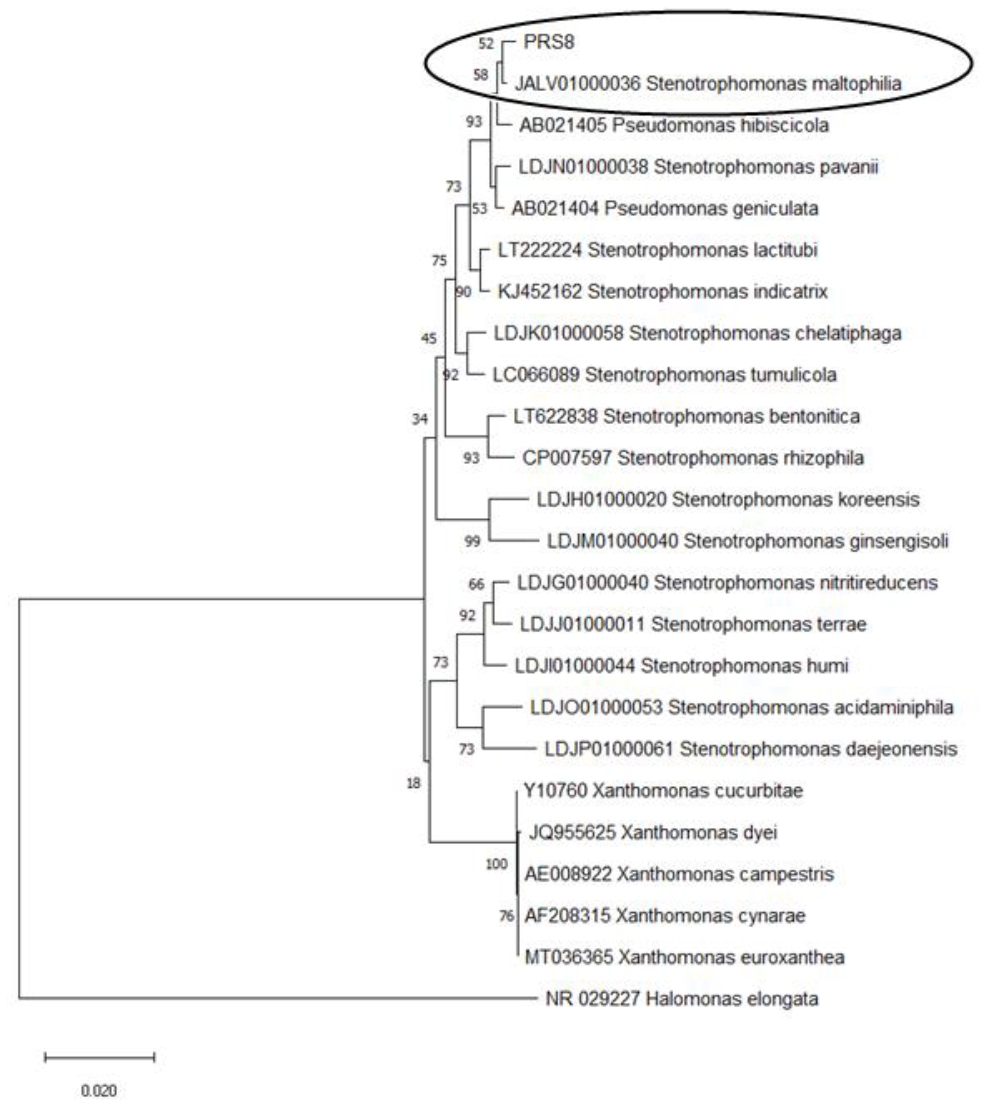
3.1. Isolation and Identification of the PET-degrading Bacteria S. maltophilia PRS8

Initially, 24 bacterial strains were screened for PET biodegradation from the plastic-contaminated soil. By the end of the experiment, only three bacterial strains, designated PRS3, PRS6, and PRS8, survived and grew very well during the rigorous screening period while utilizing PET as the sole carbon source. The strain PRS8 was selected for further study based on its rapid growth in the presence of PET.
PRS8 grew as yellowish, pointed, and small colonies on nutrient agar plates. The microscopy was found to be gram-negative. The biochemical characterization yielded positive results for citrate, urease, and catalase. The gene sequencing of 16S rRNA revealed the closest alignment to the phylum Pseudmonadota, genus Stenotrophomonas. A phylogenetic tree constructed using the neighbor-joining method presented a 99% similarity of PRS8 to Stenotrophomonas maltophilia. The sequence was submitted to the NCBI database under the accession number OK510364 (Figure 1).

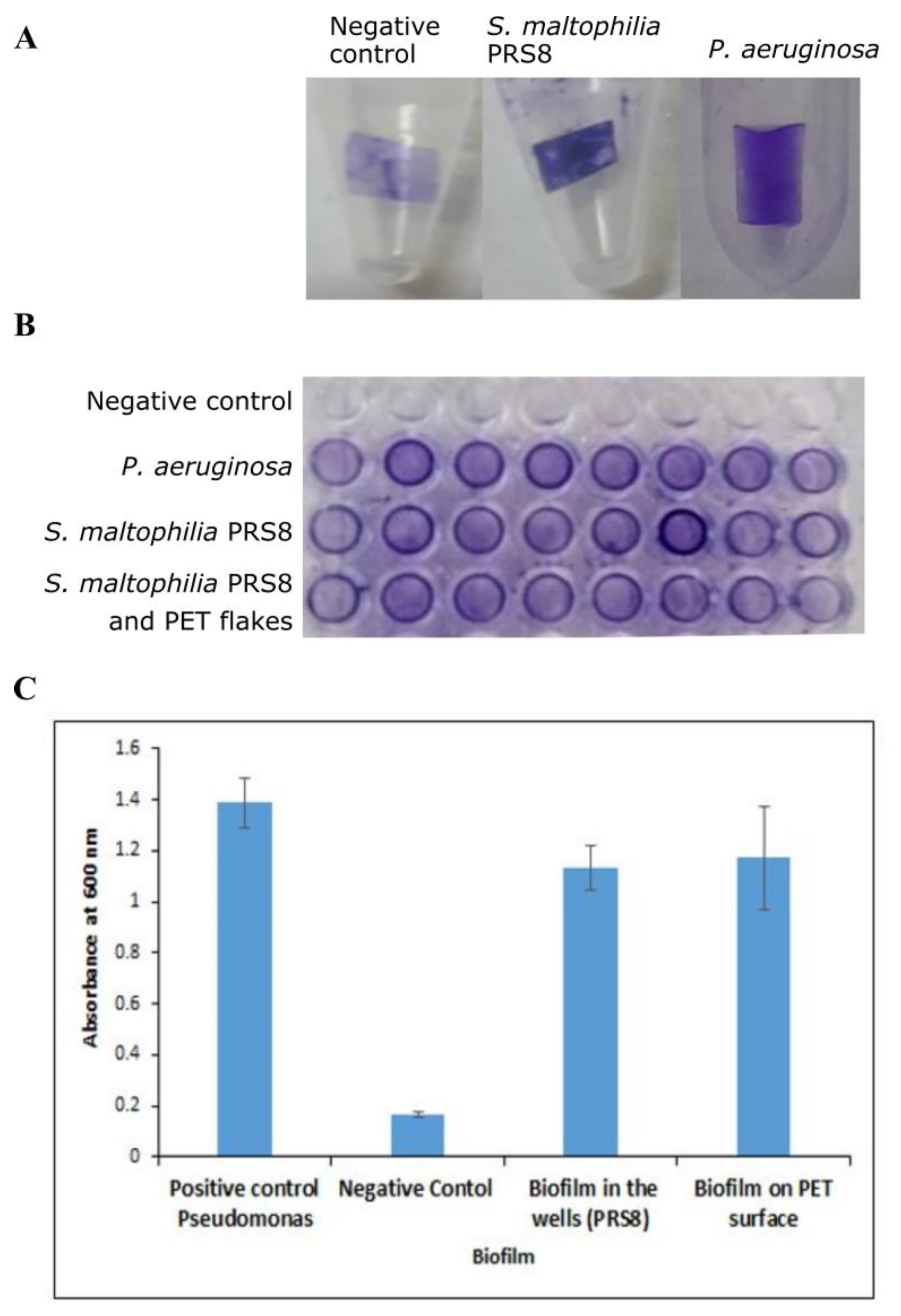
3.2. Biofilm Assay of S. maltophilia PRS8

S. maltophilia PRS8 formed significant amounts of biofilms on both a microtiter plate and the PET flakes after 48 h. It was comparable to the positive control, Pseudomonas aeruginosa (Figure 2A,B), and suggested its capability to grow attached to some plastics.

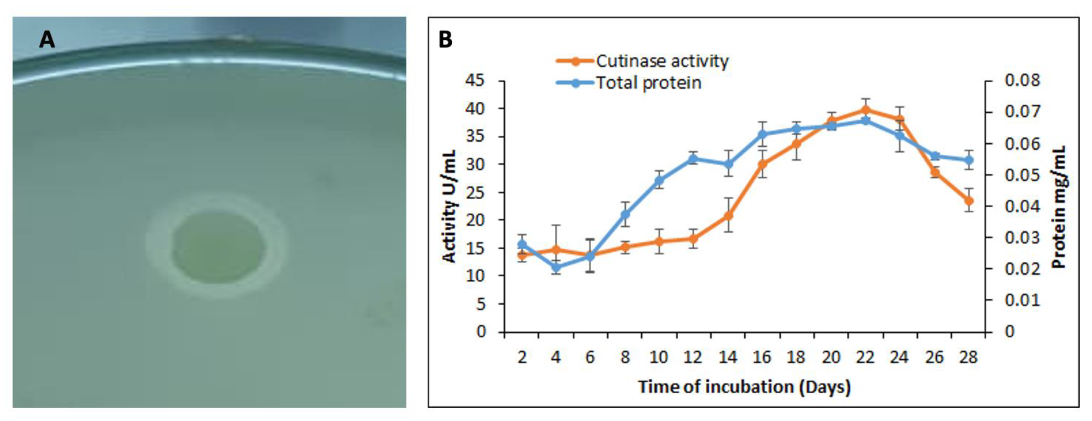
3.3. Qualitative and Quantitative Assay for the Cutinase Activity by S. maltophilia PRS8

The qualitative assays were performed by cultivating S. maltophilia PRS8 on a PCL agar plate and incubating it at 35 °C for 48 h. After incubation, the clear zones were observed around the bacterial colonies, revealing the hydrolytic action of the extracellular enzyme against PCL (Figure 3A).
The enzyme activity was determined in the samples collected from the degradation experiment every 48 h. A gradual increase in the putative cutinase activity and the protein content was observed during the first few weeks, followed by a gradual reduction at the end of the experiment. The maximum cutinase activity was measured as 39.74 U/mL with a protein content of 0.25 mg/mL after 22 days of incubation (Figure 3B).

3.4. Analysis of PET Depolymerization by S. maltophilia PRS8

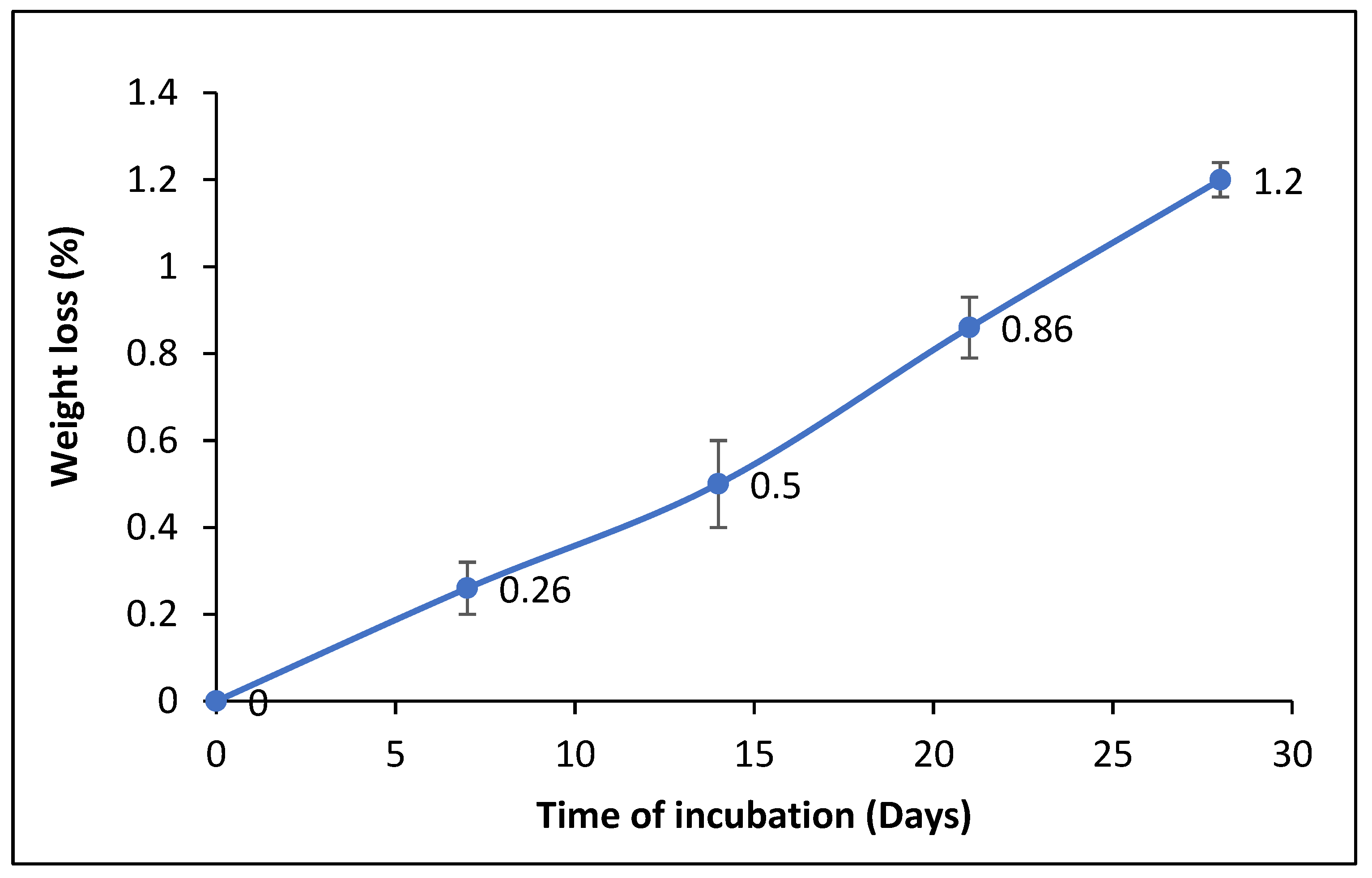
3.4.1. Determination of the Dry Weight of the Residual PET after Film Degradation

The PET flakes were recovered from both the test and control flasks at the end of the experiment after 28 d, and their weights were measured. Approximately 1.2% of the loss weight of the PET flakes was determined, while no weight loss was found (Figure 4).

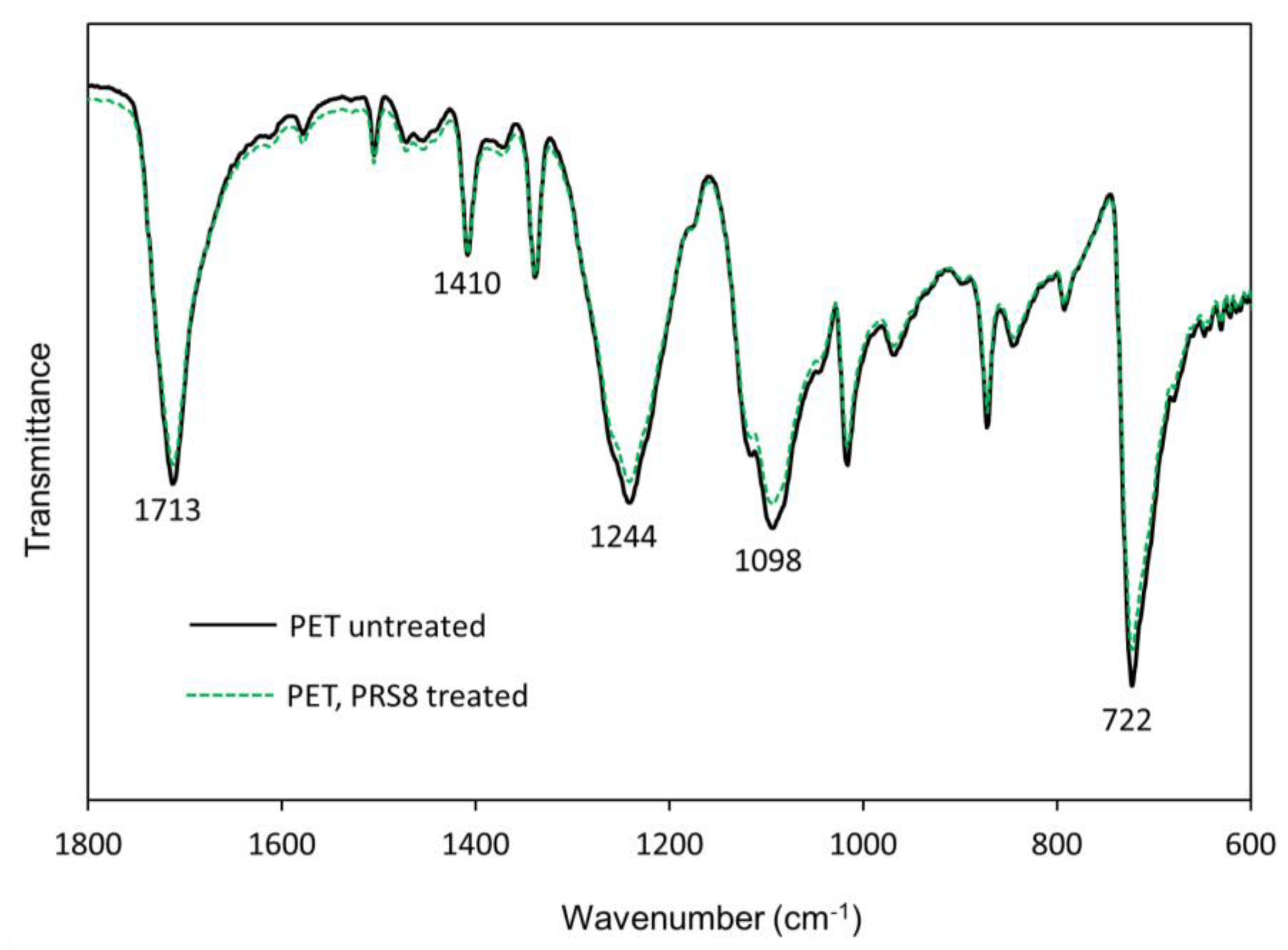
3.4.2. Fourier-Transform Infrared (FTIR) Spectroscopy

The PET flakes treated with S. maltophilia PRS8 and the untreated PET flakes (control) were analyzed using FTIR. The FTIR spectra were normalized at 1410 cm−1 (aromatic ring vibrations), according to the literature [27]. The characteristic peaks (722, 870, 1016, 1098, 1244, and 1713 cm−1) [28] were clearly observed (full spectra in the Supplementary Materials, Figure S3). The treated PET slightly reduced the intensity of the FTIR peaks at 722, 1098, 1242, and 1714 cm−1 (Figure 5) compared to the untreated PET flakes, indicating a surface degradation by S. maltophilia PRS8.

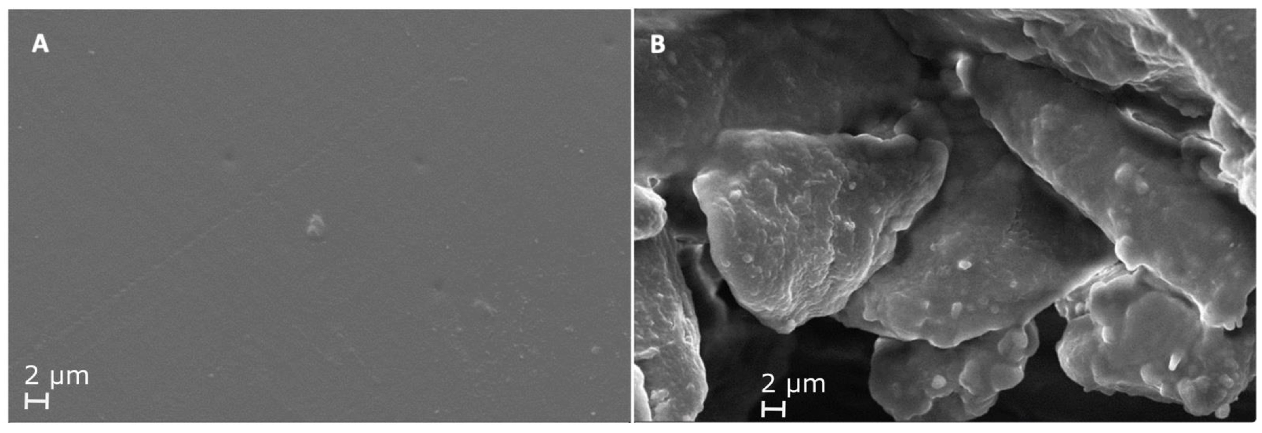
3.4.3. Scanning Electron Microscopy (SEM)

Changes in the surface morphology of the PET flakes were analyzed using SEM. PET treated with S. maltophilia PRS8 showed holes, cracks, and roughness at different points, while the untreated controls did not. These changes clearly indicated an enzymatic effect on the PET flakes during incubation with cutinase from S. maltophilia PRS8 (Figure 6A,B).

3.5. Parameter Optimization for Enzyme Production by S. maltophila PRS8

The maximum specific activity of the enzyme was obtained at 35 °C, pH 8.0, and an inoculum size of 2% after 48 h of incubation (p < 0.05).

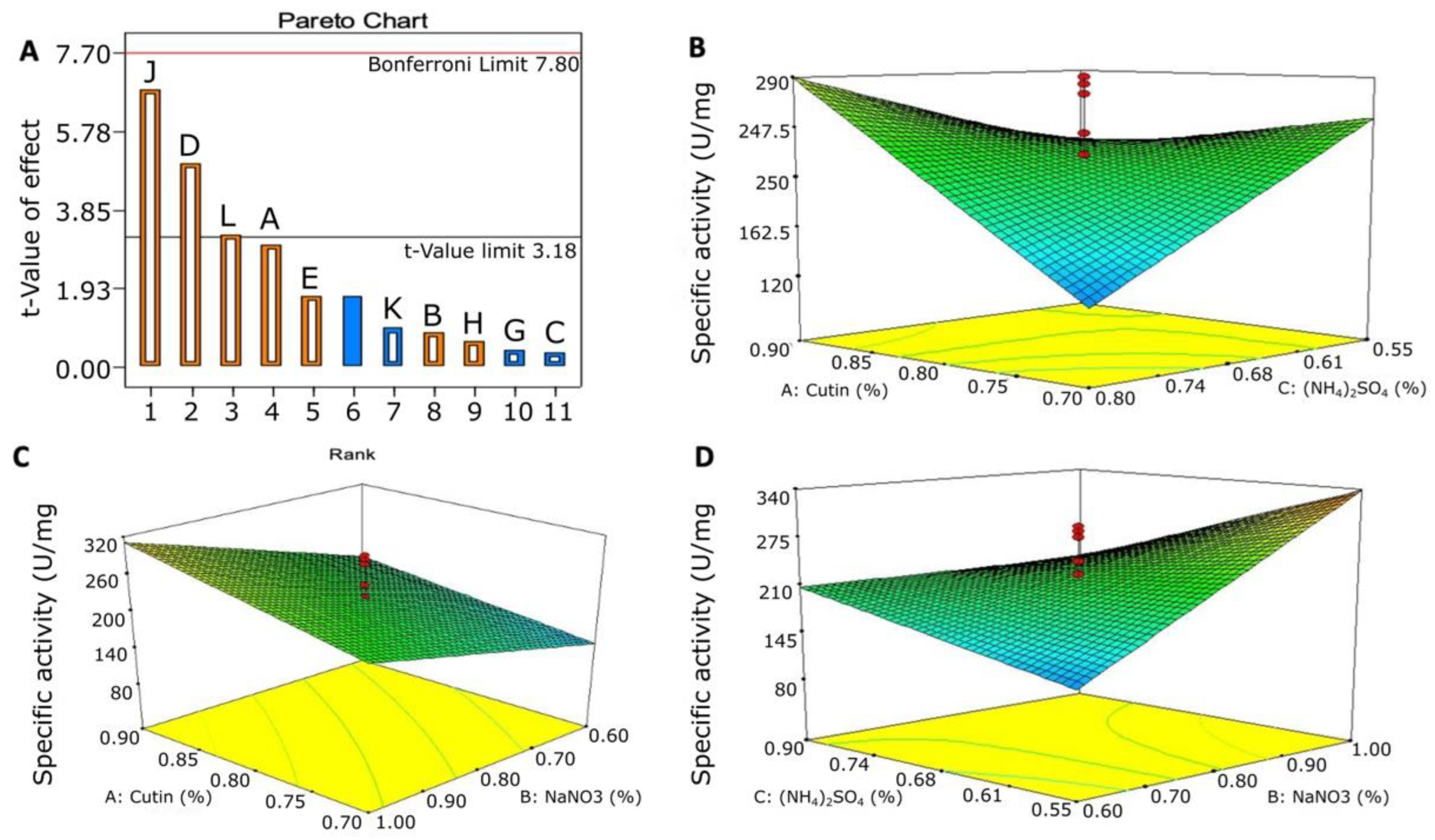
3.5.1. Plackett–Burman Design for Optimizing the Nutritional Condition

The PBD explained the influence of each element on the cutinase-like activity and its statistical significance. The maximum activity was obtained in trial 14 (313.96 U/mg) (Supplementary Materials, Table S1). Of the 11 factors, cutin, NaNO3, and (NH4)2SO4 were found to have a considerable influence on cutinase-like enzyme production, as apparent from the values of “Prob > F” for each element (Figure 7A). The model equation for the enzyme activity (U/mg) (R) is expressed as follows:
Specific activity (U/mg) = 169.39 + 20.51A + 5.78B − 2.27C + 34.34D + 11.90E + 2.74G + 4.27H + 46.82J − 6.53K + 22.26L
The calculated model F-value was 9.56 with “Prob F” values < 0.050 and identified that the model was statistically significant. According to the model, there was a high “F-Value” of 3.81% attributed to noise. The “Prob F” values < 0.050 also showed that the model was significant (Supplementary Materials, Table S2).

3.5.2. Optimization of the Significant Variable Using a Central Composite Design

A two-level CCD was used to screen the significant components obtained by the PBD. The model included three elements and their influence on the enzyme activity in twenty runs: cutin (A), NaNO3 (B), and (NH4)2SO4 (C) (Supplementary Materials, Table S3). The linear terms A, B, and C and the interactive terms AB, AC, and BC were the significant model terms (Supplementary Materials, Table S4). The following second-order polynomial equation obtained using the multiple regression analysis of the experimental data describes the effect of cutin, NaNO3, and (NH4)2SO4 in enzyme production expressed as the activity (U/mg):
Specific. Activity (U/mg) = 221.45 + 31.51A + 51.23B – 14.30C + 77.22AB + 51.02AC – 51.51BC
The model was found to be statistically significant, as implied by an F-value of 6.70 (p < 0.050), In the model, there was a 0.21% chance that an “F-value” this large could occur due to the noise. The response surface plot (AB) in (Figure 7C) showed that both factors had a potential influence on the cutinase-like activity, with a maximum regression coefficient of 77.22, followed by AC (51.02) and BC (51.19) (Figure 7B,D).

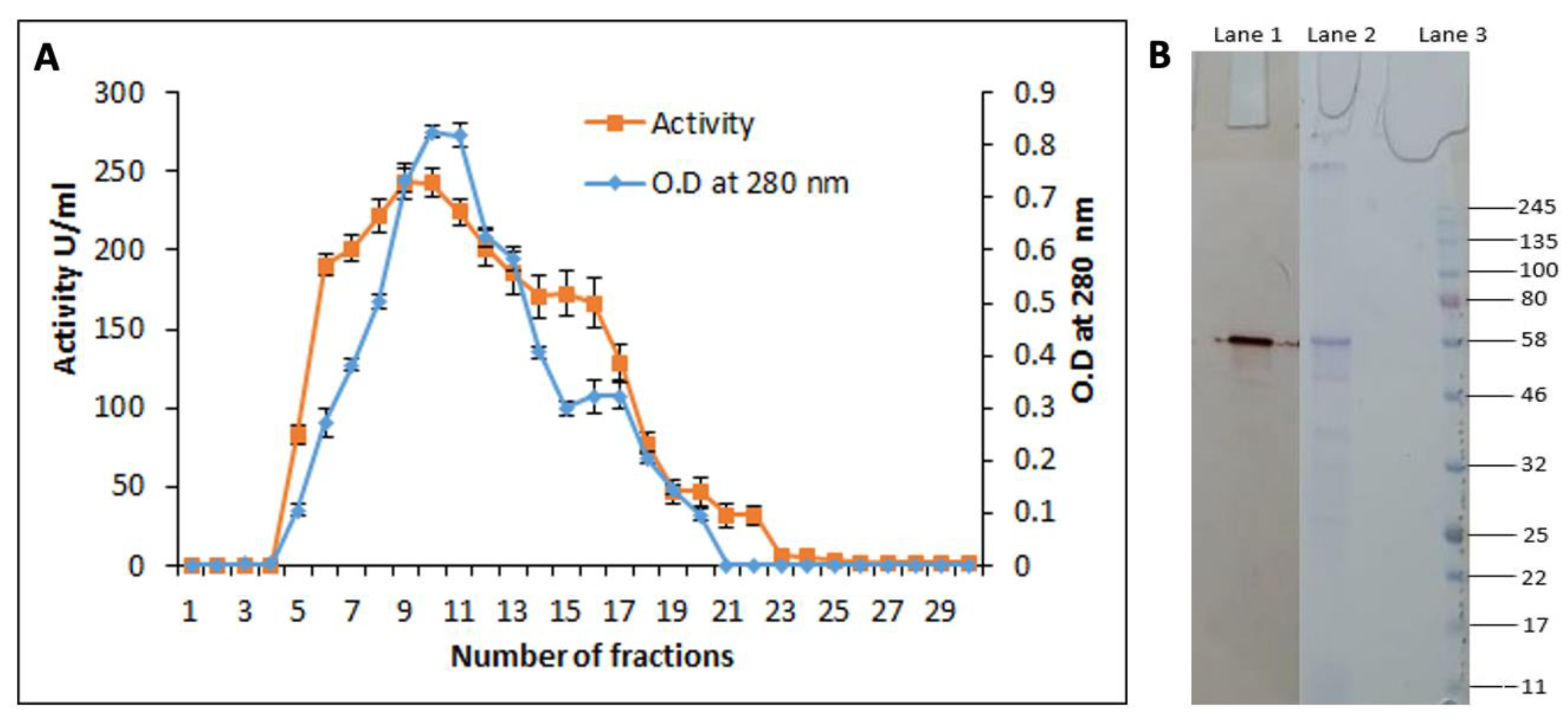
3.6. Purification of the Cutinase-like Enzyme from S. maltophilia PRS8

A cutinase-like enzyme was successfully purified using size exclusion chromatography. Its molecular mass was determined to be 58 kDa using SDS-PAGE (Figure 8B). Table 1 shows the various steps taken during the purification to measure the specific activity, purification fold, and maximum yield.

3.7. Characterization of Purified Cutinase

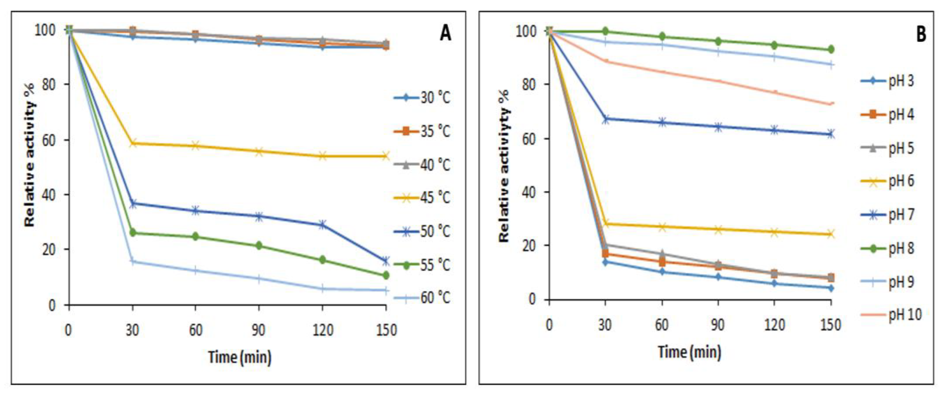
3.7.1. Effect of Temperature and pH on the Activity and Stability

The purified cutinase-like enzyme from S. maltophilia PRS8 showed a maximum activity at a temperature between 30–40 °C, with an optimum temperature achieved at 40 °C. The enzyme was found to be stable at 40 °C, retaining 95% of its activity for 150 min (Figure 9A). The purified cutinase revealed a maximum activity at a wide range of pH levels (7.0–10.0), with an optimum pH of 8.0. The enzyme was stable at pH 8.0–9.0, retaining 100% activity at pH 8.0 for 150 min (Figure 9B).

3.7.2. Effect of Metals Ions

Effects of the various monovalent and divalent metals on the cutinase activity were evaluated at 2 and 15 mM (Table 2). Ca2+, Zn2+, K+, and Mg2+ enhanced the enzyme activity at both concentrations (p < 0.005), whereas Hg2+, Ni2+, Cd2+, Ba2+, and Co2+ strongly inhibited the cutinase-like activity.

3.7.3. Effect of the Organic Solvent and Surfactants

The influence of the various organic solvents on the cutinase activity was analyzed by incubating the enzyme in a solution containing a 10% solvent for 120 min (Table 3). Among the solvents, methanol, propanol, and acetone increased the cutinase activity, whereas ethanol and butanol suppressed the enzyme activity over time (p < 0.05). The effect of the surfactants on the enzyme activity was evaluated by treating of purified enzymes with 0.5 and 5% of each surfactant. Among the surfactants, Tween-20, Triton X-100, Tween-40, Tween-60, and Tween-80 strongly enhanced the cutinase activity at both concentrations, whereas CTAB and SDS suppressed the cutinase-like activity (Table 4).

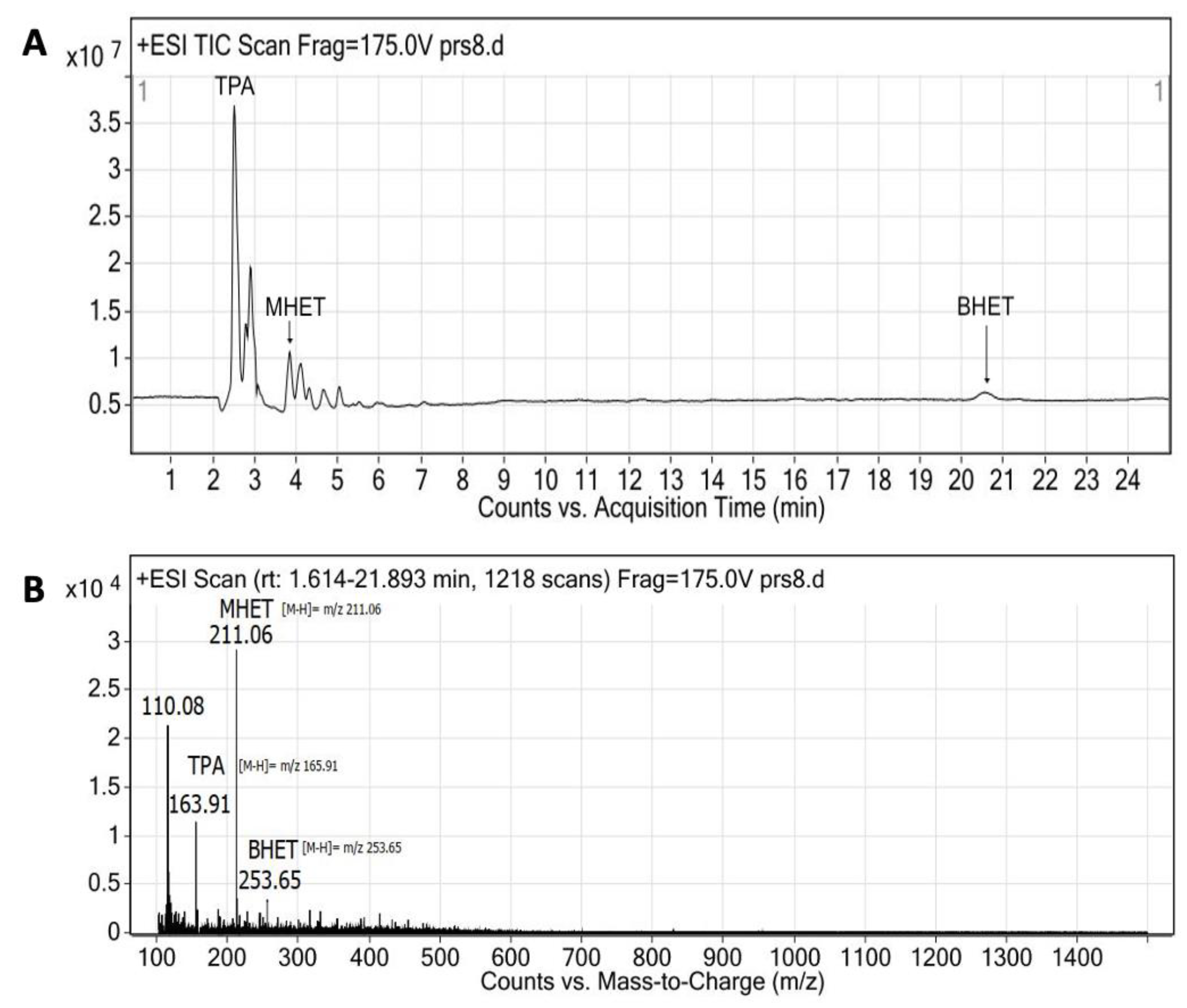
3.8. PET Bottle Flakes Degradation Products

The PET bottle flakes treated with the purified enzyme (pH 8 and 40 °C) produced terephthalic acid (TPA), mono-(2-hydroxyethyl) terephthalate (MHET), and bis-(2-hydroxyethyl) terephthalate (BHET). These products were identified using LC-MS (Figure 10). TPA eluted after ~2.51 min, while MHET and a trace amount of BHET eluted after ~3.93 and ~20.60 min, respectively. These results confirmed the PET hydrolyzing activity of the cutinase-like enzyme from S. maltophilia PRS8.

3.9. Activity on the PET Powder

The extracellular fraction (ECF) containing the cutinase-like enzyme activity against the semicrystalline PET powder was assessed after 72 h of reaction at 40 °C and a pH of 9. The product (BHET, MHET, and TPA) peaks obtained using HPLC were regarded as the terephthalic acid equivalents (TPAeq), assuming that they all had the same molar extinction coefficient e = 17.000 M−1 cm−1 due to the presence of the aromatic ring. Thus, the total amount of the degraded polymer was calculated by the TPAeq and the molecular weight 192 g/mol, corresponding to the PET repeating unit [17]. DMSO was used as the co-solvent to maximize the solubility of the depolymerized products. No products were detected in the negative controls (DMSO, PET, and the buffer) under the same conditions. The enzyme concentration in the extracellular medium was estimated from the total protein concentration and the enzyme fraction (58 kDa band) using SDS-PAGE. The PET depolymerization yield was 22.5%, with the main product being MHET, followed by TPA, and a small fraction of BHET (Table 5). Interestingly, these results were comparable with those of Ideonella sakaiensis IsPETase and superior to those of the thermostable cutinase HiCut from Humicola insolens (Table 5).

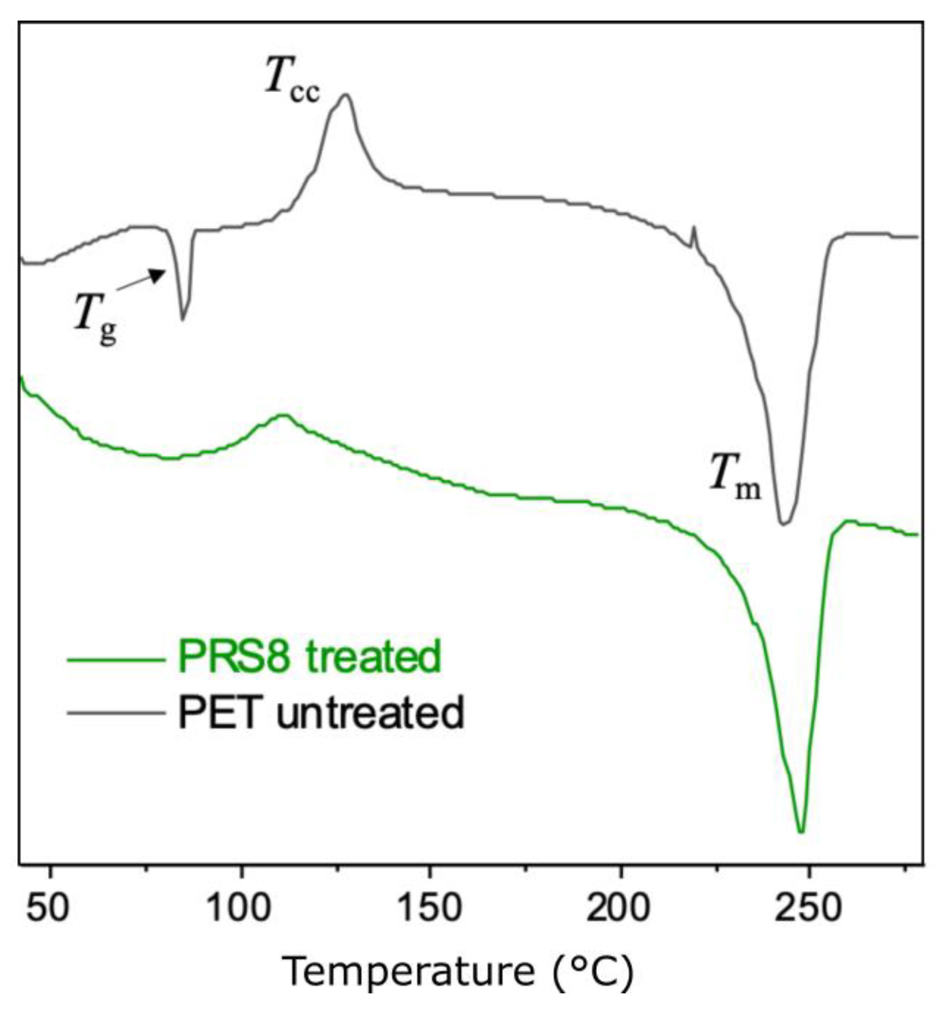
Thermal Properties and the Crystallinity of the PET Powders

The crystallinity and thermal properties of the treated (with the ECF) and untreated PET powders were analyzed using DSC (Figure 11 and Table 6). The glass transition of the untreated PET powder was clearly observed in the first heating curve (Tg ~83 °C). In the case of the treated PET powder, the Tg was not clearly observed on the first heating curve, probably due to overlapping with the onset of the cold crystallization process (~85 °C). The Tg of the treated PET powder was observed in the second heating curve (Supplementary Materials, Figure S3). It was observed that the temperature for the cold crystallization peak (Tcc) became lower after the ECF treatment (110 °C compared to 127 °C for the untreated PET powder), which was accompanied by a lowered enthalpy for the cold crystallization (ΔHcc ~2.9 J/g compared to 9.0 J/g for the pristine PET powder, Table 6). In addition, an increased Tm (247 °C) and crystallinity (21.1%) were observed after the PET powder was treated with the ECF (compared to the Tm of ~242 °C and crystallinity of 19.3% for the pristine PET powder). This result suggested that the degradation of the bulk PET materials presumably occurred (or occurred faster) in the amorphous region of the PET powder, which increased the relative content of the crystalline regions.

4. Discussion

S. maltophilia is a ubiquitous bacterium that has gained interest for its potential applications in bioremediation and agriculture, but at the same time, with concern as an emerging opportunistic pathogen with multidrug resistance to antibiotics [18]. The strain S. maltophilia PRS8, isolated from a landfill soil sample in this work, was not subjected to pathogenicity or antibiotic resistance studies. However, their ability to form biofilms was revealed using a crystal violet assay. This property was associated with the colonization of various hydrophobic surfaces and infection processes in opportunistic strains [23].
Biofilm formation seems to be an important property of plastic-degrading bacteria. For example, the biofilm formation by Rhodococcus ruber could induce a partial biodegradation of polystyrene by with a gravimetric weight loss up to 0.8% after eight weeks [34]. S. maltophilia PRS8 also formed biofilms on the polystyrene microtiter plates and PET flakes. Indeed, the PET flakes without pretreatment degraded by a gravimetric weight loss of up to 1.2% over 28 days. The chemical composition of the S. maltophilia PRS8 biofilm is not yet known. However, biofilms not only facilitate cell adhesion to the plastic flake but also provide a suitable matrix for the interaction between the extracellular enzymes and the solid polymer [34]. In fact, the interaction mechanisms between the biofilm and plastics are not yet clearly understood.
The ability of S. maltophilia PRS8 to degrade PCL indicates its production of an extracellular cutinase-like enzyme. PCL (114.1424 Da) is a biodegradable aliphatic synthetic polyester that shares some similarities with cutin, a natural polyester composed of hydroxy and epoxy fatty acids [35]. PET, however, is an aromatic polyester, and several cutinases have also shown the depolymerizing activity on PET [36]. The PET bottle flakes treated with S. maltophilia PRS8 showed a clear roughness and cracks in the SEM analysis, and a slightly reduced intensity of the characteristic PET peaks between 700 and 1800 cm−1 in the FITR analysis, compared to the non-treated PET. These results provide physical and chemical evidences of the PET degradation capability of S. maltophilia PRS8 with the participation of a cutinase-like enzyme.
The optimal nutritional conditions to produce a cutinase-like enzyme using S. maltophilia PRS8 were obtained using the Plackett–Burman and central composite experimental designs. The optimum activity of using p-NP-butyrate as a substrate was achieved at 35 °C and a pH of 8.0 after 48 h. Cutin, NaNO3, and (NH4)2SO4 showed a positive effect on the maximum production of the cutinase-like enzyme. These results revealed that cutin induced the production of the enzyme, while the salts provided the nitrogen required for the bacteria growth and the enzyme synthesis. The analysis of the reciprocal interactions showed that the specific activity of the enzyme was enhanced when cutin, NaNO3, NaNO3, and (NH4)2SO4 increased in the concentrations, and when cutin increased and (NH4)2SO4 decreased. A similar result was reported for the esterase production from Pseudomonas aeruginosa S3 [37].
A cutinase-like enzyme was successfully purified for homogeneity from the extracellular medium of S. maltophilia PRS8. It was obtained at 450.58 U/mg with a 6.39 purification fold. The molecular weight was approximately 58 kDa. The enzyme activity was optimal at 40 °C and a pH of 8.0 and was found to be stable at a wide range of temperatures (30 to 40 °C) and pH levels (8.0 to 10.0). Indeed, the S. maltophilia species was an attractive source of ester-active enzymes, and some lipases and esterases were cloned and characterized (Table 7). The first characterized lipase for this genus was named LipSM54 and was described as cold-active, solvent-tolerant, and alkaline [38]. A thermostable lipase, LipSm [39], and a thermostable-alkaline-stable esterase [40] were also cloned and characterized. The gene sequences of the cutinase-like enzyme purified in this work is unknown. However, it differs from the cloned ester-active enzymes in terms of the molecular weight and optimum temperatures. In addition, the cloned enzymes were not tested against the natural or synthetic polyesters. The purified cutinase-like enzyme from S. maltophilia PRS8 produced TPA, MHET and BHE from the PET flakes. A similar product profile was also found after the treatment of PET with PETases [17] and cutinases [33].
Some metal ions, surfactants, and organic solvents could be used as additives to increase the enzyme activity. Some metal ions could stabilize the enzyme structure, and the surfactants and organic solvents could facilitate the interaction with the hydrophobic polymers. The cutinase-like enzyme activity was slightly increased by the various metal ions (mono and divalent cationic) except for Cd2+, Co2+, and Hg2+, which strongly inhibited the activity. The activity limitation in the presence of Cd2+ and Hg2+ revealed the presence of a gathering of critical vicinal sulfhydryl [42]. However, Cu2+ and Ni2+ enhanced the relative activity [43]. Regarding the surfactants, nonionic surfactants were reported to have an important role in promoting cutinase activity [44]. Tween and Triton-100 showed a positive effect, but the SDS and CTAB strongly inhibited the enzyme activity. The enzyme was stable in the presence of the various solvents except for ethanol and butanol, which might be due to a change in the structure of the bound substrate or the active site polarity [45].
The DSC measurements of the PET powders revealed an increased crystallinity after the treatment with the extracellular fraction of S. maltophilia PRS, which suggested that the enzymatic degradation of the PET powders primarily occurred at the amorphous region, as reported in the literature [46]. Interestingly, it was previously reported that the enzymatic degradation of the PET flakes using IsPETase from Ideonella sakaiensis resulted in a slightly decreased crystallinity, which was attributed to the formation of the dimers after the degradation that might have disrupted the crystallinity [47]. In our result, no dimer (melting or crystallization) peak was observed using DSC, which was consistent with the increased crystallinity after the enzymatic degradation by S. maltophilia PRS8. Thus, the cutinase-like enzyme activity seemed restricted to the amorphous part.
Finally, the S. maltophilia PRS8 extracellular medium containing the cutinase-like enzyme showed a PET-depolymerizing activity comparable to IsPETase and significantly higher than the thermostable HiCut (HiC) from Humicola insolens. In a practical sense, these results indicated that the S. maltophilia PRS8 extracellular medium produced under optimal conditions could be used directly in the PET degradation processes, with the advantage of skipping the enzyme purification processes and costs. Of course, as an emerging opportunistic pathogen, its biotechnological use involves a safety challenge. An alternative would be using recombinant forms of S. maltophilia enzymes produced in more safe hosts. In this line of study, we are currently sequencing the genome of S. maltophilia PRS8.

5. Conclusions

In this study, a PET-active bacterium S. maltophilia PRS8 was isolated from plastic-contaminated soil that could depolymerize PET at a mesophilic temperature. The strain PRS8 was observed to develop a biofilm on the PET surface that facilitated the bacterium in the process of PET degradation. The cutinase-like activity in the degradation medium demonstrated its role in the degradation of PET. The purified enzyme was stable to various environmental factors and efficiently hydrolyzed PET at a mesophilic temperature. Considering its degradation potential against a recalcitrant polymer, S. maltophilia PRS8 and its enzymes could be potential candidates for further utilization against PET waste and the recovery of value-added products, considering bio-upcycling for the circular economy.

Supplementary Materials

The following supporting information can be downloaded at: https://www.mdpi.com/article/10.3390/app13063686/s1, S1. UV PET treatment; S2. Experimental Design; Table S1. Placket Burman design of factors with specific enzyme activity (U/mg) as response. Table S2. ANNOVA for Placket-Burman Design; Table S3. Central Composite design of factors with specific activity (U/mg); Table S4. ANNOVA for Central Composite design; Figure S1. FT-IF spectra of PET flakes before and after UV exposure; Figure S2. Fourier-transform infrared spectra; Figure S3. DSF analysis of PET samples. Reference [48] is cited in the Supplementary Materials.

Author Contributions

Conceptualization, A.A.S., F.H., S.Ç. and J.A.L.-P.; methodology, S.U.D., K., S.M.S., S.U. and E.C.; software, A.A.S., S.U.D., K. and S.M.S.; validation, S.U.D., K., S.M.S., S.K., M.B. and A.O.B.; formal analysis, A.A.S., S.U.D., F.H., K., S.M.S., S.K., M.B., S.Ç., A.O.B., S.V.M., B.Z. and J.A.L.-P.; investigation, A.A.S., S.U.D., F.H., K., S.M.S., S.K., M.B., S.Ç. and A.O.B.; resources, A.A.S., F.H., S.K., S.Ç. and J.A.L.-P.; data curation, A.A.S. and J.A.L.-P.; writing—original draft preparation, S.U.D. and A.A.S.; writing—review and editing, all authors.; visualization, A.A.S., F.H., S.K. and M.B.; supervision, A.A.S. and J.A.L.-P.; project administration, A.A.S. and J.A.L.-P. All authors have read and agreed to the published version of the manuscript.

Funding

This research received no external funding.

Institutional Review Board Statement

Not applicable.

Informed Consent Statement

Not applicable.

Data Availability Statement

The files of the models presented in this study are available on request from corresponding author. The other data are available in the article.

Acknowledgments

The authors thank the Higher Education Commission of Pakistan (HEC) for providing funds for the internships under the International Research Support Initiative Program (IRSIP), the government of Turkey for providing funds for this research, Burslari Research. The authors also thank the Royal Physiographic Society in Lund, Sweden, the Crafoord Foundation, the Swedish Research Council for Sustainable Development (Forms, no. 201-01107) and the Mistra Foundation (the “STEPS” project no. 2016/1489) for the research funding.

Conflicts of Interest

The authors have no conflict of interest.

References

  1. Müller, R.J.; Schrader, H.; Profe, J.; Dresler, K.; Deckwer, W.D. Enzymatic degradation of poly (ethylene terephthalate): Rapid hydrolyse using a hydrolase from T. fusca. Macromol. Rapid Commun. 2005, 26, 1400–1405. [Google Scholar] [CrossRef]
  2. Yoshida, S.; Hiraga, K.; Takehana, T.; Taniguchi, I.; Yamaji, H.; Maeda, Y.; Toyohara, K.; Miyamoto, K.; Kimura, Y.; Oda, K. A bacterium that degrades and assimilates poly (ethylene terephthalate). Science 2016, 351, 1196–1199. [Google Scholar] [CrossRef] [PubMed]
  3. Tournier, V.; Topham, C.; Gilles, A.; David, B.; Folgoas, C.; Moya-Leclair, E.; Kamionka, E.; Desrousseaux, M.-L.; Texier, H.; Gavalda, S. An engineered PET depolymerase to break down and recycle plastic bottles. Nature 2020, 580, 216–219. [Google Scholar] [CrossRef]
  4. PlasticsEurope, E. Plastics—The Facts 2019. An Analysis of European Plastics Production, Demand and Waste Data, PlasticEurope. 2019. Available online: https://www.plasticseurope.org/en/resources/publications/1804-plastics-facts-2019 (accessed on 17 November 2022).
  5. Nielsen, T.D.; Hasselbalch, J.; Holmberg, K.; Stripple, J. Politics and the plastic crisis: A review throughout the plastic life cycle. Wiley Interdiscip. Rev. Energy Environ. 2020, 9, e360. [Google Scholar] [CrossRef] [Green Version]
  6. Prata, J.C. Microplastics in wastewater: State of the knowledge on sources, fate and solutions. Mar. Pollut. Bull. 2018, 129, 262–265. [Google Scholar] [CrossRef] [PubMed]
  7. Cox, K.D.; Covernton, G.A.; Davies, H.L.; Dower, J.F.; Juanes, F.; Dudas, S.E. Human consumption of microplastics. Environ. Sci. Technol. 2019, 53, 7068–7074. [Google Scholar] [CrossRef] [Green Version]
  8. Pinter, E.; Welle, F.; Mayrhofer, E.; Pechhacker, A.; Motloch, L.; Lahme, V.; Grant, A.; Tacker, M. Circularity study on PET bottle-to-bottle recycling. Sustainability 2021, 13, 7370. [Google Scholar] [CrossRef]
  9. Kaabel, S.; Arciszewski, J.; Borchers, T.H.; Therien, J.D.; Friščić, T.; Auclair, K. Solid-State Enzymatic Hydrolysis of Mixed PET/Cotton Textiles. ChemSusChem 2023, 16, e202201613. [Google Scholar] [CrossRef]
  10. MacLeod, M.; Arp, H.P.H.; Tekman, M.B.; Jahnke, A. The global threat from plastic pollution. Science 2021, 373, 61–65. [Google Scholar] [CrossRef]
  11. Barboza, L.G.A.; Lopes, C.; Oliveira, P.; Bessa, F.; Otero, V.; Henriques, B.; Raimundo, J.; Caetano, M.; Vale, C.; Guilhermino, L. Microplastics in wild fish from North East Atlantic Ocean and its potential for causing neurotoxic effects, lipid oxidative damage, and human health risks associated with ingestion exposure. Sci. Total Environ. 2020, 717, 134625. [Google Scholar] [CrossRef]
  12. Zrimec, J.; Kokina, M.; Jonasson, S.; Zorrilla, F.; Zelezniak, A. Plastic-degrading potential across the global microbiome correlates with recent pollution trends. mBio 2021, 12, e0215521. [Google Scholar] [CrossRef]
  13. Danso, D.; Chow, J.; Streit, W.R. Plastics: Environmental and biotechnological perspectives on microbial degradation. Appl. Environ. Microbiol. 2019, 85, e01095-19. [Google Scholar] [CrossRef] [Green Version]
  14. Zhang, Z.; Huang, S.; Cai, D.; Shao, C.; Zhang, C.; Zhou, J.; Cui, Z.; He, T.; Chen, C.; Chen, B. Depolymerization of post-consumer PET bottles with engineered cutinase 1 from Thermobifida cellulosilytica. Green Chem. 2022, 24, 5998–6007. [Google Scholar] [CrossRef]
  15. Leitão, A.L.; Enguita, F.J. Structural insights into carboxylic polyester-degrading enzymes and their functional depolymerizing neighbors. Int. J. Mol. Sci. 2021, 22, 2332. [Google Scholar] [CrossRef]
  16. Wei, R.; Zimmermann, W. Microbial enzymes for the recycling of recalcitrant petroleum-based plastics: How far are we? Microb. Biotechnol. 2017, 10, 1308–1322. [Google Scholar] [CrossRef] [PubMed] [Green Version]
  17. Wagner-Egea, P.; Tosi, V.; Wang, P.; Grey, C.; Zhang, B.; Linares-Pastén, J.A. Assessment of Is PETase-Assisted Depolymerization of Terephthalate Aromatic Polyesters and the Effect of the Thioredoxin Fusion Domain. Appl. Sci. 2021, 11, 8315. [Google Scholar] [CrossRef]
  18. Brooke, J.S. Advances in the microbiology of Stenotrophomonas maltophilia. Clin. Microbiol. Rev. 2021, 34, e00030-19. [Google Scholar] [CrossRef] [PubMed]
  19. Arulazhagan, P.; Al-Shekri, K.; Huda, Q.; Godon, J.-J.; Basahi, J.M.; Jeyakumar, D. Biodegradation of polycyclic aromatic hydrocarbons by an acidophilic Stenotrophomonas maltophilia strain AJH1 isolated from a mineral mining site in Saudi Arabia. Extremophiles 2017, 21, 163–174. [Google Scholar] [CrossRef]
  20. Alvarado-Gutiérrez, M.L.; Ruiz-Ordaz, N.; Galíndez-Mayer, J.; Curiel-Quesada, E.; Santoyo-Tepole, F. Degradation kinetics of carbendazim by Klebsiella oxytoca, Flavobacterium johnsoniae, and Stenotrophomonas maltophilia strains. Environ. Sci. Pollut. Res. 2020, 27, 28518–28526. [Google Scholar] [CrossRef]
  21. Coleman, N.V.; Mattes, T.E.; Gossett, J.M.; Spain, J.C. Biodegradation of cis-dichloroethene as the sole carbon source by a β-proteobacterium. Appl. Environ. Microbiol. 2002, 68, 2726–2730. [Google Scholar] [CrossRef] [Green Version]
  22. Satti, S.M.; Shah, A.A.; Auras, R.; Marsh, T.L. Isolation and characterization of bacteria capable of degrading poly (lactic acid) at ambient temperature. Polym. Degrad. Stab. 2017, 144, 392–400. [Google Scholar] [CrossRef]
  23. Pompilio, A.; Piccolomini, R.; Picciani, C.; D’Antonio, D.; Savini, V.; Di Bonaventura, G. Factors associated with adherence to and biofilm formation on polystyrene by Stenotrophomonas maltophilia: The role of cell surface hydrophobicity and motility. FEMS Microbiol. Lett. 2008, 287, 41–47. [Google Scholar] [CrossRef] [PubMed] [Green Version]
  24. Kumar, S.; Stecher, G.; Li, M.; Knyaz, C.; Tamura, K. MEGA X: Molecular evolutionary genetics analysis across computing platforms. Mol. Biol. Evol. 2018, 35, 1547. [Google Scholar] [CrossRef] [PubMed]
  25. Adıgüzel, A.O.; Tunçer, M. Purification and characterization of cutinase from Bacillus sp. KY0701 isolated from plastic wastes. Prep. Biochem. Biotechnol. 2017, 47, 925–933. [Google Scholar] [CrossRef] [PubMed]
  26. Kyaw, B.M.; Champakalakshmi, R.; Sakharkar, M.K.; Lim, C.S.; Sakharkar, K.R. Biodegradation of low density polythene (LDPE) by Pseudomonas species. Indian J. Microbiol. 2012, 52, 411–419. [Google Scholar] [CrossRef] [Green Version]
  27. Drobota, M.; Persin, Z.; Zemljic, L.F.; Mohan, T.; Stana-Kleinschek, K.; Doliska, A.; Bracic, M.; Ribitsch, V.; Harabagiu, V.; Coseri, S. Chemical modification and characterization of poly (ethylene terephthalate) surfaces for collagen immobilization. Cent. Eur. J. Chem. 2013, 11, 1786–1798. [Google Scholar] [CrossRef]
  28. Ioakeimidis, C.; Fotopoulou, K.; Karapanagioti, H.; Geraga, M.; Zeri, C.; Papathanassiou, E.; Galgani, F.; Papatheodorou, G. The degradation potential of PET bottles in the marine environment: An ATR-FTIR based approach. Sci. Rep. 2016, 6, 23501. [Google Scholar] [CrossRef] [Green Version]
  29. Chaudhari, S.A.; Singhal, R.S. Cutin from watermelon peels: A novel inducer for cutinase production and its physicochemical characterization. Int. J. Biol. Macromol. 2015, 79, 398–404. [Google Scholar] [CrossRef]
  30. Determann, H. Gel Chromatography Gel Filtration·Gel Permeation Molecular Sieves: A Laboratory Handbook; Springer Science & Business Media: Berlin/Heidelberg, Germany, 2012. [Google Scholar]
  31. Karpushova, A.; Brümmer, F.; Barth, S.; Lange, S.; Schmid, R. Cloning, recombinant expression and biochemical characterisation of novel esterases from Bacillus sp. associated with the marine sponge Aplysina aerophoba. Appl. Microbiol. Biotechnol. 2005, 67, 59–69. [Google Scholar] [CrossRef]
  32. Cheng, S.Z.; Pan, R.; Wunderlich, B. Thermal analysis of poly (butylene terephthalate) for heat capacity, rigid-amorphous content, and transition behavior. Die Makromol. Chem. Macromol. Chem. Phys. 1988, 189, 2443–2458. [Google Scholar] [CrossRef]
  33. Aristizábal-Lanza, L.; Mankar, S.V.; Tullberg, C.; Zhang, B.; Linares-Pastén, J.A. Comparison of the enzymatic depolymerization of polyethylene terephthalate and Akestra TM using Humicola insolens cutinase. Front. Chem. Eng. 2022, 4, 1048744. [Google Scholar] [CrossRef]
  34. Hadar, Y.; Sivan, A. Colonization, biofilm formation and biodegradation of polyethylene by a strain of Rhodococcus ruber. Appl. Microbiol. Biotechnol. 2004, 65, 97–104. [Google Scholar]
  35. Carvalho, C.M.; Aires-Barros, M.R.; Cabral, J.M. Cutinase: From molecular level to bioprocess development. Biotechnol. Bioeng. 1999, 66, 17–34. [Google Scholar] [CrossRef]
  36. Liang, X.; Zou, H. Biotechnological Application of Cutinase: A Powerful Tool in Synthetic Biology. SynBio 2022, 1, 54–64. [Google Scholar] [CrossRef]
  37. Noor, H.; Satti, S.M.; ud Din, S.; Farman, M.; Hasan, F.; Khan, S.; Badshah, M.; Shah, A.A. Insight on esterase from Pseudomonas aeruginosa strain S3 that depolymerize poly (lactic acid)(PLA) at ambient temperature. Polym. Degrad. Stab. 2020, 174, 109096. [Google Scholar] [CrossRef]
  38. Li, M.; Yang, L.-R.; Xu, G.; Wu, J.-P. Cloning and characterization of a novel lipase from Stenotrophomonas maltophilia GS11: The first member of a new bacterial lipase family XVI. J. Biotechnol. 2016, 228, 30–36. [Google Scholar] [CrossRef]
  39. Parapouli, M.; Foukis, A.; Stergiou, P.-Y.; Koukouritaki, M.; Magklaras, P.; Gkini, O.A.; Papamichael, E.M.; Afendra, A.-S.; Hatziloukas, E. Molecular, biochemical and kinetic analysis of a novel, thermostable lipase (LipSm) from Stenotrophomonas maltophilia Psi-1, the first member of a new bacterial lipase family (XVIII). J. Biol. Res. -Thessalon. 2018, 25, 4. [Google Scholar] [CrossRef] [PubMed] [Green Version]
  40. Gao, X.; Mao, X.; Lu, P.; Secundo, F.; Xue, C.; Sun, J. Cloning, expression, and characterization of a novel thermostable and alkaline-stable esterase from Stenotrophomonas maltophilia OUC_Est10 catalytically active in organic solvents. Catalysts 2019, 9, 401. [Google Scholar] [CrossRef] [Green Version]
  41. Çolak, A.; Şişik, D.; Saglam, N.; Güner, S.; Çanakçi, S.; Beldüz, A.O. Characterization of a thermoalkalophilic esterase from a novel thermophilic bacterium, Anoxybacillus gonensis G2. Bioresour. Technol. 2005, 96, 625–631. [Google Scholar] [CrossRef]
  42. Then, J.; Wei, R.; Oeser, T.; Barth, M.; Belisário-Ferrari, M.R.; Schmidt, J.; Zimmermann, W. Ca2+ and Mg2+ binding site engineering increases the degradation of polyethylene terephthalate films by polyester hydrolases from Thermobifida fusca. Biotechnol. J. 2015, 10, 592–598. [Google Scholar] [CrossRef]
  43. Geyer, B.; Lorenz, G.; Kandelbauer, A. Recycling of poly (ethylene terephthalate)—A review focusing on chemical methods. Express Polym. Lett. 2016, 10, 559–586. [Google Scholar] [CrossRef]
  44. Degani, O. Production and purification of cutinase from fusarium oxysporum using modified growth media and a specific cutinase substrate. Adv. Biosci. Biotechnol. 2015, 6, 245. [Google Scholar] [CrossRef] [Green Version]
  45. Kim, Y.-J.; Choi, G.-S.; Kim, S.-B.; Yoon, G.-S.; Kim, Y.-S.; Ryu, Y.-W. Screening and characterization of a novel esterase from a metagenomic library. Protein Expr. Purif. 2006, 45, 315–323. [Google Scholar] [CrossRef] [PubMed]
  46. Carr, C.M.; Clarke, D.J.; Dobson, A.D. Microbial polyethylene terephthalate hydrolases: Current and future perspectives. Front. Microbiol. 2020, 11, 571265. [Google Scholar] [CrossRef] [PubMed]
  47. Menzel, T.; Weigert, S.; Gagsteiger, A.; Eich, Y.; Sittl, S.; Papastavrou, G.; Ruckdäschel, H.; Altstädt, V.; Höcker, B. Impact of enzymatic degradation on the material properties of poly (ethylene terephthalate). Polymers 2021, 13, 3885. [Google Scholar] [CrossRef]
  48. Falkenstein, P.; Gräsing, D.; Bielytskyi, P.; Zimmermann, W.; Matysik, J.; Wei, R.; Song, C. UV pretreatment impairs the enzymatic degradation of polyethylene terephthalate. Front. Microbiol. 2020, 11, 689. [Google Scholar] [CrossRef] [PubMed]
Figure 1. Neighbor-joining tree based on the 16S rRNA gene sequences showing the phylogenetic position of the Stenotrophomonas maltophilia PRS8 strain and the representatives of some other related taxa. For out group, Halomonas elongata was used.
Applsci 13 03686 g001
Figure 2. Biofilm-forming ability of S. maltophilia PRS8 compared to that of A. aeruginosa using a crystal violet assay. (A) Biofilms formed on the PET flakes. (B) Biofilms on a microtiter plate. (C) Absorbance (600 nm) of the attached cells after treatment with 30% acetic acid. The error bars represent the standard deviation.
Applsci 13 03686 g002
Figure 3. Expression of the cutinase activity by the S. maltophilia PRS8 strain. (A) Clear zone around a colony on the PCL agar showed putative cutinase activity. (B) Putative cutinase enzyme activity and the total protein in the cell-free supernatant by the S. maltophilia PRS8 strain during the biodegradation of PET as a carbon source in the MSM media.
Applsci 13 03686 g003
Figure 4. Percent of the weight reduction in the PET flakes by the S. maltophilia PRS8 strain.
Applsci 13 03686 g004
Figure 5. Expanded FTIR spectra of PET after 28 days, abiotic control untreated (black solid curve), and PET treated with the S. maltophilia PRS8 strain (green dotted curve).
Applsci 13 03686 g005
Figure 6. Effect of S. maltophilia PRS8 on the PET flakes. SEM images (×5.00 K) of bacterial-treated PET (A) compared to untreated PET after 28 days of incubation (B).
Applsci 13 03686 g006
Figure 7. Pareto chart and a three-dimensional response surface plot showing the significant factor with respect to the effect on the enzyme production. (A) Pareto chart showing the effect of the t-value by the important factors [J, cutin; D, NaNO3, and L (NH4)2SO4] generated by the PBD. (B) Design of the 3D response surface plot of cutin and (NH4)2SO4. (C) Design of the 3D response surface plot between cutin and NaNO3. (D) Design of the 3D response surface plot between NaNO3 and (NH4)2SO4.
Applsci 13 03686 g007
Figure 8. Purification profile of the cutinase-like enzyme. (A) Total protein and activity profile by Stenotrophomonas maltophilia PRS8 through the Sephadex G-100 gel filtration column (B) SDS-PAGE of cutinase by the S. maltophilia PRS8 strain after purification using column chromatography. Lane 1, Zymogram; Lane 2, Eluate from Sephadex G-100; Lane 3, Protein marker 11–245 kDa.
Applsci 13 03686 g008
Figure 9. Effect of the temperature and pH on the activity and stability of the purified cutinase-like enzyme from the S. maltophilia PRS8 strain. (A) Temperature profile. (B) Profile of the pH.
Applsci 13 03686 g009
Figure 10. Analysis of the degradation products of the PET bottle flakes using the cutinase-like enzyme. (A) Chromatogram of the product’s profile. (B) Mass spectra confirmed the identity of TA, MHET, and BHET.
Applsci 13 03686 g010
Figure 11. DSC heating curves (first cycle, exo up) of the untreated PET and PET treated with the extracellular fraction of S. maltophilia PRS8. The Tg, Tcc and Tm are shown on the PET untreated curve.
Applsci 13 03686 g011
Table 1. Purification steps of the cutinase-like enzyme from the S. maltophilia PRS8 strain.
Purification StepsVolume (mL)Activity
(U/mL)
Total Activity
(Units)
Protein
(mg/mL)
Specific Activity
(U/mg)
Yield
(%)
Purification Fold
Crude Extract500138.1369,068.181.6570.471001
(NH4)2SO4200260.4052,081.821.18153.4275.402.17
Sephadex (G-100)90373.2833,595.910.75450.5848.646.39
Table 2. Effect of metal ions on the activity of the cutinase-like enzyme from the S. maltophilia PRS8 strain.
MetalsResidual Activity (%)
15 mM2 mM
Control100 ± 0.0100 ± 0.0
Ca2+111.32 ± 0.6102.34 ± 0.5
Fe2+98.82 ± 0.7100.39 ± 0.6
Cu2+107.81 ± 1.0100.78 ± 0.2
Co2+55.85 ± 0.258.98 ± 0.3
Ni2+100.39 ± 0.4100 ± 0.1
Zn2+114.45 ± 0.5107.42 ± 0.4
K+115.23 ± 0.5111.71 ± 0.3
Cd2+1.95 ± 0.23.51 ± 0.3
Na+85.54 ± 0.191.79 ± 0.2
Mg2+107.42 ± 0.4105.07 ± 0.3
Ni2+71.48 ± 0.366.01 ± 0.5
Hg2+0 ± 0.000 ± 0.00
Ba2+54.29 ± 0.155.46 ± 0.3
Table 3. Effect of the organic solvents on the activity of the cutinase-like enzyme from the S. maltophilia PRS8 strain.
Organic Solvents (10%)Residual Activity % (Min)
30 min60 min90 min120 min
Control100100100100
Ethanol54.2654.2645.2942.60
Methanol104.03103.1398.6591.03
Acetonitrile93.2790.1391.0383.40
Acetone97.0396.8696.4195.51
Ethyl Acetate103.1398.6596.4192.82
Propanol105.82101.79101.34100
DMSO98.2095.0694.6194.17
Butanol65.0850.6749.3228.25
Formaldehyde94.6189.6885.6585.20
Table 4. Effect of the surfactant on the activity of the cutinase-like enzyme from the S. maltophilia PRS8 strain.
SurfactantsResidual Activity (%)
0.5%5%
Control100 ± 0.00100 ± 0.00
Tween-20109.41 ± 0.30106.66 ± 0.53
Tween-40112.54 ± 0.72116.47 ± 0.30
Tween-60110.98 ± 0.81110.58 ± 0.90
Tween-80113.72 ± 0.70105.09 ± 0.54
CTAB11.76 ± 0.407.84 ± 0.31
SDS0 ± 0.000 ± 0.00
Triton X-100109.80 ± 0.21112.15 ± 0.12
Table 5. PET powder depolymerization activity of the extracellular medium of S. maltophilia PRS8 compared to that of the enzymes. The reaction times were 72–73 h.
EnzymeRatio (E/PET)T (°C)pHDegradation Products (mg/L)Relative Production (%)PET
Depolymerization (%)
Ref.
TPAMHETBHET
Extracellular medium0.005 *40922463860222.5This work
IsPETase0.004377.2230424.273.72.123[17]
Trx-IsPETase0.005377.2323031.566.71.832[17]
HiCut (HiC)0.008707.5437100n.dn.d12.7[33]
* Estimated ratio based on the cutinase-like enzyme fraction present in the extracellular medium.
Table 6. Properties of the PET powder untreated and treated with the extracellular medium containing the cutinase-like enzyme.
PolymerTg
(°C)
Tcc
(°C)
Tm
(°C)
ΔHcc
(J/g)
ΔHm
(J/g)
Crystallinity *
(%)
PET (untreated)831272429.035.919.3
PET (treated)-1102472.932.421.1
* The crystallinity was calculated using Equation (1).
Table 7. Ester-active enzymes from S. maltophilia.
EnzymeStrainMW (kDa)Sequence
(GenBank)
Optimum
Temperature (°C)
Optimum pHReference
Cutinase-likePSR858Unknown408.0This work
Lipase (LipSM54)GS1152.8KX353755358.0[38]
Lipase (LipSm)Psi-140.7KC01461664.58.0–9.0[39]
Esterase (est7)OUC_Est1076.6MH253883459.0[41]
Disclaimer/Publisher’s Note: The statements, opinions and data contained in all publications are solely those of the individual author(s) and contributor(s) and not of MDPI and/or the editor(s). MDPI and/or the editor(s) disclaim responsibility for any injury to people or property resulting from any ideas, methods, instructions or products referred to in the content.

