Article

Method to Calculate Melanopic Light Reaching the Retina Depending on the Optical Density of an Aging Crystalline Lens

by
Ana Sanchez-Cano
1,2,*,
Elvira Orduna-Hospital
1,2,
Guisela Fernández-Espinosa
2 and
Justiniano Aporta
1,2
1
Applied Physics Department, University of Zaragoza, 50009 Zaragoza, Spain
2
Aragon Institute for Health Research (IIS Aragon), 50009 Zaragoza, Spain
*
Author to whom correspondence should be addressed.
Appl. Sci. 2023, 13(4), 2569; https://doi.org/10.3390/app13042569
Submission received: 19 January 2023 / Revised: 13 February 2023 / Accepted: 14 February 2023 / Published: 16 February 2023
(This article belongs to the Special Issue Research on Circadian Rhythms in Health and Disease)

Abstract

:
Lighting studies that take into account the age of the inhabitants of an area and are related to circadian light are difficult to find. This study aims to simplify a method to approximately compute the circadian light reaching the retina based on photopic illuminance reaching the corneal plane and considering the optical density of an aging crystalline lens. As an example of this proposed method, calculations were performed with both the D65 and A standard illuminants, showing how the spectral power distribution is modified by the optical density of the crystalline lens, mainly at short wavelengths. Due to these selective wavelength absorptions of the aged lens, a significant variation in the level of daylight equivalent melanopic illuminance (EDI) is present in the retina. With levels of 200 lux at the corneal plane, these variations ranged from 204 EDI lux to 178 EDI lux for the D65 standard illuminant, and from 99 EDI lux to 101 EDI lux for the A standard illuminant for observers aged 10 and 90, respectively. In this work, we aimed to simplify the greatest possible level of calculation of melanopic light, while describing simple protocols that are easy to translate into practice. Our results will allow researchers to carry out optimized lighting designs from both the photometric and circadian perspectives considering the optical density of an aging lens.

1. Introduction

Before light reaches the retina, it passes through the cornea, lens and ocular media. This prereceptoral filtering alters the spectrum relative to the light arriving at the corneal plane [1]. Figure 1, reproduced from [2], shows how a crystalline lens increases in density as a function of age and specifically attenuates short-wavelength transmitted light [3,4,5]. On the other hand, the melanopsin spectra, which absorbs light in the short-wavelength range of the visible spectrum with a maximum at or near 480 nm [6,7], is modified by the absorption spectra of the anterior media, shifted from 480 nm to 487 nm (for a 20-year-old observer) or 496 nm (for an 80-year-old observer).
As a result of the described behavior, retinal illuminance may vary between individuals due to progressive age-dependent changes in the lens, causing a reduction in the amount of short-wavelength light passing through the eye and scattering [3,4,5,8,9,10,11]. Additionally, the number of intrinsically photosensitive retinal ganglion cells (ipRGCs) drops with age, changes their distribution pattern on the retina and changes their morphology [12]. Consequently, retinal photoreceptors receive less light input at older ages, particularly short-wavelength-sensitive photoreceptors (rods, S-cones and ipRGCs).
From a technical point of view, the approach to measuring and reporting light was established by the Commission Internationale de l’Éclairage (CIE) [13,14]. The 10 nm differential between the aforementioned 480 nm peak spectral sensitivity for melanopsin and the 490 nm peak described by the CIE technical note is attributed, as has just been described, to the prereceptoral filtering of the light reaching the retinal plane, and modifications can be found depending on the age (Figure 2). In addition, the parameter of melanopic equivalent daylight illuminance (EDI) was defined by the CIE to compute the melanopic illuminance level instead of the well-known photometric illuminance quantity, since knowing the spectral power distributions (SPDs) of the source is essential to correctly compute both magnitudes, photopic and melanopic illuminances.
The specific physiological effect of light of a given melanopic irradiance, due to the wide range of individual differences to the same corneal spectra, is difficult to personalize; it is easier to estimate or predict than to accurately calculate. The melanopsin spectral sensitivity curve standardized in CIE S026/E:2018 comes closest to these types of standard observers. It shows how a spectrum should be weighted to derive an appropriately weighted quantity. In addition to a standard curve, CIE S026/E:2018 also contains information for generating age-adjusted spectral sensitivity curves, accounting for age-dependent changes in lens transmission [13]. An international group of experts led by Brown recently proposed a minimum of 250 lux (melanopic EDI) daytime level, 10 lux evening level and a 1 lux maximum as the night level for a hypothetical observer. Their results showed that a melanopic EDI below 4 lux results in minimal responses (<25% of maximum melatonin suppression), and a melanopic EDI above 300 lux strongly suppresses salivary melatonin (>75% of the maximum), depending on the exposure duration and experimental context [15]. These results have been corroborated by Gimenez et al. [16]; even with longer durations (e.g., 270 min), a 75% suppression of melatonin can already occur at a melanopic EDI of 100 lux.
Many studies have shown large individual variations in circadian photosensitivity across individuals, such as melatonin suppression, due to artificial light exposure, but recommendations for appropriate light exposure in real-life settings rarely consider such individual effects [8]. Individual differences in light sensitivity also occur at earlier developmental stages, and melatonin suppression by light is higher in children than in adults since they have large pupils and high spectral transmittance of the crystalline lens at short wavelengths; the nonvisual photoreception in 10-year-old children is twice that of 45-year-old adults [17]. Even healthy aging is associated with a progressive decrease in light transmission due to the clouding and yellowing of the natural crystalline lens, especially for short-wavelength light. When age-related cataracts develop, further worsening of ocular processes is found; specifically, they are associated with the disruption of circadian rhythms. However, when patients with cataracts undergo intraocular lens replacement with optimized spectral lens transmission, they may improve circadian photosensitivity, sleep, and cognitive function despite small pupils [8,9,18,19].
As has been intended and described by other authors [11,20,21], providing a simple process for smart or integrative lighting design is important when the process is properly integrated into a traditional workflow. Its implementation requires lighting designers to understand the tedious terminology defined, evaluated and correlated, when possible, to elucidate strengths and limitations to progress in its knowledge. Many available calculators for computing nonvisual quantities are available as a recipe to promote circadian lighting projects in parallel to real designs and without real integration in the lighting design process. Recently, Houser et al. [20] showed an implemented process to guide decisions prior to the transfer to the lighting built, and a posterior audit focused on no single solution promoting integrative points of view.
In this paper, we first summarize potential key findings related to the aging of the crystalline lens on the spectral sensitivity of the melanopsin photopigment expressed in ipRGCs [22,23,24,25]. In a second step, an easy and simplified method to calculate melanopic contribution depending on age is presented to be considered in lighting projects.

2. Background

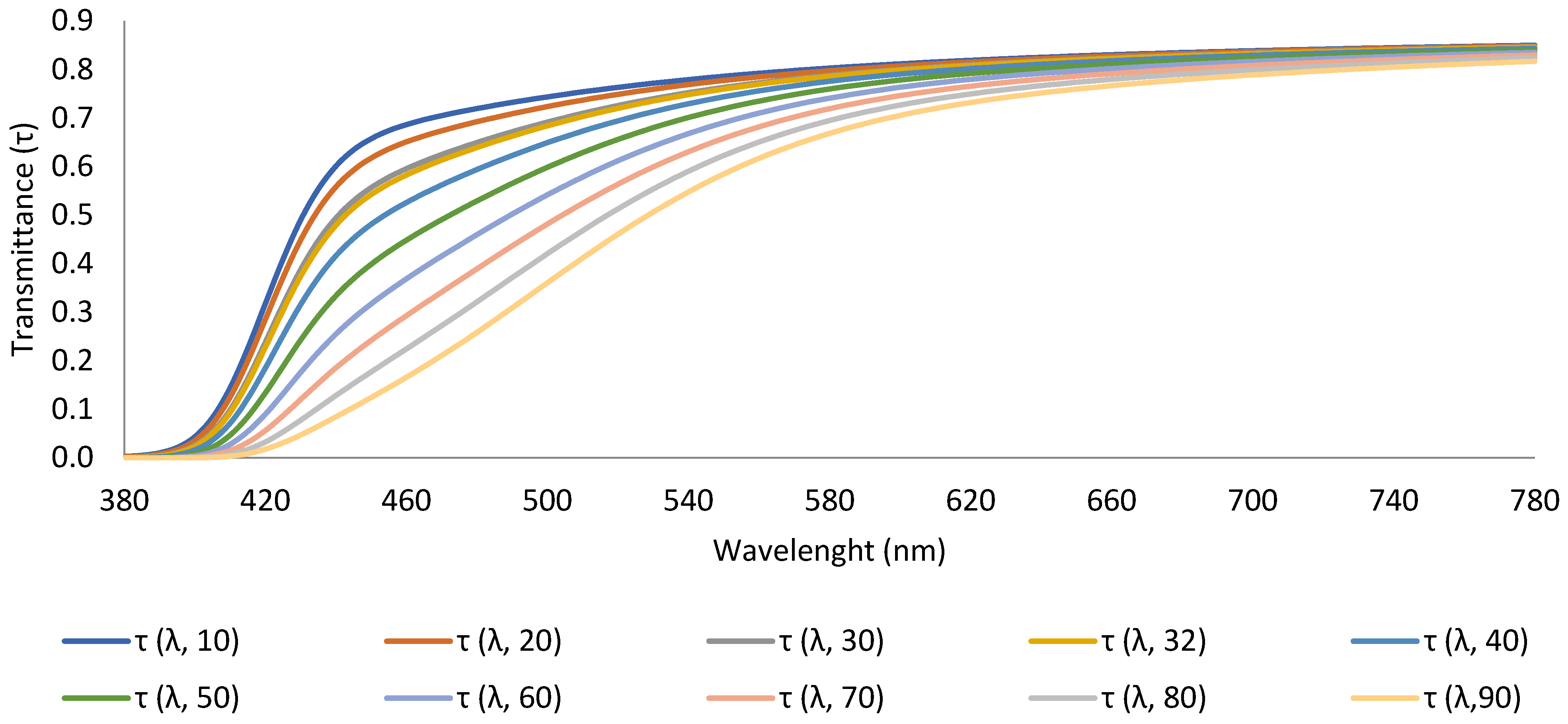
2.1. Technical Features

From a technical point of view, parameters used to describe light and lighting need to previously be defined to explain the modifications made to the calculations. The term color temperature (CT) is applied to highly selective radiators, such as standard illuminant A, when the light of this source has the same, or nearly the same, chromatic coordinates as a blackbody radiator at certain temperature. Meanwhile, the correlated color temperature (CCT) of a light radiator is not exactly equal to any of the chromaticities of a blackbody radiator, and a CCT may be found for a selective radiator by taking the nearest chromaticity match. It is computed by the iso-temperature lines for the evaluation of a CCT for a selective source; these lines are short straight lines crossing the Planckian locus perpendicularly. Light sources with high CCT seem “cool”, and those with low CCT look “warm”. A color rendering index (CRI) is a quantitative measure of the ability of a light source to reveal the colors of various objects faithfully in comparison with a natural or standard light source. The chromaticity coordinates (x,y) of a color are the ratios of each tristimulus value of the color to their sum. A diagram in which any one of the three chromaticity coordinates is plotted against any other is called a chromaticity diagram [26].
In visual photometry, spectral irradiance incident on the corneal plane is measured in the wavelength range of 380 nm to 780 nm. The CIE recommends, among others, two standard sources to be used in colorimetry specifications. One of them is the standard daylight D65 constructed to represent natural daylight having a CCT of 6500 K and, the other, the standard illuminant A intended to represent typical, tungsten-filament lighting with relative SPD, is that of a Planckian radiator at a CT of approximately 2856 K.
The CCT is recurrently described in lighting specifications, making it freely available and frequently used as the most important parameter to control lighting design. Since visual evaluation is important, not only irradiance spectra, but luminance or illuminance levels for these types of nonvisual quantifications, retinal irradiance is the most relevant parameter. Even the combination of the spectral irradiance provided by artificial sources and natural daylight, including light reflected from the surfaces or a scene, might need to be accurately measured to determine its age-related circadian effects.

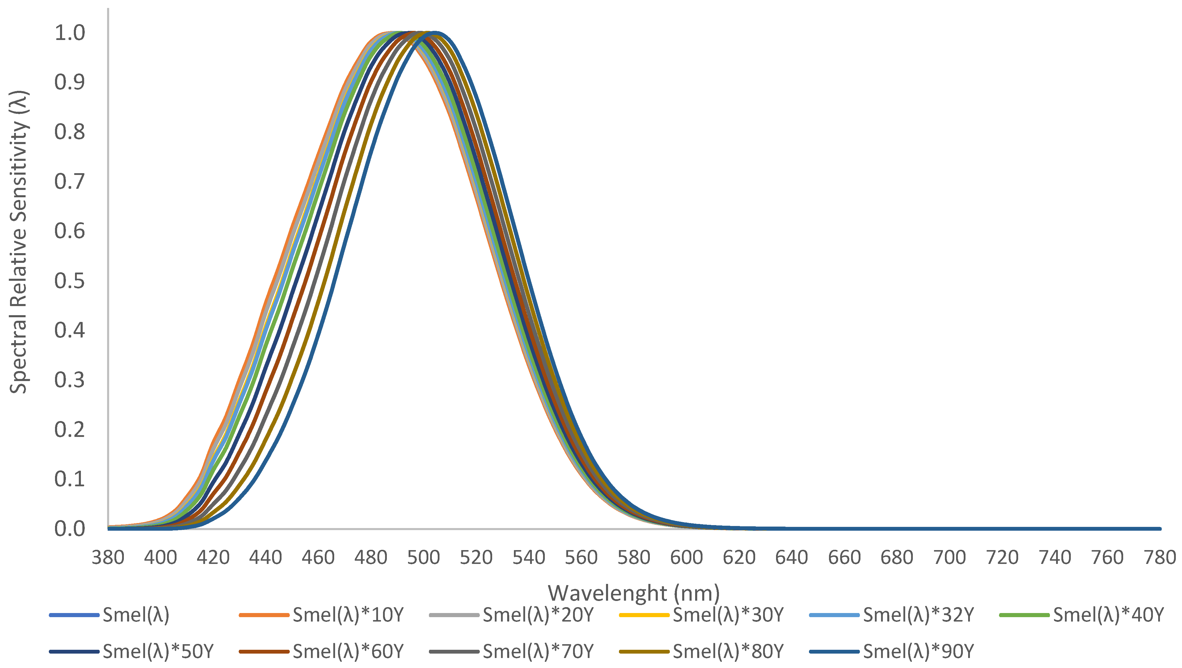
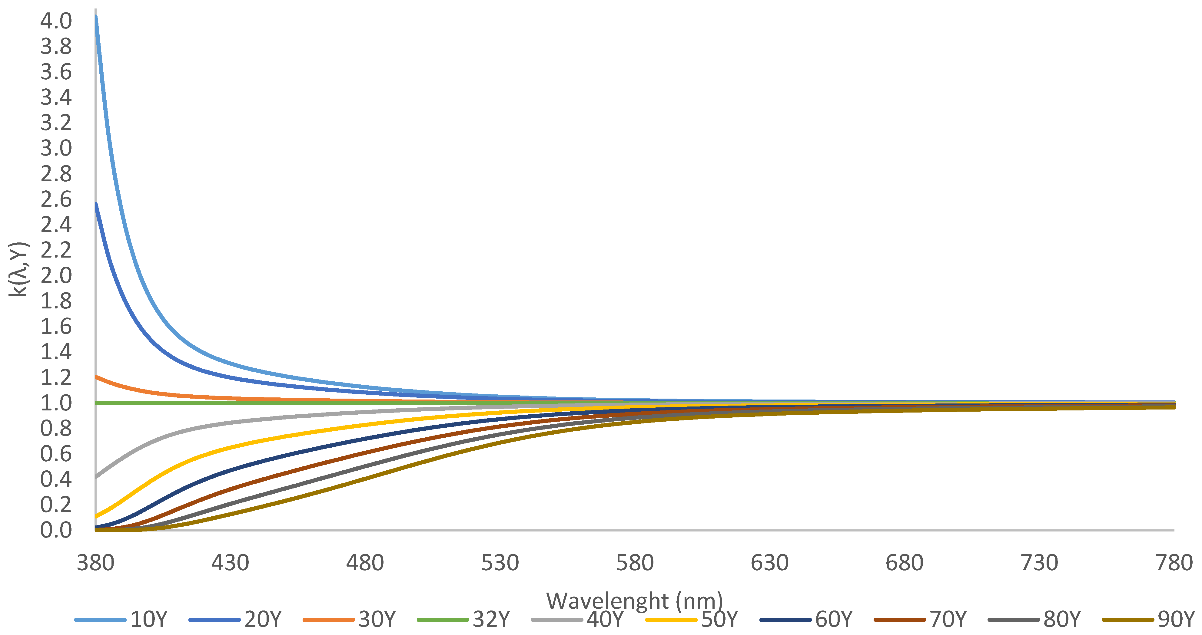
2.2. Theoretical Melanopic Considerations in Lighting for Elderly Individuals
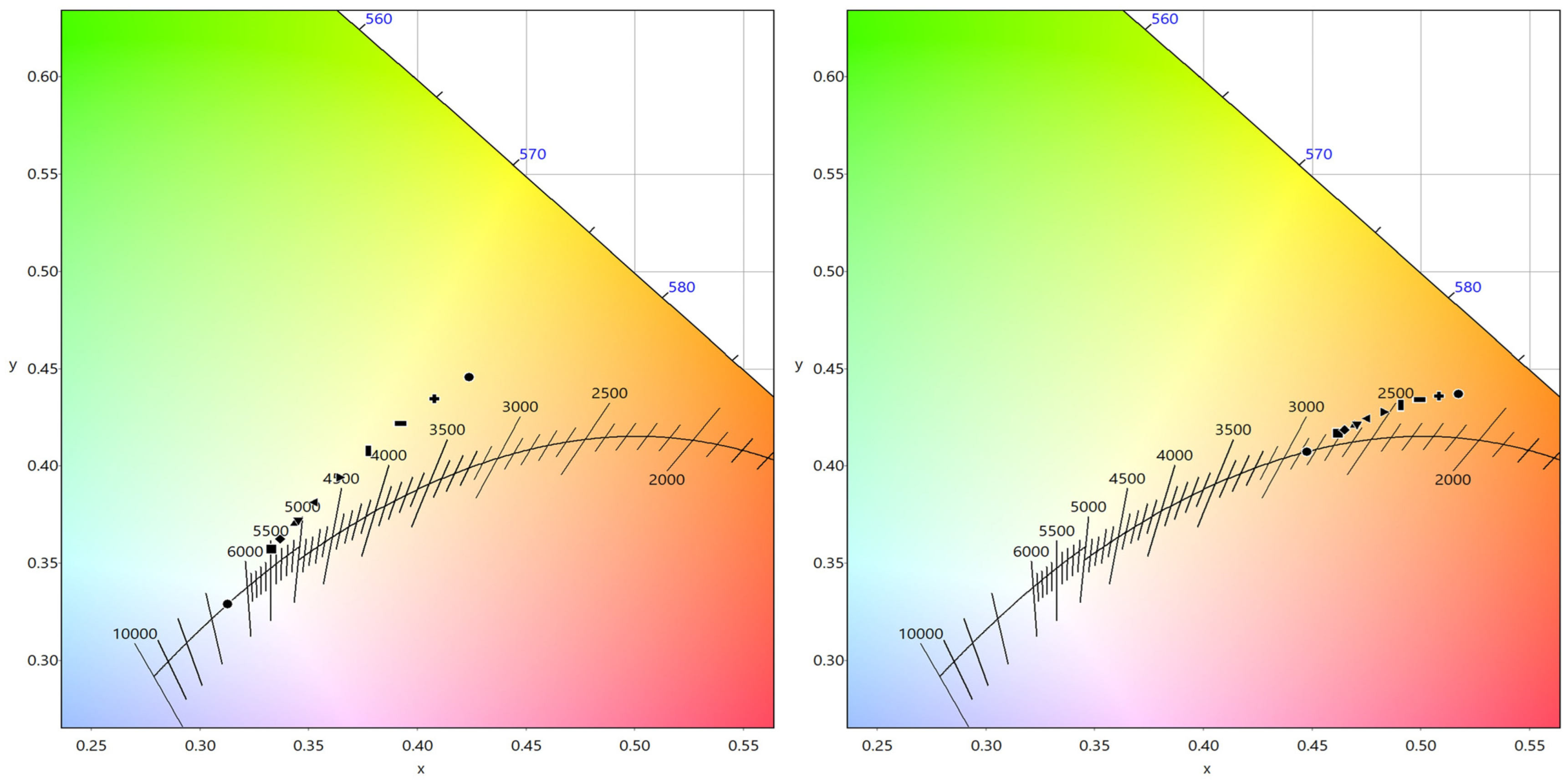
These noted modifications in photometric quantities by age should be translated and analyzed in terms of melanopic implications; data for ipRGC spectral sensitivity published by Lucas et al. [6] inherently include spectral transmittance of the human ocular media for a reference observer of age Yr = 32 years. To correct melanopic data for observers of a different age Y, an age-dependent spectral correction can be applied. This can be relevant, as mentioned, when assessing the impact of light on elderly individuals. Based on the definition for age-dependent transmittance of the human eye lens in CIE 203:2012 and its erratum, an age-related spectral correction function k(λ,Y) is defined for an observer of age Y as k(λ,Y) = τ(λ,Y)/τ(λ,Yr = 32) and plotted in Figure 3 [2].
The equation k(wavelength, age) = trans(wavelength, age)/trans(wavelength, 32 years) is defined as: k λ , Y = τ λ , Y τ λ , 32 .
In this section, basic expressions must be defined, considering both the photopic and melanopic pathways; the method we propose is calculated from the traditional relationship between radiometric and photometric magnitudes and from the current standards as defined previously in Sanchez et al. [27] and more recently in Esposito et al. [28]. The equivalent daylight illuminance (EDI) is defined by the CIE standard [13], and normalization is proposed with the melanopic illuminance provided by the standard illuminant D65 (daylight CCT = 6500 K). A light-source type D65 furnishing photopic illuminance E photopic , D 65 (E) to provide the same melanopic illuminance E melanopic , D 65 (EDI) can be calculated.
EDI = 1.104 × MAF × E
The age-dependent spectral correction factor for lens transmission k(λ,Y) defined for an observer of age Y is a parameter that should be considered to address the nonvisual effects of light in lighting designs in which the age of the user is a key factor [2]. This value is defined as k(λ,32 years) = 1.000; the higher the age is, the lower the value. This correction factor enables us to estimate the real transmission factor of the eye, the lens in particular, and to determine how much light passes through the pupil and reaches the retina. The higher the CCT is, the lower the kmel,trans. This phenomenon, combined with the age of the subject, creates a wide spectrum of possibilities that must be considered in lighting projects. As described below, equations to compute illuminance level were defined at the corneal plane or entrance of the pupil (EP); now, they must be modified to determine how much melanopic illuminance reaches the retina:
MAF = λ = 380 780 SPD λ × S mel λ d λ λ = 380 780 SPD λ × V λ d λ
k mel , trans = λ = 380 780 SPD λ × k λ , Y × S mel λ d λ λ = 380 780 SPD λ × S mel λ d λ
EDI Retina = EDI EP × k mel , trans = 1.104 × k mel , trans × MAF × E = 1.104 × λ = 380 780 SPD λ × k λ , Y × S mel λ d λ λ = 380 780 SPD λ × S mel λ d λ × λ = 380 780 SPD λ × S mel λ d λ λ = 380 780 SPD λ × V λ d λ × E = 1.104 × λ = 380 780 SPD λ × k λ , Y × S mel λ d λ λ = 380 780 SPD λ × V λ d λ × E = 1.104 × MAF trans × E
MAF trans = λ = 380 780 SPD λ × k λ , Y × S mel λ d λ λ = 380 780 SPD λ × V λ d λ = λ = 380 780 SPD λ × τ λ , Y τ λ , 32 × S mel λ d λ λ = 380 780 SPD λ × V λ d λ
The MAF trans is a new parameter that can be straightforwardly calculated from the SPD for any lamp or filter with a lamp by an easy expression taking into consideration the corrected spectral transmittance of the aged crystalline lens. It should be used, as Equation (4) shows, to effectively calculate the illumination levels not at corneal level, but at retinal plane. It allows one to know the amount of light reaching the retina to compare it to the requirements established by the standards of different hospital areas, offices, schools, or residential homes.
Using these conversion factors and photopic values of illuminance, transformations from this melanopic metric to WELL [29] can be easily calculated by:
EML = 1.104 × EDI = 1.218 × MAF trans × E

3. Results

Table 1 and Figure 4 show the modified parameters of the D65 and A standard illuminants when transmittance of the ocular media are not considered and depending on them, as the CIE described. The older the observer is, the warmer the CCT of the light becomes and the lower the chromatic reproduction. It is noted that the D65 illuminant with approximately 6500 K becomes approximately 3530 K when a 90-year-old subject is observed with this lighting, and an equivalent behavior is found for the A illuminant, falling from approximately 2850 K to approximately 2230 K for the same aged observer.
As an example of the application of the proposed method, Table 2 shows the described changes on two representative illuminants, D65 and A, according to previous calculations.

4. Discussion

It has been widely described that the effects of light on human health and well-being depend on factors such as its action spectra, intensity or timing, searching for an “integrative lighting” situation in which visual and nonvisual human behaviors are comprehended [30,31]. In this sense, the standards that regulate building lighting have been recently modified to promote the incorporation of nonvisual considerations in lighting projects, in particular, the age of the people in a specific room [32]. There are other lighting standards or recommendations integrating those effects, though these standards are in progress and not yet fully agreed on, even criticized by other stakeholders [29,33,34].
Before light excites the photoreceptors, it is modified (relative to the corneal plane) by passing through the pupil and the ocular media. These parameters depend on the eye of the population evaluated and should be taken into consideration in this type of study [35]. An ideal situation would be to perform subjective analyses of lighting scenarios in groups of observers with different age ranges [36], but lighting designers need to have a tool to predict circadian contributions in their lighting projects. Obviously, the general population does not fall near the standard observer aged 32 years, and particular characteristics are intrinsic to each eye, but a rule is needed to preview different real situations. Perhaps calculating age-dependent effects on prereceptoral filtering, along with reporting the retinal intensity of light calculated from the spectral radiance by multiplying with the pupil area, should be recommended since natural pupillary miosis is highly age dependent. In this sense, it has been described that responses to blue light, and in consequence melanopsin-dependent pupillary miosis, are preserved in healthy aging and cataracts, suggesting that both pathways (visual and nonvisual) adapt to the yellowing of the lens to maintain the constancy of the light responses [37], although when retinal or optic nerve diseases are present, it should be particularly evaluated [38].
The contributions of spectral lens transmittance to light-induced melatonin suppression are not well understood due to a lack of established methods for measuring spectral lens transmittance in vivo, although a method based on Purkinje images has been recently developed and validated. In this case, lens transmittance at 480 nm was measured with different results in children (approximately 10 years), young adults (approximately 30 years), middle-aged adults (approximately 40 years) and older adults (approximately 60 years) and was 57.6%, 52.9%, 49.1% and 35.5%, respectively [10,39]. Eto et al. [10] reported that these findings suggest that lens transmittance and pupil size may contribute to the age-related differences in melatonin suppression, and that information on both parameters may be needed to explain the age-related differences in melatonin suppression between children and adults. In a more reduced range of ages from 25 to 32 years, Yamakawa et al. [40] confirmed that there was no change in the relative relation in the three types of cone and melanopsin stimulus intensity using the model proposed by Pokorny et al. [3], although they suggested that in experiments in which brightness perception is involved, correction of the SPDs reaching the retina would be needed. We note with our calculations that variations in this age range are slight, but performing lighting projects for areas inhabited by the elderly (hospitals or nursing homes) or even by children (kindergartens or schools) should be properly evaluated and corrected when is needed, as we have proposed, with easy calculations.
Another important parameter involved in the amount of light reaching the retina is the aperture of the pupil or the visual field due to eyelids; it is different to what a standard measurement device with a 180° field of view and cosine correction can measure. Pupil size can change the retinal irradiance over a factor of 1.2 log units from approximately 8 mm at full dark adaptation to 2 mm under bright light conditions [41]. This variation in pupil size has different melatonin-suppressive effects because of the light reaching the photoreceptors; even this parameter is age dependent, in addition to the transmittance of an aged lens. Recently, a more realistic approach of the retinal illuminance level has been proposed to be performed by eye-tracker recoding, allowing us to know the dynamic behavior of the subjects [35,42]. Relative changes in pupillary area related to age have been described as being responsible for a reduction in retinal illuminance by a factor of approximately two in the eye of a 70-year-old subject with respect to the maximal value of a 15-year-old subject [19]. The total effective relative exposure will depend upon the absolute level of illumination and the duration of exposure, on the assumption that individuals of different ages are exposed under the same conditions, to compare the relative effectiveness of the exposure that they receive. The introduction of an eye-tracker to record realistic setups could be the best way to accurately compute real values. The overall relative effectiveness will be given by multiplying the spectral radiance of the source, the corneal and lenticular transmittances, the pupillary area, and the action spectrum. The literature reports that the relative effectiveness of daylight and tungsten incandescent light for individuals aged 14, 49 and 92 years with natural crystalline lenses is 1.00, 0.52 and 0.55, and 0.11 and 0.13 for daylight and tungsten, respectively [19]. These values show that despite the different spectral contents of daylight and incandescent lighting, the relative effects of the aging crystalline lens are very similar in all cases. These results partially match those found in our study, since the behavior described by the A illuminant is similar at all studied ages, approximately 100 EDI, and the D65 contributions are very different when age is considered, as can be observed in Table 2. The shorter the wavelength content of the light is, the higher the expected drop in melanopic content due to the absorption of the aged crystalline lens. Consequently, melanopic light reaching the corneal plane should be considered to promote circadian studies, but evaluating how the light could be spectrally modified passing through the lens is essential to transfer this knowledge to realistic scenarios.
As Schlangen et al. [11] stated, knowledge of the nonvisual effects of light is still developing to establish recommendations integrating visual and nonvisual scenarios. It is necessary to know how to measure specific parameters and their minimum thresholds of daily variation levels, as well as have recommendations for future buildings and lighting standards in terms of melanopic EDI values. In this sense, daylight has one of the highest potentials to induce nonvisual effects, and it has been described that in its evaluation, orientation-dependent analysis is necessary because of its different spectral properties and colorimetric characteristics. Distinct regions of the sky can vary largely; skylight from a clear north sky can have a CCT between 5000 and 100,000 K and be influenced by parameters such as orientation, seasonality or sky conditions [43,44,45]. Perhaps indoor lighting projects should have a provision for natural light reaching different areas to take advantage of its properties and provide adequate supplementary electric lighting to save energy and obtain healthy surroundings, not only with static light, but also when considering the possibility of dynamic electric light when visual comfort can be maintained [46]. There is a lack of knowledge regarding the consideration of optical characteristics of the material and the treatments of spectacles or contact lenses worn by subjects that can modify the SPD of light reaching the retina [35] that must be considered in these described scenarios.
On the other hand, the last parameter that is modified by the transmittance of the ocular media is the perceived chromaticity with both standard illuminants selected in this study, as is stated with the CRI parameter. In conventional LEDs, used in both displays and general lighting, this parameter has been substantially affected by viewer age and SPD variation, accordingly with the models in which the CIE Physiological Observer 2006 (CIEPO06) color-matching functions are involved. This takes the issue of individual differences into account by including two parameters (age, field of view) in the calculation of color matching functions [47]. The melanopic contribution of LEDs with very low emission at lower wavelengths can be significantly affected by the dependence of the short-wavelength spectrum on the subject’s age [19]. Our obtained results with D65 and A illuminants show a drift to warmer CCT not only in the A illuminant, with a blue-depleted spectrum, but also in the D65 illuminant due to the low wavelength filtering effect of the aging lens. This fact results in changes in the perceived color by the visual pathway, although a nontrivial influence of the circadian circuitry on visual contributions, such as color perception is, has been previously described [1].

5. Conclusions

In conclusion, we propose a method that provides tools for studying and evaluating real environments from visual and nonvisual perspectives to improve lighting scenarios in accordance with current standards. These recommendations and standards describe models and methods to evaluate light’s nonvisual effects providing confusing calculations and applications that will be hard-pressed to transfer to a real project. Our calculations show how the age of the people, that is reflected in the absorption of the crystalline lens, should be considered when nonvisual effects of light are evaluated. With these considerations, this paper can help designers to plan smart lighting projects in which the circadian cycle should be modified to achieve a specific effect or to avoid detriment to health. In this paper, we define the parameter MAFtrans, which is both SPD and age-dependent and which facilitates the quantification of the circadian light reaching the retina from measurements of the photopic illuminance at the corneal plane. Lighting designers can perform projects under traditional requirements based on photopic parameters such as luminaires with specific spatial distributions, illuminance levels at different planes, SPD and CCT of the lamps, or glare. Nowadays, circadian light is required to be evaluated according to the newer standards in terms of SPD, timing or dose but we have showed how age is an important parameter to be considered in these descriptions. Even for the same light, depending on the age of the occupants of an area, circadian light reaching the retina is very different and it should be considered when lighting projects are focused on personalized lighting.
To summarize, personalized lighting solutions that promote healthy spaces and quality of life should have strong support for considering individual differences when defining optimal lighting specifications in lighting projects. The consensus-based metrics from CIE S 026/E:2018 offer an efficient method for typifying light spectra based on photoreceptor responses, and we support the application of this method. Despite this, lighting designers need an easy and replicative method to implement projects and comparisons across studies as well as back-test models, and our proposal simplifies this method when aging of the crystalline lens is taken into account for promoting specific areas inhabited by people having particular age ranges.

Author Contributions

Conceptualization, A.S.-C. and J.A.; methodology, A.S.-C. and J.A.; formal analysis, A.S.-C., J.A. and E.O.-H.; investigation, A.S.-C., J.A., E.O.-H. and G.F.-E.; data curation, A.S.-C., J.A., E.O.-H. and G.F.-E.; writing—original draft preparation, E.O.-H. and G.F.-E.; writing—review and editing, A.S.-C. and J.A. All authors have read and agreed to the published version of the manuscript.

Funding

This research was funded by the Agencia Estatal de Investigación, Ministerio de Ciencia e Innovación of the Spanish Government (Grant PID2019-107058RB-I00 funded by MCIN/AEI/10.13039/501100011033), the European Union’s Horizon 2020 research and innovation programme under the Marie Skłodowska-Curie grant agreement No 956720, and Gobierno de Aragón-Departamento de Ciencia, Universidad y Sociedad del Conocimiento-No LMP39_21.

Institutional Review Board Statement

Not applicable.

Informed Consent Statement

Not applicable.

Data Availability Statement

The data sets of the current study are available from the corresponding author upon reasonable request.

Conflicts of Interest

The authors declare no conflict of interest.

References

  1. Spitschan, M. Melanopsin Contributions to Non-Visual and Visual Function. Curr. Opin. Behav. Sci. 2019, 30, 67–72. [Google Scholar] [CrossRef] [PubMed]
  2. NPR-CEN/TR 16791:2017; Quantifying Irradiance for Eye-Mediated Non-Image-Forming Effects of Light in Humans. European Committee for Standardization (CEN): Brussels, Belgium, 2017.
  3. Pokorny, J.; Smith, V.C.; Lutze, M. Aging of the Human Lens. Appl. Opt. 1987, 26, 1437. [Google Scholar] [CrossRef] [PubMed]
  4. Xu, J.; Pokorny, J.; Smith, V.C. Optical Density of the Human Lens. J. Opt. Soc. Am. A 1997, 14, 953. [Google Scholar] [CrossRef]
  5. Van de Kraats, J.; Van Norren, D. Optical Density of the Aging Human Ocular Media in the Visible and the UV. JOSA A 2007, 24, 1842–1857. [Google Scholar] [CrossRef] [PubMed]
  6. Lucas, R.J.; Peirson, S.N.; Berson, D.M.; Brown, T.M.; Cooper, H.M.; Czeisler, C.A.; Figueiro, M.G.; Gamlin, P.D.; Lockley, S.W.; O’Hagan, J.B.; et al. Measuring and Using Light in the Melanopsin Age. Trends Neurosci. 2014, 37, 1–9. [Google Scholar] [CrossRef]
  7. Bailes, H.J.; Lucas, R.J. Human Melanopsin Forms a Pigment Maximally Sensitive to Blue Light (Λmax ≈ 479 nm) Supporting Activation of Gq/11 and Gi/o Signalling Cascades. Proc. R. Soc. B Biol. Sci. 2013, 280, 20122987. [Google Scholar] [CrossRef] [Green Version]
  8. Chellappa, S.L. Individual Differences in Light Sensitivity Affect Sleep and Circadian Rhythms. Sleep 2021, 44, zsaa214. [Google Scholar] [CrossRef]
  9. Najjar, R.P.; Chiquet, C.; Teikari, P.; Cornut, P.-L.; Claustrat, B.; Denis, P.; Cooper, H.M.; Gronfier, C. Aging of Non-Visual Spectral Sensitivity to Light in Humans: Compensatory Mechanisms? PLoS ONE 2014, 9, e85837. [Google Scholar] [CrossRef] [Green Version]
  10. Eto, T.; Ohashi, M.; Nagata, K.; Shin, N.; Motomura, Y.; Higuchi, S. Crystalline Lens Transmittance Spectra and Pupil Sizes as Factors Affecting Light-induced Melatonin Suppression in Children and Adults. Ophthalmic Physiol. Opt. 2021, 41, 900–910. [Google Scholar] [CrossRef]
  11. Schlangen, L.J.; Price, L.L. The Lighting Environment, Its Metrology, and Non-Visual Responses. Front. Neurol. 2021, 12, 624861. [Google Scholar] [CrossRef]
  12. Esquiva, G.; Lax, P.; Pérez-Santonja, J.J.; García-Fernández, J.M.; Cuenca, N. Loss of Melanopsin-Expressing Ganglion Cell Subtypes and Dendritic Degeneration in the Aging Human Retina. Front. Aging Neurosci. 2017, 9, 79. [Google Scholar] [CrossRef] [Green Version]
  13. CIE S 026/E:2018; CIE System for Metrology of Optical Radiation for IpRGC-Influenced Responses to Light. CIE Central Bureau: Vienna, Austria, 2018.
  14. CIE. Position Statement on Non-Visual Effects of Light—Recommending Proper Light at the Proper Time, 2nd ed.; CIE: Vienna, Austria, 2019. [Google Scholar]
  15. Brown, T.M.; Brainard, G.C.; Cajochen, C.; Czeisler, C.A.; Hanifin, J.P.; Lockley, S.W.; Lucas, R.J.; Münch, M.; O’Hagan, J.B.; Peirson, S.N.; et al. Recommendations for Daytime, Evening, and Nighttime Indoor Light Exposure to Best Support Physiology, Sleep, and Wakefulness in Healthy Adults. PLoS Biol. 2022, 20, e3001571. [Google Scholar] [CrossRef] [PubMed]
  16. Giménez, M.C.; Stefani, O.; Cajochen, C.; Lang, D.; Deuring, G.; Schlangen, L.J.M. Predicting Melatonin Suppression by Light in Humans: Unifying Photoreceptor-Based Equivalent Daylight Illuminances, Spectral Composition, Timing and Duration of Light Exposure. J. Pineal Res. 2022, 72, e12786. [Google Scholar] [CrossRef]
  17. Turner, P.L.; Mainster, M.A. Circadian Photoreception: Ageing and the Eye’s Important Role in Systemic Health. Br. J. Ophthalmol. 2008, 92, 1439–1444. [Google Scholar] [CrossRef] [PubMed] [Green Version]
  18. Kessel, L.; Lundeman, J.H.; Herbst, K.; Andersen, T.V.; Larsen, M. Age-Related Changes in the Transmission Properties of the Human Lens and Their Relevance to Circadian Entrainment. J. Cataract Refract. Surg. 2010, 36, 308–312. [Google Scholar] [CrossRef]
  19. Charman, W.N. Age, Lens Transmittance, and the Possible Effects of Light on Melatonin Suppression. Ophthalmic Physiol. Opt. 2003, 23, 181–187. [Google Scholar] [CrossRef]
  20. Houser, K.W.; Esposito, T. Human-Centric Lighting: Foundational Considerations and a Five-Step Design Process. Front. Neurol. 2021, 12, 630553. [Google Scholar] [CrossRef] [PubMed]
  21. Gkaintatzi-Masouti, M.; van Duijnhoven, J.; Aarts, M.P.J. Simulations of non-image-forming effects of light in building design: A literature review. Light. Res. Technol. 2022, 14771535221142812. [Google Scholar] [CrossRef]
  22. Berson, D.M.; Dunn, F.A.; Takao, M. Phototransduction by Retinal Ganglion Cells That Set the Circadian Clock. Science 2002, 295, 1070–1073. [Google Scholar] [CrossRef] [Green Version]
  23. Hattar, S.; Liao, H.-W.; Takao, M.; Berson, D.M.; Yau, K.-W. Melanopsin-Containing Retinal Ganglion Cells: Architecture, Projections, and Intrinsic Photosensitivity. Science 2002, 295, 1065–1070. [Google Scholar] [CrossRef] [Green Version]
  24. Provencio, I.; Jiang, G.; De Grip, W.J.; Hayes, W.P.; Rollag, M.D. Melanopsin: An Opsin in Melanophores, Brain, and Eye. Proc. Natl. Acad. Sci. USA 1998, 95, 340–345. [Google Scholar] [CrossRef] [Green Version]
  25. Provencio, I.; Rodriguez, I.R.; Jiang, G.; Hayes, W.P.; Moreira, E.F.; Rollag, M.D. A Novel Human Opsin in the Inner Retina. J. Neurosci. 2000, 20, 600–605. [Google Scholar] [CrossRef] [PubMed] [Green Version]
  26. Wyszecki, G.; Stiles, W.S. Color Science: Concepts and Methods, Quantitative Data and Formulae; John Wiley & Sons: New York, NY, USA, 2000. [Google Scholar]
  27. Sánchez-Cano, A.; Aporta, J. Optimization of Lighting Projects Including Photopic and Circadian Criteria: A Simplified Action Protocol. Appl. Sci. 2020, 10, 8068. [Google Scholar] [CrossRef]
  28. Esposito, T.; Houser, K. Correlated Color Temperature Is Not a Suitable Proxy for the Biological Potency of Light. Sci. Rep. 2022, 12, 20223. [Google Scholar] [CrossRef] [PubMed]
  29. The WELL Building Standard. In Feature 54: Circadian Lighting Design; International WELL Building Institute: New York, NY, USA, 2020.
  30. Vetter, C.; Pattison, P.M.; Houser, K.; Herf, M.; Phillips, A.J.K.; Wright, K.P.; Skene, D.J.; Brainard, G.C.; Boivin, D.B.; Glickman, G. A Review of Human Physiological Responses to Light: Implications for the Development of Integrative Lighting Solutions. LEUKOS 2022, 18, 387–414. [Google Scholar] [CrossRef]
  31. Spitschan, M.; Santhi, N. Individual Differences and Diversity in Human Physiological Responses to Light. eBioMedicine 2022, 75, 103640. [Google Scholar] [CrossRef] [PubMed]
  32. CEN/TC 169-Light and Lighting; EN 12464-1:2021; Lighting of work places—Part 1: Indoor work places. European Committee for Standardization: Brussels, Belgium, 2011.
  33. CIBSE. Literature Review on Circadian Lighting; CIBSE: London, UK, 2017. [Google Scholar]
  34. Underwriters Laboratories (UL). Design Guideline for Promoting Circadian Entrainment with Light for Day-Active People; DG 24480; Underwriters Laboratories: Northbrook, IL, USA, 2020. [Google Scholar]
  35. CIE TN 011:2020; What to Document and Report in Studies of ipRGC-Influenced Responses to Light. CIE Central Bureau: Vienna, Austria, 2020. [CrossRef]
  36. Spitschan, M.; Stefani, O.; Blattner, P.; Gronfier, C.; Lockley, S.W.; Lucas, R.J. How to Report Light Exposure in Human Chronobiology and Sleep Research Experiments. Clocks Sleep 2019, 1, 280–289. [Google Scholar] [CrossRef] [Green Version]
  37. Rukmini, A.V.; Milea, D.; Aung, T.; Gooley, J.J. Pupillary Responses to Short-Wavelength Light Are Preserved in Aging. Sci. Rep. 2017, 7, 43832. [Google Scholar] [CrossRef] [Green Version]
  38. Rukmini, A.V.; Milea, D.; Gooley, J.J. Chromatic Pupillometry Methods for Assessing Photoreceptor Health in Retinal and Optic Nerve Diseases. Front. Neurol. 2019, 10, 76. [Google Scholar] [CrossRef] [Green Version]
  39. Eto, T.; Teikari, P.; Najjar, R.P.; Nishimura, Y.; Motomura, Y.; Kuze, M.; Higuchi, S. A Purkinje Image-Based System for an Assessment of the Density and Transmittance Spectra of the Human Crystalline Lens In Vivo. Sci. Rep. 2020, 10, 16445. [Google Scholar] [CrossRef]
  40. Yamakawa, M.; Tsujimura, S.; Okajima, K. A Quantitative Analysis of the Contribution of Melanopsin to Brightness Perception. Sci. Rep. 2019, 9, 7568. [Google Scholar] [CrossRef] [PubMed] [Green Version]
  41. Watson, A.B.; Yellott, J.I. A Unified Formula for Light-Adapted Pupil Size. J. Vis. 2012, 12, 12. [Google Scholar] [CrossRef] [PubMed]
  42. Spitschan, M. Time-Varying Light Exposure in Chronobiology and Sleep Research Experiments. Front. Neurol. 2021, 12, 654158. [Google Scholar] [CrossRef] [PubMed]
  43. Diakite-Kortlever, A.; Knoop, M. Non-Image Forming Potential in Urban Settings—An Approach Considering Orientation-Dependent Spectral Properties of Daylight. Energy Build. 2022, 265, 112080. [Google Scholar] [CrossRef]
  44. Ezpeleta, S.; Orduna-Hospital, E.; Aporta, J.; Luesma, M.J.; Pinilla, I.; Sánchez-Cano, A. Evaluation of Visual and Nonvisual Levels of Daylight from Spectral Power Distributions Considering Orientation and Seasonality. Appl. Sci. 2021, 11, 5996. [Google Scholar] [CrossRef]
  45. Diakite-Kortlever, A.; Weber, N.; Knoop, M. Reconstruction of Daylight Spectral Power Distribution Based on Correlated Color Temperature: A Comparative Study between the CIE Approach and Localized Procedures in Assessing Non-Image Forming Effects. LEUKOS 2022, 19, 118–145. [Google Scholar] [CrossRef]
  46. Kompier, M.E.; Smolders, K.C.; Kramer, R.P.; van Marken Lichtenbelt, W.; de Kort, Y.A. Contrasting Dynamic Light Scenarios in an Operational Office: Effects on Visual Experience, Alertness, Cognitive Performance, and Sleep. Build. Environ. 2022, 212, 108844. [Google Scholar] [CrossRef]
  47. CIE 170-1:2006; Fundamental Chromaticity Diagram with Physiological Axes-Part 1. CIE Central Bureau: Vienna, Austria, 2006.
Figure 1. Spectral transmittance of the crystalline lens depending on age; τ(λ,Y), being Y = years. This figure shows the transmittance for each decade of life, taking a 32-year-old subject as the standard. It can be observed that the older the individual, the more absorbed the lower wavelengths become by the crystalline lens. Reproduced from [2].
Applsci 13 02569 g001
Figure 2. Melanopsin spectral relative sensitivity Smel(λ) with a maximum at 490 nm and spectrum modified by the absorption of the anterior media of the eye shifted for aged observers (from 10 to 90 years old). Data show Smel(λmax)*10Y at 487 nm, Smel(λmax)*20Y at 488 nm, Smel(λmax)*30Y at 489 nm, Smel(λmax) and Smel(λmax)*32Y at 490 nm, Smel(λmax)*40Y at 491 nm, Smel(λmax)*50Y at 494 nm, Smel(λmax)*60Y at 496 nm, Smel(λmax)*70Y at 498 nm, Smel(λmax)*80Y at 501 nm, Smel(λmax)*90Y at 504 nm. Abbreviations: Smel(λ), melanopsin spectra modified by ipRGCs’ (intrinsically photosensitive retinal ganglion cells) absorption; Y = years.
Applsci 13 02569 g002
Figure 3. Age-related spectral correction function k(λ,Y) defined for an observer of age Y and spectrum modified by the absorption of the anterior media of the eye, shifted for different-aged observers (from 10 to 90 years old). Reproduced from [2].
Applsci 13 02569 g003
Figure 4. Chromatic diagram representing the location of the D65 and A standard illuminants by optical density and age, from 10- to 90-year-old observers. Left: D65 illuminant. Right: A illuminant.
Applsci 13 02569 g004
Table 1. D65 and A standard illuminants apparent photometric characteristics modified by crystalline lens transmittance, or perceived by retina, depending on age, from 10 to 90-year-old observers. Abbreviations: CCT, Correlated Color Temperature; CRI, Chromatic Rendering Index; R9, saturated red.
CCT (K)CRIR9xy
Standard illuminant D65:64981001000.31280.3291
Standard illuminant D65+Lens_10Years549396870.33270.3574
Standard illuminant D65+Lens_20Years534096840.33680.3625
Standard illuminant D65+Lens_30Years511295800.34350.3707
Standard illuminant D65+Lens_32Years506195790.34520.3727
Standard illuminant D65+Lens_40Years484293760.35280.3815
Standard illuminant D65+Lens_50Years455692720.36420.3941
Standard illuminant D65+Lens_60Years427491680.37750.4078
Standard illuminant D65+Lens_70Years400789640.39230.4216
Standard illuminant D65+Lens_80Years376088600.40790.4345
Standard illuminant D65+Lens_90Years353487570.42390.4457
Standard illuminant A:28561001000.44750.4075
Standard illuminant A+Lens_10Years271999970.46200.4169
Standard illuminant A+Lens_20Years269498960.46480.4186
Standard illuminant A+Lens_30Years265298940.46940.4212
Standard illuminant A+Lens_32Years264398940.47050.4218
Standard illuminant A+Lens_40Years259997920.47540.4244
Standard illuminant A+Lens_50Years253596890.48270.4279
Standard illuminant A+Lens_60Years246495860.49080.4312
Standard illuminant A+Lens_70Years238994830.49950.4340
Standard illuminant A+Lens_80Years231193800.50840.4359
Standard illuminant A+Lens_90Years223192770.51730.4370
Table 2. Changes in circadian lighting (EDI and EML) for D65 and A illuminants reaching the retinal plane depending on the age of the subjects, considering 200 photopic lux at the corneal plane. Calculations without age considerations match with a standard observed age of 32, as described in the current normative. Abbreviations: MAFtrans, Melanopic Action Factor considering transmittance of the lens; EDI, Equivalent Daylight Illuminance; EML, Equivalent Melanopic Lux.
Standard Illuminant D65Standard Illuminant A
MAFtransEDI luxEML luxMAFtransEDI luxEML lux
Without age
considerations
0.9062002210.44999109
10 Years0.9232042250.44899109
20 Years0.9172022240.44899109
30 Years0.9082002210.44999109
32 Years0.9062002210.44999109
40 Years0.8971982190.451100110
50 Years0.8831952150.452100110
60 Years0.8661912110.454100111
70 Years0.8471872070.455100111
80 Years0.8271832020.456101111
90 Years0.8061781960.457101111
Disclaimer/Publisher’s Note: The statements, opinions and data contained in all publications are solely those of the individual author(s) and contributor(s) and not of MDPI and/or the editor(s). MDPI and/or the editor(s) disclaim responsibility for any injury to people or property resulting from any ideas, methods, instructions or products referred to in the content.

