Article

Helicobacter pylori—The Bridge between Local and Systemic Inflammation in Children

by
Andreea Ligia Dincă
1,
Lorena Elena Meliț
1,*,
Simona Gurzu
2,3,4,
Simona Mocan
3,
Dana Valentina Ghiga
5 and
Cristina Oana Mărginean
1
1
Department of Pediatrics I, “George Emil Palade” University of Medicine, Pharmacy, Science, and Technology of Târgu Mureș, Gheorghe Marinescu Street No. 38, 540136 Târgu Mureș, Romania
2
Department of Pathology, “George Emil Palade” University of Medicine, Pharmacy, Science, and Technology of Târgu Mureș, Gheorghe Marinescu Street No. 38, 540136 Târgu Mureș, Romania
3
Department of Pathology, County Emergency Clinical Hospital of Târgu Mureș, Gheorghe Marinescu Street No. 50, 540139 Târgu Mureș, Romania
4
Research Center of Oncopathology and Translational Medicine (CCOMT), “George Emil Palade” University of Medicine, Pharmacy, Science, and Technology of Târgu Mureș, Gheorghe Marinescu Street No. 50, 540139 Târgu Mureș, Romania
5
Department of Scientific Medical Research Methodology, “George Emil Palade” University of Medicine, Pharmacy, Science and Technology from Târgu Mureș, Gheorghe Marinescu Street No. 38, 540136 Târgu Mureș, Romania
*
Author to whom correspondence should be addressed.
Appl. Sci. 2023, 13(4), 2162; https://doi.org/10.3390/app13042162
Submission received: 20 January 2023 / Revised: 5 February 2023 / Accepted: 7 February 2023 / Published: 8 February 2023

Abstract

:
Helicobacter pylori (H. pylori)-associated inflammatory status is no longer a debatable topic in children. The aim of our study was to to compare the inflammatory status in pediatric patients with H. pylori gastritis and non-H. pylori gastritis versus control group. We performed a prospective study on 68 children with dyspeptic symptoms which were divided into 3 groups: 14 children with H. pylori gastritis (group 1), 26 children with non-H. pylori gastritis (group 2) and 28 children with no pathological findings—control group (group 3). Several laboratory parameters, histopathological and immunohistochemistry tests were performed in all children for detecting inflammatory status. We noticed a significant difference in terms of rural area between the three groups (p = 0.0404). Comparing the laboratory parameters between the three groups, we noticed significant differences in terms of serological tests (p = 0094), and NLR (p = 0.0253), the latter being significantly higher in children with H. pylori-induced gastritis as compared to those with non-H. pylori gastritis (0.0107). According to the Dunn’s Multiple Comparison Test, we noticed a significantly elevated neutrophil level in children with H. pylori-induced gastritis when compared to non-H. pylori gastritis group (p = 0.0146), as well as a significantly increased eosinophil count in patients with non-H. pylori gastritis as compared to control group (p = 0.0417). The immunohistochemistry method pointed out no significant variation concerning interleukin (IL 6) between children with gastritis and control group [RR = 1.283, IC (95%): 0.9404–1.751, p = 0.0988]. Additionally, children with gastritis regardless of the etiology have a significant risk of associating increased gastric expression of tumor necrosis factor alpha (TNF α) [RR = 3.967; CI (95%): 1.283–12.263; p = 0.0063]. Moreover, TNF α was significantly associated with presence of H. pylori gastritis (p = 0.0002). The early detection of local inflammation triggered by this infection might preempt gastric carcinogenesis, while identifying H. pylori-induced systemic inflammation lowers the risk of severe extraintestinal manifestations.

1. Introduction

The importance of Helicobacter pylori (H. pylori), a microaerophilic, Gram-negative bacterium, is incontestable in pediatrics since most of the cases usually acquire this infection during childhood and its long-term persistence triggers or it seems to be associated with several gastrointestinal or extraintestinal complications such as chronic gastritis, peptic ulcer, gastric malignancies, iron deficiency anemia, vitamin B deficiency, thrombocytopenic purpura, growth deficiency, and less common cardiovascular diseases, diabetes mellitus, insulin resistance, obesity, autoimmune, ophthalmic, neurological or dermatological conditions [1,2]. In terms of gastric carcinogenesis, H. pylori has been well-documented to be involved in 80% of all gastric cancers and more than 5% of all malignancies worldwide resulting in aggressive tumors such as gastric-associated lymphoid tissue lymphoma or gastric adenocarcinomas [3,4]. Although the local damage caused by H. pylori is related especially to its virulence characteristics such as lipopolysaccharides, flagellin, cag-pathogenicity island, pathogen-associated molecular patterns, vacuolating cytotoxin A, and adhesins [5], the systemic negative impact seems to be related to the host’s innate and adaptive immunity which contributes to the development of subclinical systemic inflammation [6]. Thus, the host’s immune system owns a major role in triggering the wide-spectrum of extraintestinal manifestations that were suggested to be related to the presence of this bacterium at the level of the gastric mucosa [2]. Surprisingly, while gastric carcinogenesis is a complex process which requires a considerable amount of time for its development, most of the extraintestinal manifestations were reported even in pediatric patients [7] proving once more that the occurrence of the so-called devastating systemic subclinical inflammation might not depend on the length of this infection, but on relationship between the host and the bacterium itself.
In spite of its increased prevalence and the mandatory eradication, guidelines still debate who and when should benefit of H. pylori testing [8]. Thus, in order to underline its global importance as a first-line carcinogen, a recent guideline emphasized that the most accurate non-invasive test for detecting this infection are 13C-urea breath test and monoclonal stool antigen, while serum antibodies should be used to prove a past infection [8]. Moreover, the authors considered that histology, rapid urease test, culture and immunohistochemical analysis should be used only in cases that carry a strong indication for upper endoscopy with gastric biopsies. In terms of treatment, the clinicians should tailor therapy to the needs of each patient according to the peculiarities of his living area. Nevertheless, the recent guidelines recommend the use of 14-stadard triple therapy only in the areas with clarithromycin resistance <15%, otherwise recommending the administration of sequential therapy, bismuth-based quadruple therapy or concomitant therapy as first-line therapies for the eradication of this clever bacterium [8].
It was emphasized that several immune cells such as neutrophils, lymphocytes, mononuclear cells, and macrophages are proven to be contribute to the occurrence of H. pylori-triggered subclinical inflammation given their ability to synthetize and release chemotactic proteins and signal cytokines [6]. Interleukin (IL) 6 and tumor necrosis factor alpha (TNF α) are two of the most important players that contribute to this inflammatory status. IL 6 is expressed by multiple cells such as lymphocytes, macrophages, monocytes, intestinal, and endothelial ones, being located on chromosome 7 [9]. Its function reflects on the balance between immune responses and inflammation being closely related to the stimulation of acute-phase reactants such as C-reactive protein (CRP) [9,10,11]. Moreover, IL 6 might be considered a strong mediator of innate and adaptive immune responses since it is also engaged in the stimulation of proliferation and differentiation of T and B lymphocytes [10]. Aside from its proinflammatory role triggered by the trans-signaling pathway, IL 6 was also reported to have an anti-inflammatory function promoted via the classic signaling pathway [12]. Unsurprisingly, several studies involving either blood samples or gastric biopsy samples proved that IL 6 is significantly higher in individuals with H. pylori-induced gastritis [13].
TNF α is defined as an endotoxin-induced glycoprotein acting as an endogenous pyrogen and the most important coordinator of immune cells activity, and it is able to induce inflammation, apoptosis, fever and cachexia [14,15]. In the setting of H. pylori infection, this cytokine is involved in mediating inflammatory responses as a consequence of the stimulating impact of H. pylori lipopolysaccharide on macrophages promoting the local and systemic synthesis of great amounts of TNF α [16]. In addition, this cytokine expresses a major recruiting and activating effect on macrophages, monocytes and neutrophils enhancing their migration to the site of infection [17]. Therefore, TNF α is related to both local and systemic consequences induced by the presence of H. pylori within the gastric mucosa. Studies performed on individuals with H. pylori-positive gastritis underlined a strong relationship between the presence of this bacterium and the increased levels of TNF α [13]. Beyond these ‘acute’ roles of TNF α, this cytokine was also defined as a major contributor in the carcinogenesis process due to its double-edged role: the promotion angiogenesis, triggering at the same time the upregulation of the pathway related to nitric oxide which is responsible for the promotion of DNA damage and the inhibition of its repairment [9]. H. pylori relies on this cytokine for its tumorigenesis effect since it is able to synthetize and secrete a protein which stimulates TNF α production defined as TNF α-inducing protein [18].
The assessment of cellular blood count parameters has been indicated to serve as a non-expensive, widely available and easy to use tool for detecting H. pylori-associated systemic subclinical inflammation. Thus, aside from the well-known acute phase reactant—CRP, leukocytes, lymphocytes, neutrophils, and platelets have been proven to represent reliable indicators of this inflammation regardless of the patient’s age [7,19,20]. Neutrophil to lymphocyte ratio (NLR) is another minimally invasive, non-expensive, and simple inflammatory marker which can be calculated based on the complete cellular blood parameters and was suggested to indicate the systemic inflammation not solely in patients with H. pylori-induced gastritis [7], but also in those with acute coronary syndromes, obesity, or malignant disorders [6,21,22,23,24].
The aim of our study was to assess by comparison the inflammatory status in children with H. pylori gastritis and non- H. pylori gastritis versus control group.

2. Materials and Methods

2.1. Study Design

We designed and performed a prospective cross-sectional study including pediatric patients admitted to the Pediatrics Clinic 1 Târgu Mureş, Romania, between March 2019–July 2022. The children were divided according to histopathological exam into three groups: group 1—children diagnosed H. pylori gastritis, group 2—children with non- H. pylori gastritis and group 3—control group—children without any type of gastritis. The inclusion criteria for group 1 consisted of H. pylori gastritis diagnosed by histopathological exam group 2 patients which were diagnosed with non- H. pylori gastritis and group 3—children without any type of gastritis at the histopathological exam. We included in our study children aged between 1–18 years (due to the characteristics of the videoendoscope) presenting for dyspeptic symptoms (e.g., nausea, vomiting, abdominal pain, epigastric pain, heartburn, diarrhea, bloating, etc.). We excluded from the study patients with clinical or paraclinical signs of acute infectious disease, children with incomplete clinical and paraclinical data, and those whose parents/caregivers did not accept to sign the informed consent for their inclusion in this study.
The pediatric patients included in our study underwent a thorough anamnesis according to statements of their parents/caregivers, as well as a complete clinical exam. We analyzed the following laboratory parameters: complete cellular blood count, inflammatory biomarkers (erythrocyte sedimentation rate—ESR, CRP), neutrophil to lymphocyte ratio (NLR), biochemical parameters (alanine aminotransferase—ALT, aspartate aminotransferase—AST, iron), serological tests (immunoglobulin A—IgA anti-H. pylori); as well as the endoscopic aspect of the gastric mucosa.
During the upper endoscopy we took 2 biopsies from the antral and corporeal gastric mucosa which were further sent to the Pathology Department. The morphological detection of H. pylori was performed from formalin-fixed paraffin-embedded tissues (FFPE) using Giemsa staining. When necessary, immunohistochemical (IHC) detection was carried out using the polyclonal anti-Helicobacter pylori (ready-to-use) and Dako autostainer, according with the manufacturer protocol (Agilent Dako Glostrup, Denmark). In all 68 samples, FFPE tissues were also IHC processed for detecting the local gastric expression of IL 6 (clone BSB-140, dilution 1:50) and TNF α (clone BSB-141, dilution 1:50). For both antibodies the cytoplasmic stain was evaluated in the gastric mucosa using a cut-off value of 5%.
A well-trained, experienced gastroenterology specialist performed all upper digestive endoscopies.

2.2. Ethical Statements

This research was assessed and approved by the Ethics Committee of the University of Medicine, Pharmacy, Sciences and Technology in Târgu Mureș (No 28/01.03.2019 and 1457/16.09.2021). Our research followed strictly the principles of the Helsinki Declaration.

2.3. Statistical Analysis

The statistical analysis involved both descriptive statistics elements (frequency, percentage, mean, median, standard deviation), as well as inferential statistics ones. The Shapiro–Wilk test was used for determining the distribution data series which were thoroughly analyzed in this study. In order to assess two sets of data, the t-Student test for unpaired data was applied for the comparison of means, respectively the Mann–Whitney test, a non-parametric test in order to compare the medians. When we assessed more than two sets of data, the Anova test and Bonferroni’s Multiple Comparison Test for unpaired data were used for comparing the means, respectively the Kruskal–Wallis test with Dunn’s Multiple Comparison Test, a non-parametric test for medians. Non-parametric Spearman’s correlation was applied in order to identify possible correlations between inflammatory markers and immunohistochemical parameters. We chose a significance threshold for the p value of 0.05. The statistical results were obtained using the GraphPad Prism program.

3. Results

Our study included 68 children divided according to the histopathological findings in three groups: 14 children with H. pylori gastritis (group 1), 26 children with non-H. pylori gastritis (group 2) and 28 children with no pathological findings—control group (group 3). We encountered a similar mean age between the 3 groups: 14.00 ± 2.717 years for H. pylori gastritis group, 13.50 ± 2.875 years for non-H. pylori gastritis group and 15.00 ± 2.617 years for control group (p = 0.0678). We found no significant difference regarding the gender or the CRP among the three groups (p = 0.2159/p = 0.8096), but we found significant difference in terms of living area (p = 0.0404) (Table 1). Our study also pointed out that children with histologically proven H. pylori-gastritis are more commonly found to have positive immunoglobulin A (IgA) anti-H. pylori (p = 0.0094) (Table 1).
Regarding the assessed hematological, inflammatory and biochemical parameters, no significant differences were observed between the three groups, except for NLR (p = 0.0253) (Table 2).
Applying the Dunn’s Multiple Comparison test, we noticed that NLR value was higher in H. pylori-induced gastritis group versus non-H. pylori gastritis group (p = 0.0107) (Table 3 and Figure 1).
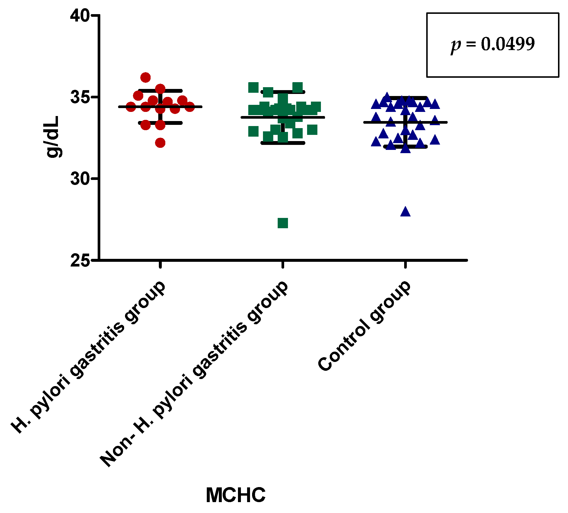
Although we found no significant differences regarding mean corpuscular hemoglobin concentration (MCHC) among the three groups, when comparing the group of H. pylori-induced gastritis group to non-H. pylori gastritis group, we noticed a significantly higher value of this parameters in H. pylori-induced gastritis group (0.0499). Similarly, we also found a significantly higher value in non-H. pylori gastritis group when compared to control group (p = 0.0489) (Table 4 and Figure 2).
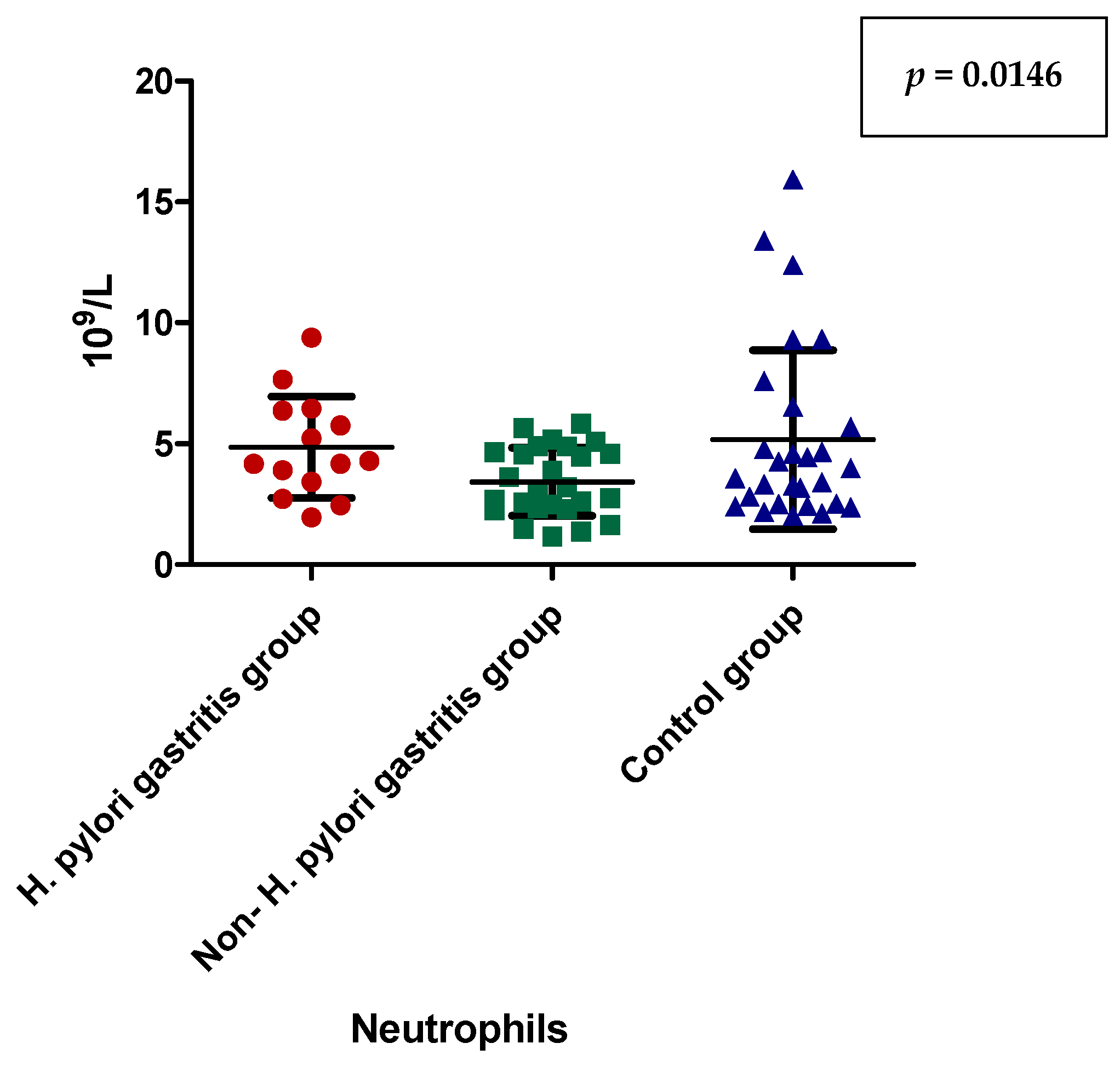
Likewise, we did not obtain any significant differences between the median values of neutrophils among the three groups, but we proved that children with H. pylori-induced gastritis had significantly higher values when compared to non-H. pylori gastritis group (p = 0.0146) (Table 5 and Figure 3).
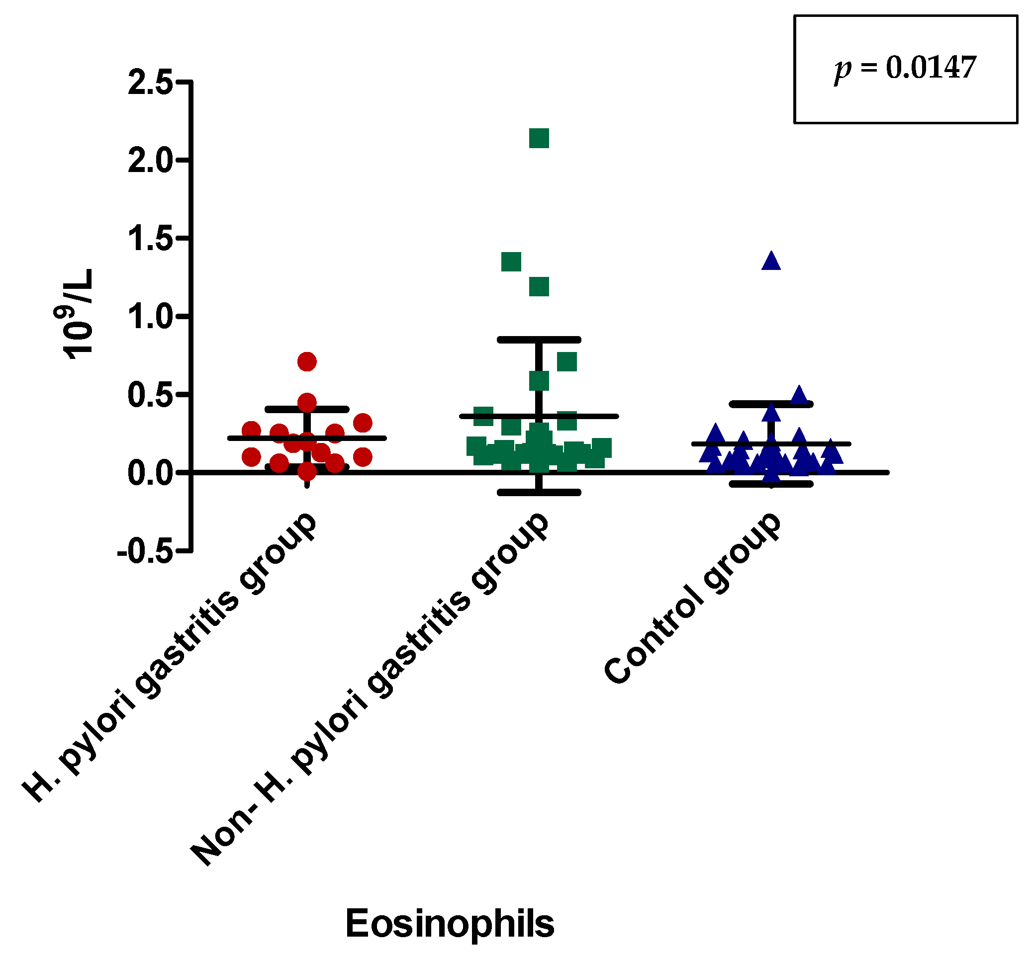
In terms of eosinophils, our study also revealed a significant difference only after applying the Dunn’s Multiple Comparison test indicating that the patients with non-H. pylori gastritis had significantly increased level in comparison to the control group (p = 0.0417) (Table 6 and Figure 4).
In terms of TNF α, which was IHC assessed from gastric samples, we obtained a statistically significant association between TNF α and gastritis in children, subjects with gastritis having a 3.967 times higher risk to express increased levels of TNF α at the site of inflammation [RR = 3.967; CI (95%): 1.283–12.263; p = 0.0063] (Table 7).
TNF α was significantly associated with presence of H. pylori gastritis (p = 0.0002) (Table 8).
Assessing the presence of IL 6 on gastric biopsy samples by immunohistochemical method, we did not obtain any correlations between gastritis in children and control group [RR = 1.283, IC (95%): 0.9404–1.751, p = 0.0988].
No correlation was identified between ESR, leukocyte, neutrophil, lymphocyte, eosinophil counts, neutrophil/lymphocyte ratio and the expression of TNF-alpha/IL 6 within the gastric mucosa.

4. Discussions

Although over 50% of the population worldwide is infected with H. pylori, its prevalence is closely related to the geographic area suggesting that other factors aside from the host and bacterium itself might be implicated in the development of H. pylori-induced gastritis [25]. Moreover, several environmental factors were related with the presence of H. pylori infection such as rural area, male gender, poor socio-economic status, poverty, educational level, poor parental education and unemployment, house crowding, birth order, bed sharing with adults, farmer profession, using a traditional pit or having no toilet, the lack of in-home water service, source of drinking water, using tanks as water supplies, active smoking, alcohol drinking, eating raw uncooked vegetables or spicy food, eating unwashed vegetables or fruits, improper hand washing after school, as well as difficult access and poor adherence to treatment [25]. Nevertheless, the majority of the studies worldwide indicated a descending prevalence trend of H. pylori infection revealing a decrease by one-third during the first decade of this century most likely due to the worldwide improvements of environmental and living conditions, as well as socio-economic status leading to better sanitation and lowering the transmission over time [26]. Unfortunately, our study revealed that in our country rural area remains a considerable risk for the presence of H. pylori infection in children raising important concerns regarding the socio-economic level and living conditions of Romanian people from rural areas.
The diagnosis of H. pylori infection in children is burdened by multiple challenges related to the specificity and sensitivity of the diagnostic method, invasiveness, interobserver-associated variability, the location and size of the biopsy sample, prior treatments, etc. Although choosing the method for diagnosing H. pylori infection in children might seem simple, we must also take into account that the parents/care-givers should also be involved in this process and most often their reluctance regarding invasive diagnostic methods will not allow the use of these methods in spite of their increased specificity and sensitivity. Therefore, the clinician should focus on identifying the most effective non-invasive method for diagnosing this infection. Serology tests represent an optimal choice due to their lack of invasiveness, wide availability, low cost, as well as relatively good specificity and sensitivity (up to 90% and 80%, respectively) [27,28]. Moreover, their accuracy is not impaired by prior administration of proton pump inhibitors, antibiotics or bismuth compounds, history of atrophic gastritis, or recent gastrointestinal hemorrhage [29]. A study involving children concluded that both IgA and IgG tests are highly accurate in diagnosing H. pylori infection, especially in younger children, under the age of 12 years [30]. Similarly, our findings also underlined that IgA test might be considered a reliable test for detecting H. pylori infection in children.
Following the same trend of non-invasiveness, recent studies focused on identifying minimally invasive tests for diagnosing the subclinical inflammatory status related to H. pylori infection which was proved to begin during early childhood [7]. Thus, complete cellular blood parameters proved to be useful in suggesting the presence of subclinical inflammation. A study involving 50 adults with H. pylori infection and 50 without this infection found a significantly higher level of leukocytes, lymphocytes and neutrophils in the first group [19]. Moreover, the authors noticed NLR was positively associated with the severity of the symptoms. These findings were also sustained by other studies indicating that both leukocytes and NLR might be reliable indicators of subclinical systemic inflammatory status [20,31]. Likewise, our study also proved a significant increase of both NLR and neutrophils in children with H. pylori-induced gastritis. Acute phase reactants might also indicate the presence of this inflammation being proved that they are increased in subjects with H. pylori infection [32]. Nevertheless, our study failed in identifying a significant association between either CRP or ESR and gastritis in children regardless of its etiology. Contrariwise, we noticed a significantly higher count of eosinophils in children with non-H. pylori gastritis when compared to control group indicating that eosinophilic gastritis might be one of the most common causes of non-H. pylori-induced gastritis in pediatric population emphasizing the role of this parameter as a screening tool for guiding the further management in children with dyspeptic symptom which are not caused by the presence of H. pylori.
The role of IL 6 in patients found with H. pylori-induced gastritis remains controversial since several studies argued regarding the association between this IL 6 and the damage caused by H. pylori on the gastric mucosa. Thus, certain authors sustained a positive association between IL 6 and H. pylori-positive gastritis [33,34,35,36], while others found no association between them [37,38,39]. Sustaining the first hypothesis, studies performed on blood samples from patients diagnosed with H. pylori-induced gastritis revealed a significant association between H. pylori antibodies and serum levels of IL 6 [40,41]. Contrariwise, the finding reported in studies performed on biopsy samples are contradictory. Thus, Sugimoto et al. pointed out an increased gastric mucosal level of IL 6 in patients with H. pylori-positive gastritis, which decreased after successful eradication of this infection [42]. Contrariwise, genetic studies found no association between IL 6 polymorphisms from H. pylori-positive gastric biopsy samples and the risk of gastric cancer [43]. Our study sustained the latter findings since we failed in finding a positive association between IL 6 from gastric biopsy samples in children with gastritis regardless of its etiology.
It is well-documented that H. pylori-infected patients, as well as those with peptic ulcer present increased levels of TNF α indicating a close collaboration between this cytokine and the bacterium for causing severe gastric injuries [16,44]. Moreover, Mansilla-Vivar et al. pointed out that the expression of this proinflammatory cytokine is closely related to bacterial load since they noticed that TNF α is significantly increased in individuals found with H. pylori nodular gastritis, a well-documented sign of great bacterial load [45]. The same authors suggested that TNF α might also be considered a reliable indicator of tissue-damage severity in patients diagnosed with peptic ulcer. Taking into account that TNF α expressed a chemotactic effect on B and T cells, we might explain its contribution in the development of peptic ulcer [46]. Other studies proved also that TNF α might be involved in the occurrence of Alzheimer’s disease most-likely due to its role in the pathogenesis of subclinical systemic inflammation [47]. A recent review of our team revealed that Alzheimer’s disease might be a possible extraintestinal manifestation of H. pylori infection [2]. Therefore, we could hypothesize that TNF α owns a complex role in triggering the inflammatory status and subsequently creating a bridge between H. pylori infection and its extraintestinal manifestations, which were proved to occur even during early childhood [2]. Furthermore, TNF α was also defined as a major contributor to gastric carcinogenesis [13]. Similarly, our study proved that children with gastritis, regardless of its etiology, present a significant risk of expressing increased gastric levels of TNF α. Additionally, according to our findings, the gastric level of this proinflammatory cytokine was significantly higher in children with H. pylori-induced gastritis as compared to those with non-H. pylori gastritis or control group.
Several limitations of this study are worth mentioning, such as the relatively small sample, especially in terms of H. pylori gastritis group, the assessment of children from a single area of our country, the lack of correlation between gastric cytokine expression and their serum levels. Nevertheless, we must underline that this is the first study from Romania and among the few reported worldwide which assessed the gastric expression of two important proinflammatory cytokines in children with H. pylori and non-H. pylori gastritis versus healthy controls. Although this is a small sample, our results might represent only the first step in a larger and more complex research.

5. Conclusions

Despite its increased prevalence worldwide, H. pylori remains a tricky bacterium. Our study revealed that rural area is a significant risk for the presence of H. pylori infection in children. Serological tests proved to be a reliable test for detecting H. pylori infection in our sample. We noticed a significant increase of NLR and neutrophils in children with H. pylori-induced gastritis suggesting the presence of systemic inflammation. Nevertheless, we found no association between either CRP or ESR and gastritis in children regardless of its etiology. Contrariwise, we noticed a significantly higher count of eosinophils in children with non-H. pylori gastritis when compared to control group suggesting that eosinophilic gastritis might be one of the most frequent causes of non-H. pylori-induced gastritis in pediatric population. In terms of gastric cytokine expression, we found no difference regarding IL 6 in children with gastritis regardless of its etiology and control group, but we noticed a significant risk of expressing increased gastric levels of TNF α in these patients. Moreover, the gastric level of this cytokine was significantly higher in children with H. pylori-induced gastritis as compared to those with non-H. pylori gastritis or control group. Furter studies on larger samples from different geographical area are needed to draw relevant conclusions.

Author Contributions

A.L.D., L.E.M. and C.O.M. conceptualized and designed the study, drafted the initial manuscript, and reviewed and revised the manuscript. A.L.D. and L.E.M. collected the data. C.O.M. performed all the endoscopies. S.M. and S.G. performed the histopathological exam. D.V.G. performed the statistical analysis, C.O.M., S.G. and L.E.M. supervised the study. All authors have read and agreed to the published version of the manuscript.

Funding

This research received no external funding.

Institutional Review Board Statement

The study was approved by the Ethics Committee of the University of Medicine, Pharmacy, Sciences and Technology in Târgu Mureș (No 28/01.03.2019 and 1457/16.09.2021). The study was performed according to the principles of the Helsinki Declaration.

Informed Consent Statement

Moreover, we obtained the signed informed consent of all the parents/caregivers and the assent of all children prior to their inclusion in the study.

Data Availability Statement

The data presented in this study are available on request from the corresponding author.

Acknowledgments

This research was partially supported by the doctoral scholarship of “George Emil Palade” University of Medicine, Pharmacy, Science, and Technology of Târgu Mureș, Romania, entitled “Evaluation of the inflammatory status in children with gastritis”. We thanks to Genoveva Rigmanyi for technical support. The immunostains were performed at the Research Center for Oncopathology and Translational Medicine (CCOMT).

Conflicts of Interest

The authors declare no conflict of interest.

References

  1. Salama, N.R.; Hartung, M.L.; Müller, A. Life in the Human Stomach: Persistence Strategies of the Bacterial Pathogen Helicobacter Pylori. Nat. Rev. Microbiol. 2013, 11, 385–399. [Google Scholar] [CrossRef] [PubMed]
  2. Mărginean, C.D.; Mărginean, C.O.; Meliț, L.E. Helicobacter Pylori-Related Extraintestinal Manifestations-Myth or Reality. Children 2022, 9, 1352. [Google Scholar] [CrossRef] [PubMed]
  3. Amieva, M.; Peek, R.M. Pathobiology of Helicobacter Pylori-Induced Gastric Cancer. Gastroenterology 2016, 150, 64–78. [Google Scholar] [CrossRef] [PubMed]
  4. Parkin, D.M.; Bray, F.; Ferlay, J.; Pisani, P. Global Cancer Statistics, 2002. CA Cancer J. Clin. 2005, 55, 74–108. [Google Scholar] [PubMed]
  5. Săsăran, M.O.; Meliț, L.E.; Dobru, E.D. MicroRNA Modulation of Host Immune Response and Inflammation Triggered by Helicobacter Pylori. Int. J. Mol. Sci. 2021, 22, 1406. [Google Scholar] [CrossRef] [PubMed]
  6. Guclu, M.; Faruq Agan, A. Association of Severity of Helicobacter Pylori Infection with Peripheral Blood Neutrophil to Lymphocyte Ratio and Mean Platelet Volume. Euroasian J. Hepatogastroenterol. 2017, 7, 11–16. [Google Scholar] [CrossRef]
  7. Meliţ, L.E.; Mărginean, M.O.; Mocan, S.; Mărginean, C.O. The Usefulness of Inflammatory Biomarkers in Diagnosing Child and Adolescent’s Gastritis: STROBE Compliant Article. Medicine 2019, 98, e16188. [Google Scholar] [CrossRef]
  8. Romano, M.; Gravina, A.G.; Eusebi, L.H.; Pellegrino, R.; Palladino, G.; Frazzoni, L.; Dajti, E.; Gasbarrini, A.; Di Mario, F.; Zagari, R.M.; et al. Management of Helicobacter Pylori Infection: Guidelines of the Italian Society of Gastroenterology (SIGE) and the Italian Society of Digestive Endoscopy (SIED). Dig. Liver Dis. 2022, 54, 1153–1161. [Google Scholar] [CrossRef]
  9. Bagheri, V.; Memar, B.; Momtazi, A.A.; Sahebkar, A.; Gholamin, M.; Abbaszadegan, M.R. Cytokine Networks and Their Association with Helicobacter Pylori Infection in Gastric Carcinoma. J. Cell. Physiol. 2018, 233, 2791–2803. [Google Scholar] [CrossRef]
  10. Taniguchi, K.; Karin, M. IL-6 and Related Cytokines as the Critical Lynchpins between Inflammation and Cancer. Semin. Immunol. 2014, 26, 54–74. [Google Scholar] [CrossRef]
  11. Gabay, C.; Kushner, I. Acute-Phase Proteins and Other Systemic Responses to Inflammation. N. Engl. J. Med. 1999, 340, 448–454. [Google Scholar] [CrossRef] [PubMed]
  12. Qu, D.; Liu, J.; Lau, C.W.; Huang, Y. IL-6 in Diabetes and Cardiovascular Complications. Br. J. Pharmacol. 2014, 171, 3595–3603. [Google Scholar] [CrossRef] [PubMed]
  13. Dincă, A.L.; Meliț, L.E.; Mărginean, C.O. Old and New Aspects of H. Pylori-Associated Inflammation and Gastric Cancer. Children 2022, 9, 1083. [Google Scholar] [CrossRef] [PubMed]
  14. Fond, G.; Hamdani, N.; Kapczinski, F.; Boukouaci, W.; Drancourt, N.; Dargel, A.; Oliveira, J.; Le Guen, E.; Marlinge, E.; Tamouza, R.; et al. Effectiveness and Tolerance of Anti-Inflammatory Drugs’ Add-on Therapy in Major Mental Disorders: A Systematic Qualitative Review. Acta Psychiatr. Scand. 2014, 129, 163–179. [Google Scholar] [CrossRef] [PubMed]
  15. Güzel, M.; Sönmez, M.F.; Baştuğ, O.; Aras, N.F.; Öztürk, A.B.; Küçükaydın, M.; Turan, C. Effectiveness of Lycopene on Experimental Testicular Torsion. J. Pediatr. Surg. 2016, 51, 1187–1191. [Google Scholar] [CrossRef] [PubMed]
  16. Moradipour, A.; Khosravi, A.; Piri, F. Fecal Helicobacter Pylori GlmM and 16S RRNA Genes Correlate with Serum TNF-α and IL-1β Cytokine Fluctuations. Acta Microbiol. Immunol. Hung. 2018, 65, 489–499. [Google Scholar] [CrossRef]
  17. Kusters, J.G.; van Vliet, A.H.M.; Kuipers, E.J. Pathogenesis of Helicobacter Pylori Infection. Clin. Microbiol. Rev. 2006, 19, 449–490. [Google Scholar] [CrossRef]
  18. Suganuma, M.; Watanabe, T.; Sueoka, E.; Lim, I.K.; Fujiki, H. Role of TNF-α-Inducing Protein Secreted by Helicobacter Pylori as a Tumor Promoter in Gastric Cancer and Emerging Preventive Strategies. Toxins 2021, 13, 181. [Google Scholar] [CrossRef]
  19. Farah, R.; Khamisy-Farah, R. Association of Neutrophil to Lymphocyte Ratio with Presence and Severity of Gastritis Due to Helicobacter Pylori Infection. J. Clin. Lab. Anal. 2014, 28, 219–223. [Google Scholar] [CrossRef]
  20. Atayan, Y.; Hacisalihoglu, P. The Correlation between Tissue Helicobacter Pylori Severity and the Increase in Serum Neutrophil/Lymphocyte Ratio in Patients with Active Chronic Gastritis. Biomed. Res. 2017, 28, 4874–4877. [Google Scholar]
  21. Tamhane, U.U.; Aneja, S.; Montgomery, D.; Rogers, E.-K.; Eagle, K.A.; Gurm, H.S. Association between Admission Neutrophil to Lymphocyte Ratio and Outcomes in Patients with Acute Coronary Syndrome. Am. J. Cardiol. 2008, 102, 653–657. [Google Scholar] [CrossRef] [PubMed]
  22. Kemal, Y.; Yucel, I.; Ekiz, K.; Demirag, G.; Yilmaz, B.; Teker, F.; Ozdemir, M. Elevated Serum Neutrophil to Lymphocyte and Platelet to Lymphocyte Ratios Could Be Useful in Lung Cancer Diagnosis. Asian Pac. J. Cancer Prev. 2014, 15, 2651–2654. [Google Scholar] [CrossRef] [PubMed]
  23. Feng, J.-F.; Huang, Y.; Chen, Q.-X. Preoperative Platelet Lymphocyte Ratio (PLR) is Superior to Neutrophil Lymphocyte Ratio (NLR) as a Predictive Factor in Patients with Esophageal Squamous Cell Carcinoma. World J. Surg. Oncol. 2014, 12, 58. [Google Scholar] [CrossRef] [PubMed]
  24. Ardelean, C.L.; Pescariu, S.; Lighezan, D.F.; Pleava, R.; Ursoniu, S.; Nadasan, V.; Mihaicuta, S. Particularities of Older Patients with Obstructive Sleep Apnea and Heart Failure with Mid-Range Ejection Fraction. Medicina 2019, 55, 449. [Google Scholar] [CrossRef]
  25. Borka Balas, R.; Meliț, L.E.; Mărginean, C.O. Worldwide Prevalence and Risk Factors of Helicobacter Pylori Infection in Children. Children 2022, 9, 1359. [Google Scholar] [CrossRef]
  26. Mehata, S.; Parajuli, K.R.; Pant, N.D.; Rayamajhee, B.; Yadav, U.N.; Mehta, R.K.; Jha, P.; Mehta, N.; Dhimal, M.; Singh, D.R. Prevalence and Correlates of Helicobacter Pylori Infection among Under-Five Children, Adolescent and Non-Pregnant Women in Nepal: Further Analysis of Nepal National Micronutrient Status Survey 2016. PLoS Negl. Trop. Dis. 2021, 15, e0009510. [Google Scholar] [CrossRef]
  27. Bordin, D.S.; Voynovan, I.N.; Andreev, D.N.; Maev, I.V. Current Helicobacter Pylori Diagnostics. Diagnostics 2021, 11, 1458. [Google Scholar] [CrossRef]
  28. Stefano, K.; Rosalia, A.; Chiara, B.; Federica, G.; Marco, M.; Gioacchino, L.; Fabiola, F.; Francesco, D.M.; Gian, L. de’Angelis Non-Invasive Tests for the Diagnosis of Helicobacter Pylori: State of the Art. Acta Biomed. 2018, 89, 58–64. [Google Scholar] [CrossRef]
  29. Malfertheiner, P.; Megraud, F.; O’Morain, C.A.; Gisbert, J.P.; Kuipers, E.J.; Axon, A.T.; Bazzoli, F.; Gasbarrini, A.; Atherton, J.; Graham, D.Y.; et al. Management of Helicobacter Pylori Infection-the Maastricht V/Florence Consensus Report. Gut 2017, 66, 6–30. [Google Scholar] [CrossRef]
  30. Serrano, C.A.; Gonzáez, C.G.; Rollan, A.R.; Duarte, I.; Torres, J.; Peña, A.J.; Harris, P.R. Lack of Diagnostic Utility of Specific Immunoglobulin M in Helicobacter Pylori Infection in Children. J. Pediatr. Gastroenterol. Nutr. 2008, 47, 612–617. [Google Scholar] [CrossRef]
  31. Papa, A.; Emdin, M.; Passino, C.; Michelassi, C.; Battaglia, D.; Cocci, F. Predictive Value of Elevated Neutrophil-Lymphocyte Ratio on Cardiac Mortality in Patients with Stable Coronary Artery Disease. Clin. Chim. Acta 2008, 395, 27–31. [Google Scholar] [CrossRef] [PubMed]
  32. Jackson, L.; Britton, J.; Lewis, S.A.; McKeever, T.M.; Atherton, J.; Fullerton, D.; Fogarty, A.W. A Population-Based Epidemiologic Study of Helicobacter Pylori Infection and Its Association with Systemic Inflammation. Helicobacter 2009, 14, 108–113. [Google Scholar] [CrossRef]
  33. Kinjo, K.; Sato, H.; Sato, H.; Shiotani, I.; Kurotobi, T.; Ohnishi, Y.; Hishida, E.; Nakatani, D.; Mizuno, H.; Sasaki, T.; et al. Prevalence of Helicobacter Pylori Infection and Its Link to Coronary Risk Factors in Japanese Patients with Acute Myocardial Infarction. Circ. J. 2002, 66, 805–810. [Google Scholar] [CrossRef] [PubMed]
  34. Osawa, H.; Kawakami, M.; Fujii, M.; Kubo, N.; Iwanaka, H.; Yamamoto, W.; Saitoh, M.; Yaginuma, T.; Sugano, K. Helicobacter Pylori Infection and Coronary Heart Disease in Japanese Patients. Cardiology 2001, 95, 14–19. [Google Scholar] [CrossRef] [PubMed]
  35. Mayr, M.; Kiechl, S.; Mendall, M.A.; Willeit, J.; Wick, G.; Xu, Q. Increased Risk of Atherosclerosis is Confined to CagA-Positive Helicobacter Pylori Strains: Prospective Results from the Bruneck Study. Stroke 2003, 34, 610–615. [Google Scholar] [CrossRef] [PubMed]
  36. Franceschi, F.; Niccoli, G.; Ferrante, G.; Gasbarrini, A.; Baldi, A.; Candelli, M.; Feroce, F.; Saulnier, N.; Conte, M.; Roccarina, D.; et al. CagA Antigen of Helicobacter Pylori and Coronary Instability: Insight from a Clinico-Pathological Study and a Meta-Analysis of 4241 Cases. Atherosclerosis 2009, 202, 535–542. [Google Scholar] [CrossRef]
  37. Pellicano, R.; Parravicini, P.P.; Bigi, R.; Gandolfo, N.; Aruta, E.; Gai, V.; Figura, N.; Angelino, P.; Rizzetto, M.; Ponzetto, A. Infection by Helicobacter Pylori and Acute Myocardial Infarction. Do Cytotoxic Strains Make a Difference? New Microbiol. 2002, 25, 315–321. [Google Scholar]
  38. Whincup, P.; Danesh, J.; Walker, M.; Lennon, L.; Thomson, A.; Appleby, P.; Hawkey, C.; Atherton, J. Prospective Study of Potentially Virulent Strains of Helicobacter Pylori and Coronary Heart Disease in Middle-Aged Men. Circulation 2000, 101, 1647–1652. [Google Scholar] [CrossRef]
  39. Murray, L.J.; Bamford, K.B.; Kee, F.; McMaster, D.; Cambien, F.; Dallongeville, J.; Evans, A. Infection with Virulent Strains of Helicobacter Pylori is Not Associated with Ischaemic Heart Disease: Evidence from a Population-Based Case-Control Study of Myocardial Infarction. Atherosclerosis 2000, 149, 379–385. [Google Scholar] [CrossRef]
  40. Nakagawa, H.; Tamura, T.; Mitsuda, Y.; Goto, Y.; Kamiya, Y.; Kondo, T.; Wakai, K.; Hamajima, N. Significant Association between Serum Interleukin-6 and Helicobacter Pylori Antibody Levels among H. Pylori-Positive Japanese Adults. Mediat. Inflamm. 2013, 2013, 142358. [Google Scholar] [CrossRef]
  41. Yudkin, J.S.; Stehouwer, C.D.; Emeis, J.J.; Coppack, S.W. C-Reactive Protein in Healthy Subjects: Associations with Obesity, Insulin Resistance, and Endothelial Dysfunction: A Potential Role for Cytokines Originating from Adipose Tissue? Arterioscler. Thromb. Vasc. Biol. 1999, 19, 972–978. [Google Scholar] [CrossRef] [PubMed]
  42. Hull, J.; Thomson, A.; Kwiatkowski, D. Association of Respiratory Syncytial Virus Bronchiolitis with the Interleukin 8 Gene Region in UK Families. Thorax 2000, 55, 1023–1027. [Google Scholar] [CrossRef] [PubMed] [Green Version]
  43. Ramis, I.B.; Vianna, J.S.; Gonçalves, C.V.; von Groll, A.; Dellagostin, O.A.; da Silva, P.E.A. Polymorphisms of the IL-6, IL-8 and IL-10 Genes and the Risk of Gastric Pathology in Patients Infected with Helicobacter Pylori. J. Microbiol. Immunol. Infect. 2017, 50, 153–159. [Google Scholar] [CrossRef] [PubMed]
  44. Bayraktaroğlu, T.; Aras, A.S.; Aydemir, S.; Davutoğlu, C.; Ustündağ, Y.; Atmaca, H.; Borazan, A. Serum Levels of Tumor Necrosis Factor-Alpha, Interleukin-6 and Interleukin-8 Are Not Increased in Dyspeptic Patients with Helicobacter Pylori-Associated Gastritis. Mediat. Inflamm. 2004, 13, 25–28. [Google Scholar] [CrossRef]
  45. Mansilla-Vivar, R.; Serrano, C.A.; Palma, C.; Vera, M.; Hernandez, C.; Pizarro, M.; Torres, J.; Harris, P.R.; Fuentes-López, E.; Riquelme, A.; et al. High Helicobacter Pylori Bacterial Load and Low Cytokine Expression Levels Are Associated with Nodular Gastropathy. Dig. Dis. Sci. 2020, 65, 565–575. [Google Scholar] [CrossRef]
  46. Larussa, T.; Leone, I.; Suraci, E.; Imeneo, M.; Luzza, F. Helicobacter Pylori and T Helper Cells: Mechanisms of Immune Escape and Tolerance. J. Immunol. Res. 2015, 2015, 981328. [Google Scholar] [CrossRef]
  47. Tourani, M.; Habibzadeh, M.; Karkhah, A.; Shokri-Shirvani, J.; Barari, L.; Nouri, H.R. Association of TNF-α but Not IL-1β Levels with the Presence of Helicobacter Pylori Infection Increased the Risk of Peptic Ulcer Development. Cytokine 2018, 110, 232–236. [Google Scholar] [CrossRef]
Figure 1. The difference of median values of NLR between the three groups.
Applsci 13 02162 g001
Figure 2. The difference of median values of MCHC between the three groups.
Applsci 13 02162 g002
Figure 3. The difference of median values of neutrophils between the three groups.
Applsci 13 02162 g003
Figure 4. The difference of median values of eosinophils between the three groups.
Applsci 13 02162 g004
Table 1. Comparison of three groups according to age, gender, area and CRP.
ParametersH. pylori Gastritis Group (n = 14)
n (%)
Non-H. pylori Gastritis Group (n = 26)
n (%)
Control Group (n = 28)
n (%)
p Value
Age (years)14.00 ± 2.71713.50 ± 2.87515.00 ± 2.6170.0678
GenderFemale7 (50.00%)15 (57.69%)21 (75.00%)0.2159
Male7 (50.00%)11 (42.31%)7 (25.00%)
AreaRural11 (78.57%)11 (42.31%)11 (39.29%)0.0404
Urban3 (21.43%)15 (57.69%)17 (60.71%)
CRPNegative5 (55.56%)10 (58.82%)14 (66.67%)0.8096
Positive4 (44.44%)7 (41.18%)7 (33.33%)
IgA anti-H. pyloriPositive5 (55.56%)2 (12.50%)1 (6.67%)0.0094
Negative4 (44.44%)14 (87.50%)14 (93.33%)
Legend: CRP—C reactive protein. n—number, Mann–Whitney test was used.
Table 2. Demographic characteristics of the three groups.
ParametersH. pylori Gastritis Group (n = 14)
Mean ± SD, (Median)
Non-H. pylori Gastritis Group (n = 26)
Mean ± SD, (Median)
Control Group (n = 28)
Mean ± SD, (Median)
p Value
Erythrocytes (×106/µL)4.779 ± 0.4147 (4.685)4.782 ± 0.5510 (4.865)4.600 ± 0.5893 (4.750)0.3918
MPV (fL)83.00 ± 5.088 (82.65)81.37 ± 4.522 (81.30)82.52 ± 5.080 (81.70)0.5365
Htc (%)39.58 ± 3.081 (39.30)38.94 ± 4.809 (39.70)37.87 ± 5.601 (39.30)0.5653
MCHC (g/dL)34.41 ± 0.9856 (34.40)33.75 ± 1.562 (34.20)33.46 ± 1.486 (33.80)0.0803
Hb (g/dl)13.64 ± 1.235 (13.75)13.18 ± 1.885 (13.55)12.74 ± 2.115 (13.25)0.3549
Leukocytes (number/µL)8.106 ± 2.567 (7.630)7.104 ± 1.433 (6.925)8.290 ± 3.696 (6.870)0.6541
Lymphocytes (number/µL)2.259 ± 0.5576 (2.100)2.660 ± 0.8269 (2.565)2.287 ± 0.5990 (2.160)0.1819
Neutrophils (number/µL)4.855 ± 2.097 (4.235)3.431 ± 1.413 (3.075)5.177 ± 3.696 (3.780)0.1307
NLR2.223 ± 1.034 (1.980)1.444 ± 0.8351 (1.260)2.263 ± 1.791 (1.790)0.0253
Eosinophils (number/µL)0.2214 ± 0.1841 (0.1950)0.3615 ± 0.4889 (0.1550)0.1836 ± 0.2542 (0.1250)0.1017
ESR (mmHg)15.75 ± 13.19 (14.00)11.91 ± 14.38 (8.000)15.00 ± 21.46 (11.00)0.2863
Iron (µmol/L)18.91 ± 7.898 (20.25)16.16 ± 8.775 (14.15)13.84 ± 5.749 (14.68)0.2499
AST (U/L)23.48 ± 8.569 (20.90)23.04 ± 16.14 (19.95)22.19 ± 11.16 (19.50)0.7679
ALT (U/L)13.82 ± 4.853 (12.20)15.95 ± 12.09 (12.60)18.81 ± 24.45 (13.70)0.9612
GGT (U/L)16.20 ± 6.546 (14.50)17.45 ± 20.19 (13.00)20.76 ± 30.70 (13.00)0.6024
Legend: ESR—erythrocyte sedimentation rate, AST = aspartate aminotransferase, ALT = alanine aminotransferase, GGT = gamma-glutamyl transpeptidase, Hb—hemoglobin, Htc—hematocrit, MCHC—mean corpuscular hemoglobin concentration, MPV—mean platelet volume, NLR—neutrophil to lymphocyte ratio, n—number, SD—standard deviation, Mann–Whitney test was used.
Table 3. The difference of median values of NLR between the three groups.
NLRDunn’s Multiple Comparison TestSignificant? p < 0.05?
H. pylori-induced gastritis vs. non-H. pylori gastritis0.0107
H. pylori-induced gastritis vs. control group0.3716
non-H. pylori gastritis vs. control group0.0581
Legend: NLR—neutrophil to lymphocyte ratio; statistical significance (p < 0.05).
Table 4. The difference of median values of MCHC between the three groups.
MCHC (g/dL)Dunn’s Multiple Comparison TestSignificant? p < 0.05?
H. pylori-induced gastritis vs. non-H. pylori gastritis0.0499
H. pylori-induced gastritis vs. control group0.0489
non-H. pylori gastritis vs. control group0.5211
Legend: MCHC—mean corpuscular hemoglobin concentration; statistical significance (p < 0.05), Kruskal–Wallis test.
Table 5. The difference of median values of neutrophils between the three groups.
Neutrophils (number/µL)Dunn’s Multiple Comparison TestSignificant? p < 0.05?
H. pylori-induced gastritis vs. non-H. pylori gastritis0.0146
H. pylori-induced gastritis vs. control group0.5662
non-H. pylori gastritis vs. control group0.1769
Table 6. The difference of median values of eosinophils between the three groups.
Eosinophils(number/µL)Dunn’s Multiple Comparison TestSignificant? p < 0.05?
H. pylori-induced gastritis vs. non-H. pylori gastritis0.0147
H. pylori-induced gastritis vs. control group0.5662
non-H. pylori gastritis vs. control group0.1769
Table 7. The association of TNF α and gastritis in children.
Presence of TNF α (n/%)Absence of TNF α (n/%)Statistical Parameters
Gastritis (H. pylori-induced gastritis + non-H. pylori gastritis) group 17 (85.00%)23 (47.92%)RR = 3.967
IC (95%): 1.283–12.263
p = 0.0063
Control goup3 (15.00%)25 (52.08%)
Legend: CI—confidence interval, n—number; statistical significance (p < 0.05), RR—relative risk.
Table 8. Correlation between the presence of TNF α in the three groups.
Presence of TNF α (n/%)Absence of TNF α (n/%)p Value
H. pylori-induced gastritis group10 (50.00%)4 (8.33%)0.0002
Non-H. pylori gastritis group7 (35.00%)19 (39.58%)
Control group3 (15.00%)25 (52.08%)
Disclaimer/Publisher’s Note: The statements, opinions and data contained in all publications are solely those of the individual author(s) and contributor(s) and not of MDPI and/or the editor(s). MDPI and/or the editor(s) disclaim responsibility for any injury to people or property resulting from any ideas, methods, instructions or products referred to in the content.