Abstract
Knowledge of anatomical variations in the base of the skull and anatomical landmarks is crucial for clinical procedures by surgeons, ENT physicians, and radiologists. This study investigated morphometric and anatomical variations in the foramen magnum, occipital condyles, hypoglossal canals, and jugular foramina to improve knowledge of the base of the skull’s complex anatomy and consider the anatomical variations via a morphometric study. One hundred and sixty intact skulls were investigated. Morphometric measurements showed that the foramen magnum, occipital condyles, hypoglossal canals, and jugular foramina were all significantly larger in males than females and could be useful for sex determination. Increased awareness of morphological location and anatomical landmark variation can improve surgical proficiency.
1. Introduction
The foramen magnum is the largest central outlet of the base of the skull and derives from the occipital bone. The medulla oblongata and spinal cord, spinal arteries, vertebral arteries, and the spinal accessory nerve (cranial nerve XI) all pass through the foramen magnum [1]. This structure varies in size and shape according to age, ethnicity, and sex [2]. Morphometric shape variability can be divided into oval, egg-shaped, irregular, round, tetragonal, pentagonal, and hexagonal [3]. This region is neurosurgically complex because of its closeness to neurovascular structures and the deep positioning of the foramen. The occipital condyles are vital structures of the craniovertebral junction, located anterolaterally to the foramen magnum and laterally to the jugular foramen [4]. Understanding variations in the hypoglossal canal is crucial to anatomists, anthropologists, forensic scientists, and physicians. The hypoglossal canal is a small opening in the occipital bone, located over the occipital condyles and above the foramen magnum anterolaterally. It carries the hypoglossal nerve (cranial nerve XII), a meningeal branch of the ascending pharyngeal artery, which also transmits motor innervations to the tongue, and an emissary vein from the basilar plexus [5,6]. Knowledge of the aspects of this canal is important for radiologists and neurosurgeons in surgical planning procedures of the posterior cranial fossa for tumors, such as schwannoma of the hypoglossal nerve, to ensure safe surgical intervention in the craniovertebral region [7]. The complex irregular bony canal of the posterior base of the skull, situated between the medial petrous pyramid of the temporal bone and the anteromedial portion of the occipital bone, called the jugular foramen, contains numerous vital structures. The jugular foramen has complex anatomical relationships with important neurovascular structures passing through it, such as the glossopharyngeal nerve (cranial nerve IX), the vagus nerve (cranial nerve X), the spinal accessory nerve, and the sigmoid sinus vein, which becomes the internal jugular vein [8]. Normally, there are two par structures in this foramen: the pars vascularis (or pars venosa), located in the posterolateral section of the jugular foramen and involving the vagus nerves, spinal accessory nerves, the sigmoid sinus, and the ascending pharyngeal artery. The ascending pharyngeal artery becomes the posterior meningeal artery in the dura region, and the pars nervosa resides in the anteromedial region of the jugular foramen and includes the glossopharyngeal nerve, the inferior petrosal sinus, and the ascending pharyngeal artery [9]. Some exceptional cases demonstrate integration of the vagus nerves and hypoglossal nerves immediately after departing the hypoglossal canal and jugular foramen, respectively [10]. The jugular foramen is diverse and critical in otorhinolaryngology, radiology, and neurosurgical procedures involving the base of the skull and the middle ear. This foramen is also challenging to enter surgically [11]. Multiple techniques for approaching the jugular foramen are considered case by case because of their anatomical variations. The anteroposterior and mediolateral widths are factors that determine the neurovascular content, which passes through this foramen. Variations in neurovascular structures and their complexity make each surgical procedure unique, and knowledge of the anatomy is essential [12]. Intracranial or extracranial lesions in this section may occur, such as a metastatic lesion, a schwannoma or intracranial meningioma [13]. The jugular ganglion of the vagus nerve can also develop slow-growing glomus jugulare tumors that generate multiple cranial nerve palsies, such as jugular foramen syndrome or paresis of the glossopharyngeal nerve, vagus nerve, and spinal accessory nerve [14,15,16]. The dome of the jugular foramen presents a superior bulb of major paired venous return from the brain, called the internal jugular vein [13]. The dome characteristic is related to clinical implications because kinking of the vein can occur, leading to thrombosis. Obstruction from thrombosis or cancer metastasis can also occur and depress the cranial nerve and adjacent vessels. This study considered the morphometric and anatomical disparities in the foramen magnum, occipital condyles, hypoglossal canal, and jugular foramen to improve knowledge of the base of the skull anatomy and optimize surgical procedures for anatomical variations.
2. Materials and Methods
Data were collected via a cross-sectional study that investigated the base of the skull using samples from the bone bank of the Forensic Osteology Research Center (FORC), Anatomy Department, Chiang Mai University. Samples with a deteriorated base of the skull or pathological bone congenital anomalies were excluded. Eighty adult male and eighty adult female cadaveric skulls were selected (between 20 and 80 years old). Ethics approval was received from the Research Ethics Committee of Chiang Mai University (CODE: ANA-2564-08283).
Morphological parameters of the foramen magnum, occipital condyles (Figure 1), hypoglossal canals (Figure 2), and jugular foramina (Figure 3) were measured by a Vernier caliper to determine the mean ± standard deviation in millimeters of the maximum transverse diameter of the foramen magnum, maximum anteroposterior diameter of the foramen magnum, posterior tip of occipital condyles width, anterior tip of occipital condyles width, maximum transverse length of occipital condyles and maximum anteroposterior length of occipital condyles, distance from the posterior occipital condyles to the internal hypoglossal canal, distance from posterior occipital condyles to the external hypoglossal canal, anteroposterior diameter of the jugular foramen, and mediolateral diameter of the jugular foramen. All measurement parameters were analyzed using SPSS version 26 (SPSS Inc., Chicago, IL, USA) and Microsoft Excel 2016 (Microsoft Corp., Redmond, WA, USA). Furthermore, the visual comparison was used to assess the prevalence and existence of anatomical variation in the shape of the foramen magnum (Figure 4), septum, and spur of the hypoglossal canals (Figure 2), as well as the presentation of the dome characteristics in the superior jugular bulb on both sides. The comparison of anatomical variation was shown as a percentage.
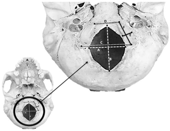
Figure 1.
Anatomical landmarks of the foramen magnum and occipital condyles: (A) maximum transverse diameter of the foramen magnum, (B) maximum anteroposterior diameter of the foramen magnum, (C) posterior tip of occipital condylar width, (D) anterior tip of occipital condylar width, (E) maximum transverse length of occipital condyles and (F) maximum anteroposterior length of occipital condyles.
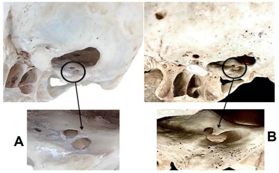
Figure 2.
Anatomical characteristics of the hypoglossal canals: (A) present septal structure, (B) present spur structure.
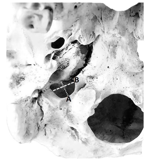
Figure 3.
Measurements of jugular foramen parameters: (A) anteroposterior diameter of the jugular foramen, (B) mediolateral diameter of the jugular foramen.

Figure 4.
Anatomical variation of shape of the foramen magnum under the base of the skull: (A) oval shape, (B) round shape, (C) tetragonal shape, (D) pentagonal shape and (E) hexagonal shape.
To locate the hypoglossal canals (Figure 2), two parameters were measured on the specific landmarks by a digital Vernier caliper as follows:
- The anteroposterior diameter of the hypoglossal canals: the maximum distance from the anterior border to the posterior border perpendicularly of each hypoglossal canal.
- The mediolateral diameter of the hypoglossal canals: the perpendicular maximum distance from the medial border to the lateral border of each hypoglossal canal.
In addition, to locate the jugular foramen (Figure 3) precisely, two parameters were measured on the specific landmarks by a digital Vernier caliper as follows:
- The anteroposterior diameter of the jugular foramina: the maximum distance from the border of the anterior border to the posterior border perpendicularly of each jugular foramen
- The mediolateral diameter of the jugular foramina: the perpendicular maximum distance from the medial border to the lateral border of each jugular foramen.
3. Results
Measurement parameters of the foramen magnum, occipital condyles, and hypoglossal canals
In Table 1, measurement parameters showed statistically significant differences. The anteroposterior diameter of the left occipital condyle was greater than on the right side, and the transverse diameter of the left occipital condyle was also greater than on the right side. The distance of the left posterior occipital condyle to the left internal hypoglossal canal was greater than on the right side, and the distance of the left posterior occipital condyles to the left external hypoglossal canal was greater than on the right side. Sexual dimorphism in measured parameters was also investigated. The anteroposterior and transverse diameters of the foramen magnum of the male specimens were greater than the females.

Table 1.
Morphometric measurements related to the foramen magnum, occipital condyle, and hypoglossal canal.
An oval shape of the foramen magnum was the most common in both sexes. Other shapes such as round, tetragonal, pentagonal, and hexagonal were also present. The internal septum of the hypoglossal canal was most commonly present on both sides, as shown in Table 2.
Table 2.
Anatomical variation in the foramen magnum and hypoglossal canal.
Distance from the anterior to the posterior border of the jugular foramen on both sides in different sexes. A comparison between different sexes showed statistically significant differences in the width of the left side of the mediolateral jugular foramen. Comparison between different sides of the foramen also showed statistically significant differences in the anteroposterior jugular foramen width in males and females and the mediolateral jugular foramen width in females. In males, the right side of the anteroposterior diameter and the mediolateral diameter were statistically significantly larger than in females, as shown in Table 3.
Table 3.
Morphometric study of jugular foramen.
Prevalence of the Dome Characteristic Covered Jugular Foramen
Observations of the structural bony roof associated with the superior jugular bulb are called dome characteristics. In males, an extended superior jugular roof or dome characteristics occurred bilaterally in 81.25% of the skulls, right side only in 2.5% of the skulls and on the left side one in 1.25% of the skulls were without a dome bilaterally in 15% of the skulls. In females, an extended superior jugular roof or dome characteristics occurred bilaterally in 85% of the skulls, on the right side only in 2.5% of the skulls, and on the left side, one in 4% of the skulls were without a dome bilaterally in 8.75% of the skulls.
4. Discussion
This study in the Thai population contributes to enlightening the anatomical variations through a morphometric assessment. The evaluation of the foramen magnum, occipital condyles, hypoglossal canals, and jugular foramina in this study reveals that the diameter of males is greater than that of females. In Table 4, the studies of Lyrtzis et al., (2017) [7] and Natsis et al., (2013) [17] also demonstrated the same trend as in this study. Muthukumar et al., (2005) [18] showed the linear measurements of the foramen magnum, occipital condyles, and hypoglossal canals, but there is no comparison between sexes. Due to sexual dimorphism in the measured traits and structural diversity of the components of the skull base, the surgical approaches should differ for females and males, at least in the studied population. The impact of sexual dimorphism may be related to hormones and the pattern of growth and development [19,20].
Table 4.
Comparison of foramen magnum, occipital condyle and hypoglossal canals among several studies (diameters are expressed in mm).
Knowing the precise position and trend of anatomical variation of base of skull can reduce the error of surgical procedure and prevent the neurovascular structure of skull base. For example, opportunities for compression of the neurovascular system at the left jugular foramen are common because the diameters of the left-sided jugular foramen are less than on the right side [15]. This study in Thai subjects elucidated the anteroposterior diameter of the left side to be significantly less than on the right side (Table 5). Other studies [8,16,21,22,23,24] have described differences in diameters, including those of the anteroposterior and mediolateral jugular foramina. Knowledge of the precise anatomical morphology of the jugular foramina would increase surgical awareness to avoid internal jugular vein damage.
Table 5.
Previous study morphometric study in the jugular foramen.
The percentage of bilateral presence of the dome structure in the jugular foramen (Table 6) is common [22,25,26] except in the study of Patel and Singel (2007) [27]. In addition, the absence of a domed bony roof implies that the superior jugular bulb is destitute in development and may not form the floor of the middle ear cavity. Thus, there is a slight risk of infiltration of tumors into the middle ear [15]. The dome demonstrates a superior jugular bulb that may be a dural septum to partition cranial nerve IX from cranial nerves X and XI [28]. A conspicuous domed bony should not be misinterpreted for a malignancy in radiological technique and should be considered during surgical operation [29]. Despite extensive cadaveric dissection studies or morphometric studies in the dry skull, accurate data on the base of the skull is uncertain. Some interventions, such as ultrasound procedures and computerized tomography scans, can contribute to more definitive data.
Table 6.
Comparison of the dome of jugular fossa with other studies.
5. Conclusions
This morphometric study of variations in the foramen magnum, occipital condyles, and hypoglossal canals is useful for surgical approaches. For example, knowing the precise location and anatomical variation can reduce the error of surgical approach, such as neurosurgery. In the Thai population, the anteroposterior and transverse diameter of the foramen magnum, the anteroposterior diameter and transverse diameter of the occipital condyles, the distance between the anterior tip and distance between the posterior tip of the occipital condyles, the distance between the posterior border of each occipital condyle to the internal border of each hypoglossal canal, and distance between the posterior border of each occipital condyle to the external border of each hypoglossal canal as parameters in males were significantly greater than in females. The anatomical shapes of these structures varied. An oval shape of the foramen magnum was the most common, while the prevalence of the spur and septum structure of the hypoglossal canal also varied. Surgical approach should be considered regarding right side because several right parameters are greater than left side. This information contributes to the protection of neurovascular structures. Furthermore, the sexual dimorphism of the skull needs to be thoroughly evaluated in terms of the morphological features in different populations [30]. The sexual dimorphism may correlate with endocrine and pattern of development [19,20]. This study presents novel morphometric data to elucidate anatomical variations and contributes the anatomical knowledge of base of skull.
Author Contributions
Conceptualization, S.T. and P.M.; methodology, S.T.; formal analysis, S.T.; resources, P.M.; writing—original draft preparation, S.T.; writing—review and editing, P.M.; supervision, P.M.; funding acquisition, P.M. All authors have read and agreed to the published version of the manuscript.
Funding
This research was funded by from the Excellence Center in Osteology Research and Training Center (ORTC) with partial support from Chiang Mai University.
Institutional Review Board Statement
Ethics approval was received from the Research Ethics Committee of Chiang Mai University (CODE: ANA-2564-08283).
Informed Consent Statement
Not applicable.
Conflicts of Interest
The authors declare no conflict of interest.
References
- Degno, S.; Abrha, M.; Asmare, Y.; Muche, A. Anatomical Variation in Morphometry and Morphology of the Foramen Magnum and Occipital Condyle in Dried Adult Skulls. J. Craniofacial. Surg. 2019, 30, 256–259. [Google Scholar] [CrossRef] [PubMed]
- Bhatnagar, S.; Iwanaga, J.; Decater, T.; Loukas, M.; Tubbs, R.S. Foramen Magnum Variant with Elongation of the Anterior Notch. Cureus 2020, 12, e8506. [Google Scholar] [CrossRef]
- Krishna, K.; Nag, A.; Prasad, R. Morphometric Study of Foramen Magnum and Variation in Its Shape. IOSR J. Dent. Med. Sci. 2016, 15, 120–123. [Google Scholar] [CrossRef]
- Pal, A.; Aggarwal, P.; Ghosal, A.K.; Datta, I.; Ganguly, S. A Study on the Anatomical Variations of Occipital Condyles in the Dry Adult Human Occipital Bones. J. Med. Res. 2019, 9, 43–47. [Google Scholar]
- Yadav, S.; Pandey, P.; Pasricha, N.; Bhatnagar, R. Variant Anatomy of Hypoglossal Canal: An Osteological Study in North Indian Population. Acad. Anat. Int. 2020, 6, 40–42. [Google Scholar] [CrossRef]
- Roopali, D.N.; Dhiraj, B.N.; Rohini, R.K.; Avinash, D.S. Morphological Study of Hypoglossal Canal and Its Anatomical Variation. Int. J. Health Sci. Res. 2013, 3, 54–58. [Google Scholar]
- Lyrtzis, C.; Piagkou, M.; Gkioka, A.; Anastasopoulos, N.; Apostolidis, S.; Natsis, K. Foramen magnum, occipital condyles and hypoglossal canals morphometry: Anatomical study with clinical implications. Folia Morphol. 2017, 76, 446–457. [Google Scholar] [CrossRef] [PubMed]
- Gupta, C.; Kurian, P.; Seva, K.N.; Kalthur, S.G.; D’Souza, A.S. A morphological and morphometric study of jugular foramen in dry skulls with its clinical implications. J. Craniovertebral Junction Spine 2014, 5, 118–121. [Google Scholar] [CrossRef]
- Caldemeyer, K.S.; Mathews, V.P.; Azzarelli, B.; Smith, R.R. The jugular foramen: A review of anatomy, masses, and imaging characteristics. Radiographics 1997, 17, 1123–1139. [Google Scholar] [CrossRef] [PubMed]
- Kikuta, S.; Jenkins, S.; Kusukawa, J.; Iwanaga, J.; Loukas, M.; Tubbs, R.S. Ansa cervicalis: A comprehensive review of its anatomy, variations, pathology, and surgical applications. Anat. Cell Biol. 2019, 52, 221–225. [Google Scholar] [CrossRef] [PubMed]
- Freitas, C.; Santos, L.; Santos, A.N.; Amaral Neto, A.; Brandão, L.G. Anatomical study of jugular foramen in the neck. Braz. J. Otorhinolaryngol. 2020, 86, 44–48. [Google Scholar] [CrossRef] [PubMed]
- Navsa, N.; Kramer, B. A quantitative assessment of the jugular foramen. Ann Anat. 1998, 180, 269–273. [Google Scholar] [CrossRef] [PubMed]
- Amudha, G.; Aishwarya, C.N.; Hepzibah, D.J.; Kesavan, V.A.; Manicka Vasuki, A.K. Morphometric study of jugular foramen and jugular fossa of dried adult human skulls and its clinical significance. Natl. J. Clin. Anat. 2019, 8, 160–164. [Google Scholar] [CrossRef]
- Griessenauer, C.J.; McGrew, B.; Matusz, P.; De Caro, R.; Loukas, M.; Tubbs, R.S. Surgical Approaches to the Jugular Foramen: A Comprehensive Review. J. Neurol. Surg. Part B Skull Base 2016, 77, 260–264. [Google Scholar]
- Das, S.S.; Saluja, S.; Vasudeva, N. Complete morphometric analysis of jugular foramen and its clinical implications. J. Craniovertebral Junction Spine 2016, 7, 257–264. [Google Scholar] [CrossRef]
- Sheetal, K.; Athavale, S.A. Morphometric study of jugular foramen in adult South Indian skulls. J. Anat. Soc. India 2014, 62, 166–169. [Google Scholar]
- Natsis, K.; Piagkou, M.; Skotsimara, G.; Piagkos, G.; Skandalakis, P. A morphometric anatomical and comparative study of the foramen magnum region in a Greek population. Surg. Radiol. Anat. 2013, 35, 925–934. [Google Scholar] [CrossRef] [PubMed]
- Muthukumar, N.; Swaminathan, R.; Venkatesh, G.; Bhanumathy, S.P. A morphometric analysis of the foramen magnum region as it relates to the transcondylar approach. Acta Neurochir. 2005, 147, 889–895. [Google Scholar] [CrossRef] [PubMed]
- Techataweewan, N.; Hefner, J.T.; Freas, L.; Surachotmongkhon, N.; Benchawattananon, R.; Tayles, N. Metric sexual dimorphism of the skull in Thailand. Forensic Sci. Int. Rep. 2021, 4, 1–12. [Google Scholar] [CrossRef]
- France Diane, L. Observational and metric analysis of sex in the skeleton. In Forensic Osteology: Advances in the Identification of Human Remains; Charles C Thomas: Springfield, IL, USA, 1998; pp. 163–186. [Google Scholar]
- Pereira, G.A.M.; Lopes, P.T.C.; Santos, A.M.P.V.; Krebs, W.D. Morphometric aspects of the jugular foramen in dry skulls of adult individuals in Southern Brazil. J. Morphol. Sci. 2010, 27, 3–5. [Google Scholar]
- Lovely Jain, S.R.; Rajendra Singh Kushwah. Study of Morphometric Variations in Jugular Foramen and Jugular Fossa of Dried Adult Human Skulls. Int. J. Contemp. Med. Res. 2018, 5, E6–E10. [Google Scholar]
- Saheb, H.S.; Mavishetter, G.F.; Thomas, S.T.; Prasanna, L.C.; Muralidhar, P.A. Morphometric study of the jugular foramen in human adult skulls of South India. J. Biomed. Sci. Res. 2010, 2, 240–243. [Google Scholar]
- Ekinci, N.; Unur, E. Macroscopic and morphometric investigation of the jugular foramen of the human skull. Kaibogaku Zasshi. 1997, 72, 525–529. [Google Scholar] [PubMed]
- Hatiboğlu, M.T.; Anil, A. Structural variations in the jugular foramen of the human skull. J. Anat. 1992, 180, 191–196. [Google Scholar] [PubMed]
- Sturrock, R.R. Variations in the structure of the jugular foramen of the human skull. J. Anat. 1988, 160, 227–230. [Google Scholar] [PubMed]
- Patel, M.M.; Singel, T.C. Variations in the structure if the jugular foramen of the human skull of the Saurashtra region. J. Anat. Soc. India 2007, 56, 34–37. [Google Scholar]
- Tekdemir, I.; Tuccar, E.; Aslan, A.; Elhan, A.; Deda, H.; Ciftci, E.; Akyar, S. The jugular foramen: A comparative radioanatomic study. Surg. Neurol. 1998, 50, 557–562. [Google Scholar] [CrossRef] [PubMed]
- Aseta, F.B.; Mwachaka, P.M.; Mandela, P.I.; Ogeng’o, J. Variant Anatomy of the Jugular Foramen: An Osteological Study. Acad. Anat. Int. 2016, 2, 38–43. [Google Scholar] [CrossRef]
- Cappella, A.; Bertoglio, B.; Di Maso, M.; Mazzarelli, D.; Affatato, L.; Stacchiotti, A.; Sforza, C.; Cattaneo, C. Sexual Dimorphism of Cranial Morphological Traits in an Italian Sample: A Population-Specific Logistic Regression Model for Predicting Sex. Biology 2022, 11, 1202. [Google Scholar] [CrossRef] [PubMed]
Disclaimer/Publisher’s Note: The statements, opinions and data contained in all publications are solely those of the individual author(s) and contributor(s) and not of MDPI and/or the editor(s). MDPI and/or the editor(s) disclaim responsibility for any injury to people or property resulting from any ideas, methods, instructions or products referred to in the content. |
© 2023 by the authors. Licensee MDPI, Basel, Switzerland. This article is an open access article distributed under the terms and conditions of the Creative Commons Attribution (CC BY) license (https://creativecommons.org/licenses/by/4.0/).