Featured Application
An LED strip with 405 nm LEDs can be developed and installed in places such as inside the shelves in a retail store. The LED will provide continuous disinfection of the highly touched areas. Installing on the shelves will ensure faster disinfection, as the distance between the LED strip and surface will be approximately 10~20 cm.
Abstract
The ongoing coronavirus pandemic requires more effective disinfection methods. Disinfection using ultraviolet light (UV), especially longer UVC wavelengths, such as 254 and 270/280 nm, has been proven to have virucidal properties, but its adverse effects on human skin and eyes limit its use to enclosed, unoccupied spaces. Several studies have shown the effectiveness of blue light (405 nm) against bacteria and fungi, but the virucidal property at 405 nm has not been much investigated. Based on previous studies, visible light mediates inactivation by absorbing the porphyrins and reacting with oxygen to produce reactive oxygen species (ROS). This causes oxidative damage to biomolecules, such as proteins, lipids, and nucleic acids, essential constituents of any virus. The virucidal potential of visible light has been speculated because the virus lacks porphyrins. This study demonstrated porphyrin-independent viral inactivation and conducted a comparative analysis of the effectiveness at 405 nm against other UVC wavelengths. The betacoronavirus 1 (strain OC43) was exposed to 405, 270/280, 254, and 222 nm, and its efficacy was determined using a median tissue culture infectious dose, i.e., TCID50. The results support the disinfection potential of visible light technology by providing a quantitative effect that can serve as the basic groundwork for future visible light inactivation technologies. In the future, blue light technology usage can be widened to hospitals, public places, aircraft cabins, and/or infectious laboratories for disinfection purposes.
Keywords:
visible light; 405 nm; virucidal; inactivation; betacoronavirus; irradiation; far-UVC; 222 nm 1. Introduction
The severe acute coronavirus 2 (SARS-CoV-2) pandemic has been an ongoing concern since its major outbreak in 2019, affecting the health and lives of millions of people across the world. Several unprecedented measures have been taken to control its spread, such as vaccinating people against the virus and strict sanitization measures, which have been quite effective in combatting the ill effects of the virus. As of February 2022, more than 10 billion doses of vaccines have been administered across the globe [1]. In addition, rigorous sanitization, which uses 60 to 95% ethyl alcohol, has well-established benefits and has thus been adopted by most households to clean surfaces and hands. However, despite strict sanitization, the virus is still spreading and affecting millions of people globally, indicating the need for more effective disinfection technologies. Ultraviolet (UV) is an invisible part of the electromagnetic spectrum that ranges from 100 to 400 nm. The UV range is mainly categorized into three sections based on wavelength. The range from 100 to 280 nm is termed UVC, from 280 to 320 nm as UVB, and from 320 to 400 nm as UVA [2]. The lower range of UVC wavelengths, between 200 and 222 nm, is termed far-UVC. The ozone layer absorbs UVC, but due to the fact of its antimicrobial properties, it is artificially created and has been used for disinfection purposes over the past few centuries. The most used UVC wavelength has been 254 nm, and it has well-established efficacy against bacteria, fungi, and viruses. Other UVC wavelengths, such as 260–280 nm, have also shown the potential to inactivate viruses. A study conducted in the past used 260 nm, a combination of 260/280 nm, and 280 nm, to inactivate viruses. The fluences (UV doses) used were approximately 8 mJ/cm2 for coxsackievirus A10 and poliovirus 1, 10 mJ/cm2 for enterovirus 70, and 13 mJ/cm2 for echovirus 30 [3]. Amongst these three, 260 nm was more effective for enteroviruses, and 280 nm showed relatively higher efficacy for human adenovirus (a DNA virus) [3]. Though UVCs have proven inactivation potential, they can cause severe burns on the skin and eye injuries such as photokeratitis [4]. Additionally, the effect of UVC radiation on materials raises concerns about compromising the structural integrity or the premature aging of the products [5].
Over the last decade, far-UVC has gained huge popularity for its higher irradiation power and lower penetration in human live tissue compared to 254 nm, making the inactivation process quicker and safer for humans [6]. Past studies on the antimicrobial property of far-UVC have demonstrated its capacity to inactivate influenza and coronaviruses in the air at a dose that does not damage human cells [7,8]. Prior research has a dearth of data on the long-term effects of far-UVC and its exposure to human eyes.
The CoVs belong to the family of nidovirales, coronaviridae, and the subfamily coronavirinae. On the basis of genetics, they can be categorized as Alphacoronavirus, Betacoronavirus, Gammacoronavirus, and Deltacoronavirus in which the former two are known to infect mammals and the latter two infect birds [9]. The Betacoronavirus can be divided into three categories based on lineage: SARS CoV, MERS CoV, and SARS CoV-2. The SARS-CoV-2 virus is very closely related to the genus Betacoronavirus. The SARS CoV-2 shares more than 70% genetic similarity with SARS CoV-1 [10]. The coronavirus contains structural proteins, i.e., spike (S), envelope (E), membrane (m), and nucleocapsid (N) proteins [11]. The virus uses its spike proteins to attach to the host cell. Once inside the host cell, it multiplies and kills the cells [12]. UVC exposure can cause either genome damage (DNA/RNA) or protein damage. The 254 nm is absorbed by the deoxyribonucleic acid (DNA) and ribonucleic acid (RNA) through the formation of pyrimidine dimers [13]. However, the DNA can repair itself in the presence of blue light through a process called photoreactivation [14]. The 222 nm, however, can cause both genome and protein damage because structural proteins more readily absorb 222 nm in contrast to 254 nm [15]. In terms of the shorter wavelength (200 to 230 nm) range, 222 nm is proven to be more effective than 210 and 230 nm [15].
Visible blue light (400–470 nm), especially 405 nm, has shown to have germicidal properties. Unlike UVC, blue light is considered safe for human exposure [16] and has no degrative effect on material [17]. A past study [17] conducted a comparative analysis of the degrative effects of UVC and 405 nm on endoscope tube material. The UVC caused substantial photodegradation damage, whereas 405 nm showed no notable detrimental effect [17] on the endoscope tube material. Low blue light doses have shown inactivation for bacteria such as Clostridium spp. and Listeria spp. [18,19], fungal species such as Saccharomyces spp. and Candida spp. [19], and a higher dose has shown inactivation for viruses such as feline calcivirus (FCV) 30 [20] and viral hemorrhagic septicemia virus (VHSV) [21].
The inactivation mechanism of blue light is by absorbing the light via photosensitizers such as porphyrins and reacting with oxygen or other cell components [22,23]. This reaction produces reactive oxygen species (ROS), which cause oxidative damage [22,23]. The nonselective nature of ROS can cause direct damage to biomolecules, such as proteins, lipids, and nucleic acids, which are essential constituents of bacteria, fungi, and viruses [22,23]. As the virus lacks porphyrins, the virucidal property of blue light is highly speculated. Most of the research conducted used porphyrins to inactivate the virus in media. In this study, the media had no porphyrins, and the test was conducted without the use of external photosensitizers. A recent study also showed the inactivation of SARS-CoV-2 and influenza A H1N1 without using a photosensitizer [24]. Nevertheless, mechanisms by which blue light causes nonselective damage to the virus is still unknown. A future investigation is required to understand how blue light causes inactivation.
The dose requirement varies with wavelengths and pathogens. The wavelengths below 240 nm can more readily generate ozone [25] and, thus, should be a deciding factor for dose determination. The dose of 222 nm used was 3 mJ/cm2 for this study. Whereas for 254 and 270/280 nm, a slightly higher dose of 10 mJ/cm2 was used. For 405 nm, a dose of 17,280 mJ/cm2 was used. The dose chosen for 405 nm was considerably low, safe, and can be generated from commercially available products.
The primary goal of this study was to investigate blue light’s (405 nm) viral efficacy.
2. Materials and Methods
2.1. Blue Light and UV Light Equipment
The 405 nm LED (Figure 1a) used in this study was from DIEHL Aviation (Sterrett, AL, USA) [26]. The LED prototype used the same mechanical component as the regular mini spotlight. The light had a peak wavelength of 405 nm and had a single mode with a constant power wattage. The 222 nm light was from the manufacturer Ushio Care222 (Oude Meer, The Netherlands) (Figure 1b), krypton-chloride (Kr-Cl) excimer lamp module. The typical excimer lamp emits peak irradiation at 222 nm alongside a longer UVC wavelength. In contrast, Ushio uses a special short-pass filter to block other UVC wavelengths that are above 230 nm [27]. The Ushio germicidal low-pressure mercury arc lamp emits a peak wavelength of 253.7 nm (Figure 1c). The 270/280 nm was from the manufacturer GMKJ (Massapequa Park, NY, USA). Each LED module shown in Figure 2 was connected in a three-corner arrangement. Each module had the same peak wavelength and the same irradiance power. A DC voltage from the main power supply was applied to each UV-LED module in accordance with the available current. The manufacturers of all of the light sources certified their emission spectrum, and the lights were tested multiple times to determine the irradiance reproducibility when the light was positioned 10 cm above the test area.

Figure 1.
The light sources used in the study: (a) DIEHL 405 nm LED; (b) Ushio Excimer Lamp; (c) low-pressure mercury Bulb. All light sources were calibrated and certified by the manufacturers for peak wavelength emission.

Figure 2.
The CAD model and overall LED setup: (a) the three LED modules were connected in a three-corner arrangement to form a UVC LED system; (b) final UVC PCB board with a toggle switch.
2.2. Experimental Setup
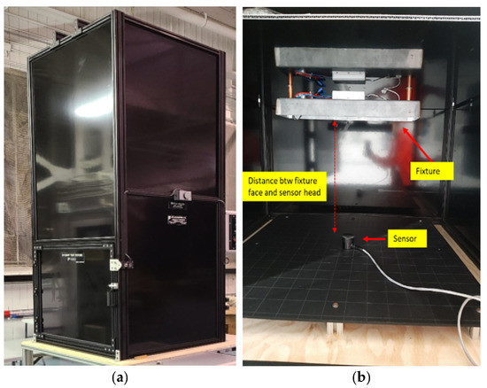
Figure 3 shows the test rig where the experiments were conducted. The test rig was designed in a vertical fashion with a platform to hold the viral sample and a fixture to install the light. The rig was designed in such a way that the distance between the light source and the viral sample could be adjusted. An ILT2500-UVGI-X UVC Flash Meter [28] was used to measure the irradiance in mW/cm2. The ILT2500-UVGI-X UVC Flash Meter had a sensor with the peak calibration at 280 and in the 200~450 nm range. This was used for measuring the irradiance of the mercury bulb (254 nm) and UVC LED (270/280 nm) light source. As the sensor had a peak wavelength of 280 nm, the correction calibration factor was used to ensure the irradiance’s accuracy, which was obtained from the manufacturer. The built-in sensor was replaced by the SED240/FUVC/W and XSD140A sensors to measure the irradiance of the Ushio Care222 (Oude Meer, The Netherlands) (222 nm) and DIEHL blue LED (405 nm), respectively. The irradiance was measured at varying distances from 10 to 50 cm for a full examination. The distance between the fixture face and the head of the sensor was changed by raising the fixture platform manually. For the viral testing, the distance between the viral sample and the face of the fixture was fixed at 10 cm. Based on the measured irradiance (mW/cm2) and required dose (mJ/cm2), which is described above, the exposure time was calculated using Equation (1):
Dose (mJ/cm2) = Irradiance (mW/cm2) × Exposure time (s)

Figure 3.
The test rig setup and characterization: (a) enclosed test rig; (b) test rig setup showing the fixture to install the light source and the sensor to measure the irradiance. After the irradiance measurement, the sensor was replaced with viral sample in the actual experiment, and the distance was adjusted to 10 cm.
The irradiances were measured at varying distances from 10 to 50 cm and the measured values can be used for interpolating the irradiances at a desired distance.
2.3. Cells and Virus Preparation
The HCT8 cells (CCL244) purchased from ATCC were cultured according to the manufacturer’s protocol. The cells were grown in a complete medium prepared by supplementing RPMI 1640 medium with 10% horse serum (Gibco; 26050070) and 1% penicillin–streptomycin (Gibco; 15 140122). The cells’ culture flask was placed in the incubator and maintained at 37 °C with 5% CO2. Betacoronavirus 1 strain OC43 (VR-1558), purchased from ATCC, was propagated using host cell HCT8. All the experiments involving OC43 were conducted within the biosafety level 2 safety cabinet at the Toronto St. Michaels Hospital facility. Viral stocks were prepared by infecting the confluent monolayers of HCT8 cells with OC43. The initial viral absorption was allowed for 1–2 h, with continuous rocking, and then the cells were supplied with an infection medium consisting of RPMI1640 supplemented with 2% horse serum. The virus-infected cells were incubated at 37 °C with 5% CO2 and monitored for 4–6 days or until achieving an 80% cytopathic effect. The produced viral stocks were stored at −80 °C for further use.
2.4. TCID50
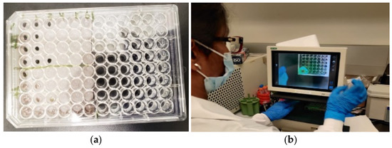
The commonly used polymerase chain reaction (PCR) test did not apply to our test, as it measures virus infection. Instead, the TCID50 assay was used to measure the infectivity of the virus, i.e., virus inactivation. TCID50 represents a dilution of virus that makes 50% of the test wells show cell detachment. The infectivity titer is expressed as TCID50/mL. To perform the TCID50, the host cells (HCT-8) (1.5 × 10⁴ cells in 100 μL) in complete medium were plated in a 96-well plate (Cryo Vial Holder) (Figure 4a). The plates were incubated for 16–24 h at 37 °C in a 5% CO2 incubator to achieve 80% confluency of the cells. To prepare the virus for infection, 100 μL of virus from stock was diluted to 900 μL of serum-free media (10-1 dilution) and 10-fold serial dilutions of the virus (10-2 to 10-10 dilution) were subsequently prepared. On day 1, the culture medium was removed from each well, and 100 μL of the virus solution was inoculated to each well. Aliquots of the same sample were inoculated in multiples of 4 wells. The plates were incubated for 1 h at 37 °C in a 5% CO2 incubator. After incubation, the inoculum was removed and an overlay infection medium containing 2% horse serum was added. The plates were incubated at 37 °C in a 5% CO2 incubator, and the cytopathic effect (CPE) was observed for 6–12 days. Using an inverted microscope (Figure 4b), the number of wells with or without CPE was counted. The TCID50/mL was then calculated using the Reed–Muench method [29].

Figure 4.
Betacoronavirus 1 was plated in a 96-well plate and incubated for 16–24 h. An inverted microscope was used to count the number of wells with and without CPE, followed by calculating the TCID50 using the Reed–Muench method: (a) performing TCID50 on host cells in a 96-well plate; (b) using an inverted microscope to count the number of wells with and without CPE.
3. Results
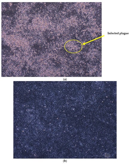
Dose-dependent inactivation of Betacoronavirus 1 without any external photosensitizer. The viral sample was exposed to 405 nm with a 17,280 mJ/cm2 dose for 2 h and 40 min, and an inactivation of log10 2.84 was observed. For 222 nm, the virus sample was exposed to 3 mJ/cm2 for 3.27 s, which resulted in a log10 2.50 reduction. For ultraviolet rays (i.e., 254 and 270/280 nm), a 10 mJ/cm2 dose was exposed for 3.26 and 288.18 s, respectively, which resulted in a log10 3.17 and log10 3.37 reduction. The summarized results are shown in Table 1. The basic infectivity test was conducted on a 270/280 nm exposed sample, and a significant reduction could be observed in comparison to the control sample, as shown in Figure 5. This viral sample was randomly chosen to observe the light exposure’s efficacy.

Table 1.
Showing the TCID50 results for the exposed virus sample, applied dose, and exposure time for different UVC radiation and blue light.

Figure 5.
The basic infectivity test result of the control sample, i.e., the viral sample not exposed to any UVC radiation or visible blue light and the viral sample exposed to UVC. (a) Control sample showing the infected plague of the virus. The control sample was kept at 4 °C at the testing facility. (b) Viral sample exposed to a 10 mJ/cm2 dose of 270/280 nm, showing much less infected plague compared to the control sample.
Irradiation measurements at varying distances. The irradiances at varying distances from 10 to 50 cm were measured to draw a relationship using the inverse square law. Figure 6 shows the relationship between the irradiance and distance for the DIEHL blue light. The inverse square law was used to draw irradiance and the distance dependence assuming the constant angular position (θ = 0°). From Figure 6, the curve equation can be used to calculate the intermittent irradiance value at different distances for the same blue light source used in this study. Based on the calculated irradiance and desired dose, the exposure time could be calculated using Equation (1). Table 2 shows the irradiance at varying distances for all of the different lights used in this study.

Figure 6.
The graph shows the relationship between the irradiance and distance for the DIEHL 405 nm LED. Illustration of the inverse square law for calculating the irradiance at varying distances.

Table 2.
Irradiances (W/cm2) for different UVCs radiations and blue light.
4. Discussion
The ongoing pandemic is in dire need of uninterrupted inactivation technology. In this study, we wanted to explore the impact of 405 nm visible light technology for inactivation of betacoronavirus 1 and compared its efficacy with the other germicidal UVC radiation.
The 254 and 270/280 nm resulted in the highest log reduction of log10 3.17 and log10 3.37 at a 10 mJ/cm2 dose for 3.26 and 288.18 s, respectively. A lower dose of 222 nm (i.e., 3 mJ/cm2) resulted in a log10 2.50 reduction. The 405 nm visible light resulted in a log10 2.84 reduction after 2 h and 40 min with a dose of 17,280 mJ/cm2. The inactivation dose of 405 nm was achieved from a commercially available light source, and the fluence was well within the safe level.
The use of UVC for disinfection purposes has been in practice for centuries but has adverse effects on humans. UVC (200–280 nm) is well known for damaging human tissue, particularly skin. A 254 nm radiation is absorbed by both DNA and RNA, causing severe damage to human cells and tissues. It can cause photokeratitis and erythema (Fields, A., 2019), leading to severe skin burn. This restricts UVC usage to enclosed and unoccupied spaces, making it futile for our desired application. Far-UV 222 nm has a higher irradiation power, which makes the inactivation much faster, and its lower penetration into human skin makes its safe for human exposure [6]. However, a study showed the formation of erythema and cyclobutene pyrimidine dimers (CPDs) after 222 nm irradiation [13]. The use of 222 nm raises concern of ozone generation. Ozone is an unstable gas and can affect the respiratory, cardiovascular, and central nervous systems [25]. The limited studies on the effects of far-UV make its use risky for human exposure. Visible blue light has the potential to be used in occupied spaces. This study showed a significant reduction of betacoronavirus 1 with a very low and safe dose of 405 nm. In past studies, external photosensitizers were used in the medium for inactivation by 405 nm. In this study, 405 nm caused inactivation of the Betacoronavirus without any external photosensitizers.
The TCID50 method was selected for determining the viral infectivity over a plague assay, as it allows for a faster and more straight forward sample preparation and handling. The PCR technique was not used, as it detects both infectious and noninfectious viruses, which can provide an incorrect estimation.
Before using visible light for continuous inactivation, its photobiological hazards to humans should be studied. The IEC 62471 [30] standards have considered the skin, cornea, and retinal hazards. These hazards depend on wavelengths, physiological sensitivity to various wavelengths, angular subtense, and exposure time. The brief mention of eye anatomy is pertinent for understanding the angular subtense. The basic parts of the eyes are the retina, cornea, pupil, lens, and conjunctiva. The pupil is the small opening that allows the light into the eye, and behind the pupil is the lens that focuses the light energy density on the retina. The superficial part of the eye is the cornea and conjunctiva. The amount of light entering the eye depends on the pupil’s size. The pupil’s average maximum constriction and dilation are approximately a diameter of 3 and 7 mm, respectively. This diameter is a limiting factor corresponding to the field of view (FOV). It is assumed that the pupil remains constricted to a diameter of 3 mm for visible light, whereas the pupil is dilated for wavelengths outside the visible range. The exposure time also determines the field of view. For disinfection purposes, the exposure time is more than 10,000 s. The exposed area of the retina will be approximately a FOV angle of 0.1 radians, which is over 5 degrees. By IEC 62471 [30], the skin or cornea hazard by blue light is denoted by EB. The equation provided by the IEC standard for EB is for small sources, i.e., an aperture less than 0.011 [30]. The skin and cornea hazard (EB) ranges from low risk at 1.0 W.m−2 and moderate risk at 400 W.m−2. LB denotes retinal hazard, and the value is based on radiance (W.m−2.sr1). The LB limit ranges from low risk at 10,000 W/(m2.sr) to moderate risk at 4,000,000 W/(m2.sr). A human exposed to a light source with a radiance lower than 100 W/(m2.sr) will receive a dose of greater than 106 J/(m2.sr) in 10,000 s exposure time (2 h and 45 min), and this is classified as no risk [31]. If exposed to a light source of a radiance higher than 100 W/(m2.sr) but less than 10,000 W/(m2.sr), the person will receive a dose greater than 106 J/(m2.sr) within a 100 s exposure time, this is classified as low risk [31]. The light source with a radiance that is higher than 10,000 W/(m2.sr) is classified as medium to high risk, and that light source is prohibited from domestic general lighting [31]. There are many commercial lights such as vital vio [32] that have been tested for IEC 62471 and have been placed in the Exempt Group (RG O), i.e., poses no optical hazard for continuous, unrestricted exposure to humans. The skin and cornea hazard are determined by irradiance, whereas the retinal hazard is by radiance. Assuming that only domestic general visible blue light is used for disinfection means the retinal hazard is already considered. Only skin and cornea hazards must be calculated for uninterrupted disinfection purposes.
For safety reasons, the betacoronavirus was used and not SARS-CoV-2, as there have been many reported accidental cases of the usage of SARS-CoV-2 [33].
This study was conducted to achieve surface disinfection through static liquid media. Air disinfection through visible light has been very briefly studied, and future work will focus on using an aerosolized medium to test the efficacy of visible light for air disinfection.
Based on the results and discussed advantages of using 405 nm over UVC, the 405 nm deployment in public arenas seems pragmatic and doable. The dose used in this study is safe and can be achieved from commercially available LEDs. This technology can be used to disinfect surfaces continuously in public areas, such as hospitals, schools, aircraft cabins, and laboratories. The potential of 405 nm LED in reducing the betacoronavirus 1 is significant and should be explored further.
5. Conclusions
In this study, we demonstrated the inactivation of the betacoronavirus using 405 nm light without the use of any external photosensitizers. The inactivation was achieved using the safe dose level from the commercially available light. Using 405 nm light can have an impact on COVID-19’s spread and can be the first of its kind for uninterrupted disinfection.
Author Contributions
Conceptualization, K.V., P.D., F.X. and A.D.; methodology, K.V. and F.X.; validation, K.V., F.X. and P.D.; formal analysis, K.V. and P.D.; investigation, P.D.; resources, F.X. and P.D.; data curation, K.V.; writing—original draft preparation, K.V. and P.D.; writing—review and editing, K.V., F.X. and P.D.; visualization, K.V.; supervision, F.X. and A.D.; project administration, F.X. and A.D. All authors have read and agreed to the published version of the manuscript.
Funding
This research received no external funding.
Institutional Review Board Statement
Not applicable.
Informed Consent Statement
Not applicable.
Data Availability Statement
The data presented in this study are available upon request from the corresponding authors.
Conflicts of Interest
The authors declare no conflict of interest.
References
- Randall, T.; Cedric, S.; Tartar, A. Murray, Vaccine Tracker. 1 February 2022. Available online: https://www.bloomberg.com/graphics/covid-vaccine-tracker-global-distribution/ (accessed on 20 January 2023).
- What is Ultraviolet Radiation? 11 July 2017. Available online: https://www.canada.ca/en/health-canada/services/sun-safety/what-is-ultraviolet-radiation.html (accessed on 20 January 2023).
- Woo, H.; Beck, E.; Boczek, A.; Carlson, M.; Brinkman, E.; Linden, G.K. Efficacy of Inactivation of Human Enteroviruses by Dual-Wavelength Germicidal Ultraviolet (UV-C) Light Emitting Diodes (LEDs). Water 2019, 11, 1131. [Google Scholar] [CrossRef] [PubMed]
- Ultraviolet (UV) Radiation. 19 August 2020. Available online: https://www.fda.gov/radiation-emitting-products/tanning/ultraviolet-uv-radiation (accessed on 20 January 2023).
- Matthew, M. Testing the Effects of UV-C Radiation on Materials. Int. Surf. Technol. 2021, 14, 46–47. [Google Scholar]
- Childress, J.; Roberts, J.; King, T. Disinfection with Far-UV (222 nm Ultraviolet Light). 2020. Available online: https://www.boeing.com/confident-travel/research/CAP-3_Disinfection_with_Far-UV.html (accessed on 20 January 2023).
- Buonanno, M.; Welch, D.; Shuryak, I. Far-UVC light (222 nm) efficiently and safely inactivates airborne human coronaviruses. Sci. Rep. 2020, 10, 10285. [Google Scholar] [CrossRef]
- David, W.; Buonanno, M.; Grilj, V.; Shuryak, I.; Crickmore, C.; Bigelow, A.; Randers-Pehrson, G.; Johnson, G.; Brenner, J.D. Far-UVC light: A new tool to control the spread of airborne-mediated microbial diseases. Sci. Rep. 2018, 8, 2752. [Google Scholar]
- Coronavirus Disease (COVID-19). Available online: https://www.who.int/emergencies/diseases/novel-coronavirus-2019 (accessed on 8 January 2023).
- Rat, P.; Olivier, E.; Dutot, M. SARS-CoV-2 vs. SARS-CoV-1 management: Antibiotics and inflammasome modulators potential. Eur. Rev. Med. Pharmacol. Sci. 2020, 14, 7880–7885. [Google Scholar] [CrossRef]
- Fatimab, K.; Mohammada, T.; Singh, I.; Singh, A.; Atif, M.; Hariprasad, G.; Hasan, M.G. Insights into SARS-CoV-2 genome, structure, evolution, pathogenesis and therapies: Structural genomics approach. Biochim. Biophys. Acta Mol. Basis Dis. 2020, 1866, 165878. [Google Scholar]
- Simmons, G.; Reeves, J.; Rennekamp, A.; Amberg, S.; Piefer, A.; Bates, P. Characterization of severe acute respiratory syndrome-associated coronavirus (SARS-CoV) spike glycoprotein-mediated viral entry. Proc. Natl. Acad. Sci. USA 2004, 101, 4240–4245. [Google Scholar] [CrossRef]
- Pfeifer, G.; You, Y.; Besaratinia, A. Mutations induced by ultraviolet light. Mutat. Res. 2005, 571, 19–31. [Google Scholar] [CrossRef]
- Oguma, K.O.; Katayama, H.; Ohgaki, S. Photoreactivation of Escherichia coli after Low- or Medium-Pressure UV Disinfection Determined by an Endonuclease Sensitive Site Assay. J. Clin. Microbiol. 2022, 68, 6029–6035. [Google Scholar] [CrossRef]
- Hessling, M.; Haag, R.; Sieber, N.; Vatter, P. The impact of far-UVC radiation (200–230 nm) on pathogens, cells, skin, and eyes—A collection and analysis of a hundred years of data. GMS Hyg. Infect. Control 2021, 16, Doc07. [Google Scholar]
- Irving, D.; Lamprou, D.A.; Maclean, M.; MacGregor, S.J.; Anderson, J.G.; Grant, M.H. A comparison study of the degradative effects and safety implications of UVC and 405 nm germicidal light sources for endoscope storage. Polym. Degrad. Stab. 2016, 133, 249–254. [Google Scholar] [CrossRef]
- Murrell, L.J.; Hamilton, E.; Johnson, H.B. Influence of a visible-light continuous environmental disinfection system on microbial contamination and surgical site infections in an orthopedic operating room. Am. J. Infect. Control 2019, 47, 804–810. [Google Scholar] [CrossRef] [PubMed]
- Maclean, M.; Murdoch, L.E.; MacGregor, S.; Anderson, J.G. Sporicidal effects of high-intensity 405 nm visible light on endospore-forming bacteria. Photochem. Photobiol. 2013, 89, 120–126. [Google Scholar] [CrossRef] [PubMed]
- Murdoch, L.E.; Mckenzie, K.; Maclean, M.; Macgregor, S.; Andersonj, J.G. Lethal effects of high-intensity violet 405-nm light on Saccharomyces cerevisiae, Candida albicans, and on dormant and germinating spores of Aspergillus niger. Fungal Biol. 2013, 117, 519–527. [Google Scholar] [CrossRef]
- Tomb, R.M.; Maclean, M.; Coia, J.E. New Proof-of-Concept in Viral Inactivation: Virucidal Efficacy of 405 nm Light Against Feline calicivirus as a Model for Norovirus Decontamination. Food Environ. Virol. 2017, 9, 159–167. [Google Scholar] [CrossRef]
- Ho, D.T.D.T.; Ahran, K.; Nameun, K. Effect of blue light emitting diode on viral hemorrhagic septicemia in olive flounder (Paralichthys olivaceus). Aquaculture 2020, 521, 735019. [Google Scholar] [CrossRef]
- Tianhong, D.; Asheesh, G.; Clinton, M.K. Blue light for infectious diseases: Propionibacterium acnes, Helicobacter pylori, and beyond? Drug Resist. Updates 2012, 15, 223–236. [Google Scholar]
- Bumah, V.V.; Aboualizadeh, E.; Masson-Meyers, D.S. Spectrally resolved infrared microscopy and chemometric tools to reveal the interaction between blue light (470 nm) and methicillin-resistant Staphylococcus aureus. J. Photochem. Photobiol. 2016, 167, 150–157. [Google Scholar] [CrossRef]
- Rathnasinghe, R.; Jangra, S.; Miorin, L.; Schotsaert, M.; Yahnke, C.; Garc, A. The Virucidal effects of 405 nm visible light on SARS-CoV-2 and influenza A virus. Sci. Rep. 2021, 11, 19470. [Google Scholar] [CrossRef]
- Claus, H. Ozone Generation by Ultraviolet Lamps. Photochem. Photobiol. 2021, 97, 471–476. [Google Scholar] [CrossRef]
- DIEHL Aviation. Available online: https://www.diehl.com/aviation/de/portfolio/cabin-lighting/ (accessed on 25 March 2022).
- Care222 Science. 2022. Available online: https://care222.com/care222-science/ (accessed on 25 March 2022).
- International Light Technologies. Available online: https://www.intl-lighttech.com/products/ilt2400-xsd140a (accessed on 25 March 2022).
- Ramakrishnan, M.A.; Dhanavelu, M. Influence of Reed-Muench Median Dose Calculation Method in Virology in the Millennium. Antivir. Res. 2018, 28, 16–18. [Google Scholar]
- "CLC EN 62471:2008 Photobiological Safety of Lamps and Lamp Systems," 11 September 2008. [Online]. Available online: https://standards.iteh.ai/catalog/standards/clc/a50af9ae-9590-4282-ba82-91f666fe52a7/en-62471-2008 (accessed on 20 January 2023).
- Point, S. Blue Light Hazard: Are exposure limit values protective enough for newborn infants? Radioprotection 2018, 53, 219–224. [Google Scholar] [CrossRef]
- Fields, A. The Impact of Vital Vio Antibacterial Light. 2019. Available online: https://vyv.tech/wp-content/uploads/2020/04/The-Impact-of-Vital-Vio-Technology.pdf (accessed on 20 January 2023).
- Lim, W.; Ng, K.-C.; Tsang, D.N.C. Laboratory Containment of SARS Virus; Annals of the Academy of Medicine: Singapore, 2006; Volume 35, pp. 354–360. [Google Scholar]
Disclaimer/Publisher’s Note: The statements, opinions and data contained in all publications are solely those of the individual author(s) and contributor(s) and not of MDPI and/or the editor(s). MDPI and/or the editor(s) disclaim responsibility for any injury to people or property resulting from any ideas, methods, instructions or products referred to in the content. |
© 2023 by the authors. Licensee MDPI, Basel, Switzerland. This article is an open access article distributed under the terms and conditions of the Creative Commons Attribution (CC BY) license (https://creativecommons.org/licenses/by/4.0/).