Communication

Innovative Alveolar Socket Preservation Procedure Using Demineralized Tooth Dentin as Graft Biomaterial Covered with Three Reabsorbable Membranes: Human Histological Case Series Evaluation

1
Department of Biomedical, Surgical, Dental Science, University of Milan, 20161 Milan, Italy
2
Independent Researcher, 41121 Reggio Emilia, Italy
3
Independent Researcher, 28001 Madrid, Spain
4
College of Medicine and Dentistry, Birmingham B4 6BN, UK
5
Clinical and Experimental Medicine Department, University of Foggia, 65121 Pescara, Italy
*
Author to whom correspondence should be addressed.
Appl. Sci. 2023, 13(3), 1411; https://doi.org/10.3390/app13031411
Submission received: 6 December 2022 / Revised: 11 January 2023 / Accepted: 13 January 2023 / Published: 20 January 2023

Abstract

:
Background: Extracted tooth material has been seen as a valuable alternative to synthetic biomaterials. Aims: A novel flapless alveolar socket preservation (ASP) procedure with demineralized extracted tooth dentin graft material covered by three reabsorbable membranes was histologically and clinically evaluated 4 months after bone healing. Methods: Ten patients were enrolled and separated into two groups. Five post-extractive sites without buccal and/or palatal bone walls were treated with the flapless ASP procedure with demineralized tooth dentin covered with three reabsorbable membranes (Group A—GA). Five patients were treated with primary wound closure covered only with a reabsorbable membrane (Group B—GB). Bone biopsies were performed for histologic and histomorphometric analyses. Results: In both procedures, all clinical outcomes showed good healing of hard and soft tissue and a good maintenance of maxillary architecture. The histological analysis showed no necrosis or inflammatory areas in either group. The histomorphometric analysis showed higher total bone volume in GA (62.78 ± 7.97%) compared to GB (48.04 ± 9.32%), higher vital new bone in GA (57.53 ± 11.16%) compared to GB (42.41 ± 13.06%) and similar values for residual graft in GA (5.24 ± 5.82%) compared to GB (5.29 ± 4.83%). Conclusions: The data obtained show how this novel technique, mixed with the dentin-derived graft material, seems to promote higher bone regeneration.

1. Introduction

Several bone biological processes after tooth extraction have been described in the literature. All events related to the bundle-bone resorption during alveolar bone healing promote a clinical bone volume shrinkage and higher buccal bone resorption. All clinical evidence shows an unfavorable crown–implant ratio and several esthetic and bio-mechanical complications that could represent the potential failure of final dental implant rehabilitation. In the last 15 years, several studies have been carried out to understand the bone loss process after tooth extraction and to promote surgical procedures and autologous or heterologous biomaterials to reduce the volume decrease after tooth extraction and support bone regeneration at the end of the bone healing period [1,2,3]. Therefore, initially, guided bone regeneration (GBR) and, recently, alveolar socket preservation (ASP) procedures, were applied to decrease bone loss and promote alveolar preservation, obtaining adequate horizontal/height bone volume, simple implant placement, favorable crown–implant prosthetic ratio and predictable prognosis of rehabilitation [4,5,6,7]. To evaluate the ideal biomaterial in ASP, several autologous and heterologous materials with biocompatibility, osteogenesis, osseoinduction, and different osseoconduction characteristics have been used [8]. Reabsorbable or non-reabsorbable membranes used in the GBR technique are crucial in the bone regeneration and avoid the alveolar socket space colonization by fibroblasts that have a greater cellular turn-over than osteoblast cells. In 2011, Heggeler et al., in a systematic review, observed better results in ASP cases compared with spontaneous bone healing, concluding that ASP is useful to reduce bone loss but is unable to prevent it [9]. In 2014, Lindhe et al. showed that non-reabsorbed heterologous particles surrounded by bone in a post-extraction socket after healing were able to occupy the space (osseoconduction), promote bone growth and prevent further bone resorption [10]. In 1967, Yeomans and Urist showed the demineralized dentin matrix osteoinductive potential, promoting the tooth as autologous bio-material for bone reparation and regeneration [11,12]. In 2015, Kurmar showed a similar chemical composition for the hydroxyapatite inorganic portion of bone (95%) and dentin (65%) as well as the Col-1 protein and other secondary molecules [13].
The aim of the present study was to assess, in compromised wall socket sites, the clinical, histological and histomorphometrical values for a novel, minimally invasive flapless ASP technique using three reabsorbable membranes, compared to classic open-flap elevation ASP procedures covered with a single reabsorbable membrane.

2. Materials and Methods

General inclusion criteria (asa-1 and asa-2) were adopted. The exclusion criteria were: patients with a history of allergies, diabetes, HIV, cancer, bone or metabolic diseases, pregnancy, use of immunosuppressive agents, systemic corticosteroids, intramuscular/intravenous bisphosphonates, tobacco, radio or chemotherapy patients (within the last six months). Ten patients [average age 57 years (from 50 to 73)—5 male and 5 female] without buccal or palatal bone defects after tooth extraction were enrolled and divided into two groups in four different dental clinics in Italy. All healthy patients were selected and a written informed consent was subscribed for enrollment in the study. The patients were treated for alveolar socket preservation procedures according to the 2013 Helsinki protocols and ethical requirements. After extraction, socket pre-surgical CBCT (cone-beam computed tomography) measurement evaluation was performed in all patients and before extraction.

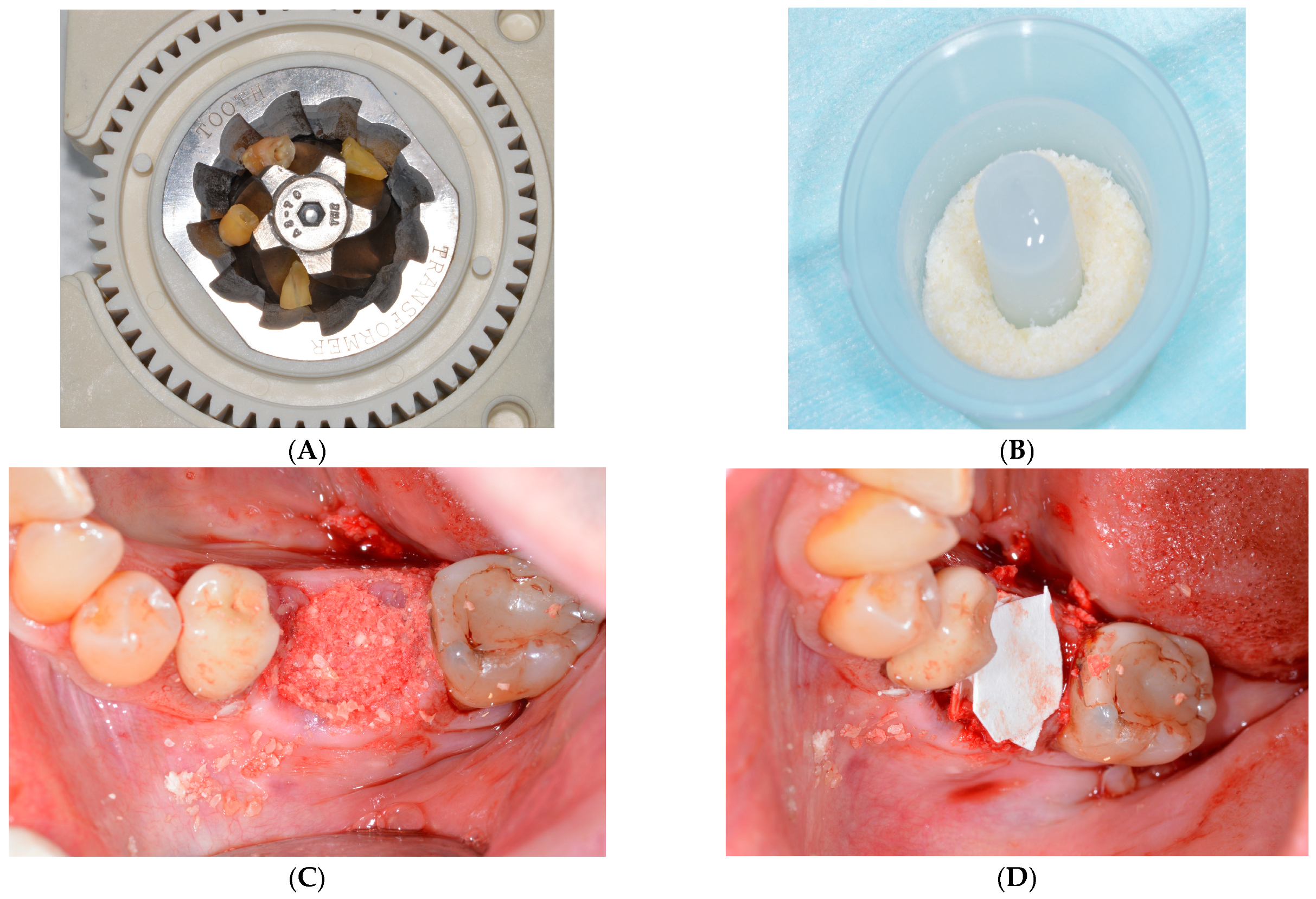
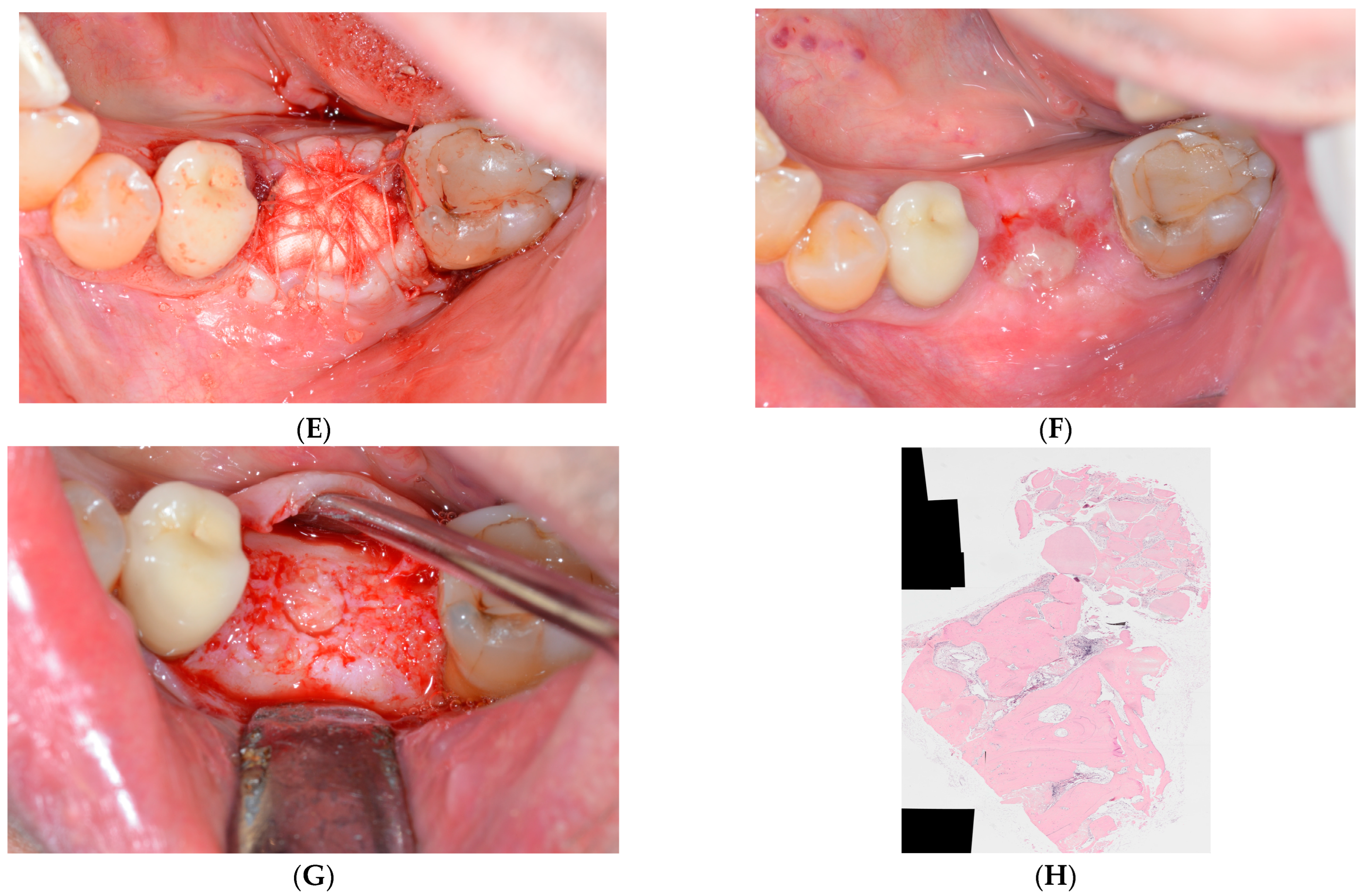
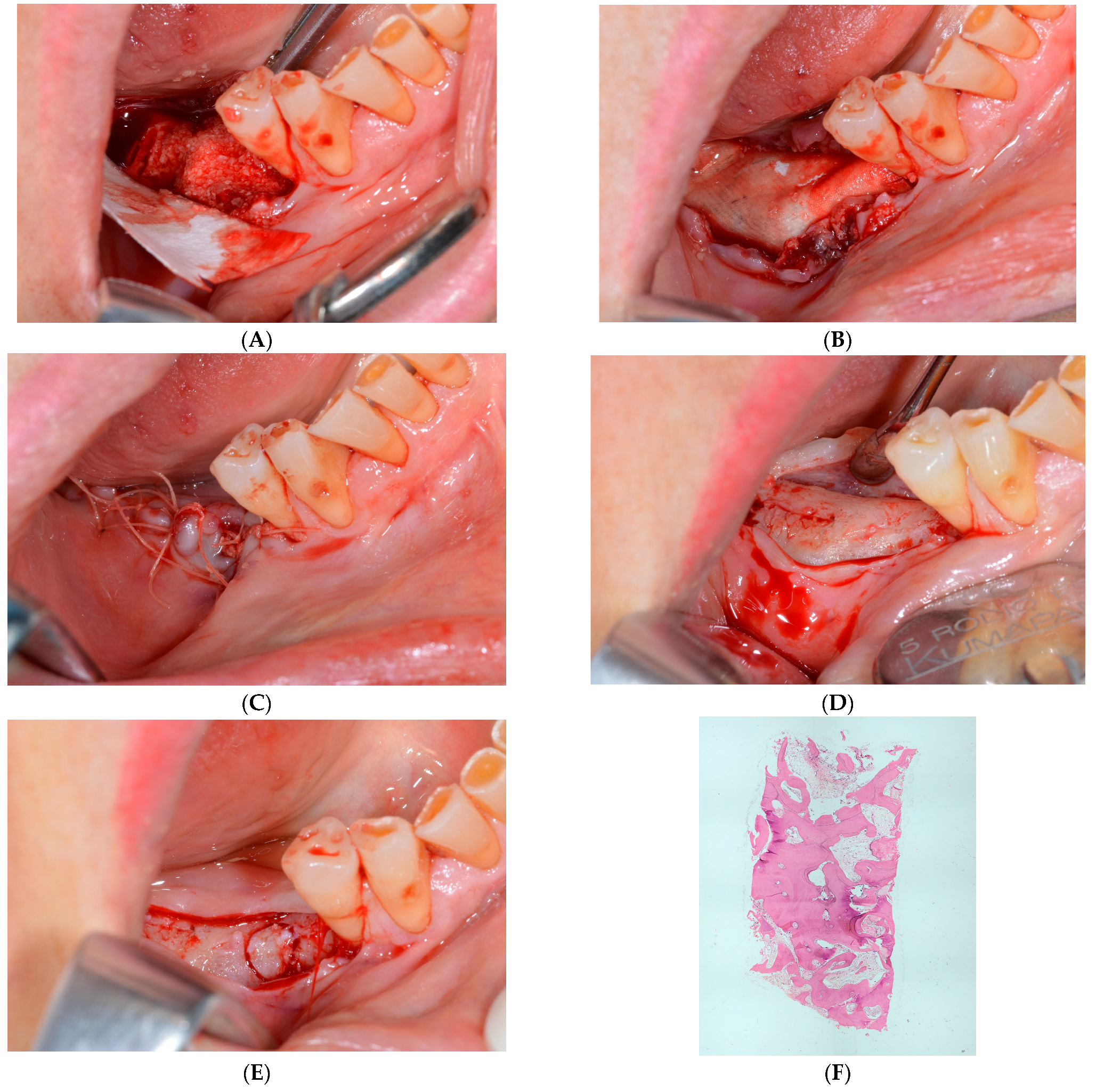
2.1. Surgical Protocol

In accordance with the University of Chieti Ethics Committee, protocol N_1869 12 December 2018 (request ID richhtnc4—approved 17 verb 21 March 19 St. 638—P.I. Perfetti), ten (10) healthy patients needing mandibular molar extraction were enrolled in four private dental clinics in Italy.
Two weeks before the surgery treatment, a professional oral hygiene session was administered, and twice a day, for all patients, chlorhexidine 0.2% mouth rinses were prescribed.
After atraumatic tooth extraction, the baseline socket buccal-lingual and vertical morphology dimensions were recorded using CBCT measurement, and again after the healing period (4 months) before the insertion of the implants. After tooth extraction, the absence of palatal or buccal bone walls prompted the specific clinician’s attention to preserve the keratinized soft tissue. The whole extracted tooth was firstly cleaned in abundant water irrigation, using a diamond drill (ref. 6855 Dentsply Maillefer, Ballaigues, Switzerland), and endodontic filling materials (gutta-percha, composite, etc.) were carefully removed. Also, the tooth was cut into small pieces (6–6 mm) and inserted into the device (Tooth Transformer, Milan, Italy), and according to the manufacturer’s instructions, a single-use box (containing 0.1 M hydrochloric acid, 10% hydrogen peroxide and demineralized water) was inserted. After 25 min, the particle graft biomaterials obtained were placed in the alveolar post-extractive socket sites and covered with a reabsorbable collagen membrane (Osseoguard, Zimmer Biomet, Warsaw, IN, USA). The graft material was placed into the existing alveolar ridge dimensions, making no attempt to go outside the confines of the ridge. In order to increase keratinized tissue width, the membrane was hole punched to produce three round membrane layers, they covered the defect, and only the third superficial membrane was sutured. The sites were allowed to heal without primary wound closure. Amoxicillin 2 gr × 7 days (Pfizer, New York, NY, USA) was administred and after 14 days all sutures were removed. Four months after surgery, through a periodontal probe measurement with the mesial tooth, into the center of the original extraction socket, specific bone biopsies were obtained using graduated trephine cylindrical drills (4 × 18 mm Meisinger, Neuss, Germany). Then, ten dental implants (CEA, Geneva, Switzerland) were placed in the maxillary alveolar regenerated sites.
Group A: The graft material was condensed in the defect. The membrane was hole-punched to produce three round membrane layers, all membranes covered the defect, and only the third superficial membrane was secured with 5-0 sutures. The socket was allowed to heal without primary wound closure in order to increase keratinized tissue width (Figure 1C–H).
Group B: Tooth particle graft biomaterials were placed in the alveolar post-extractive socket sites and covered with only one resorbable collagen membrane. To allow a primary closure of the tissues, a surgical flap was elevated. The marginal tissues were placed near and fixed with single stitches. Two weeks post-surgery, the sutures were removed and, after the healing period, (according to the patient’s requirements) bone biopsies using 4 × 18 mm graduated trephine cylindrical drills (Meisinger, Neuss, Germany) were performed, matching the center of the original extraction socket in the area indicated for implant placement according to prosthetic requirements; dental implants (CEA, Geneva, Switzerland) were then placed in alveolar regenerated sites. After the implant healing period, a ceramic prosthetic was placed. Before and after the healing period, X-rays were performed (Figure 2A–F).

2.2. Histology

All specimens were firstly decalcified, paraffin-embedded and, finally, sliced thinly. All specimens were buffered in formalin (7 days), and afterwards disodium EDTA (pH 7) was used to obtain a total decalcification of the samples. After this, the samples were dehydrated in ethanol (concentration from 70% to 100%), cleared with xylene, and embedded in paraffin (Carlo Erba reagents, Firenze, Italy). Then, the paraffin slides were obtained with a Lecia RM2245 rotatory microtome (Wetzlar, Germany) and colored with hematoxylin and eosin. The histological images made with transmitted light microscope (TLM, Olympus, Tokyo, Japan) were analyzed by IAS 2000 software (QEA, Billerica, MA, USA). Each specimen was split into nine sub-sections to evaluate the differences between different portions (lateral vs. central, or upper vs. bottom). A total of 909 sub-sections were detected and measured using ImageJ program. The mineralized bone volume percentage (BV%), the residual graft percentage (RG%) and the vital bone percentage (VB%) were evaluated.

2.3. Statistical Analysis
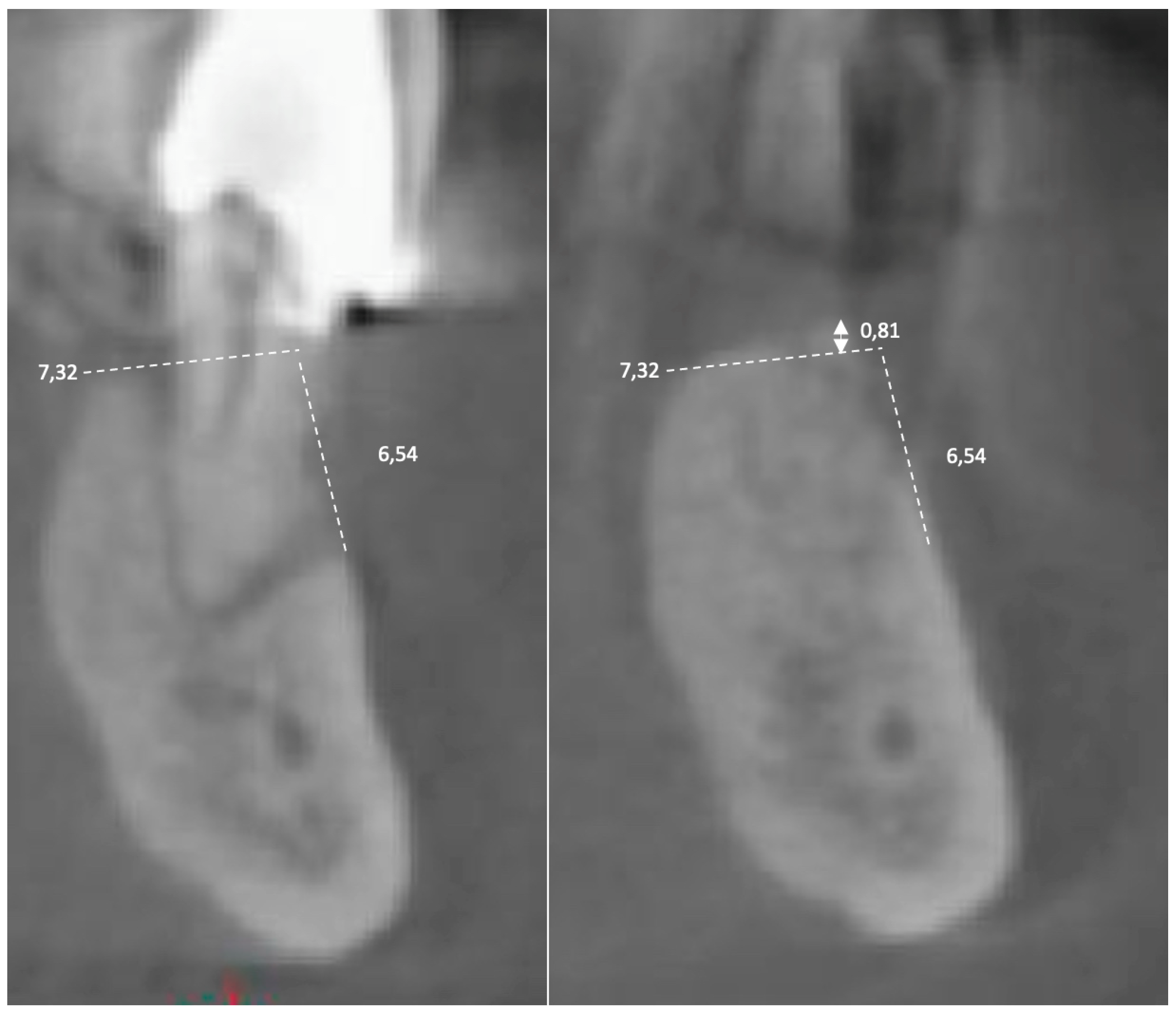
To compare the test vs. control groups behavior, statistical values analysis was carried out. Using Statistical Package for Social Sciences software (SPSS for Windows, Version 11.5, Chicago, IL, USA), the t-test for paired samples in pre–post differences was performed with time as the co-factor, to detect significant divergences between pre and post-test scores.

3. Results

All surgical extraction sockets healed uneventfully. After eight weeks, in all GA patients, clinical complete re-epithelialization healing sites over the reabsorbable barriers were observed. Four months after surgery, maxillary ridge CBCT measurements of GA patients showed adequate bone width for 3D implant placement. Clinical differences were observed between the two groups in the healing of the coronal parts of the regenerations. In Group A (test), the vertical variation (difference between initial volume and the volume measured at the end of the therapy) was 89%, and in Group B (control) it was 102%. The horizontal variation was 92% in Group A (test) and 100% in FC + Group B (control) (Table 1).
The volume was measured before surgery with CBCT and again after 4 months and the images were superimposed. During the measurement (millimeters) before surgery, the dimensions (vertical and horizontal) were recorded, and they were then compared with the same dimensions (vertical and horizontal) when superimposing the image taken after 4 months (Figure 3). The differences in percentages are shown in Table 1.
The histomorphometric analysis showed different bone behavior between the two groups (Figure 1H and Figure 2F): in GA, a higher bone volume (BV) (62.78 ± 7.97%) mean value was observed compared to Group B (48.04 ± 9.32%). A higher vital bone (VB) mean value was detected in GA (57.53 ± 11.16%) compared to GB (42.41 ± 13.06%). Interestingly, similar results for the demineralized dentin residual graft in GA (5.24 ± 5.82%) compared to GB (5.29 ± 4.83%) were observed in all specimens analyzed (Table 1).

4. Discussion

In the last ten years, different procedures have been suggested to preserve the bone volume after tooth extraction, and different biomaterials have been promoted for ASP surgical treatments, such as bovine bone particles (BBP) + socket sealing (SS), 90% bovine bone granules and 10% porcine collagen (BBG/PC) + SS, cortico-cancellous porcine bone particles (CPBP) + SS, allograft particles (AG) + SS, alloplastic material (AP) with or without SS, autologous blood-derived products (ABDP), cell therapy (CTh), recombinant morphogenic protein-2 (rhBMP-2) and SS alone [14]. In a recent histomorphometrical literature review, the different market graft materials were compared without showing the best biomaterial [15]. Recently, a three-punch alveolar ridge reconstruction (ARR) technique was clinically proposed to obtain predictable bone volume maintenance results. In 2022, Grassi proposed using a xenograft material covered with three reabsorbable membranes in ARP maxillary treatment [16]. Recently, in several ASP or ARP human studies, autologous demineralized dentin, obtained after tooth extraction, was used as a graft material and was investigated with clinical and histomorphometrical evaluation. Encouraging histological and histomorphometrical results of the bone biopsies have been shown in several publications [17,18,19]. Moreover, the differences between non-vital endodontic treated and vital whole teeth used as autologous graft materials in ASP procedures were described [17]. Also, Minetti et al. in 2021 showed a high implant survival rate 1 year after loading (98.2%) in maxillary regenerated sites with tooth used as a graft material [18]. Several studies have shown how the buccal plate thickness, the absence or the highly compromised buccal plate influence, the ridge augmentation shape and the dimensions of the autologous tooth matrix conditioned buccal bone resorption, and in one systematic review the buccal plate was related to different biomaterials [20,21,22,23,24]. No inflammation, infection or graft rejection were present after 14 implants without a bone buccal wall and filled using autologous tooth extracted-derived material. After 6 months, the bone resorption was measured: mesial 0.39 ± 1.19 mm and distal 0.42 ± 0.90 mm [25].
Murata and Kim, using a different demineralization procedure, have achieved similar results in terms of quality and the amount of newly formed bone tissue [26,27].
In a recent literature review, the conclusions were: autogenous teeth have superior clinical performance when compared with other grafts.
In 2022, a histological and histomorphometric study showed the different outcomes of tooth-derived materials used as bone substitute in ASP [19]. The present study showed a high global bone volume in both groups evaluated. Moreover, significant differences were detected in vertical bone variation percentage in GB (102 mean value) compared to GA (89 mean value), as well as in horizontal bone variation, which was higher in GB (100% mean vale) compared to GA (92 mean value). The similar histomorphometrical results of the global residual graft in GA (5.24 ± 5.82%) compared to GB (5.29 ± 4.83%), observed in all specimens analyzed, suggested the hypothesis that the biomaterials seem to be extremely bio-compatible to the patient bone, promoting high value of bone regeneration. Furthermore, the present study showed very promising histomorphometrical results with higher NB (62.78) and VB (57.53) percentage mean values in GA compared to GB (NB 48.08–VB 42.41). In GA, on the bone surface it is possible to observe 1–2 mm where the residual granules are the majority of the volume, and the new bone is reduced, probably influencing the buccal plate elevated resorption. In Group B (control group), the residual graft is scattered in all the total histological core. While the test group has a high density but loses 15% of volume, the control group maintains the volume but has a lower density. It was possible to draw these conclusions as the volume, measured with the superimposition, was connected with the histological and histomorphometrical results.
This article compares this new technique with a traditional technique using a patient’s tooth as biomaterial. It is also possible to compare the results obtained by Grassi [15], who used the same new technique but with a market xenograft biomaterial. It is interesting to observe that the mean value among eight patients showed by Grassi is very different from that found in this research. The residual graft was 31.8 ± 8.3 and the vital bone was 25.4 ± 8.7. In our study, the obtained results, as shown above, were better. The residual graft was 5.24 (GA) and 5.29 (GB) and the vital bone was 57.53 (GA) and 42.41 (GB). Probably, the difference is due to the autologous graft material, wich is very similar to the bone.
This study has some limitations, the first being the small number of samples and therefore the poor statistics; other limitations could be the lack of comparison with different biomaterials, as well as the use of only one type of absorbable membrane. Therefore, new randomized clinical trials with a larger sample size and different defect shapes are required to evaluate the potential properties of the present bone preservation technique.

5. Conclusions

The preliminary clinical and histological results of the new three-punch ARR technique combined with autologous tooth-derived dentin graft materials showed predictable outcomes in terms of clinical bone volume and new vital bone volume.
Despite the variability of the results, the extremely simple procedure, the possibility to expand the mucosa volume during the second surgical stage and the interesting results in bone volume improvement, induce us to evaluate positively the three-punch membrane approach in ARP bone treatments.

Author Contributions

Conceptualization, E.M. and F.M.; methodology, A.P.; investigation, A.P.; data curation, T.B.C.; writing—original draft preparation, E.M.; writing—review and editing, E.M.; supervision, A.G. All authors have read and agreed to the published version of the manuscript.

Funding

This research received no external funding.

Institutional Review Board Statement

The study was conducted in accordance with the Declaration of Helsinki, and approved in accordance with the protocol recorded at the University of Chieti (Ethical Committee request ID richhtnc4, protocol N_1869 12 December 2018, approved 17 verb 21 March 2019 St.638—PI Perfetti).

Informed Consent Statement

Informed consent was obtained from all subjects involved in the study.

Conflicts of Interest

The authors declare no conflict of interest.

References

  1. Araujo, M.G.; Lindhe, J. Dimensional ridge alterations following tooth extraction. An experimental study in the dog. J. Clin. Periodontol. 2005, 32, 212–218. [Google Scholar] [CrossRef] [PubMed]
  2. Lekholm, U.; Grondahl, K.; Jemt, T. Outcome of oral implant treatment in partially edentulous jaws followed 20 years in clinical function. Clin. Implant Dent. Relat. Res. 2006, 8, 178–186. [Google Scholar] [CrossRef] [PubMed]
  3. Park, J.B. Healing of extraction socket grafted with deproteinized bovine bone and acellular dermal matrix: Histomorphometric evaluation. Implant. Dent. 2010, 19, 307–313. [Google Scholar] [CrossRef]
  4. Hammerle, C.H.; Jung, R.E. Bone augmentation by means of membrane membranes. Periodontol. 2000 2003, 33, 36–53. [Google Scholar] [CrossRef] [PubMed] [Green Version]
  5. Rakhmatia, Y.D.; Ayukawa, Y.; Furuhashi, A.; Koyano, K. Current membrane membranes: Titanium mesh and other membranes for guided bone regeneration in dental applications. J. Prosthodont. Res. 2013, 57, 3–14. [Google Scholar] [CrossRef] [Green Version]
  6. Chiapasco, M.; Zaniboni, M. Clinical outcomes of gbr procedures to correct peri-implant dehiscences and fenestrations: A systematic review. Clin. Oral Implants Res. 2009, 20 (Suppl. 4), 113–123. [Google Scholar] [CrossRef]
  7. Benic, G.I.; Hammerle, C.H. Horizontal bone augmentation by means of guided bone regeneration. Periodontol. 2000 2014, 66, 13–40. [Google Scholar] [CrossRef]
  8. Corbella, S.; Taschieri, S.; Francetti, S.; Weinstein, R.; Del Fabbro, M. Histomorphometric results after postextraction socket healing with different biomaterials: A systematic review of the literature and meta-analysis. Int. J. Oral Maxilofac. Implants 2017, 32, 1001–1017. [Google Scholar] [CrossRef]
  9. Ten- Heggeler, J.M.; De Van Der Weijden, G.A. Effect of socket preservation therapies following tooth extraction in non-molar regions in humens. A systematic review. Clin. Oral Implants Res. 2011, 22, 779–788. [Google Scholar] [CrossRef]
  10. Lindhe, J.; Cecchinato, D.; Tomasi, C.; Donati, M.; Liljenberg, B. Ridge preservation with the use of deproteinized bovine bone mineral. Clin. Oral Implants Res. 2014, 25, 786–790. [Google Scholar] [CrossRef]
  11. Yeomans, J.D.; Urist, M.R. Bone induction by decalcified dentine implanted into oral, osseous and muscle tissues. Arch. Oral Biol. 1967, 12, 999–1008. [Google Scholar] [CrossRef] [PubMed]
  12. Bang, G.; Urist, M.R. Bone induction in excavation chambers in matrix of decalcified dentin. Arch. Surg. 1967, 94, 781–789. [Google Scholar] [CrossRef] [PubMed]
  13. Kumar, G.S. Orban’s Oral Histology & Embryology, 14th ed.; Elsevier: New Delhi, India, 2015; pp. 40–71. [Google Scholar]
  14. Manavella, V.; Romani, F.; Corano, L.; Bignardi, C.; Aimetti, M. Three dimensional volumetric changes in severely resorbed alveolar sockets after ridge augmentation with bovine derived xenograft and resorbable membrane: A preliminary study on CBCT imaging. Int. J. Oral Maxillofaccial Implants 2018, 33, 373–382. [Google Scholar] [CrossRef]
  15. Grassi, A.; Bernardello, F.; Cavani, F.; Palumbo, C.; Spinato, S. Three-punch alveolar ridge reconstruction technique: A novel flapless approach in eight consecutive cases. Int. J. Periodontics Restor. Dent. 2021, 41, 875–884. [Google Scholar] [CrossRef]
  16. Minetti, E.; Giacometti, E.; Gambardella, U.; Contessi, M.; Ballini, A.; Marenzi, G.; Celko, M.; Mastrangelo, F. Alveolar Socket Preservation with Different Autologous Graft Materials: Preliminary Results of a Multicenter Pilot Study in Human. Materials 2020, 13, 1153. [Google Scholar] [CrossRef] [PubMed] [Green Version]
  17. Minetti, E.; Celko, M.; Contessi, M.; Carini, F.; Gambardella, U.; Giacometti, E.; Santillana, J.; Beca Campoy, T.; Schmitz, J.H.; Libertucci, M.; et al. Implants Survival Rate in Regenerated Sites with Innovative Graft Biomaterials: 1 Year Follow-Up. Materials 2021, 14, 5292. [Google Scholar] [CrossRef] [PubMed]
  18. Minetti, E.; Corbella, S.; Taschieri, S.; Canullo, L. Tooth as graft material: Histologic study. Clin. Implant Dent. Relat. Res. 2022, 24, 488–496. [Google Scholar] [CrossRef]
  19. Hazballa, D.; Inchingolo, A.D.; Inchingolo, A.M.; Malcangi, G.; Santacroce, L.; Minetti, E.; Di Venere, D.; Limongelli, L.; Bordea, I.R.; Scarano, A.; et al. The effectiveness of autologous demineralized tooth graft for the bone ridge preservation: A systematic review of the literature. J. Biol. Regul. Homeost. Agents 2020, 35 (Suppl. 1), 283–294. [Google Scholar]
  20. Spinato, S.; Galindo-Moreno, P.; Zaffe, D.; Bernardello, F.; Soardi, C.M. Is pocket healing conditioned by buccal pate thickness?a clinical and histologic study 4-months after mineralized human allografting. Clin. Oral Implants Res. 2014, 25, e120–e126. [Google Scholar] [CrossRef]
  21. Nevins, M.; Camelo, M.; De Paoli, S. A study of the fate of the buccal wall of extraction sockets of the teeth with prominent roots. Int. J. Periodontics Restor. Dent. 2006, 26, 19–29. [Google Scholar] [CrossRef]
  22. Ferrus, J.; Cecchinato, D.; Pietursson, E.; Iang, N.P.; Sanz, M.; Lindhe, J. Factors influencing ridge alterations following immediate implant placement into extraction socket. Clin. Oral Implants Res. 2012, 21, 22–29. [Google Scholar] [CrossRef] [PubMed]
  23. Avila-Ortiz, G.; Chambrone, L.; Vignoletti, F. Effect of alveolar ridge preservation interventions following tooth extraction: A systematic review and meta-analysis. J. Clin. Periodontol. 2019, 46 (Suppl. 21), 195–223. [Google Scholar] [CrossRef] [PubMed] [Green Version]
  24. Taschieri, S.; Morandi, B.; Alberti, A.; Tarasenko, S.; Diachkova, E.; Francetti, L.; Corbella, S. Immediate implant positioning using tooth-derived bone substitute material for alveolar ridge preservation: Preliminary results at 6 months. Clin. Exp. Dent. Res 2022, 1–8, online ahead of print. [Google Scholar] [CrossRef]
  25. Murata, M.; Kabir, A.M.D.; Hirose, Y.; Ochi, M.; Okubo, N.; Akazawa, T.; Kashiwazaki, H. Histological Evidences of Autograft of Dentin/Cementum Granules into Unhealed Socket at 5 Months after Tooth Extraction for Implant Placement. J. Funct. Biomater. 2022, 13, 66. [Google Scholar] [CrossRef] [PubMed]
  26. Kim, Y.; Ku, J.-K.; Um, I.W.; Seok, H.; Leem, D.H. Impact of Autogenous Demineralized Dentin Matrix on Mandibular Second Molar after Third Molar Extraction: Retrospective Study. J. Funct. Biomater. 2023, 14, 4. [Google Scholar] [CrossRef]
  27. Cenicante, J.; Botelho, J.; Machado, F.; Mendes, J.J.; Mascarenhas, P.; Alcoforado, G.; Santos, A. The Use of Autogenous Teeth for Alveolar Ridge Preservation: A Literature Review. Appl. Sci. 2021, 11, 1853. [Google Scholar] [CrossRef]
Figure 1. (A) Tooth Transformer device. Blade box with the tooth specimen after surgical extraction and before shredding and demineralization treatment. (B) Tooth graft materials after shredding and demineralization. (C) Autologous tooth-derived graft material placed in alveolar post-extractive socket. (D) Three reabsorbable membrane placements to cover the tooth graft materials. (E) Suture. (F) ASP technique healing period after suture excision. (G) Surgical bone biopsy excision after 4-month healing period. (H) Bone histological specimens for Group A.
Applsci 13 01411 g001aApplsci 13 01411 g001b
Figure 2. (A) Autologous tooth-derived graft material placed in alveolar post-extractive socket. (B) Tooth graft materials covered with a resorbable collagen membrane. (C) Suture. (D,E) Surgical bone biopsy excision after 4-month healing period before implant placement. (F) Group B bone histological specimens.
Applsci 13 01411 g002
Figure 3. The superimposition of these two CBCT sections allows seeing the regeneration was 0.81 mm greater than expected.
Applsci 13 01411 g003
Table 1. Histological and histomorphometrical variations of bone biopsies in Group A (GA) and Group B (GB).
Group APatients% Vertical Bone Variation% Horizontal Bone Variation% New Bone% Vital Bone% Residual Graft
A658554.1451.7602.38
B979961.6961.5200.17
C999857.1542.7214.43
D908666.6759.0607.60
E929274.2572.6301.62
Mean Value899262.7857.5305.24
Group BPatients% Vertical Bone Variation% Horizontal Bone Variation% Bone Volume% Vital Bone% Residual Graft
F1179146.5642.1104.44
G978538.4229.9708.45
H11013863.5963.4300.16
I1009844.9933.1711.82
L1028246.6443.3701.59
Mean Value10210048.0842.4105.29
Disclaimer/Publisher’s Note: The statements, opinions and data contained in all publications are solely those of the individual author(s) and contributor(s) and not of MDPI and/or the editor(s). MDPI and/or the editor(s) disclaim responsibility for any injury to people or property resulting from any ideas, methods, instructions or products referred to in the content.

