Abstract
Kinematic and kinetic changes in the lower extremities occur as an athlete becomes fatigued during vertical jumping; however, the specifics of these changes are not well-understood. Therefore, the purpose of this study was to quantify the influence of a sport-specific, vertical jumping fatigue protocol on the biomechanics of the ankle, knee, and hip joint. Twenty male varsity athletes performed repetitive standing countermovement squat jumps every 20 s until fatigued (vertical jump and reach height decreased to 88% of their maximum height for three consecutive jumps). The kinematics and kinetics of their lower extremities (ankle, knee, and hip) were quantified, and the ankle, knee, and hip joint’s moments, angular velocity, and joint power were compared. The participants performed an average of 175 jumps before they were classified as being fatigued. When they became fatigued, the peak power of the ankle and hip joints were substantially reduced due to a decrease in the angular velocity at both joints. Ankle and hip joint moments were unchanged. Peak power at the knee joint was also unchanged over the course of the jumping protocol. To maintain vertical jumping performance over the course of a game or to delay the influence of fatigue, training should be targeted at maintaining the angular velocity of the ankle and hip joints.
1. Introduction
Vertical jumping is integral to the performance of many sports, such as volleyball and basketball, requiring the coordination of the ankle, knee, and hip joints. While the ability to perform a single maximal effort jump is important, over the course of a basketball or volleyball game, an athlete may perform hundreds of jumps [1,2], signifying the importance of an athlete’s ability to maintain their vertical jumping performance for a prolonged duration.
Fatigue is generally defined as a decrease in peak muscle tension and power output resulting in reduced work capacity [3]. As athletes become fatigued, changes in their movement patterns may occur [4], and increases in movement in the frontal plane during running fatigue have been reported [5]. With respect to vertical jumping, fatigue has been shown to reduce an athlete’s vertical jumping performance [6,7,8,9]; however, little is known about the underlying changes in mechanics behind the degradation in jumping performance. Studies investigating changes in vertical jump height incorporate many different methods to render athletes fatigued. These methods include long-duration running [7], knee extensor fatigue [9], plantarflexor fatigue [6], continuous jumping protocols [8,10,11], and different jumping styles such as drop jumps [12] and squat jumps [13]. Each protocol is undoubtedly effective at achieving fatigue; however, none can be assured to fatigue the participant in the same manner as a sport with frequent jumping.
Freitas et al. (2014) [14] performed a kinematic analysis of the lower extremities with a sport-specific jumping fatigue protocol in which athletes performed vertical jumps every 7 s until their performance could no longer be maintained. When comparing the athletes’ vertical jumps prior to and following fatigue, increases in jump time and an increase in the thigh segment angle upon movement reversal were reported [14]. While these data provide some insight into the changes in the lower extremities that occur during sport-specific fatigue, mechanistically, it remains unclear how the ankle, knee, and hip joint were influenced throughout the fatiguing protocol. Further insight can be gained by reviewing studies that indicated that both knee and ankle power are decreased following continuous stretch-shortening cycle exercises using a sled apparatus [10,11,15]. However, this measurement device (the sled apparatus) imposes limitations on the hip joint, one of the main contributors to jumping performance. Additionally, sled pushing is more synonymous with continuous jumping, which does not equate to sport-specific jumping performance. During sport, delays between jumps occur, and alterations in the rest interval between jumps have been shown to postpone fatigue and change its source [16].
Overall, kinematic and kinetic changes in the lower extremities will occur as an athlete becomes fatigued during vertical jumping. However, the specifics of these changes are unclear. No previous studies have quantified how the biomechanics of the ankle, knee, and hip joint change as an athlete becomes fatigued during a sport-specific fatiguing protocol. If the changes in the mechanics of the joints of the lower extremities during a sport-specific fatiguing protocol are better understood, targeted strength- and conditioning-training programs can focus on these joints to prolong athletic jumping performance and reduce the onset or detrimental impact of fatigue. Therefore, the purpose of this study was to quantify the influence of a sport-specific, vertical jumping fatigue protocol on the biomechanics of the ankle, knee, and hip joint.
2. Materials and Methods
2.1. Participants
Twenty male varsity athletes participated in the study (mean ± SD: height = 1.91 ± 0.12 m, mass = 88.1 ± 9.2 kg). All participants were 18+ years of age, familiar with a countermovement jump, and a member of a varsity basketball or volleyball team. Before testing, informed written consent was obtained from each participant in accordance with the University’s Ethics Committee (Ethics ID: REB16-1133).
2.2. Procedures
For the study, participants were required to perform repetitive standing countermovement squat jumps, which were defined as follows: starting from an erect position, a downward movement is performed by flexing the knee and hip joint, and then the knee and hip joint are rapidly extended [17]. The goal of the countermovement jump was for the athlete to reach as high on the Vertec jump height measurement system (Sports Imports, Hilliard, OH, USA)—used to determine the athletes’ vertical jump and reach height—as possible. Prior to testing, athletes performed a self-selected warmup and performed submaximal practice countermovement squat jumps as described above. During these practice jumps, each athlete became accustomed to the jumping protocol and the use of the Vertec jump system.
Following the warmup, each athlete performed maximum effort countermovement squat jumps every twenty seconds until they were fatigued. This protocol was based on the study by Viitasalo et al. (1987) [18], wherein it was determined that volleyball players perform high intensity activities every 24–44 s during gameplay. The rest time between jumps (20 s) employed in this study was selected because it was the shortest time between trials in which the data could be recorded and the Vertec jump meter could be reset.
For the purposes of this investigation, an athlete was classified as fatigued it they met one of the following criteria:
- (1)
- The athlete could no longer maintain 88% of their maximum jump heights for three consecutive jumps (failure jumps). This was determined through pilot testing, where participants performed the jump protocol and verbally quantified their level of fatigue.
- (2)
- The athlete verbally stated they were fatigued and no longer wished to continue jumping.
During each jump of the fatiguing protocol, kinetic ground reaction force data were collected on the right leg at 2400 Hz via a force platform embedded in the lab floor (Kistler AG, Winterthur, Switzerland) while kinematic data of the right foot, lower leg, upper leg and sacrum were recorded using an 8-camera motion capture system capturing images at 240 Hz (Motion Analysis Corp., Rohnert Park, CA, USA). The motion capture system was calibrated to an accuracy defined by a 3D residual below 0.6 mm. The foot, shank, thigh, and hips were defined by attaching retro-reflective markers measuring 19 mm in diameter to each segment using double-sided tape, with three markers attached per segment in the following locations: fibula, upper tibial crest, distal lateral leg to define the shank segment, posterior shoe heel, distal shoe heel, lateral shoe heel to define the rearfoot segment, lower lateral thigh, upper lateral thigh and anterior thigh to define the thigh segment and left/right anterior superior iliac spine, and left and right posterior superior iliac spine (Figure 1). The ankle, knee, and hip joint centers were determined using a standing neutral trial with additional markers placed on the medial and lateral malleolus (ankle), medial, and lateral epicondyles (knee) and greater trochanter. Joint centers were identified as the midpoint of the two joint markers for each joint. The participants were asked to stand with their feet hip-width apart and with their knee and hips fully extended. Prior to analysis, the kinematic and kinetic data were filtered using a low-pass, fourth-order Butterworth filter with a cut-off frequency of 15 Hz. The kinematic and kinetic data were analyzed first with Kintrak 7.0.25 software (Human Performance Laboratory, University of Calgary, Calgary, AB, Canada), which calculated the ankle, knee, and hip joint moment; angular velocity; joint power; and joint energetics, which were then imported into MATLAB (MathWorks, Natick, MA, USA) for further analysis (normalization of the fatigue protocol, etc.).
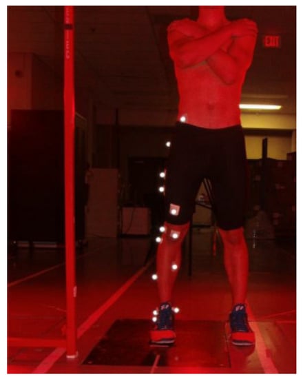
Figure 1.
Photograph of the marker setup.
2.3. Statistics
For kinematic and kinetic analyses, to normalize the fatigue protocol between participants, the total number of jumps each participant performed was divided into 5 sections, with each section representing 20% of the fatiguing protocol. Within each section, ten vertical jump trials were randomly selected for kinematic and kinetic analyses. If any section contained fewer than ten jumps, all jumps were used for analysis. Comparisons were made using a paired t-test (α < 0.05) on previous 20% sections (jumps from 1–20% were compared to jumps from 21–40%, jumps from 21–40% were compared to jumps from 41–60%, and so on). Lastly, the three failure jumps were also analyzed and compared to the data from 81–100%. Effect size (ES) was reported and calculated [19] if a significant difference existed.
3. Results
Participants performed 175 ± 85 jumps (mean ± standard deviation) before they were classified as being fatigued. A sample fatigue jump curve is shown in Figure 2, with the total number of jumps divided into five different sections and the three consecutive jumps below 88% of their maximum jump height highlighted in red.
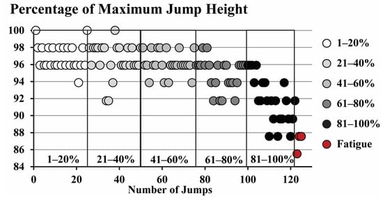
Figure 2.
A jump fatigue curve of a representative athlete. The three red dots represent the three consecutive jumps that were below 88% of the participant’s maximum jump height, indicating the athlete was fatigued.
Peak joint power, angular velocity at peak power, and joint moment at peak power of the ankle, knee, and hip are shown in Figure 3. At the ankle joint, a significant decrease in peak power (p = 0.011, ES = 0.627) occurred during the fatigued jumps due to a significant decrease in ankle plantarflexion angular velocity at peak power (p = 0.003, ES = 0.747). A trend was also present in the 81–100% segment, with peak ankle power being decreased (p = 0.063, ES = 0.442), and with a decrease in the ankle plantarflexion velocity at peak power (p = 0.053, ES = 0.460). At the knee joint, a significant increase in the knee extension moment at peak power occurred at 21–40% (p < 0.001, ES = 1.823) with a corresponding increase in the knee joint peak power at 21–40% (p = 0.020, ES = 0.569). At the hip joint, a significant decrease in the hip extension angular velocity at peak power occurred at 61–80% (p = 0.004, ES = 0.730), at 81–100% (p = 0.040, ES = 0.494), and during the final three fatigued jumps (p = 0.049, ES = 0.470). The peak power at the hip was significantly reduced at 61–80% (p = 0.004, ES = 0.757) and during the fatigued jumps (p = 0.044, ES = 0.481), with the trend of being reduced during 81–100% of the jump protocol (p = 0.057, ES = 0.452).
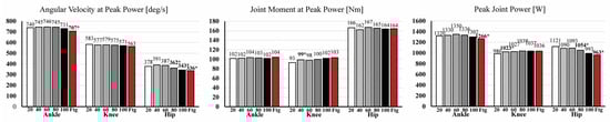
Figure 3.
Ankle, knee, and hip joint angular velocity at peak power (left), joint moment at peak power (center), and peak power (right) during the different phases of the vertical jump fatigue protocol. Bolded asterisks represent a significant difference from the previous jump phase.
Joint energetics are shown in Figure 4. The ankle joint had a significant increase in the energy absorbed at 61–80% (p = 0.028, ES = 0.533), an increase in the energy generated (p = 0.006, ES = 0.693), and an increase in net energy (p = 0.045, ES = 0.599) at 21–40%, while the energy generated (p = 0.010, ES = 0.640 and p = 0.045, ES = 0.479) and net energy (p = 0.001, ES = 0.829 and p = 0.011, ES = 0.629) decreased at 81–100% and during the final fatigued jumps. At the knee joint, significant increases in the energy absorbed (p = 0.005, ES = 0.494) and energy generated (p = 0.039, ES = 0.710) were present at 21–40%, while a significant decrease in the energy absorbed (p = 0.020, ES = 0.566) occurred at 81–100%. At the hip joint, no changes in joint energy absorbed were present, while a significant reduction in the energy generated (p = 0.024, ES = 0.548 and p = 0.047, ES = 0.475) and net energy (0.049, ES = 0.469 and p = 0.005, ES = 0.705) occurred at 81–100% and during the three final fatigued jumps.
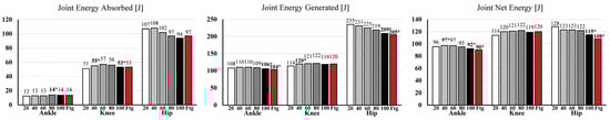
Figure 4.
Ankle, knee, and hip joint energy absorbed (left), energy generated (center), and net energy (right) during the different phases of the vertical jump fatigue protocol. Bolded asterisks represent a significant difference from the previous jump phase.
Additional kinetic data are shown in Table 1. The vertical ground reaction impulse was significantly reduced during the final fatigued jumps (p = 0.0329, ES = 0.514). Jump time was significantly reduced during 20–40% (p = 0.049, ES = 0.471), with trends of having increased during 60–80% (p = 0.080, ES = 0.414) and then reduced during the final three fatigued jumps (p = 0.085, ES = 0.406). The peak ankle plantarflexor moment and angular impulse values were not different during any of the jumping phases, while the athletes increased their peak knee extension moment (p = 0.003, ES = 0.769) and angular impulse (p = 0.032, ES = 0.519) during the 21–40% phase. A significant decrease in the peak hip extension moment occurred at the 61–80% (p = 0.027, ES = 0.534) and 81–100% (p = 0.049, ES = 0.470) phases; however, no significant differences in hip angular impulse were detected.

Table 1.
Vertical ground reaction impulse, peak joint moments, and angular impulses during the fatigue protocol. Bolded asterisks represent a significant difference from the previous jump phase.
4. Discussion
The purpose of this study was to determine how the lower extremities’ biomechanics were altered as the athletes became fatigued. When fatigued, the ankle and hip power and net joint energy were reduced, and the athletes attempted to compensate this loss through an increase in their knee joint power and net energy.
At the ankle joint, from 1–80% of the jump-based fatiguing protocol, the athletes were able to maintain ankle joint power, with no changes in the ankle moment or ankle angular velocity at peak power. However, during the last phase of the jump protocol (81–100%), a reduction in ankle angular velocity occurred, which caused a decrease in peak ankle power. Interestingly, while ankle angular velocity decreased, participants were able to maintain a consistent ankle plantarflexion joint moment (both peak moment and moment at peak power). Although previous studies have shown reductions in the ankle joint’s peak power and angular velocity during vertical jumping following a fatiguing protocol [8,11], the influence of fatigue during countermovement, double-legged jumps on the ankle plantarflexion moment has not been previously reported. Research on single-legged hopping has reported a reduction in the ankle moment; however, this was following a fatigue-to-failure protocol of weighted knee extensions [20]. During a sport-specific jump fatigue protocol, it appears that athletes can maintain their ankle joint moment but are not able to maintain their ankle joint angular velocity. This result is similar to those found by Yu et al. (2020) [21], where no significant difference in ankle joint moment was observed during a double-leg jump following a running-based fatiguing protocol. From a muscular perspective, while direct in vivo measurements were not taken, it has been shown that when fatigued, the activity of ATPase, enzymes that catalyze the decomposition of ATP into ADP and a free phosphate ion, is reduced [22,23], which would decrease the rate of force production but allow for the muscle cross-bridges to stay attached longer to produce force.
At the hip joint, similar peak power reductions in the ankle joint occurred; however, the decrease in hip power occurred much earlier during the jump fatigue protocol, becoming prominent at the 61–80% phase with a continued decrease until the final three fatigued jumps. While the peak hip moments athletes could attain were reduced, the participants were able to maintain the same magnitude of hip moment at peak power, with the reduction in hip power occurring due to a decrease in hip angular velocity. A previous study has shown a reduction in hip joint angular velocity following a fatiguing protocol; however, no significant decreases in peak power at the hip joint were reported [8]. There are differences in the methodologies employed in the previous study and our own, with the previous study measuring kinematics and kinetics following a continuous jumping fatiguing protocol to 70% of the jump height of the unfatigued condition. With a substantially greater reduction in jumping performance and significantly shortened and more intense fatiguing protocol compared to the current study, it may be that the other joints of the lower extremities were more affected by that protocol compared to the hip joint. At the hip joint, during a dynamic sport-specific jump fatigue protocol, a reduction in peak hip power was observed to be due to reduced hip angular velocity and not due decreased hip joint moment.
Conversely, at the knee joint, the athletes compensated for the decreased ankle and hip joint power by increasing their knee joint power, which occurred as early as at 21–40% of the fatiguing protocol. The increased knee joint power was due to the athletes increasing their knee joint moment, which offset the subtle decrease that occurred in knee joint angular velocity. This resulted in a greater knee joint power during the final three fatigued jumps than during the start of the jump-based fatiguing protocol. These data suggest a general trend towards athletes having the capacity to increase their knee joint moment during prolonged periods of repeated jumping, which is facilitated by an increase in knee joint power to compensate for decreased ankle and hip joint power. This result is in direct contrast to previous studies, which indicated a reduction in knee joint peak power following a fatiguing protocol [8,10,11,15]. However, those previous studies performed more rapid fatiguing protocols that are substantially different from those of the current study, with some directly targeting the knee extensors. Our study indicated that during a game-specific fatigue protocol, the knee joint can maintain peak power and athletes can even increase their knee moment as they become fatigued.
Regarding joint energetics, the hip joint was the greatest contributor of energy at the beginning of the fatigue protocol. However, during the last phase (81–100%), as well for the final fatigued jumps, the knee joint generated a greater amount of net energy. At the end of the fatigue protocol, decreases in the net joint energy at the ankle and hip were present, which were due to decreases in the amount of energy generated at each joint and not to an increase in energy absorption. Comparing the magnitudes of net energy from the start to the end of the jump protocol, the ankle joint had a decrease of 6 J, the knee joint had an increase in net energy of 6 J, and the hip joint had a decrease of 20 J. Overall, the knee joint was able to compensate and offset the decreased net energy of the ankle joint but could not compensate for the greater energy decrease seen at the hip joint as well. A decrease in the total net energy of the lower extremities of 3.6% occurred at the 81–100% phase, but it did not result in consistent decreases in jumping performance (<88% of initial jump height) during that phase of the protocol, with participants being able to recover in subsequent jumps. However, during the final three fatigued jumps, the total net energy was reduced by 5.9%.
Many questions arise from the results of this study. For example, why did the participants not utilize their maximal knee joint moment when jumping to achieve maximal height during the early phases of the protocol? The exact reasoning behind this is currently unknown; however, the required coordination of the joints of the lower extremities during jumping [24] or the many bi-articular muscles of the knee joint may provide some insight. Specifically, the main ankle plantarflexor muscle, the gastrocnemius, also contributes to knee flexion, while one of the main knee extensors, the rectus femoris, has been labelled as a hip flexor muscle [25]. During the push-off phase of a countermovement jump, the primary actions of the joints of the lower extremities are ankle plantarflexion, knee extension, and hip extension. With some muscles supporting seemingly opposing actions throughout the movement, it follows that an intricate coordination of muscle activity is required to effectively perform a maximum vertical jump.
5. Conclusions
The results of this study indicate that fatigue during a sport-specific jumping protocol results in a deterioration in jumping performance due to biomechanical changes at the ankle and hip joints. Based on the current results, we conclude that if athletes want to maintain their vertical jumping performance over the course of a game or to delay the onset of fatigue, training should be targeted towards maintaining the angular velocity of the ankle and hip joints. This would allow athletes to maintain ankle and hip joint power, thereby maintaining their net joint energetics.
Author Contributions
Conceptualization, J.W. and D.S.; methodology, J.W. and D.S.; formal analysis, N.S., M.-L.W. and R.M.; investigation, N.S., M.-L.W. and R.M.; writing—original draft preparation, J.W. and Z.B.; writing—review and editing, D.S., N.S., M.-L.W., R.M. and Z.B. All authors have read and agreed to the published version of the manuscript.
Funding
This study was funded by adidas. Adidas had no involvement in the data collection or in the interpretation of the results.
Institutional Review Board Statement
The study was conducted in accordance with the Declaration of Helsinki and approved by the Ethics Committee of the University of Calgary (REB16-1133).
Informed Consent Statement
Informed consent was obtained from all subjects involved in the study.
Data Availability Statement
Data from this study is available from the corresponding author upon reasonable request.
Conflicts of Interest
The authors declare no conflict of interest.
References
- Mori, Y.; Yamada, Y.; Umezaki, S.; Kida, N.; Nomura, T. A Study on the Number of Jumps and Jump Height in Volleyball: From a Mock Game of College Men Players. Adv. Phys. Educ. 2022, 12, 1–10. [Google Scholar] [CrossRef]
- Talpey, S.; Smyth, A.; O’Grady, M.; Morrison, M.; Young, W. The Occurrence of Different Vertical Jump Types in Basketball Competition and their Relationship with Lower-Body Speed-Strength Qualities. Int. J. Strength Cond. 2021, 1, 1–7. [Google Scholar] [CrossRef]
- Fitts, R.H. Muscle fatigue: The cellular aspects. Am. J. Sport. Med. 1996, 24, 9–13. [Google Scholar] [CrossRef]
- Moreira, A.; Aoki, M.S.; Franchini, E.; da Silva Machado, D.G.; Paludo, A.C.; Okano, A.H. Mental fatigue impairs technical performance and alters neuroendocrine and autonomic responses in elite young basketball players. Physiol. Behav. 2018, 196, 112–118. [Google Scholar] [CrossRef] [PubMed]
- Willwacher, S.; Sanno, M.; Brüggemann, G.P. Fatigue matters: An intense 10 km run alters frontal and transverse plane joint kinematics in competitive and recreational adult runners. Gait Posture 2020, 76, 277–283. [Google Scholar] [CrossRef] [PubMed]
- Bobbert, M.F.; Van Der Krogt, M.M.; Van Doorn, H.; De Ruiter, C.J. Effects of fatigue of plantarflexors on control and performance in vertical jumping. Med. Sci. Sport. Exerc. 2011, 43, 673–684. [Google Scholar] [CrossRef]
- Cooper, C.N.; Dabbs, N.C.; Davis, J.; Sauls, N.M. Effects of Lower-Body Muscular Fatigue on Vertical Jump and Balance Performance. J. Strength Cond. Res. 2020, 34, 2903–2910. [Google Scholar] [CrossRef]
- Rodacki, L.F.; Fowler, N.E.; Bennett, S.J.; Bota, J. Multi-segment coordination: Fatigue effects. Appl. Sci. Biodyn. 2001, 5, 1157–1167. [Google Scholar] [CrossRef]
- Rodacki, L.F.; Fowler, N.E.; Bennett, S.J.; Bota, J. Vertical jump coordination: Fatigue effects. Appl. Sci. Biodyn. 2002, 7, 105–116. [Google Scholar] [CrossRef]
- Horita, T.; Komi, P.V.; Nicol, C.; Kyröläinen, H. Effect of exhausting stretch-shortening cycle exercise on the time course of mechanical behaviour in the drop jump: Possible role of muscle damage. Eur. J. Appl. Physiol. Occup. Physiol. 1999, 79, 160–167. [Google Scholar] [CrossRef]
- Kuitunen, S.; Avela, J.; Kyröläinen, H.; Nicol, C.; Komi, P.V. Acute and prolonged reduction in joint stiffness in humans after exhausting stretch-shortening cycle exercise. Eur. J. Appl. Physiol. 2002, 88, 107–116. [Google Scholar] [CrossRef] [PubMed]
- Dabbs, N.C.; Espericueta, S.; Bonilla, S.; Jones, M.T. The Effects of Whole-Body Vibration on Fatigue in Vertical Jump Performance and Isometric Mid-Thigh Pull Measures. Vibration 2021, 4, 759–767. [Google Scholar] [CrossRef]
- Ondatje, W.C.; Noffal, G.J.; Costa, P.B.; Coburn, J.W. The Biomechanical Effects of Fatigue on Drop-Jump Performance in Basketball Athletes. Med. Sci. Sport. Exerc. 2019, 51, 633. [Google Scholar] [CrossRef]
- Pereira, G.; De Freitas, P.B.; Barela, J.A.; Ugrinowitsch, C.; Rodacki, L.F.; Kokubun, E.; Fowler, N.E. Vertical jump fatigue does not affect intersegmental coordination and segmental contribution. Motriz 2014, 20, 303–309. [Google Scholar] [CrossRef]
- Horita, T.; Komi, P.V.; Hämäläinen, I.; Avela, J. Exhausting stretch-shortening cycle (SSC) exercise causes greater impairment in SSC performance than in pure concentric performance. Eur. J. Appl. Physiol. 2003, 88, 527–534. [Google Scholar] [CrossRef]
- Pereira, G.; Morse, C.; Ugrinowitsch, C.; Rodacki, A.; Kokubun, E.; Fowler, N. Manipulation of rest period length induces different causes of fatigue in vertical jumping. Int. J. Sport. Med. 2009, 30, 325–330. [Google Scholar] [CrossRef]
- Bobbert, M.F.; Gerritsen, K.G.; Litjens, M.C.; Van Soest, A.J. Why is countermovement jump height greater than squat jump height? Med. Sci. Sport. Exerc. 1996, 28, 1402–1412. [Google Scholar] [CrossRef]
- Viitasalo, J.T.; Österback, L.; Alen, M.; Rahkila, P.; Havas, E. Mechanical jumping power in young athletes. Acta Physiol. Scand. 1987, 131, 139–145. [Google Scholar] [CrossRef] [PubMed]
- Cohen, J. A power primer. Psychol. Bull. 1992, 112, 155–159. [Google Scholar] [CrossRef]
- Augustsson, J.; Thomeé, R.; Lindén, C.; Folkesson, M.; Tranberg, R.; Karlsson, J. Single-leg hop testing following fatiguing exercise: Reliability and biomechanical analysis. Scand. J. Med. Sci. Sport. 2006, 16, 111–120. [Google Scholar] [CrossRef] [PubMed]
- Yu, P.; Gong, Z.; Meng, Y.; Baker, J.S.; István, B.; Gu, Y. The Acute Influence of Running-Induced Fatigue on the Performance and Biomechanics of a Countermovement Jump. Appl. Sci. 2020, 10, 4319. [Google Scholar] [CrossRef]
- Fowles, J.R.; Green, H.J.; Tupling, R.; O’Brien, S.; Roy, B.D. Human neuromuscular fatigue is associated with altered Na+-K+-ATPase activity following isometric exercise. J. Appl. Physiol. 2002, 92, 1585–1593. [Google Scholar] [CrossRef] [PubMed]
- Fraser, S.F.; Li, J.L.; Carey, M.F.; Wang, X.N.; Sangkabutra, T.; Sostaric, S.; Selig, S.E.; Kjeldsen, K.; McKenna, M.J. Fatigue depresses maximal in vitro skeletal muscle Na+-K+-ATPase activity in untrained and trained individuals. J. Appl. Physiol. 2002, 93, 1650–1659. [Google Scholar] [CrossRef] [PubMed]
- Bobbert, M.; Schenau, G. Coordination in vertical jumping. J. Biomech. 1988, 21, 249–262. [Google Scholar] [CrossRef] [PubMed]
- Landin, D.; Thompson, M.; Reid, M. Actions of Two Bi-Articular Muscles of the Lower Extremity: A Review Article. J. Clin. Med. Res. 2016, 8, 489–494. [Google Scholar] [CrossRef]


Disclaimer/Publisher’s Note: The statements, opinions and data contained in all publications are solely those of the individual author(s) and contributor(s) and not of MDPI and/or the editor(s). MDPI and/or the editor(s) disclaim responsibility for any injury to people or property resulting from any ideas, methods, instructions or products referred to in the content. |
© 2023 by the authors. Licensee MDPI, Basel, Switzerland. This article is an open access article distributed under the terms and conditions of the Creative Commons Attribution (CC BY) license (https://creativecommons.org/licenses/by/4.0/).