Article

Severity of Temporomandibular Joint Disc Displacement and Generalized Joint Hypermobility in Growing Patients: A Cross-Sectional Magnetic Resonance Image Study

by
Adriana Assunta De Stefano
,
Ana Maria Boboc
,
Martina Horodynski
*,
Alessandra Impellizzeri
,
Emanuela Serritella
and
Gabriella Galluccio
Department of Oral and Maxillofacial Sciences, Sapienza University of Rome, 00161 Rome, Italy
*
Author to whom correspondence should be addressed.
Appl. Sci. 2023, 13(22), 12495; https://doi.org/10.3390/app132212495
Submission received: 3 October 2023 / Revised: 16 October 2023 / Accepted: 8 November 2023 / Published: 20 November 2023

Abstract

:
Background: This study aimed to investigate the association between the internal derangement of the temporomandibular joint (TMJ), in particular the severity of disc displacement (DD), and the presence of generalized joint hypermobility (GJH) in growing patients evaluated on magnetic resonance images (MRIs). The study also investigated the relationship between the stages of DD severity and age. Methods: This cross-sectional study has been conducted on a pre-orthodontic population (8–16 years). The GJH was assessed with the Beighton test (cutoff ≥ 4) and the final sample of 56 patients was divided into two groups: a study group, 30 subjects with GJH, and a control group, 26 subjects without GJH. The disc position was evaluated on MRI and the severity of DD was classified in four stages. Pearson’s χ2 Test was performed for the analysis of the statistical correlation. Results: A statistically significant relationship between GJH and disc position was found, in particular, a disc displacement with reduction (DDwR) and DD severity. A positive association also emerged between the severity of DD and age, that increased with increasing age. Conclusions: The research results suggest that there is an association between the presence of generalized joint hypermobility (GJH) and disc displacement (DD) in growing patients. Growing patients with GJH showed a higher severity of DD. In older patients, greater severity of DD was evident, suggesting that this condition may worsen with age during growth.

1. Introduction

Temporomandibular disorders (TMDs) are a heterogeneous group of pathologies affecting either the temporomandibular joint (TMJ), masticatory muscles, or associated structures [1]. The prevalence of TMDs is approximately 31% for adults/elderly [2] and in children and adolescence varies between 20% and 60% [3]. TMJ clicking sounds, pain, limitation of mandibular movements, and headache are the most common signs and symptoms of TMDs [4,5,6,7]. In children and adolescents, signs and symptoms of TMD seem to increase with age with a higher incidence in middle age [2,3,8,9,10].
TMDs have been classified into two majors categories: pain-related disorders (e.g., myalgia, headache attributed to TMD, and arthralgia) and disorders associated with the TMJ (primarily disc displacements and degenerative diseases) [1].
Disc displacement (DD) is one of the disorders associated with the TMJ that can occur also in the children and adolescent population with a click sound as the most frequent sign [7,8,11]. DD is a frequent finding among adults, children, and adolescents who search for an orthodontic clinical examination [12] with a higher incidence of symptomatic TMJ DD in late teenager years [13].
The diagnosis of DD is predominantly clinical, through the clinical evaluation protocol of the diagnostic criteria for TMDs (DC/TMD). Clinically, DDs can be classified according to the DC/TMD into disc displacement with reduction, disc displacement with reduction with intermittent locking, disc displacement without reduction with limited opening, and disc displacement without reduction without limited opening [1]. The diagnosis of DD can be confirmed by MRI, considered the gold standard for the evaluation of the articular disc. On MRI images, DD can be classified into disc displaced with reduction (DDwR) and disc displaced without reduction (DDwoR) according to the position the disc takes in a closed mouth and an open mouth [1].
Several studies have investigated the etiopathogenesis of DD, including macro- and microtrauma in the joint, lack of muscle coordination, changes in TMJ lubrication, postural disorders, stress, and depression [11,14,15]. Likewise, the anatomical and functional changes of the osseous structures of the TMJ have been associated with disc displacement [16,17,18,19,20,21,22]. Joint hypermobility has also been associated with DD [23,24,25]. Joint hypermobility (JH) is a term that describes the ability of a singular or multiple joints to move beyond the normal physiological limits, and it is considered more as a descriptor rather than a diagnosis; it can be observed as localized joint hypermobility or as general hypermobility (GJH) when it affects multiple joints [26]. The most common method used for assessing GJH is the Beighton Score (BS), a set of tests in a nine-point scoring system. It was initially developed as an epidemiological tool used for screening large populations, but later, it has been utilized as a clinical tool for diagnosis of GJH [27,28].
In a review of the clinical presentation of DD, it is suggested that in patients with DD the structures of the TMJ can adapt and that the progression is extremely benign in most cases [29]. However, the studies included in the review are of adult patients and in no case was the presence of GJH considered. Adolescents with GJH seem to have a greater predisposition to present DD, especially disc displacement without reduction [23]. To the best of our knowledge, there are no studies in the literature on the correlation between different degrees of severity of DD and GJH in growing patients.
It would be interesting to know if the severity of disc displacement assessed on MRI is associated with the presence of GJH in growing patients and if this severity changes with the age of the patients evaluated which could indicate a progression of the condition.
The aims of the present study, therefore, were (1) to assess if there is an association between internal derangements of TMJ, in particular the severity of DD and the presence of GJH in pre-orthodontic growing patients evaluated on MRI and, (2) to determine the relationship between the severity stages of DD and age in growing patients.

2. Materials and Methods

2.1. Study Population

This observational cross-sectional study followed the Helsinki Declaration on medical protocol and recommendations for research on humans and was approved by the Institutional Ethics Committee of Policlinico Umberto I (N.47/19/0001155) [30]. All patients and their parents were informed about the risks and benefits of participating in this study, as well as the potential use of the data in future research studies, and written informed consent forms were obtained from parents who agreed to participate.
This study has been conducted on pre-orthodontic patients visiting an Orthodontics Operational Unit of the Department of Odontostomatological and Maxillofacial Sciences of the University “La Sapienza” of Rome for an initial examination. Patients aged between 8 and 16 years, both sexes, diagnosed with one of the four types of DD (unilateral or bilateral) according to the DC/TMD and with indications for further diagnosis by MRI by the treating clinician and not as part of this study, were included after informed consent of all the subjects or their parents was obtained. Patients with systemic pathologies, clinical history of craniofacial traumas, syndromic patients, history of orthodontic or gnathological treatment, history of surgical operations of TMJ or maxillofacial, inflammatory TMJ pathologies, and, finally, motion artifacts in MRIs that were avoiding a proper evaluation of the MRI were excluded. The patients were divided into three age groups: Group I from 8 to 9 years (GR-I), Group II from 10 to 12 years (GR-II), and Group III from 13 to 16 years (GR-III).

2.2. GJH Evaluation

The Beighton score [24] was applied to determine the presence of GJH, it consists of five tests, of which four are bilateral: (1) passive abduction of the thumb towards the front of the forearm; (2) passive dorsiflexion of the metacarpophalangeal joint of the 5th finger > 90°; (3) hyperextension of the elbow joint > 10°; (4) hyperextension of the knee joint > 10°; (5) contact of the palms of the hands on the floor with the extended lower limbs. For each positive test, 1 point was assigned.
Considering a cut-off value of ≥4 in the Beighton score [27,28], the final sample was divided into two groups: a study group (SG), subjects with GJH, and a control group (CG), subjects without GJH.

2.3. MRI Evaluation

The TMJ MRIs were acquired with a bilateral 7.5 cm TMJ antenna on an MRI scanner OpticaMR360® GE 1.5-Tesla model (General Electric Medical System®, Chicago, IL, USA). The images were obtained in maximum intercuspation and in maximum mouth opening in both the sagittal and coronal projection (parallel to the long axis of the condyle) with 3 mm thick sections. For each TMJ, (right and left) oblique sagittal T1-w, oblique sagittal T2-w, and oblique coronal T1-w in closed-mouth position and oblique sagittal PD in opened-mouth sequences were obtained.

2.3.1. Disc Position

The disc position, evaluated in the sagittal and coronal projection of MRI, was classified into three categories: Refs. [1,31,32].
Normal disc position (N), when in closed-mouth position, the posterior band of the disc was positioned above the apex of the condylar head (in a 12 o’clock position), while in an open-mouth position the intermediate zone of the disc was interposed between the condyle and the articular eminence; in coronal projection, the disc was located over the head of the condyle; in open-mouth, the disc remained interposed between the condyle and the articular surface of the articular eminence.
Disc displacement with reduction (DDwR), when in sagittal projection, in a closed-mouth position, the posterior band of the disc was anteriorly displaced relative to the head of the condyle and in coronal projection could appear in a normal position over the head of the condyle and/or displaced medially or laterally. However, the articular disc was reduced on mouth opening.
Disc displacement without reduction (DDwoR), when in sagittal and/or coronal planes, in both closed-mouth position and open-mouth position, the posterior band of the disc was displaced relative to the head of the condyle, and in an open-mouth position, the correct relationship of the condyle–disc complex was not restored (Figure 1).

2.3.2. Disc Displacement Severity

According to the method developed by Ikeda et al. [32], the severity of DD was assessed in MRI sagittal and coronal planes, to classify the disc position into four different severity stages of DD, from stage 0 to stage 4. In the sagittal plane, the position of the disc in each joint was evaluated in three sagittal MRI slices (medial, middle, and lateral) and based on the location of the posterior band (PB) of the articular disc related to the condyle apex was rated as level 0 (normal), level 1, level 2, and level 3 when PB is at 12 o’clock, 11 o’clock, 10 o’clock, and 9 o’clock or below, respectively. In the coronal plane, the mediolateral DD was assessed and assigned the rating of Level 0 when mediolateral disc displacement was not present and Level 1 when the disc was wider on the medial or lateral side or when the disc was displaced until one-sixth of its mediolateral breadth; Level 2 when the disc was displaced till one-third of its mediolateral breadth with a drop-like form; and, finally, Level 3 when the disc was displaced half or more of its mediolateral breadth.
Based on the assessment of the emerged data from the sagittal and coronal plane, a severity stage was assigned to the DD [32]:
  • Stage 0 (normal): disc in a normal position, both in a sagittal and coronal plane.
  • Stage 1: the position of the disc in the sagittal planes and the coronal plane up to level 1.
  • Stage 2: the disc position in sagittal planes of level 2 or sagittal planes of level 1 and the coronal plane up to level 2.
  • Stage 3: disc position in sagittal planes of level 3 and the coronal plane of level up to 3 with disc reduction in mouth opening.
  • Stage 4: sagittal planes of level 3 and the coronal plane of level up to 3 without disc reduction in mouth opening.

2.4. Statistical Analysis
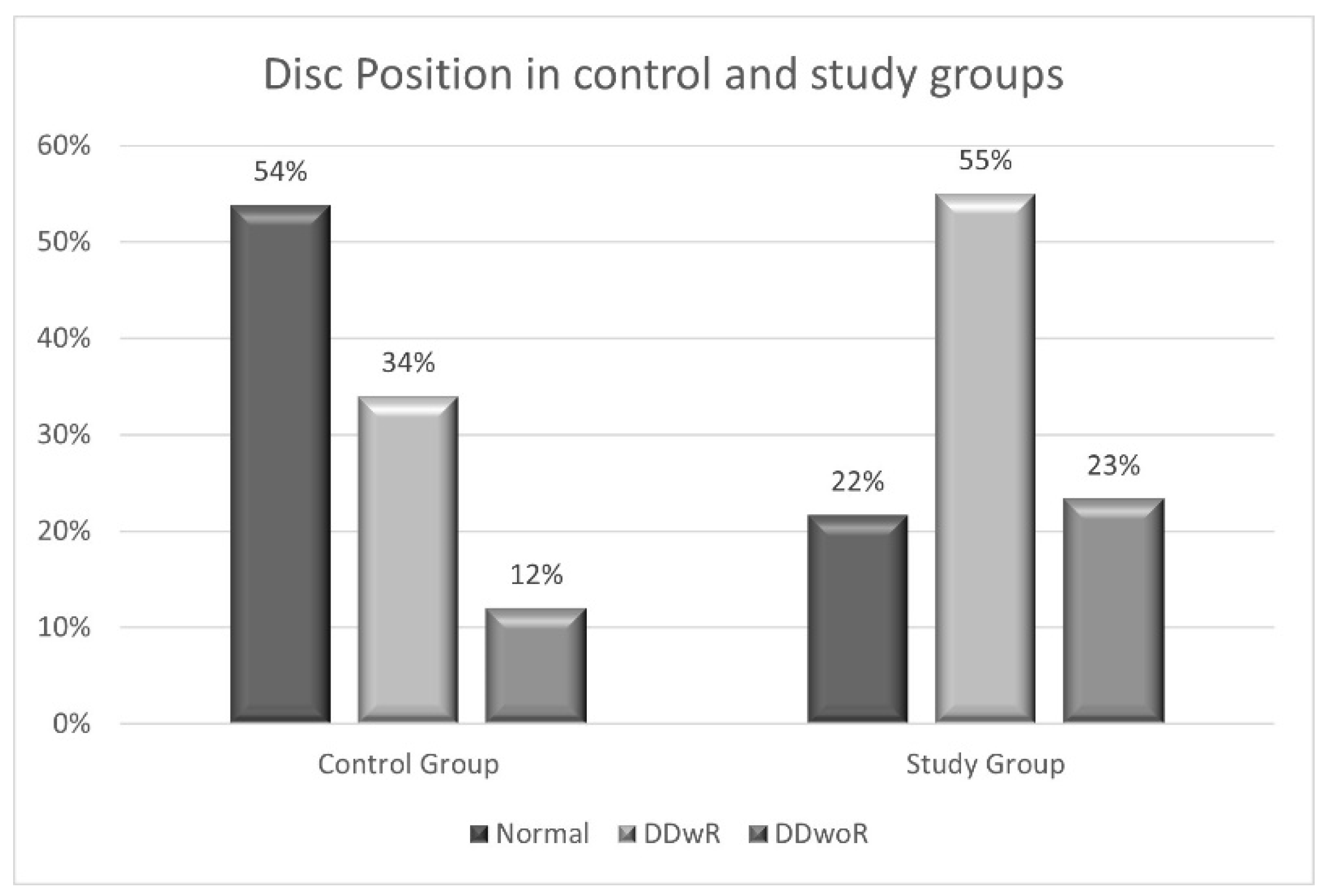
For statistical descriptive purposes, each TMJ was considered as a statistical unit. The statistical analysis was conducted using the IBM SPSS version 24 software and Pearson’s χ2 Test was performed for the analysis of the statistical correlation to compare the frequency of events of the two groups. The statistical significance level of the correlations was set at 5% (p ≤ 0.05).

3. Results

The final sample was composed of 56 individuals, of which there were 36 females (64.3%) and 20 males (35.7%), ranging in age from 8 to 16 years with a mean age of 12.46 ± 2.28 years.
The sample was divided, based on the presence or absence of GJH, in a study group of 30 subjects with GJH, 22 females (39.3%) and 8 males (14.3%), and a control group without GJH composed of 26 subjects: 14 females (25%) and 12 males (21.4%). No statistically significant association was observed between the presence of GJH and sex (p > 0.05).
Furthermore, the sample was divided into three age groups: group I (GR-I) from 8 to 9 years, composed of 7 subjects (12.5%), group II (GR-II) from 10 to 12 years, were 23 subjects (41.1%), and group III (GR-III) from 13 to 16 years of 26 subjects (46.4%). No statistical association between age groups and the presence of GJH was evident (p > 0.05).

MRI Assessment

A total of one hundred twelve (112) TMJs were examined and each TMJ was treated as a single statistical unit.
The study of MRI images revealed the following percentages of disc position in the total sample: normal disc position 41%, DDwR 51%, DDwoR 20%. Therefore, a higher prevalence of DDwR was observed. There was no statistically significant association between disc position and sex (p > 0.05) or age groups (p > 0.05). However, DDwoR did not occur in age group I, but was more frequent in GR-III (Table 1).
A statistically significant association emerged between GJH and disc position, in the study group, 78% of patients showed a DD, compared to 46% of the control group (p = 0.00; χ2 = 12.431; OR = 4.218). DDwR and DDwoR showed a higher presence in the study group (55% and 23%, respectively) compared to the control group (34% and 12%, respectively) (p = 0.002; χ2 = 12.592) (Figure 2).
The analysis of the association between the GJH and DD severity showed statistically significant results (p < 0.010; χ2 = 13.196); the most severe DD showed a frequency of 70% in the study group compared to 30% in the control group. The specific percentages of each stage are illustrated in Figure 3.
Regarding the severity of DD and sex, there was no statistical correlation (p > 0.05), but stage 3 and 4 of severity DD was more frequent in the female participants.
A statistically significant association was observed between DD severity and age groups (p = 0.017; χ2 = 18.707), with a higher frequency of stages 2, 3, and 4 severity in age groups II and III, showing that with increasing age, the greater the severity of DD. The specific percentages of each stage are represented in Table 2.

4. Discussion

This cross-sectional study was conducted to investigate the association between the severity of DD and the presence of GJH in pre-orthodontic growing patients evaluated with MRI, and to investigate the relationship between the stages of DD and age.
MRI is an important tool for the imaging diagnosis of TMDs and has been considered the gold standard for the evaluation of the TMJ, because it provides detailed images of soft tissues and anatomical structures of the TMJ, especially of the articular disc and its position [33,34,35]. MRI can identify disc displacements in their early stages, enabling early and preventive treatment [33,36]. In growing patients, it is crucial to detect issues in the TMJ as early as possible to address them effectively. Unlike other imaging techniques like computed tomography (CT), MRI does not use ionizing radiation, this makes it safe for use in young patients and avoids unnecessary radiation exposure [37,38].
The DD is the most common TMJ internal damage condition. It was diagnosed in the general population and is also present in children and adolescents [7,39,40,41,42,43]. In the present study, the most common DD was DDwR, a result that is in agreement with several authors. Valesan et al. [2], in their systematic review, reported a higher prevalence of DDwR than DDwoR in developmental individuals of the general population. While Nebbe et al. [44] evaluated pre-orthodontic adolescents with a high prevalence of DDwR.
Various hypotheses have been put forward to explain the etiopathogenesis of abnormal disc position, including ligamentous hypermobility [45]. The TMJ can be affected by GJH, receiving an overload, and, as a consequence, suffers degenerative changes which can produce internal derangements [46]. The articular disc is maintained in the normal position on the condyle thanks to the action of the collateral ligaments [3]. The higher prevalence in this study of DD in growing patients with GJH could suggest greater laxity of the ligaments that hold the disc in its normal anatomic relationship with the condyle, which over time could lead to continued damage to the ligaments. Some studies show that in the TMJ of GJH subjects there is an increased risk of articular disc displacement and a lower risk of reduced mouth opening capacity [47,48]. These data agree with the results of this research; subjects with GJH, compared to the control group, have a higher percentage of DD, with a 4-fold increased risk of presenting DD for patients with GJH. Other authors suggest that there is no such correlation between GJH and DD; however, they did not study growing patients [46,49].
DDwR was the most frequent DD in the whole sample, as reported also by other authors [7,8,9,15]. However, in the age distribution, it is important to note that DDwR occurred only in age group III from 12 to 14 years. This result could be explained by the suggestion of some authors who propose that DDwR would be the first stage of disc displacement, which could evolve into DDwoR [11,15,40]. However, the literature is controversial on this aspect, as some authors report that this evolution is not evident. In the systematic review conducted by Naeije et al. [50], it emerges that DDwR is usually a stable, painless condition and accompanies the patient throughout life depending on the adaptive physiological processes that may occur.
Regarding the severity of DD, patients with GJH had a 2-fold higher frequency of DD of greater severity (stage 3 and 4) than patients in the control group. Furthermore, there was a higher frequency of 2, 3, and 4 severity stages in age groups II and III, while group I showed the presence stage 1 of DD, considered incipient, showing that with increasing age, the greater the severity of DD. These results, which are consistent with those reported by Ikeda et al. [32], suggest that it is possible that intra-articular disorder may begin before the age of 10 years and that it is clinically desirable to check the condition of the articular disc at an early age. To the best of the authors’ knowledge, there are no articles in the literature that consider the evolution of DD in growing patients with GJH.
This study has some limitations. The sample size did not allow for a proportionate distribution by age groups, so the results do not allow conclusions to be made without risk of bias. Furthermore, the clinical diagnoses of DD according to the DC/TMD were not considered separately, this could represent a limitation if the results are to be transferred to the clinical evaluation alone.
Despite the limitations of this study, the results obtained provide evidence of an association between a generalized condition such as GJH and DD in growing patients. This could contribute to understanding the risk factors associated with the development of DD in children and adolescents.
The knowledge that the presence of GJH in growing patients could represent an increased risk of developing and having a negative outcome for DD implies that the diagnosis of GJH should be included in the evaluation of TMDs in growing patients. Furthermore, it would be advisable to develop specific indications for the prevention and treatment of DD in patients growing up with GJH.
To expand this line of research and confirm the results of this study, future research on the effect of GJH on TMJ tissues in growing patients should be conducted. Longitudinal studies on the progression of DD in children and adolescents with GJH are also needed in order to know the prognosis of the condition in these patients and to treat early if necessary.

5. Conclusions

The research results suggest that there is an association between the presence of generalized joint hypermobility (GJH) and disc displacement (DD) in growing patients. Growing patients with GJH showed a higher severity of DD. In adolescent patients, greater severity of DD was evident, suggesting that this condition may worsen with age during growth.

Author Contributions

Conceptualization, A.M.B. and A.A.D.S.; methodology, M.H.; software, A.M.B.; validation, A.A.D.S. and M.H.; formal analysis, A.I.; investigation, E.S.; data curation, A.M.B.; writing—original draft preparation, A.M.B. and A.A.D.S.; writing—review and editing, M.H.; visualization and supervision, G.G.; project administration, G.G.; funding acquisition, A.A.D.S. All authors have read and agreed to the published version of the manuscript.

Funding

This research was funded by projects to “Avvio alla Ricerca—Tipo 2” La Sapienza, protocol number: AR2221816B427F66.

Institutional Review Board Statement

The study was conducted in accordance with the Declaration of Helsinki and approved by the Institutional Ethics Committee of Policlinico Umberto I (N. 47/19/0001155).

Informed Consent Statement

Informed consent was obtained from all subjects involved in the study.

Data Availability Statement

The data presented in this study are available on request from the corresponding author. The data are not publicly available due to privacy and ethical restrictions.

Conflicts of Interest

The authors declare no conflict of interest. The funders had no role in the design of the study; in the collection, analyses, or interpretation of data; in the writing of the manuscript; or in the decision to publish the results.

References

  1. Schiffman, E.; Ohrbach, R.; Truelove, E.; Look, J.; Anderson, G.; Goulet, J.-P.; Odont, T.L.; Odont, P.S.; Gonzalez, Y.; Lobbezzoo, F.; et al. Diagnostic Criteria for Temporomandibular Disorders (DC/TMD) for Clinical and Research Applications: Recommendations of the International RDC/TMD Consortium Network* and Orofacial Pain Special Interest Group†. J. Oral Facial Pain Headache 2014, 28, 6–27. [Google Scholar] [CrossRef] [PubMed]
  2. Valesan, L.F.; Da-Cas, C.D.; Réus, J.C.; Denardin, A.C.S.; Garanhani, R.R.; Bonotto, D.; Januzzi, E.; de Souza, B.D.M. Prevalence of temporomandibular joint disorders: A systematic review and meta-analysis. Clin. Oral Investig. 2021, 25, 441–453. [Google Scholar] [CrossRef] [PubMed]
  3. Minervini, G.; Franco, R.; Marrapodi, M.M.; Fiorillo, L.; Cervino, G.; Cicciù, M. Prevalence of temporomandibular disorders in children and adolescents evaluated with Diagnostic Criteria for Temporomandibular Disorders: A systematic review with meta-analysis. J. Oral. Rehabil. 2023, 50, 522–530. [Google Scholar] [CrossRef] [PubMed]
  4. Minervini, G.; Marrapodi, M.M.; Fiorillo, L.; Franco, R.; Cicciù, M.; Cervino, G. Temporomandibular disorders and orofacial neuropathic pain in children and adolescents: A systematic review. J Clin Pediatr Dent. 2023, 47, 26–38. [Google Scholar] [CrossRef]
  5. Bonjardim, L.R.; Gavião, M.B.D.; Pereira, L.J.; Castelo, P.M.; Garcia, R.C.M.R. Signs and symptoms of temporomandibular disorders in adolescents. Braz. Oral. Res. 2005, 19, 93–98. [Google Scholar] [CrossRef]
  6. Kohler, A.; Helkimo, N.; Magnusson, T.; Hugoson, A. Prevalence of symptoms and signs indicative of temporomandibular disorders in children and adolescents. A cross-sectional epidemiological investigation covering two decades. Eur. Arch. Paediatr. Dent. 2009, 10, 16–25. [Google Scholar] [CrossRef] [PubMed]
  7. Da Silva, C.G.; Pachêco-Pereira, C.; Porporatti, A.L.; Savi, M.G.; Peres, M.A.; Flores-Mir, C.; De Luca Canto, G. Prevalence of clinical signs of intra-articular temporomandibular disorders in children and adolescents A systematic review and meta-analysis. J. Am. Dent. Assoc. 2015, 147, 10–18e8. [Google Scholar] [CrossRef]
  8. Christidis, N.; Lindström Ndanshau, E.; Sandberg, A.; Tsilingaridis, G. Prevalence and treatment strategies regarding temporomandibular disorders in children and adolescents-A systematic review. J. Oral Rehabil. 2019, 46, 291–301. [Google Scholar] [CrossRef]
  9. De Stefano, A.; Guercio-Mónaco, E.; Uzcátegui, A.; Boboc, A.M.; Barbato, E.; Galluccio, G. Temporomandibular disorders in Venezuelan and Italian adolescents. CRANIO® 2022, 40, 517–523. [Google Scholar] [CrossRef]
  10. Song, Y.L.; Yap, A.U.; Türp, J.C. Association between temporomandibular disorders and pubertal development: A systematic review. J. Oral. Rehabil. 2018, 45, 1007–1015. [Google Scholar] [CrossRef]
  11. Impellizzeri, A.; Di Benedetto, S.; De Stefano, A.; Monaco Guercio, E.; Barbato, E.; Galluccio, G. General health & psychological distress in children with temporomandibular disorder. Clin. Ter. 2019, 170, e321–e327. [Google Scholar] [PubMed]
  12. Lai, Y.C.; Yap, A.U.; Türp, J.C. Prevalence of temporomandibular disorders in patients seeking orthodontic treatment: A systematic review. J. Oral Rehabil. 2020, 47, 270–280. [Google Scholar] [CrossRef] [PubMed]
  13. LeResche, L.; Mancl, L.A.; Drangsholt, M.T.; Saunders, K.; Von Korff, M. Relationship of pain and symptoms to pubertal development in adolescents. Pain 2005, 118, 201–209. [Google Scholar] [CrossRef] [PubMed]
  14. Manfredini, D. Etiopathogenesis of disk displacement of the temporomandibular joint: A review of the mechanisms. Indian. J. Dent. Res. 2009, 20, 212–221. [Google Scholar] [CrossRef] [PubMed]
  15. Romani, V.; Di Giorgio, R.; Castellano, M.; Barbato, E.; Galluccio, G. Prevalence of craniomandibular disorders in orthodontic pediatric population and possible interactions with anxiety and stress. Eur. J. Paediatr. Dent. 2018, 19, 317–323. [Google Scholar]
  16. Aranha, R.L.B.; Martins, R.C.; de Aguilar, D.R.; Moreno-Drada, J.A.; Sohn, W.; Martins, C.C.; de Abreu, M.H.N.G. Association between Stress at Work and Temporomandibular Disorders: A Systematic Review. Biomed. Res. Int. 2021, 2021, 2055513. [Google Scholar] [CrossRef]
  17. Mortazavi, N.; Tabatabaei, A.H.; Mohammadi, M.; Rajabi, A. Is bruxism associated with temporomandibular joint disorders? A sys-tematic review and meta-analysis. Evid. Based Dent. 2023, 24, 144. [Google Scholar] [CrossRef] [PubMed]
  18. De-Stefano, A.A.; Di-Chicco, A.; Impellizzeri, A.; Serritella, E.; Guercio-Mónaco, E.; Galluccio, G. Unilateral Condylar Hyperplasia: A Thee-Dimensional CBCT Morphometric and Volumetric Evaluation of Mandibular Condyle by Open-Source Softwares. Int. J. Morphol. 2021, 39, 1164–1170. [Google Scholar] [CrossRef]
  19. De Stefano, A.; Guercio-Monaco, E.; Hernández-Andara, A.; Galluccio, G. Association between temporomandibular joint disc position evaluated by magnetic resonance imaging and mandibular condyle inclination evaluated by computed tomography. J. Oral. Rehabil. 2020, 47, 743–749. [Google Scholar] [CrossRef]
  20. Guercio-Monaco, E.; De Stefano, A.; Impellizzeri, A.; Galluccio, G. Association between the temporomandibular joint disc position on magnetic resonance imaging and the mandibular deviation on posteroanterior cephalogram: A cross-sectional study in adolescents. Clin. Ter. 2020, 171, e509–e516. [Google Scholar]
  21. Almășan, O.; Leucuța, D.C.; Buduru, S. Disc Displacement of the Temporomandibular Joint and Facial Asymmetry in Children and Adolescents: A Systematic Review and Meta-Analysis. Children 2022, 9, 1297. [Google Scholar] [CrossRef] [PubMed]
  22. Guercio Monaco, E.; De Stefano, A.; Hernandez-Andara, A.; Galluccio, G. Correlation between condylar size on CT and position of the articular disc on, M.R.I.; of the temporomandibular joint. CRANIO® 2022, 40, 64–71. [Google Scholar] [CrossRef] [PubMed]
  23. Boboc, A.M.; De Stefano, A.; Impellizzeri, A.; Barbato, E.; Galluccio, G. Correlation between generalized joint hypermobility and temporomandibular joint disc displacement in adolescent patients: Magnetic ResonanceImaging study. Eur. J. Paediatr. Dent. 2022, 23, 106–110. [Google Scholar] [PubMed]
  24. Bin Abd Razak, H.R.; Bin Ali, N.; Howe, T.S. Generalized ligamentous laxity may be a predisposing factor for musculoskeletal injuries. J. Sci. Med. Sport. 2014, 17, 474–478. [Google Scholar] [CrossRef]
  25. Chang, C.L.C.; Wang, D.D.H.; Yang, M.C.M.; Hsu, W.E.W.; Hsu, M.L.M. Functional disorders of the temporomandibular joints: Internal derangement of the temporomandibular joint. Kaohsiung J. Med. Sci. 2018, 34, 223–230. [Google Scholar] [CrossRef] [PubMed]
  26. Castori, M.; Tinkle, B.; Levy, H.; Grahame, R.; Malfait, F.; Hakim, A. A framework for the classification of joint hypermobility and related conditions. Am. J. Med. Genet. C Semin. Med. Genet. 2017, 175, 148–157. [Google Scholar] [CrossRef]
  27. Remvig, L.; Jensen, D.V.; Ward, R.C. Epidemiology of general joint hypermobility and basis for the proposed criteria for benign joint hypermobility syndrome: Review of the literature. J. Rheumatol. 2007, 34, 804–809. [Google Scholar]
  28. Bockhorn, L.N.; Vera, A.M.; Dong, D.; Delgado, D.A.; Varner, K.E.; Harris, J.D. Interrater and Intrarater Reliability of the Beighton Score: A Systematic Review. Orthop. J. Sport. Med. 2021, 9, 2325967120968099. [Google Scholar] [CrossRef]
  29. Poluha, R.L.; Canales, G.T.; Costa, Y.M.; Grossmann, E.; Bonjardim, L.R.; Conti, P.C.R. Temporomandibular joint disc displacement with reduction: A review of mechanisms and clinical presentation. J. Appl. Oral. Sci. 2019, 27, e20180433. [Google Scholar] [CrossRef]
  30. N.47/19/0001155; Institutional Ethics Committee of Policlinico Umberto I. PubMed Central: Tucson, AZ, USA, 2021.
  31. Tasaki, M.M.; Westesson, P.L.; Isberg, A.M.; Ren, Y.F.; Tallents, R.H. Classification and prevalence of temporomandibular joint disk displacement in patients and symptom-free volunteers. Am. J. Orthod. Dentofac. Orthop. 1996, 109, 249–262. [Google Scholar] [CrossRef]
  32. Ikeda, K.; Kawamura, A.; Ikeda, R. Prevalence of Disc Displacement of Various Severities among Young Preorthodontic Population: A Magnetic Resonance Imaging Study. J. Prosthodont. 2014, 23, 397–401. [Google Scholar] [CrossRef] [PubMed]
  33. Ahmad, M.; Hollender, L.; Anderson, Q.; Kartha, K.; Ohrbach, R.; Truelove, E.L.; John, T.; Schiffman, E.L. Research diagnostic criteria for temporomandibular disorders (RDC/TMD): Development of image analysis criteria and examiner reliability for image analysis. Oral Surg. Oral Med. Oral Pathol. Oral Radiol. Endodontology 2009, 107, 844–860. [Google Scholar] [CrossRef] [PubMed]
  34. Pupo, Y.M.; Pantoja, L.L.Q.; Veiga, F.F.; Stechman-neto, J.; Zwir, L.F.F.; Farago, P.V.; De Luca Canto, G. Diagnostic validity of clinical protocols to assess temporomandibular disk displacement disorders: A meta-analysis. Oral. Surg. Oral. Med. Oral. Pathol. Oral. Radiol. 2016, 122, 572–586. [Google Scholar] [CrossRef] [PubMed]
  35. Vogl, T.J.; Lauer, H.C.; Lehnert, T.; Naguib, N.N.N.; Ottl, P.; Filmann, N.; Soekamto, H.; Nour-Eldin, N.-E.A. The value of, M.R.I.; in patients with temporomandibular joint dysfunction: Correlation of, M.R.I.; and clinical findings. Eur. J. Radiol. 2016, 85, 714–719. [Google Scholar] [CrossRef]
  36. Talmaceanu, D.; Lenghel, L.M.; Bolog, N.; Hedesiu, M.; Buduru, S.; Rotar, H.; Baciut, M.; Baciut, G. Imaging modalities for temporomandibular joint disorders: An update. Dent. Med. 2018, 91, 280–287. [Google Scholar] [CrossRef]
  37. Shen, S.; Ye, M.; Wu, M.; Zhou, W.; Xu, S. MRI and DC/TMD Methods Analyze the Diagnostic Accuracy of the Change in Articular Disc of Temporomandibular Joint. Comput. Math. Methods Med. 2022, 2022, 1770810. [Google Scholar] [CrossRef]
  38. Whyte, A.; Boeddinghaus, R.; Bartley, A.; Vijeyaendra, R. Imaging of the temporomandibular joint. Clin. Radiol. 2021, 76, 76.e21–76.e35. [Google Scholar] [CrossRef]
  39. Paesani, D.; Salas, E.; Martinez, A.; Isberg, A. Prevalence of temporomandibular joint disk displacement in infants and young children. Oral Surg. Oral Med. Oral Pathol. Oral Radiol. Endodontol. 1999, 87, 15–19. [Google Scholar] [CrossRef] [PubMed]
  40. De Melo, D.P.; Sousa Melo, S.L.; De Andrade Freitas Oliveira, L.S.; Ramos-Perez, F.M.D.M.; Campos, P.S.F. Evaluation of temporomandibular joint disk displacement and its correlation with pain and osseous abnormalities in symptomatic young patients with magnetic resonance imaging. Oral. Surg. Oral. Med. Oral. Pathol. Oral. Radiol. 2015, 119, 107–112. [Google Scholar] [CrossRef]
  41. Marpaung, C.; van Selms, M.K.A.; Lobbezoo, F. Temporomandibular joint anterior disc displacement with reduction in a young population: Prevalence and risk indicators. Int. J. Paediatr. Dent. 2019, 29, 66–73. [Google Scholar] [CrossRef]
  42. Kellenberger, C.J.; Bucheli, J.; Schroeder-Kohler, S.; Saurenmann, R.K.; Colombo, V.; Ettlin, D.A. Temporomandibular joint magnetic resonance imaging findings in adolescents with anterior disk displacement compared to those with juvenile idiopathic arthritis. J. Oral. Rehabil. 2019, 46, 14–22. [Google Scholar] [CrossRef] [PubMed]
  43. Devaraj, S.D.; Pradeep, D. Internal Derangement of Temporomandibular Joint—A Review. IOSR J. Dent. Med. Sci. 2014, 13, 66–73. [Google Scholar] [CrossRef]
  44. Nebbe, B.; Major, P.W. Prevalence of TMJ Disc Displacement in a Pre-Orthodontic Adolescent Sample. Angle Orthod. 2000, 70, 454–463. [Google Scholar] [PubMed]
  45. Chiodelli, L.; Pacheco, A.B.; Missau, T.S.; Silva, A.M.; Corrêa, E.C. Influence of generalized joint hypermobility on temporomandibular joint and dental occlusion: A cross-sectional study. Codas 2016, 28, 551–557. [Google Scholar] [CrossRef] [PubMed]
  46. Sáez-Yuguero, R.; Linares-Tovar, E.; Calvo-Guirado, J.L.; Bermejo-Fenoll, A.; Rodríguez-Lozano, F.J. Joint hypermobility and disk displacement confirmed by magnetic resonance imaging: A study of women with temporomandibular disorders. Oral. Surg. Oral. Med. Oral. Pathol. Oral. Radiol. Endod. 2009, 107, e54–e57. [Google Scholar] [CrossRef]
  47. Kavuncu, V.; Sahin, S.; Kamanli, A.; Karan, A.; Aksoy, C. The role of systemic hypermobility and condylar hypermobility in temporomandibular joint dysfunction syndrome. Rheumatol. Int. 2006, 26, 257–260. [Google Scholar] [CrossRef]
  48. Hirsch, C.; John, M.T.; Stang, A. Association between generalized joint hypermobility and signs and diagnoses of temporomandibular disorders. Eur. J. Oral. Sci. 2008, 116, 525–530. [Google Scholar] [CrossRef]
  49. Berger, M.; Szkutnik, J.; Szalewski, L.; Wójcik, D.; Bakalczuk, M.; Ginszt, M. Correlation between generalized joint laxity and symptoms of temporomandibular disorders. Pol. Merkur. Lek. 2016, 40, 248–251. [Google Scholar]
  50. Naeije, M.; te Veldhuis, A.H.; te Veldhuis, E.C.; Visscher, C.M.; Lobbezoo, F. Disc displacement within the human temporomandibular joint: A systematic review of a “noisy annoyance. J. Oral. Rehabil. 2013, 40, 139–158. [Google Scholar] [CrossRef]
Figure 1. Disc position based on open-mouth and closed-mouth. DDwR: disc displacement with reduction; DDwoR: disc displacement without reduction.
Applsci 13 12495 g001
Figure 2. Distribution of the disc position in control group and study group.
Applsci 13 12495 g002
Figure 3. Distribution of the DD severity stage in control group and study group.
Applsci 13 12495 g003
Table 1. Distribution of disc position by age groups. (p = 0.369; χ2 = 4.285).
Age GroupsDisc Position
NormalDDwRDDwoR
N%N%N%
GR-I
(8 to 9 years)
717%714%00%
Gr-II
10 to 12 years
1741%2141%840%
Gr-III
13 to 16 years
1741%2345%1260%
Total41100%51100%20100%
Table 2. Distribution of severity of DD by age groups. (p = 0.017; χ2 = 18.707).
Age GroupsDD Severity
Stage 0Stage 1Stage 2Stage 3Stage 4
N%N%N%N%N%
GR-I717%728%00%00%00%
(8 to 9 years)
Gr-II1741%1352%729%150%840%
10 to 12 years
Gr-III1741%520%1771%150%1260%
13 to 16 years
Total41100%25100%24100%2100%20100%
Disclaimer/Publisher’s Note: The statements, opinions and data contained in all publications are solely those of the individual author(s) and contributor(s) and not of MDPI and/or the editor(s). MDPI and/or the editor(s) disclaim responsibility for any injury to people or property resulting from any ideas, methods, instructions or products referred to in the content.

