Abstract
The characteristics of 3-dimensional (3D) cultured periodontal ligament stem cells derived from permanent teeth (pPDLSCs) and supernumerary teeth (sPDLSCs) were compared and evaluated in this study. pPDLSCs and sPDLSCs were cultured in an ultra-low attachment 6-well plate, and the changes in morphology and size were observed under an optical microscope for 14 days. Cell counting kit-8 was used to quantitatively evaluate cell viability, and a live/dead assay was performed for the qualitative assessment. The degrees of osteogenic, chondrogenic, and adipogenic differentiation of pPDLSCs and sPDLSCs were also assessed. The pPDLSCs and sPDLSCs were initially spherical, and sPDLSCs were smaller than pPDLSCs. The viability of pPDLSCs decreased by 47.9% (day 3) and 10.4% (day 7), whereas that of sPDLSCs decreased by 34.3% (day 3) and 2.5% (day 7) compared to the percentage on Day 1. No significant difference in osteogenic differentiation was found (p = 0.94); however, significant differences in adipocyte and cartilage differentiation were observed (p = 0.003 and p = 0.013, respectively). Within the limitations of this study, sPDLSCs and pPDLSCs exhibited multilineage differentiation capacities, including osteogenic, chondrogenic, and adipogenic differentiation, in 3D culture. Overall, pPDLSCs should be further investigated as a source of stem cells for tissue engineering and regenerative medicine.
1. Introduction
Mesenchymal stem cells (MSCs) have self-renewal and multilineage differentiation capabilities. Consequently, MSCs have been evaluated for regeneration and repair. To date, stem cells of dental origin have been demonstrated to express MSC characteristics [1]. Dental MSCs are harvested from different dental tissues, including dental pulp stem cells (DPSCs), stem cells from apical papilla (SCAP), periodontal ligament stem cells (PDLSCs), alveolar bone-derived mesenchymal stem cells (ABMSCs), gingival-derived mesenchymal stem cells (GMSCs), and dental follicle stem cells (DFSCs) [2]. These tissues were analyzed to determine their marker gene expression and their potential to promote the regeneration of the pulp-dentin complex and differentiation of MSC lineages.
The periodontal ligament (PDL) is a multifunctional connective tissue composed of osteoblasts, osteoclasts, fibroblasts, epithelial rests of Malassez cells, odontoblasts, cementoblasts, macrophages, and undifferentiated mesenchymal cells [3]. Notably, PDLSCs can differentiate into multilineage cells such as osteogenic, chondrogenic, adipogenic and neurogenic. Furthermore, PDLSCs possess immunosuppressive and less inflammatory functions when compared to bone marrow MSCs, making them crucial in the field of regenerative medicine [4].
Supernumerary teeth (ST) are excess odontogenic masses or extra teeth in the dental arch [5]. ST can lead to delayed or interrupted permanent tooth eruption, displacement, crowding, midline diastema, root resorption in adjacent teeth, and cyst formation [6]. Therefore, it is recommended to extract ST early and then manage them properly. Various theories exist regarding the development of ST, including overstimulated dental lamina [7], division of developing tooth germs, remnants of epithelial cells [6], and genetic factors. ST, the discarded tissues, are also composed of tissues, such as DPSCs, SCAPs, dental follicles, and PDL, which are easily obtained, not contaminated, and contain sufficient tissue. However, research on the PDL of ST remains limited.
Three-dimensional (3D) cell culture has emerged as a revolutionary paradigm in the fields of cell biology and biomedical research, ushering in a new era of experimental techniques that bridge the gap between traditional two-dimensional (2D) cell cultures and in vivo models [8,9]. Unlike 2D cultures, where cells grow on flat surfaces, 3D cell cultures involve the cultivation of cells within 3D matrices. These matrices can simulate the intricate architecture and microenvironment of living tissues [10]. This approach has gained increasing attention in recent years because of its potential to provide physiologically relevant insights into cellular behavior, drug responses, and disease mechanisms [11].
Dental research has embraced 3D cell culture as a valuable tool for studying various aspects of oral health and dental disease [12]. Researchers have used 3D cell culture techniques to model the complex microenvironment of the oral cavity and have investigated areas such as tooth development and regeneration [13,14], periodontal disease [15], orthodontics [16], oral cancer [17], drug testing and development [18], bacterial biofilms [19], and salivary gland research [20]. These applications of 3D cell cultures in dental research have contributed to a deeper understanding of oral health, disease mechanisms, and potential therapeutic interventions [21]. More accurate modeling could lead to more effective treatments and interventions in dentistry.
Three-dimensional cell culture techniques encompass a diverse range of methods, namely: 1. scaffold-based 3D cell culture systems; 2. hydrogel-based 3D cell culture systems; 3. spheroid formation tools; 4. microfluidic-based systems; 5. bioprinting technology; 6. an organic culture kit; 7. hanging drops and magnetic levitation systems; and 8. 3D cell culture media and reagents [22,23]. These methodologies allow the cultivation of cells in structures resembling organs, tissues, or specific microenvironments. This offers researchers the ability to investigate intricate cellular processes and responses in a more contextually accurate manner.
An ultralow attachment plate system is one of the spheroid formation tools [24]. These plates are used to promote spheroid formation by inhibiting cell attachment, and they have many advantages. The key characteristics and principles of ultraslow-attachment 3D cell cultures include reduced attachment, spheroid formation, and mimicking in vivo conditions. They can also be used in a wide range of applications. However, studies in the dental field are rare [25].
The purpose of this study was to grow PDLSCs using ultralow-attachment plates. Two types of PDLSCs originating from supernumerary and permanent teeth were used. We aimed to confirm the possibility of 3D cell culture in stem cell research in the dental field and to compare the characteristics of the two cell types in a 3D culture based on cell morphology and multilineage differentiation.
2. Materials and Methods
2.1. Cell Culture
The pPDLSC and sPDLSC lines were purchased from Cell Engineering for Origin (CEFO Co., Ltd., Seoul, Republic of Korea). After thawing, the cells were cultured in Dulbecco’s modified Eagle’s medium (DMEM; Gibco, Waltham, MA, USA) and incubated at 37 °C with 5% CO2. Ultra-low attachment 6-well plates (ULA; Corning, NY, USA) and 3D scaffold-free culture plates were prepared. The bubbles were removed using EtOH and replaced with DMEM. pPDLSCs and sPDLSCs were seeded, and 3D spheroids were formed after 24 h. The culture medium was replaced every 2 days.
2.2. Morphological Assessment
The morphologies of pPDLSCs and sPDLSCs were observed on days 1, 2, 4, 7, 10, and 14 under a fluorescence microscope (JuLI; Nanoentek Inc., Seoul, Republic of Korea). The cells were monitored for 14 days to determine the long-term stability of the PDLSC spheroids. Cell size was measured using ImageJ software (version 1.53, NIH, Bethesda, MD, USA).
2.3. Cell Proliferation Assessment
To evaluate cell proliferation, the cell counting kit-8 assay (CCK-8; Dojindo Molecular Technologies, Kumamoto, Japan) and live and dead assay (Cytotoxicity kit for mammalian cells; Invitrogen, Carlsbad, CA, USA) were performed. The PDLSCs were sub-cultured at a seeding density of 1.5 × 10⁵/well, with growth media (GM) containing 10% fetal bovine serum (FBS) and 1% gentamycin for 1, 3, 5, and 7 days. The absorbance of CCK-8 was measured at 450 nm using a Benchmark Plus Multiplate Spectrophotometer (Bio-Rad, Hercules, CA, USA).
The live and dead assays were performed on days 1, 5, and 7 and staining with calcein AM and ethidium homodimer-1 (Thermo Fisher Scientific, Waltham, MA, USA) was performed for the live and dead cells, respectively. Fluorescence was detected using a fluorescence microscope (IX 71; Olympus, Tokyo, Japan) at 450 nm and 559 nm.
2.4. In Vitro Multilineage Differentiation Assays
For osteogenic differentiation, cells were cultured in a basal medium at the appropriate confluence. The cells were incubated for 48 h. Thereafter, the medium was changed to the osteogenic supplementation medium, which contained α-minimum essential medium (MEM), 15% FBS, gentamycin (5 μg/mL), L-ascorbic acid (100 μM/L), dexamethasone (10 nM/L), and β-glycerolphosphate (10 mM/L). The cells were then cultured for 2 weeks. To evaluate the extent of mineralization, Alizarin Red S (ARS) staining was performed to detect calcium formation. Microscopic images were captured for morphological evaluation. The basal medium without any supplements was used as a control.
For chondrogenic differentiation, the medium was replaced with a commercial chondrogenic supplementation medium (StemPro Chondrogenesis Differentiation Kit; Gibco, Grand Island, NY, USA) and maintained for up to 23 days. Alcian Blue (AB) staining was performed to evaluate the extent of mineralization, and microscopic images were obtained for morphological evaluation.
For adipogenic differentiation, the medium was changed to adipogenic supplementation medium, which is part of a commercial StemPro Adipogenesis Differentiation Kit (Gibco, Grand Island, NY, USA). Thereafter, the cells were observed for up to 28 days. Oil Red O (ORO) staining was performed to evaluate the extent of mineralization, and microscopic images were obtained for morphological evaluation.
As all images were colored, they were separated according to red-green-blue (RGB) channels using ImageJ. The channel appropriate for each staining material was selected from the red, green, and blue channel images. Red channel images were selected to quantify odontogenic and adipogenic differentiation, while blue channel images were used to quantify chondrogenic differentiation. As the degree of differentiation was expressed as the intensity of each pixel, a region of interest (ROI) was established around the area of pixels with high intensity. On each image, ten square regions, 32 pixels wide and 32 pixels high (1024 pixels), were randomly selected without overlapping within the ROI. The mean intensity of each squared region was calculated.
2.5. Statistical Analysis
The cell differentiation data were analyzed using SPSS software (version 26.0; IBM Corp., Armonk, NY, USA). One-way ANOVA followed by Scheffé post-hoc test was used for analyzing cell proliferation. Considering the sample size, the Mann–Whitney U test was performed to compare the degree of cell differentiation between the groups. The level of significance was set at p < 0.05.
3. Results
3.1. Cell Morphology
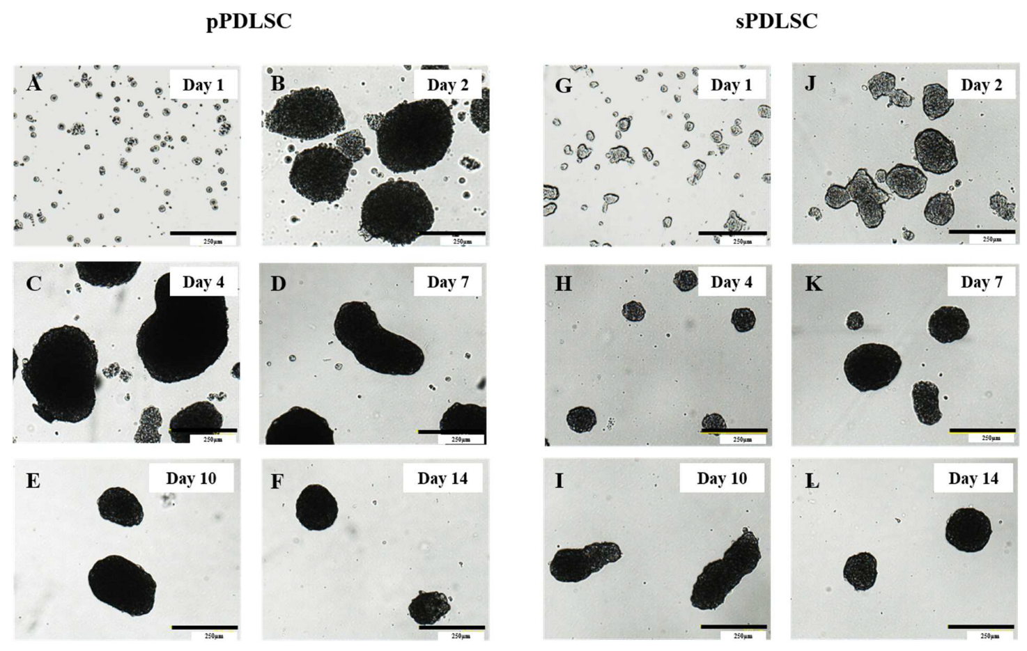
The 3D cultured cell spheroids of pPDLSCs in ULA were observed. Based on the microscopic images (Figure 1), the spheroid sizes increased until day 4. The largest cell size was observed on day 4. Cell-like substances that were scattered around the spheroids appeared on day 14 and were clearly observed over time.
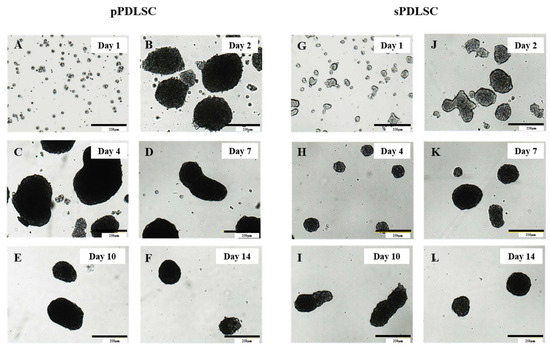
Figure 1.
Cell morphology and size. (A–F) Three-dimensional cultured cells of the permanent periodontal ligament stem cell in ULA on days 1, 2, 4, 7, 10, and 14. (G–L) Three-dimensional cultured cells of the supernumerary periodontal ligament stem cell in ULA on days 1, 2, 4, 7, 10, and 14. Scale bar: 250 μm.
The 3D cultured cell spheroids of sPDLSCs in ULA were observed. Based on the microscopic images, the spheroid sizes increased and became denser over time until day 7. From day 7, the cell size was maintained; however, the circular form showed irregularities on day 10. On day 10, hairy substances were observed around the spheroids.
The sPDLSCs were relatively smaller and more constant in size than pPDLSCs.
3.2. Cell Proliferation
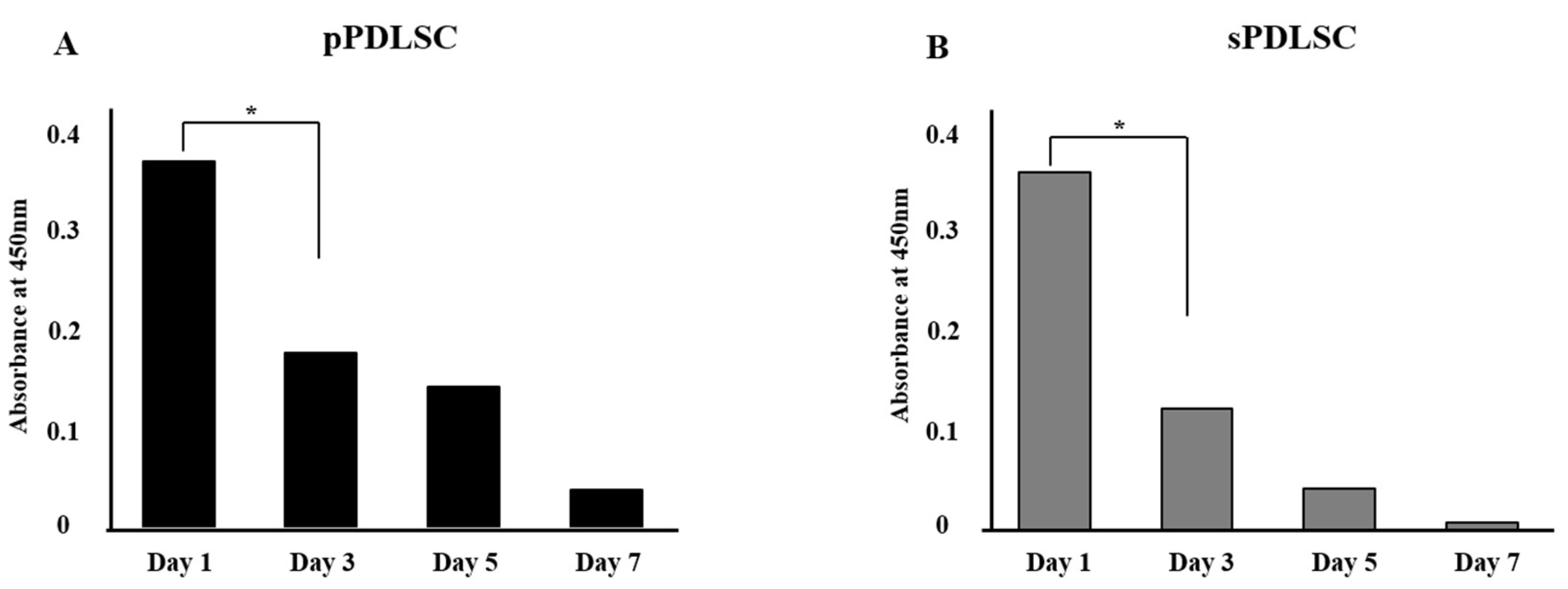
Based on the CCK-8 assay, the two groups displayed a decrease in proliferation. In particular, the proliferation of the pPDLSC group decreased from 100% on day 1 to 10.4% on day 7, while the sPDLSC group decreased from 100% on day 1 to 2.5% on day 7 (Figure 2). This result indicated that the culture time affected the absorbance. The pPDLSCs had a better viability than sPDLSCs each day.

Figure 2.
Cell proliferation analysis using cell counting kit-8 (CCK-8). (A) Results of CCK-8 assay of pPDLSC on 1, 3, 5, and 7 days. The proliferation of the pPDLSC decreased from 100% on day 1 to 10.4% on day 7. (B) Results of CCK-8 assay of sPDLSC on 1, 3, 5, and 7 days. The proliferation of the pPDLSC decreased from 100% on day 1 to 2.5% on day 7. * p < 0.05; Scheffé post hoc test following one-way ANOVA.
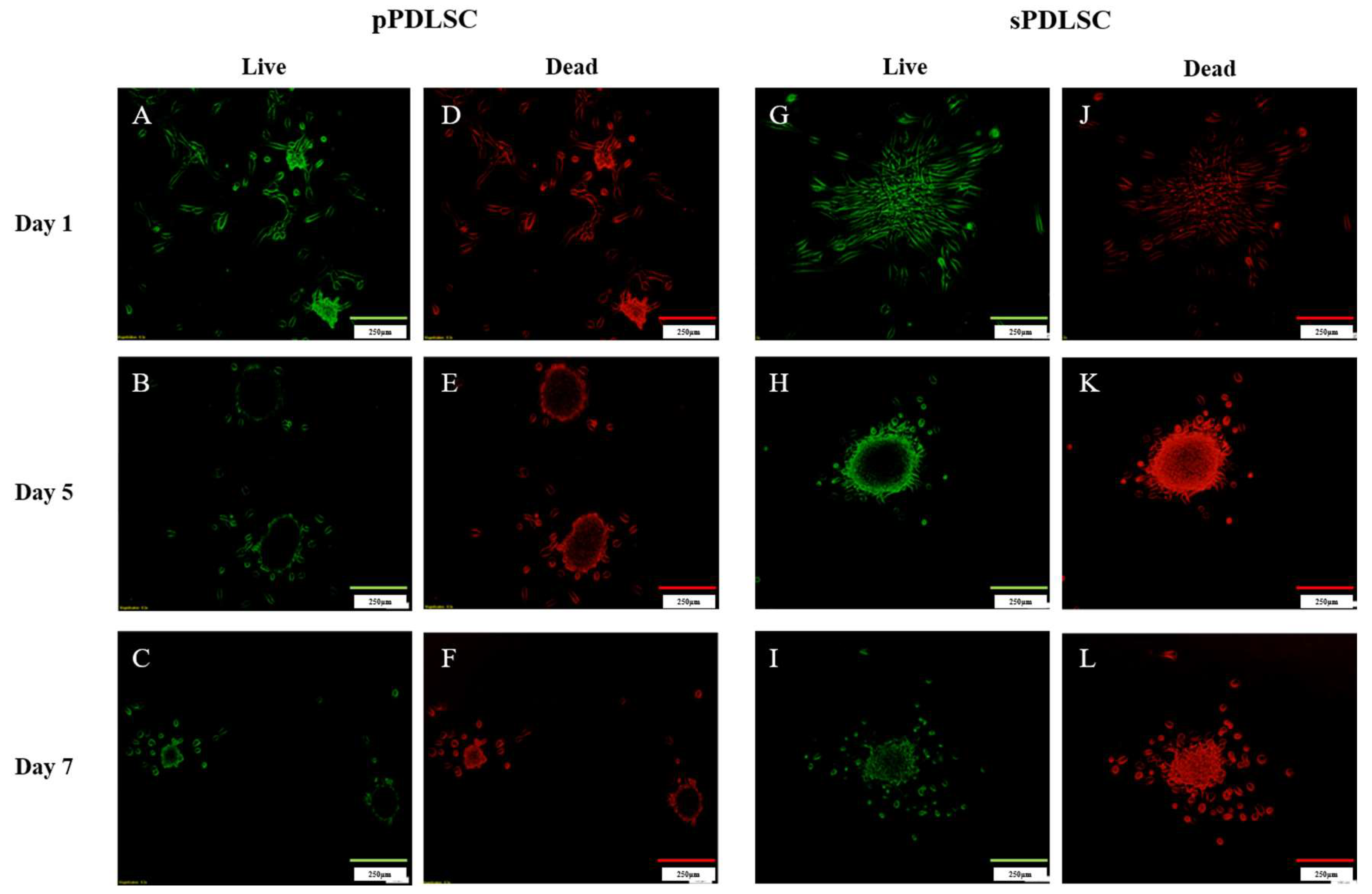
The live and dead pPDLSC assay results over 7 days are shown in Figure 3. pPDLSCs exhibited bright green fluorescence on day 1; however, this color faded on day 7. The number of dead cells increased in the center of the spheroids and appeared red on day 5. The green fluorescence exhibited by the cells and red blood cells was similar on day 7.
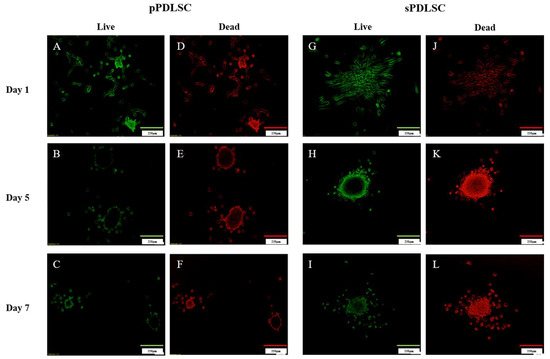
Figure 3.
Cell proliferation analysis using Live and Dead assay. (A–F) Live and dead assay results of pPDLSCs at days 1, 5, and 7. (G–L) Live and dead assay results of sPDLSCs at days 1, 5, and 7.
The live and dead sPDLSC assay results over 7 days are shown in Figure 3. sPDLSCs exhibited bright green fluorescence. Green fluorescence indicates the cytoplasm of live cells, whereas red fluorescence indicates the nuclei of dead cells. The number of dead cells increased in the center of the spheroids and appeared red on day 5. The green fluorescence of cells decreased and the number of red cells increased on day 7.
3.3. Cell Differentiation
3.3.1. Osteogenic Differentiation
ARS staining revealed significant mineralization, which is defined as extracellular calcium deposition during cell culture. Red staining was well observed in osteogenic differentiation media, unlike in basal media. Calcium was present in both sPDLSCs and pPDLSCs, and osteogenic differentiation was confirmed in both cell types (Figure 4A). The difference in osteogenic differentiation between pPDLSCs and sPDLSCs (p = 0.94) (Figure 4B).
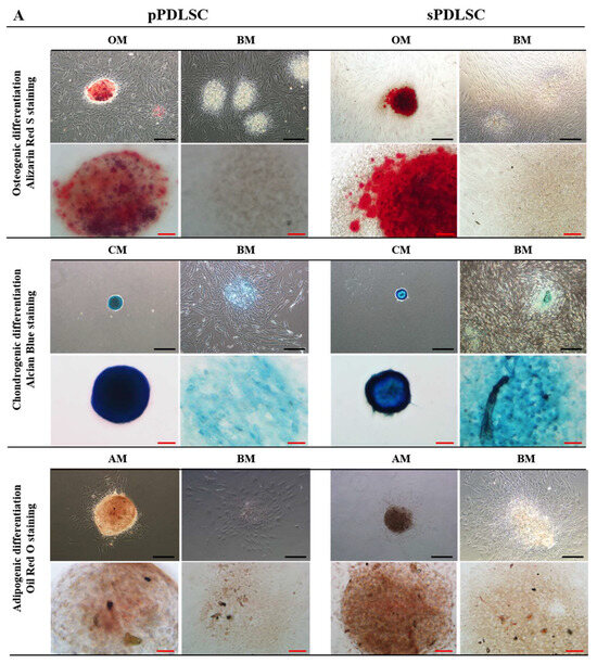
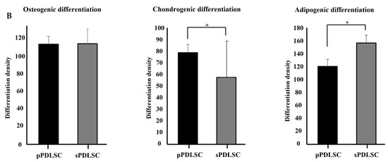
Figure 4.
(A) Evaluation of osteogenesis, chondrogenesis and adipogenesis in 3D culture of pPDLSC and sPDLSC. BM: Basal media, OM: Osteogenic media, CM: Chondrogenic media, AM: Adipogenic media. Black scale bar: 200 μm, Red scale bar: 40 μm (B) No significant difference in osteogenic differentiation between pPDLSCs and sPDLSCs (p = 0.94). Significant differences in adipogenic (p = 0.003) and chondrogenic (p = 0.013) differentiation between pPDLSCs and sPDLSCs. * p < 0.05; Mann–Whitney U test was performed.
3.3.2. Chondrogenic Differentiation
Both sPDLSCs and pPDLSCs had positive signals for AB staining, indicating chondrogenic differentiation. The pPDLSCs displayed typical characteristics of a glycosaminoglycan matrix when stained with AB at 16 days after induction. In addition, the sPDLSCs exhibited mineralization at 23 days after induction (Figure 4A). pPDLSCs showed a significantly higher chondrogenic differentiation than sPDLSCs (p = 0.003) (Figure 4B).
3.3.3. Adipogenic Differentiation
4. Discussion
In this study, similarities and differences were found between pPDLSCs and sPDLSCs grown in a 3D culture medium. In scaffold-free 3D media, pPDLSCs and sPDLSCs developed into spheroids. However, after one week, their proliferative ability was significantly decreased when compared to that observed after 24 h of culture. There was no significant difference between the two groups in terms of differentiation into bone tissue. pPDLSCs were more dominant in cartilage, while sPDLSCs were more dominant in fat.
According to Zhang et al. [26], factors related to the properties of PDLSCs are typically determined by tissue origin, donor age, culture methods, and growth factors.
The most commonly used tissue sources for PDLSCs include permanent teeth (3rd molar, extracted orthodontic tooth) [27], deciduous teeth [28], granulation tissue [29], and ST [30]. Among these teeth, PDLSCs obtained from ST may be useful candidates for use in regenerative medicine for several reasons. First, STs are defined as those in addition to the normal series of deciduous or permanent dentition and are an ethical material for stem cells as they are tissues that must be removed [31]. Second, the recovered state is useful for cryopreservation without contamination, such as dental caries and periodontitis. Third, STs have rich sources of stem cells, such as the dental pulp, periodontal ligament, apical papilla, and dental cysts, enabling their greater utility for biological use and potential therapeutic applications.
Many studies have identified the characteristics of PDLSCs; however, it is important to note that culturing methods also play a crucial role. The culture environment influences the growth rate of cells, their differentiation characteristics, and the direction of cell size [32]. In culture, the important factors include the composition and form of the culture medium, concentration of oxygen [33], and number of cells during culture [34]; however, recent reports have highlighted the critical role of 3D culture [35].
In a 3D culture setting, cells aggregate to form spheroids, which offer advantages in terms of cell–cell interactions, better replication in the in vivo microenvironment, and improved cell signaling pathways [36]. In our study, pPDLSCs and sPDLSCs also formed spheroid-like structures in the initial stage; however, as the number of cells increased, the spheroid shape became more irregular. The shape of 3D cell aggregates can change over time owing to various factors, such as cell proliferation, cell death, mechanical stress, and oxygen and nutrient gradients. As cells grow, the diffusion of oxygen and nutrition are limited, affecting the apoptotic cell cycle. Moreover, degradation products may influence the shape of the spheroids. According to Start et al. [36], the growing mass increases the mechanical stress, which is also a factor of irregularity.
The ULA plates used in this study showed different characteristics from those previously used in the 3D cell culture system, StemFit [37]. In our study, ULA plates served as a versatile tool for 3D cell culture by preventing cell attachment and facilitating spheroid formation. In contrast, StemFit, a tissue culture adhesive primarily designed for stem cell culture, which is currently not widely associated with dental research, could be relevant in dental stem cell applications or tissue engineering. The choice between ULA plates and StemFit depends on the specific research objectives and cell types employed, with ULA plates offering a broader versatility in dental 3D culture applications.
The proliferation rate of the 3D PDLSC aggregates declined over time. The CCK-8 assay results of a previous study [37] revealed an increase in cell proliferation over time in two-dimensional cultures and a decrease in 3D cultures. Our experimental results also revealed a decrease in cell proliferation in 3D cultures. In particular, the live/dead assay confirmed that the outer surface and center of the sPDLSCs were quickly filled with dead cells. A study on DPSCs revealed opposing results, as the proliferative ability of ST cells was reported to be significantly better than that of permanent teeth [38]. This result was observed in the culture system and ST. Scaffold-free 3D culture is more physiologically relevant with improved cell–cell interactions and is simpler to set up; however, owing to weak structural support for proliferation and differentiation, it results in limited scalability.
Recently, PDLSCs were demonstrated to have a high potential for multilineage differentiation into osteogenic, chondrogenic, and adipogenic cells [39]. PDLSCs close to the bone have a stronger capacity to differentiate into bone tissue. This process is induced by various growth factors and signaling molecules, such as bone morphogenetic proteins (BMPs), transforming growth factor-beta (TGF-β), and Wnt signaling [40]. Our finding revealed that sPDLSCs and pPDLSCs displayed similar osteogenic differentiation. Chondrogenic differentiation, which has a similar inducing growth factor as osteogenic differentiation (ex, TGF-β and BMPs), was superior to pPDLSC. In contrast, the adipogenic differentiation tendency was higher for ST than for permanent teeth. Similar reports by Lu et al. [38], who compared DPSC from ST and permanent teeth, revealed that osteogenic and chondrogenic differentiation were stronger in pDPSCs while adipogenic differentiation was superior in sDPSCs. Further studies on stem cells originating from the ST are needed.
This study had some limitations. First, due to the spontaneous spheroid formation technique used in this study, it was challenging to control the composition of the spheroids and their size and establish the right ratio of two different cell types in the spheroid for co-culture. Second, the assays were not optimized for cells cultured in a 3D environment. For example, the CCK-8 assay requires a media change, which increases the risk of loss of floating cells. Therefore, further research is necessary to establish a suitable method for analyzing 3D cultured cells. Third, this evaluation was a pilot study comparing 3D cultured sPDLSCs and pPDLSCs. The study also had an insufficient sample size; thus, additional studies are required to determine the genetic function using RT-PCR. In the future, it will be essential to determine the expression characteristics of the sPDLSCs. Despite these limitations, we confirmed that sPDLSCs could be an alternative source of pPDLSCs.
5. Conclusions
In this study, we aimed to reveal the characteristics of 3D cultured pPDLSCs and sPDLSCs in a 3D culture setting. Our findings confirm that sPDLSCs and pPDLSCs possess multilineage differentiation capacities, including osteogenic, chondrogenic, and adipogenic differentiation. ST, which are extracted and discarded, may be a more ethically feasible source for clinical use than permanent teeth. Thus, ST could serve as one of the future sources of stem cells. Therefore, a further study of sPDLSCs is required for clinical applications as their use can be applied in tissue engineering and regenerative medicine.
Author Contributions
Conceptualization, H.-S.L.; methodology, K.E.L.; validation, H.K., H.-S.L. and K.E.L.; formal analysis, H.K.; investigation, O.H.N.; resources, S.C.C.; data curation, H.K. and Y.K.C.; writing—original draft preparation, H.K. and K.E.L.; writing—review and editing, Y.K.C., M.S.K., O.H.N., J.-H.J. and S.C.C.; visualization, J.-H.J.; supervision, H.-S.L.; project administration, H.-S.L.; funding acquisition, H.-S.L. and J.-H.J. All authors have read and agreed to the published version of the manuscript.
Funding
This research was supported by the Bio and Medical Technology Development Program of the National Research Foundation (NRF) and funded by the Korean government (MSIP and MOHW) NRF (No.2022R1C1C1007851).
Institutional Review Board Statement
Not applicable.
Informed Consent Statement
Not applicable.
Data Availability Statement
The data presented in this study are openly available.
Conflicts of Interest
The authors declare no conflict of interest.
References
- Gronthos, S.; Mankani, M.; Brahim, J.; Robey, P.G.; Shi, S. Postnatal human dental pulp stem cells (dpscs) in vitro and in vivo. Proc. Natl. Acad. Sci. USA 2000, 97, 13625–13630. [Google Scholar] [CrossRef]
- Smojver, I.; Katalinić, I.; Bjelica, R.; Gabrić, D.; Matišić, V.; Molnar, V.; Primorac, D. Mesenchymal stem cells based treatment in dental medicine: A narrative review. Int. J. Mol. Sci. 2022, 23, 1662. [Google Scholar] [CrossRef]
- Chen, F.-M.; Jin, Y. Periodontal tissue engineering and regeneration: Current approaches and expanding opportunities. Tissue Eng. Part B Rev. 2010, 16, 219–255. [Google Scholar] [CrossRef]
- Wada, N.; Menicanin, D.; Shi, S.; Bartold, P.M.; Gronthos, S. Immunomodulatory properties of human periodontal ligament stem cells. J. Cell. Physiol. 2009, 219, 667–676. [Google Scholar] [CrossRef]
- Omer, R.S.; Anthonappa, R.P.; King, N.M. Determination of the optimum time for surgical removal of unerupted anterior supernumerary teeth. Pediatr. Dent. 2010, 32, 14–20. [Google Scholar]
- Mallineni, S.K. Supernumerary Teeth: Review of the Literature with Recent Updates; Conference Papers in Science; Hindawi: London, UK, 2014. [Google Scholar]
- Anthonappa, R.; King, N.; Rabie, A. Aetiology of supernumerary teeth: A literature review. Eur. Arch. Paediatr. Dent. 2013, 14, 279–288. [Google Scholar] [CrossRef]
- Duval, K.; Grover, H.; Han, L.H.; Mou, Y.; Pegoraro, A.F.; Fredberg, J.; Chen, Z. Modeling physiological events in 2d vs. 3d cell culture. Physiology 2017, 32, 266–277. [Google Scholar] [CrossRef]
- Habanjar, O.; Diab-Assaf, M.; Caldefie-Chezet, F.; Delort, L. 3d cell culture systems: Tumor application, advantages, and disadvantages. Int. J. Mol. Sci. 2021, 22, 12200. [Google Scholar] [CrossRef]
- Ravi, M.; Paramesh, V.; Kaviya, S.R.; Anuradha, E.; Solomon, F.D. 3d cell culture systems: Advantages and applications. J. Cell. Physiol. 2015, 230, 16–26. [Google Scholar] [CrossRef]
- Wang, H.; Brown, P.C.; Chow, E.C.Y.; Ewart, L.; Ferguson, S.S.; Fitzpatrick, S.; Freedman, B.S.; Guo, G.L.; Hedrich, W.; Heyward, S.; et al. 3d cell culture models: Drug pharmacokinetics, safety assessment, and regulatory consideration. Clin. Transl. Sci. 2021, 14, 1659–1680. [Google Scholar] [CrossRef]
- Tanaka, H. Modeling and analysis of disease microenvironments with 3d cell culture technology. Yakugaku Zasshi 2021, 141, 647–653. [Google Scholar] [CrossRef]
- Itoh, Y.; Sasaki, J.I.; Hashimoto, M.; Katata, C.; Hayashi, M.; Imazato, S. Pulp regeneration by 3-dimensional dental pulp stem cell constructs. J. Dent. Res. 2018, 97, 1137–1143. [Google Scholar] [CrossRef]
- Visakan, G.; Su, J.; Moradian-Oldak, J. Ameloblastin promotes polarization of ameloblast cell lines in a 3-d cell culture system. Matrix Biol. 2022, 105, 72–86. [Google Scholar] [CrossRef]
- Basso, F.G.; Soares, D.G.; Pansani, T.N.; Cardoso, L.M.; Scheffel, D.L.; de Souza Costa, C.A.; Hebling, J. Proliferation, migration, and expression of oral-mucosal-healing-related genes by oral fibroblasts receiving low-level laser therapy after inflammatory cytokines challenge. Lasers Surg. Med. 2016, 48, 1006–1014. [Google Scholar] [CrossRef]
- Brezulier, D.; Pellen-Mussi, P.; Tricot-Doleux, S.; Novella, A.; Sorel, O.; Jeanne, S. Development of a 3d human osteoblast cell culture model for studying mechanobiology in orthodontics. Eur. J. Orthod. 2020, 42, 387–395. [Google Scholar] [CrossRef]
- Zhao, H.; Jiang, E.; Shang, Z. 3d co-culture of cancer-associated fibroblast with oral cancer organoids. J. Dent. Res. 2021, 100, 201–208. [Google Scholar] [CrossRef]
- Shehzad, A.; Ravinayagam, V.; AlRumaih, H.; Aljafary, M.; Almohazey, D.; Almofty, S.; Al-Rashid, N.A.; Al-Suhaimi, E.A. Application of three-dimensional (3d) tumor cell culture systems and mechanism of drug resistance. Curr. Pharm. Des. 2019, 25, 3599–3607. [Google Scholar] [CrossRef]
- Mountcastle, S.E.; Cox, S.C.; Sammons, R.L.; Jabbari, S.; Shelton, R.M.; Kuehne, S.A. A review of co-culture models to study the oral microenvironment and disease. J. Oral Microbiol. 2020, 12, 1773122. [Google Scholar] [CrossRef]
- Charbonneau, A.M.; Tran, S.D. 3d cell culture of human salivary glands using nature-inspired functional biomaterials: The egg yolk plasma and egg white. Materials 2020, 13, 4807. [Google Scholar] [CrossRef]
- Baena, A.R.; Casasco, A.; Monti, M. Hypes and hopes of stem cell therapies in dentistry: A review. Stem. Cell. Rev. Rep. 2022, 18, 1294–1308. [Google Scholar] [CrossRef]
- Ryu, N.-E.; Lee, S.-H.; Park, H. Spheroid culture system methods and applications for mesenchymal stem cells. Cells 2019, 8, 1620. [Google Scholar] [CrossRef]
- Klotz, B.J.; Gawlitta, D.; Rosenberg, A.J.; Malda, J.; Melchels, F.P. Gelatin-methacryloyl hydrogels: Towards biofabrication-based tissue repair. Trends Biotechnol. 2016, 34, 394–407. [Google Scholar] [CrossRef]
- Malhão, F.; Macedo, A.C.; Ramos, A.A.; Rocha, E. Morphometrical, morphological, and immunocytochemical characterization of a tool for cytotoxicity research: 3d cultures of breast cell lines grown in ultra-low attachment plates. Toxics 2022, 10, 415. [Google Scholar] [CrossRef]
- Luo, L.; Zhang, W.; Wang, J.; Zhao, M.; Shen, K.; Jia, Y.; Li, Y.; Zhang, J.; Cai, W.; Xiao, D. A novel 3d culture model of human ascs reduces cell death in spheroid cores and maintains inner cell proliferation compared with a nonadherent 3d culture. Front. Cell Dev. Biol. 2021, 9, 737275. [Google Scholar] [CrossRef]
- Zhu, W.; Liang, M. Periodontal ligament stem cells: Current status, concerns, and future prospects. Stem Cells Int. 2015, 2015, 972313. [Google Scholar] [CrossRef]
- Ji, K.; Liu, Y.; Lu, W.; Yang, F.; Yu, J.; Wang, X.; Ma, Q.; Yang, Z.; Wen, L.; Xuan, K. Periodontal tissue engineering with stem cells from the periodontal ligament of human retained deciduous teeth. J. Periodontal. Res. 2013, 48, 105–116. [Google Scholar] [CrossRef]
- Silvério, K.G.; Rodrigues, T.L.; Coletta, R.D.; Benevides, L.; Da Silva, J.S.; Casati, M.Z.; Sallum, E.A.; Nociti, F.H., Jr. Mesenchymal stem cell properties of periodontal ligament cells from deciduous and permanent teeth. J. Periodontol. 2010, 81, 1207–1215. [Google Scholar] [CrossRef]
- Ronay, V.; Belibasakis, G.N.; Attin, T.; Schmidlin, P.R.; Bostanci, N. Expression of embryonic stem cell markers and osteogenic differentiation potential in cells derived from periodontal granulation tissue. Cell Biol. Int. 2014, 38, 179–186. [Google Scholar] [CrossRef]
- Park, J.H.; Song, J.S.; Kim, S.; Kim, S.O.; Choi, B.J.; Park, K.H.; Jung, H.S.; Lee, J.H. Characteristics of stem cells derived from the periodontal ligament of supernumerary teeth. Tissue Eng. Regen. Med. 2011, 8, 123–131. [Google Scholar]
- Garvey, M.T.; Barry, H.J.; Blake, M. Supernumerary teeth--an overview of classification, diagnosis and management. J. Can. Dent. Assoc. 1999, 65, 612–616. [Google Scholar]
- Agata, H.; Kagami, H.; Watanabe, N.; Ueda, M. Effect of ischemic culture conditions on the survival and differentiation of porcine dental pulp-derived cells. Differentiation 2008, 76, 981–993. [Google Scholar] [CrossRef]
- Mohyeldin, A.; Garzón-Muvdi, T.; Quiñones-Hinojosa, A. Oxygen in stem cell biology: A critical component of the stem cell niche. Cell Stem Cell 2010, 7, 150–161. [Google Scholar] [CrossRef]
- Feng, F.; Akiyama, K.; Liu, Y.; Yamaza, T.; Wang, T.M.; Chen, J.H.; Wang, B.B.; Huang, G.T.; Wang, S.; Shi, S. Utility of pdl progenitors for in vivo tissue regeneration: A report of 3 cases. Oral Dis. 2010, 16, 20–28. [Google Scholar] [CrossRef]
- Yu, H.; Zhang, X.; Song, W.; Pan, T.; Wang, H.; Ning, T.; Wei, Q.; Xu, H.H.K.; Wu, B.; Ma, D. Effects of 3-dimensional bioprinting alginate/gelatin hydrogel scaffold extract on proliferation and differentiation of human dental pulp stem cells. J. Endod. 2019, 45, 706–715. [Google Scholar] [CrossRef]
- Sart, S.; Tsai, A.C.; Li, Y.; Ma, T. Three-dimensional aggregates of mesenchymal stem cells: Cellular mechanisms, biological properties, and applications. Tissue Eng. Part B Rev. 2014, 20, 365–380. [Google Scholar] [CrossRef]
- Jeong, Y.Y.; Kim, M.S.; Lee, K.E.; Nam, O.H.; Jang, J.-H.; Choi, S.-C.; Lee, H.-S. In vitro characterization of periodontal ligament stem cells derived from supernumerary teeth in three-dimensional culture method. Appl. Sci. 2021, 11, 6040. [Google Scholar] [CrossRef]
- Lu, X.; Liu, S.F.; Wang, H.H.; Yu, F.; Liu, J.J.; Zhao, Y.M.; Zhao, S.L. A biological study of supernumerary teeth derived dental pulp stem cells based on rna-seq analysis. Int. Endod. J. 2019, 52, 819–828. [Google Scholar] [CrossRef]
- Coura, G.; Garcez, R.; De Aguiar, C.M.; Alvarez-Silva, M.; Magini, R.; Trentin, A. Human periodontal ligament: A niche of neural crest stem cells. J. Periodontal Res. 2008, 43, 531–536. [Google Scholar] [CrossRef]
- Hakki, S.S.; Bozkurt, B.; Hakki, E.E.; Kayis, S.A.; Turac, G.; Yilmaz, I.; Karaoz, E. Bone morphogenetic protein-2,-6, and-7 differently regulate osteogenic differentiation of human periodontal ligament stem cells. J. Biomed. Mater. Res. Part B Appl. Biomater. 2014, 102, 119–130. [Google Scholar] [CrossRef]


Disclaimer/Publisher’s Note: The statements, opinions and data contained in all publications are solely those of the individual author(s) and contributor(s) and not of MDPI and/or the editor(s). MDPI and/or the editor(s) disclaim responsibility for any injury to people or property resulting from any ideas, methods, instructions or products referred to in the content. |
© 2023 by the authors. Licensee MDPI, Basel, Switzerland. This article is an open access article distributed under the terms and conditions of the Creative Commons Attribution (CC BY) license (https://creativecommons.org/licenses/by/4.0/).