Article

Antioxidant Properties of Selenium Nanoparticles Synthesized Using Tea and Herb Water Extracts

by
Aleksandra Sentkowska
1,* and
Krystyna Pyrzyńska
2
1
Heavy Ion Laboratory, University of Warsaw, Pasteura 5A, 02-093 Warsaw, Poland
2
Department of Chemistry, University of Warsaw, Pasteura 1, 02-093 Warsaw, Poland
*
Author to whom correspondence should be addressed.
Appl. Sci. 2023, 13(2), 1071; https://doi.org/10.3390/app13021071
Submission received: 12 December 2022 / Revised: 11 January 2023 / Accepted: 11 January 2023 / Published: 13 January 2023
(This article belongs to the Special Issue New Insights into Natural Antioxidants in Foods)

Abstract

:

Featured Application

Practical tips that can be used in the green synthesis of SeNPs.

Abstract

Selenium nanoparticles (SeNPs) are the object of great interest due to their potential to be used in many areas of industry and medicine. Work is still underway on their synthesis methods; however, green methodologies are becoming more and more popular. In this study, aqueous extracts of black and green tea as well as infusions of popular herbs such as chamomile and mint were used in the synthesis of SeNPs serving as the Se(IV) reducers to its nano form. The influence of the reagents concentration ratios on the properties of the obtained nanoparticles were examined. SeNPs showed a greater ability to neutralize hydroxyl radicals compared to tea extracts. It has been proven that in the infusion of selenium-containing tea, the formation of SeNPs occurs.

1. Introduction

Selenium, which is an essential element for human health, plays a key role in several major metabolic pathways such as antioxidant defense systems, thyroid hormone metabolism, DNA synthesis, and protection from oxidative damage and infection [1,2,3,4]. As selenium deficiency remains a problem in many human populations, Se-enriched foods and supplements have been proposed to circumvent this problem [5,6,7]. In dietary supplementation, the inorganic forms of selenium (selenite and selenate) or selenomethionine are mainly present. However, various selenium species have different chemical properties, environmental effects, biological utilizations, toxicities, and nutrition values. Selenium organic compounds are less toxic and more bioavailable than inorganic forms [8]. Insufficient supplementation of this element results in an increased risk of developing many chronic degenerative diseases [2,7].
In recent years, selenium nanoparticles (SeNPs) have attracted great attention due to their higher bioactivity and lower toxicity in comparison to inorganic and organic selenium species [8]. They offer great potential in various biomedical applications and food science [9,10,11,12,13,14,15]. SeNPs have important roles in scavenging free radicals such as reactive oxygen and nitrogen species, regulating their content produced during biochemical reactions, and protecting cells from damage and oxidative stress [11]. Their antibacterial and antifungal activity can be used as antimicrobial agents [12]. Several reports have also confirmed the anticancer effect of SeNPs [13,14]. SeNPs could be used in the drug delivery system due to easy manipulation in their size and low toxicity as they are taken up by cells more efficiently than larger biomolecules [15].
Selenium nanoparticles can be synthesized by physical, chemical, and biological methods [16]. However, the biologically synthesized SeNPs demonstrate greater compatibility with human organs and tissues and do not require chemical reagents which may be toxic and can hinder their utilization in biological systems [17,18]. This green synthesis approach via plant extract involves many secondary metabolites such as flavonoids, alkaloids, saponins, carbohydrates, proteins, tannins, and steroids as natural reducers and/or stabilizers. The mechanism of their action in the synthesis of NPs is complex. According to Marslin [19], when selenium salt dissociates, cations are saturated to form hydroxyl complexes. After that, crystallite growth of metal with oxygen species starts to originate. At this stage of the NPs synthesis, heating is playing a key role in providing energy to the reaction system. The process continues until activation of the capping agent from the plant extract, which eventually stops the growth of high-energy atomic growth planes. The result of this process is formation of NPs. Reducing agents from plant extract are electron donors to the metals ions and convert them to the nanoparticles. From this point of view, secondary plant metabolites are very important in green SeNPs synthesis; they act as selenium reductors, but also prevent the aggregation of NPs and promotes production of smaller particles. The use of green synthesis was used to obtain many metallic nanoparticles, including AgNPs [20], ZnO NPs [21,22], or PdNPs [23].
Green synthesis of selenium nanoparticles is also described in the literature. Generally, selenium precursor (most often Na2SeO3) is mixed with the plant extract solution at different ratios and stirred for different periods, sometimes with heating. During this process, the colour of the reaction media changes to red, indicating the formation of Se nanoparticles. In the synthesis of SeNPs, the main goal is their formation with minimum particle size and maximum stability as a smaller size can guarantee more powerful antioxidant activities [24].
In the present study, the potential of water extracts of popular black (Lipton Yellow label) and green tea (Lipton Green Tea Classic) as well as chamomile (Matricaria recutita) and mint (Mentha), commonly used to make herbal infusions, were compared in the biosynthesis of SeNPs. The dependence of the ratio of reagents on the properties of the obtained SeNPs has been investigated. The content of polyphenolic compounds, which can potentially act as selenium reducing reagents, was measured using liquid chromatography operating in HILIC mode. The antioxidant activity of extracts themselves, as well as obtained nanoparticles, were determined using a hydroxyl radical scavenging assay. The reductive properties were evaluated by the Folin–Ciocalteu method. An attempt has been made to link the properties of the obtained SeNPs with the ratios of the reagents used for the synthesis.

2. Materials and Methods

2.1. Reagents

Sodium selenite (Na2SeO3) used as selenium precursor in the synthesis of SeNPs was purchased from Merck-Sigma (Steinheim, Germany). All of the polyphenolics standards used during calibration curves development in HPLC analysis, acetonitrile (ACN), and methanol (MeOH) used as eluents in HPLC analysis were also by Merck-Sigma production. All of the reagents used for antioxidant activity measurements were obtained from the same producer, Merck-Sigma. During the experiments, water obtained from Milli-Q system (Millipore, Bedford, MA, USA) was used.

2.2. Tea, Herbal Samples, and Supplement

Black and green tea used in the experiment were purchased in the local grocery store. Both were produced by Lipton (Unilever, UK). Chamomile and mint teas were purchased from Polish producer Herbapol (Lublin, Poland) and came from the Zielnik Polski line. All teas were brewed in the same way. A single tea bag (~2 g) was poured with 200 mL of boiling water and brewed for 10 min. The bag was then removed and the extract after filtration was used for further research. The concentration of obtained extract was 0.01 mg L−1.
Zhejiang tea was purchased from Manufaktura herbaty (Piła, Polska).
The dietary supplement used in the research is commercially offered by Aliness (Karczew, Poland). According to the label, it contains 100 µg of Na2SeO3 per tablet (weight of one tablet~313 mg). The extraction procedure consisted of grinding the tablet in a mortar, quantitative transfer to a volumetric flask, and adding 50 mL of boiling water. The sample was stirred for an hour on a magnetic stirrer, then filtered and used for the experiment.

2.3. Chromatographic Analysis of Polyphenols in the Studied Infusions

Chromatographic analysis of polyphenolic compounds in studied infusions was performed using a Shimadzu LC system coupled to an 8030 triple quadrupole mass spectrometer (Kyoto, Japan). The chromatographic separation was performed in HILIC mode, using ZIC-HILIC column (100 × 2.2 mm, 3 µm) from Merck. The mobile phase was delivered at 0.2 mL min−1 in gradient mode: 0–4 min 98% B, 6–7 min 90% B, 8–8.4 min 80% B, 8.4–12 min 50% B, and 13–20 min 98% B, where B is acetonitrile and A- water. All of the compounds were identified based on the knowledge of their fragmentation patterns and retention times, as we described earlier [25]. Qualitative and quantitative determination of polyphenolic compounds was carried out immediately after brewing and after SeNPs synthesis in order to compare the concentrations of these compounds before and after the synthesis of SeNPs.

2.4. The Synthesis and Characterization of Obtained SeNPs

The green synthesis of selenium nanoparticles was based on the reduction of sodium selenite with tea or herbal extract. Briefly, 2.5 mL of selenium solution (0.1 mol L−1) was placed on the magnetic stirrer and 15 mL of deionized water was added. After that, 2.5 mL of tea infusion (Se/infusion ratio 1:1) or 5 mL (Se/infusion ratio 1:2) or 7.5 mL (Se/infusion ratio 1:3) or 10 mL (Se/infusion ratio 1:4) was added dropwise. The concentration of plant extract in the reaction mixture was: 1.25 × 10⁻³ (1:1), 2.50 × 10⁻³ (1:2), 3.75 × 10⁻³ (1:3), and 5.00 × 10⁻³ (1:4). The reaction mixture was intensively stirred for 60 min. The analogical reaction was also performed with the excess of selenium; in that case, the volumes were reversed.
The obtained nanoparticles were so strongly stabilized that it was impossible to isolate them from the post-reaction mixture using e.g., centrifugation. This problem concerned tea extracts. In the case of herbal extracts, centrifugation with the intensity of 12,000 rpm enabled their isolation; however, due to the difficulties with repeating the procedure for teas, the separation of SeNPs in post-reaction mixtures was abandoned.
The UV–Vis absorption spectra in the range of 250–900 nm were recorded after the synthesis to predict the sizes of obtained SeNPs. This was conducted using Perkin Elmer spectrophotometer (model Lambda 20) with cuvettes of 1 cm in length. In order to avoid interference related to the color of the brew used for synthesis, the spectra were recorded using the appropriate brew as a blank. The infusion used had the same concentration as that used for the given synthesis. For measurements, the sample was diluted 50 times.
The size of the obtained nanoparticles was also investigated by dynamic light scattering (DLS). For this purpose, Mastersizer 2000 (Malvern Panalytical, UK) was equipped with the wet samples dispersion unit (Hydro 2000 MU). The device was controlled by standard operating procedure, which is a part of Malvern SOP software. The apparatus allows the measurement of particle sizes larger than 0.01 um, which can be valuable support for the measurement of synthesized nanoparticles if they are larger than 10 nm.

2.5. Antioxidant Activity Measurements

The ability to neutralize hydroxyl radicals by both tea infusions and selenium nanoparticles was performed according to the procedure described by Smirnoff and Cumbes [26]. The analytical performance was as followed: 1 mL of tea extract or SeNPs solution was mixed with the reaction mixture, which consisted of 1 mL iron sulfate (1.5 × 10⁻³ mol L−1), 0.7 mL hydrogen peroxide (6 × 10⁻³ mol L−1), and 0.3 mL sodium salicylate (2 × 10⁻² mol L−1). The sample was incubated for one hour at 37 °C and then the absorbance was measured at 562 nm. Obtained results are expressed as the percentage of hydroxyl radical scavenging.
The reducing power of the studied tea infusions and SeNPs was evaluated by Folin–Ciocalteu (FC) assay. We mixed 1 mL of the sample with 0.1 mL of FC reagent and 0.9 mL of water. After 5 min, 1 mL of 7% solution of Na2CO3 and 0.4 mL of water were added. After 10 min, the absorbance was measured at 765 nm. The results were expressed as gallic acid (GA) equivalent.

2.6. Statistical Analysis

The experimental results from antioxidant activity measurements as well as HPLC analysis were obtained from at least three parallel measurements and are presented as average ± SD. The significance of differences among means was carried out at a 5% probability level using one-way ANOVA and Tukey’s test.

3. Results and Discussion

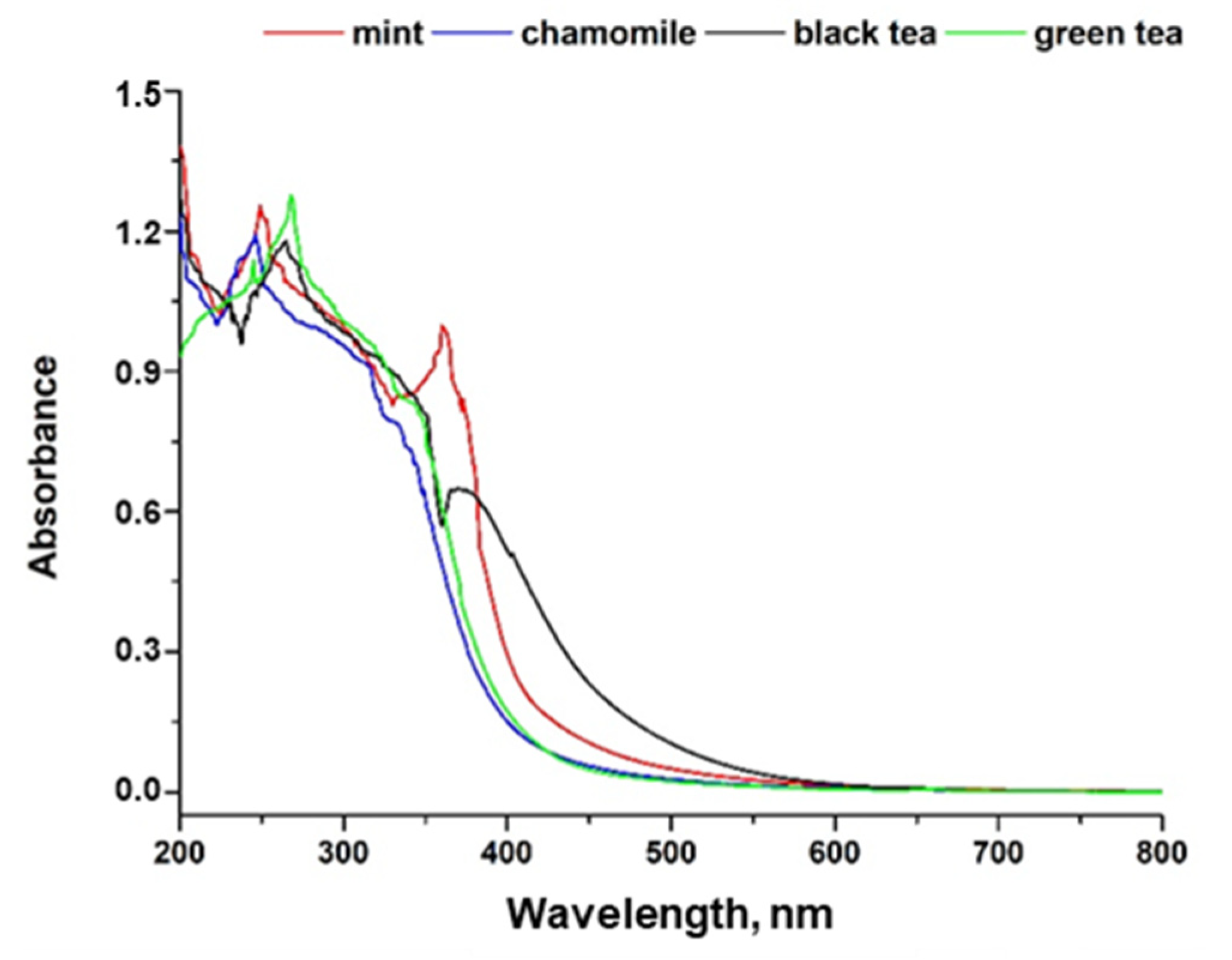
3.1. HPLC Analysis ad UV–Vis Spectroscopy of Used Plant Extracts

Chromatographic analysis of polyphenols and ascorbic acid in all tested infusions was performed in HILIC mode and its results are presented in Table 1. Ascorbic acid was included in the research because it is often used as a selenium reducer in the chemical synthesis of SeNPs [18]. Its presence in extracts can significantly affect the course of the synthesis reaction, as well as the properties of the obtained selenium nanoparticles.
Catechins (flavan-3-ols) are the dominant polyphenolic compounds present in tea leaves and their infusions. These compounds were detected in high amounts in both green and black tea. However, green tea infusion seems to be a better source of catechins. According to the literature, total catechins decreases in amount with increasing fermentation from green to oolong and black tea, which can also be seen in our results [27]. The levels of epigallocatechin-3-gallate (EGCG) ranged from 1.69 to 90.1 mg L−1, making it the major polyphenolic constituent of green tea. Antioxidant and health-promoting effects of EGCG have been widely described in the literature. It was reported that EGCG acts as an angiogenesis inhibitor by modulating protease activity during endothelial morphogenesis and thus inhibits the growth of cancer [28]. It should be mentioned that, in addition to the catechins designated by us, tea infusions also contain other compounds from this group, e.g., epigallocatechin, epicatechin gallate, gallocatechin, and gallocatechin gallate. MS spectra confirmed their presence in the infusions, but their quantitative analysis was not performed.
In general, the content of polyphenols in tea leaves varies with climate, season, horticultural practices, and leaf age [29]. Rutin (quercetin-3-rutinoside) was found in the studied tea extracts at the highest level, but its concentration in herbal infusions was below the limit of detection. On the other hand, luteolin was detected only in chamomile and mint infusions. The presence of luteolin, rutin, and chlorogenic acid in chamomile infusion was also reported in our previous work [30]. Mint was described in the literature as a good source of luteolin and caffeic acid, which was also confirmed by our results [31]. All of the studied extracts were good sources of polyphenolic acids. p-Hydroxybenzoic acid (pHBA) was present in all studied infusions in high concentrations and caffeic acid was detected only in herbal infusions. In plants, caffeic acid is formed from p-coumaric acid and then transformed into ferulic acid [32]. In the case of our infusions, p-coumaric acid was present in all samples, with a significantly higher concentration in teas. The concentrations of ferulic acid in all studied samples were below the limit of detection.
The high quantity of polyphenolic compounds is crucial from the point of view of selenium nanoparticles synthesis, as they are responsible for the reduction of selenium salt and also can stabilize obtained SeNPs. However, the presence of other compounds in plant extracts can affect the efficiency of the nanoparticle synthesis. Ascorbic acid is an example of such a compound due to its reductive properties. Its concentration in all studied infusions was increasing in the order: mint < chamomile < green tea < black tea (Table 1).
The absorbance spectra of studied infusions were recorded in the range of 200–850 nm, which is shown in Figure 1. It has been proven that the analyzed herbal extracts contain similar compounds as it was observed that spectra curve over the same wavelength range [33]. According to the literature, mint and chamomile extracts are characterized by a high content of terpenes, terpenoids, and phenolic compounds [34,35]. This was confirmed by UV–Vis spectra, namely by the peak registered at 270 nm and as a band for 290–320 nm [36]. Characteristic for plants, chlorophyll content was also observed as an extensive peak in the range of 360 to 450 nm [37]. An intensive band recorded in the range 380–450 nm, which was the most intensive for mint and black tea infusion, can be associated with the presence of carotenoids [38].
UV–Vis spectra can also be the basis for distinguishing black tea from green tea. Catechins and methylxanthines, which show absorbance in the region from 250 to 350 nm (related to the η→π electronic transition) can be chemical descriptors to distinguish these two tea types [39]. Other groups of compounds which can be useful in the interpretation of tea spectra are theaflavins and thearubigins, typical for black tea as a result of fermentation processes. Their presence in the infusion can be examined at 380 and 460 nm [40]. As can be seen on the recorded spectra, green tea infusion shows higher absorbance in the region from 250 to 350 nm than black tea due to the higher content of the catechins, which was found in green tea. On the other hand, the band characteristic for theaflavins and thearubigins is significantly higher for black tea, as was expected. Additionally, green tea infusion presents the lowest absorbance in the region 450–600 nm, which was also reported earlier [41]. The obtained spectra confirmed that the analyzed extracts come from two different types of tea, which should translate into their polyphenolics content. This is very important from the point of view of their application to the green synthesis of SeNPs. As they act simultaneously as selenium reducers and stabilizers of the obtained nanoparticles, their concentration can affect the physical properties of the nanoparticles, such as size and shape, but also potentially their stability.

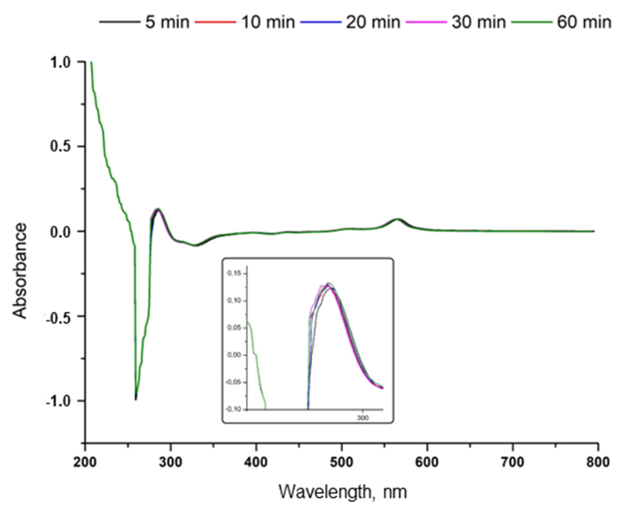
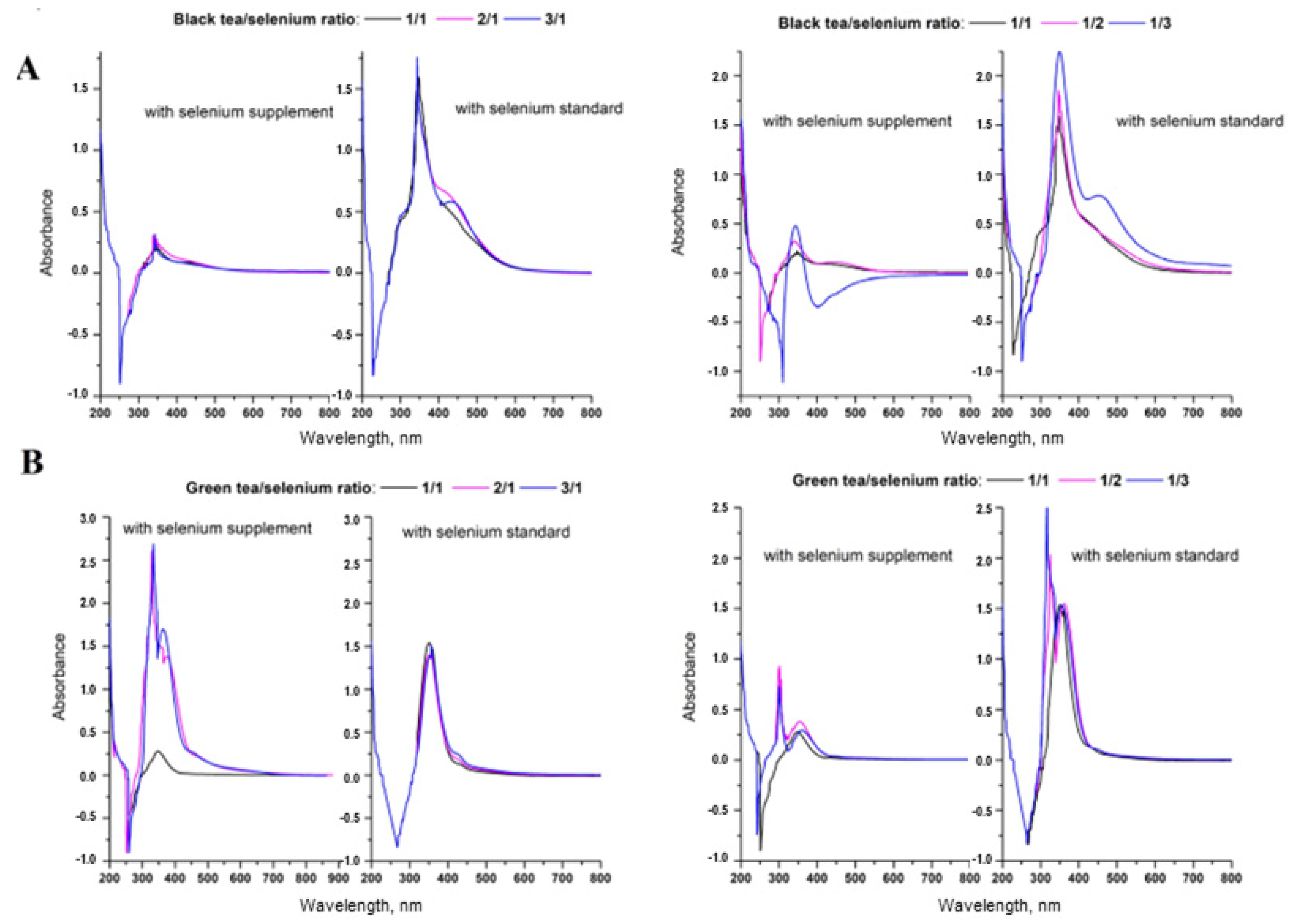
3.2. Synthesis and Characterization of SeNPs

The synthesis of SeNPs was conducted parallel by the reduction of sodium selenite with tea or herbal infusion. The synthesis was carried out in two ways: with an increasing concentration of the plant extract with a constant concentration of selenium salts, and in the second variant with an increasing concentration of selenium salts and a constant concentration of the infusion. The recorded UV–Vis spectra of obtained nanoparticles are shown in Figure 2. The presented spectra are similar to those described by us earlier [42]. We also observed a decrease in absorbance at the wavelength of 330 nm. It should be noted that the spectra were recorded using a tea infusion as a blank. The infusion was of the same concentration as that in the reaction mixture. Absorbance less than zero may suggest that the blank absorbs more light than the sample; in other words, the blank has a more intense colour than the sample. In the case of the study of the post-reaction mixture, this is understandable, as the compounds determining the color of the tea may be consumed during the synthesis of SeNPs. The main pigments in fresh tea leaves are chlorophyll and carotenoids. These pigments may condense as they wither and oxidize, becoming darker. During oxidation, the green color of tea’s chlorophyll turns black. This leads to the dark color of oxidized teas. The negative absorbance may be evidence that these two groups of compounds are also involved in the reduction of selenium salts to selenium in the form of nanoparticles.
It has been proven that UV–Vis spectra of nanoparticles can provide valuable information regarding their size [38]. The basis of this method is the fact that with the increase in the size of nanoparticles, the colour of their suspension changes from yellow (for SeNPs with dimensions of about 20 nm) to red (size of about 100 nm). This entails a shift of the absorption maximum in the UV–Vis spectrum towards higher wavelengths. UV–Vis spectra recorded for SeNPs suspensions obtained by synthesis using chamomile infusion show a maximum at a wavelength of about 400 nm. According to the method proposed by Lin [43], it should be assumed that they have dimensions of about 120 nm. The performed DLS analysis confirmed these assumptions; the suspension is dominated by the fraction with dimensions of 125.9 ± 5.31 nm at a 1:1 reagent ratio. The increasing ratio of reactants does not change the size of the obtained nanoparticles. Only with the increase in the concentration of chamomile infusion, the intensity of the absorption band at the wavelength of 400 nm increases, which may suggest an increase in the concentration of nanoparticles with dimensions of about 120 nm (Figure 2). With the increase in the concentration of the extract, the homogeneity of the obtained nanoparticles increases, but the formation of those with dimensions larger than 100 nm is preferred. In our previous work, it has been proven that the antioxidant capacity of nanoparticles is influenced not only by their size but also by their homogeneity [44]. Thus, obtaining nanoparticles with similar dimensions seems to be beneficial from the point of view of their antioxidant capacity but not necessarily from the point of view of their potential biomedical applications as they must be smaller than 100 nm to cross cell membranes.
In the case of mint extract at a 1:1 reagent ratio, larger nanoparticles of 188.4 ± 6.35 nm were found. By doubling the concentration of mint extract (1:2 ratio), nanoparticles with much smaller dimensions of 63.1 ± 3.0 nm are obtained. A further increase in the concentration of the extract increased the dimensions of the obtained SeNPs (ratio 1:3- 72.4 ± 4.20 nm and 1:4 nm 95.5 ± 6.31). These results come from the DLS analysis; it should be emphasized that this is not visible on the UV–Vis spectrum. In each case, there are also particles with dimensions of about 100 nm in the suspension, which is consistent with the spectrum, but the fraction of dominant SeNPs is significantly smaller than this value. With increasing concentration of the extract, an increase in the dimensions of the SeNPs was observed. The value of R2 for this dependency is 0.735. When the synthesis is carried out at a constant concentration of mint extract and increasing concentration of selenium, a decrease in the size of SeNPs was recorded. There is a clear relationship with the coefficient R2 equal to 0.709. In each case, however, the obtained nanoparticles are larger than 100 nm and are, respectively: for Se/mint ratio 1:1- 188.4 ± 6.35 nm, 2:1- 166.6 ± 4.75 nm, 3:1- 144.5 ± 5.10 nm, and 4:1- 125.9 ± 3.95 nm. The determined SeNPs values from DLS analysis are consistent with the obtained UV–Vis spectrum. The spectra of the nanoparticle suspensions obtained from the synthesis with the use of tea infusions suggest a high inhomogeneity. Using green tea extract, only at a 1:1 reagent ratio, the size of the dominant fraction is below 100 nm, namely 50.0 ± 3.20 nm. The remaining fraction of nanoparticles has particles with sizes larger than 120 nm. Increasing the concentration of the extract in the reaction mixture increases the size of the obtained nanoparticles, from 125.9 ± 4.3 nm (1:2 ratio) up to 288 ± 10.5 nm (1:4 ratio). The correlation coefficient R2 for this relationship is high and amounts to 0.806. Increasing the concentration of selenium also causes a significant increase in the size of the nanoparticles, but in this case, there is no clear correlation between these parameters. In all cases, their sizes are significantly higher than 100 nm (for a 4:1 ratio, size of 125.9 ± 11.3 nm) or even higher than 200 nm (at 2:1 and 3:1 ratios). These relationships were quite visible in the UV–Vis spectrum, where an increase in the intensity of the absorption band was observed at wavelengths higher than 300 nm (it is λmax for SeNPs with dimensions of 100 nm according to Lin [38]). Synthesis with black tea produced nanoparticles smaller than 100 nm only in the case of a four-fold excess of Se and the size of their dominant fraction was 95.5 ± 3.75 nm. With the reagent concentration ratio of 1:1, nanoparticles with dimensions of 131.8 ± 10.2 were observed in DLS analysis. Further increasing the concentration of reagents increases the size of the SeNPs (except for the aforementioned 4:1 ratio). It should be noted that the main problem of the synthesis using black tea infusion is the lack of homogeneity of the obtained nanoparticles. They are differentiated in terms of size, with the predominance of those remarkably larger than 100 nm, which significantly limits their potential use in medicine.
Apart from size, another important parameter characterizing SeNPs is polydispersity index (PDI). It is used estimate the average uniformity of a particle solution. From the point of view of antioxidant capacity, it is also very important. In our previous work, we postulated that the greater the homogeneity of nanoparticles, the higher their antioxidant capacity [44]. According to the theory, the sample can be considered as monodisperse when the PDI value is less than 0.1. The PDI values for all SeNPs obtained are summarized in the Table 2.
For most of the obtained SeNPs, the PDI values were high, which proves their lack of monodispersity. There is no relationship between the concentration of the infusion used for SeNPs synthesis and the PDI value. Surprisingly, the lowest PDI values were obtained for SeNPs synthesized at a two-fold excess of plant extract in relation to selenium. These values were close to 0.1 and amounted to 0.129 for SeNPs obtained from green tea, 0.109 from chamomile, 0.105 from black tea, and 0.082 from mint, respectively. Figure 3 shows the SEM and TEM images obtained for the most homogeneous SeNPs (synthesis in the infusion/selenium ratio 2:1). The obtained results allowed us to assess the size and shape of the synthesized SeNPs. In each case, spherical nanoparticles were obtained. In the case of those obtained by synthesis using teas, they had larger dimensions (108 nm for green and 102 nm for black tea, respectively). The smallest nanoparticles were obtained from the synthesis using mint; they had a size of 54.8 nm. Replacing mint with chamomile resulted in SeNPs with dimensions of 95.4 nm. From the point of view of potential biomedical applications, only those obtained with the use of herbal extracts have dimensions that allow them to cross the cell membrane barrier (i.e., less than 100 nm).
The stability of the obtained SeNPs was investigated within a period of 48 h. The observed changes in the average diameter obtained by DLS analysis were not significant, indicating good stability of the investigated nanoparticles. It should be mentioned that SeNPs obtained by synthesis using tea infusions were so strongly stabilized by the extract components that it was impossible to separate them from the solution by centrifugation. Surface complexation is reported to affect the intrinsic stability of nanoparticles by regulating its colloidal stability. Singh [45] postulated that the nature and stability of NPs can be theoretically predicted through a mechanistic understanding of the surface complexation processes. Moreover, the colloidal stability (or rate of dissolution) of nanoparticles can be regulated by controlling the particle size and surface capping or through functionalization techniques. In the case of our studies, further investigation involving the measurement of zeta potential is planned. The zeta potential of the nanoparticles, indicating their surface charge density, affects the stability of the nanoparticles, but also determines the way in which they interact with biological systems.

3.3. Antioxidant Activity of Obtained SeNPs

Selenium nanoparticles are also gaining great popularity due to their antioxidant properties. Many studies proved that SeNPs possess higher antioxidant activity than the plant extract, which was used for their synthesis [46,47]. Thus, the ability of the synthesized selenium nanoparticles to neutralize hydroxyl radicals was determined. This is a very important aspect as OH· radicals are the major members of the reactive oxygen species (ROS). In a living organism, ROS can contribute to oxidative stress, i.e., an imbalance between the systemic production of ROS and the ability of cells to instantly detoxify the reactive intermediates or to restore the resultant impairment [48]. Increased oxidative stress is associated with metabolic risk factors and may contribute to the development of several diseases [49]. Nanoparticle antioxidants, called “nanoantioxidants” constitute a new wave of antioxidant therapies for the prevention and treatment of diseases involving oxidative stress. Showing strong and persistent interactions with biomolecules, they would be more effective against free radical induced damage [50]. The obtained results confirmed the high antioxidant activities of SeNPs, which are presented in Table 3.
In each case, the SeNPs showed a higher ability to scavenge hydroxyl radicals than the extracts that were used for their synthesis. In the case of selenium nanoparticles synthesized using Lipton Yellow Label (black tea), the highest antioxidant capacity was shown by nanoparticles obtained after mixing the reagents in a 1:1 ratio (97.3 ± 3.21%). Increasing the concentration of any of the used reagents resulted in a decrease in their activity. The same relationship was observed for Lipton Green Tea Classic. However, the determined maximum ability to neutralize OH radicals was in this case close to 100% and significantly higher than that determined for black tea. Herbal infusions showed significantly lower antioxidant capacity compared to tea extracts. However, some of the SeNPs obtained with their participation showed the OH radical scavenging capacity significantly above 90%. With the use of mint and chamomile extracts, no clear trend was observed between the increase in the concentration of reagents used for synthesis and the antioxidant capacity of the obtained SeNPs. In the case of chamomile, the highest antioxidant capacity was exhibited by nanoparticles obtained with a fourfold excess of tea extract to selenium (99.10 ± 4.10%). In the case of mint, SeNPs obtained with a four-fold excess of any of the reagents showed similar values of antioxidant capacity and these values were close to 100% (99.9 ± 3.70% for the excess of the extract and 99.95 ± 2.20 for excess of selenium salt).
For all of the analyzed samples, the Folin–Ciocalteu (FC) method was also applied. This method, based on a single electron transfer and very often called the total phenolic content (TPC), is an indirect measurement in terms of antioxidant activity. It measures the reductive capacity of an antioxidant. It is assumed that the antioxidant activity is equal to the reducing capacity of a given sample and there is a direct correlation between antioxidant activity and TPC value [51]. However, it should be remembered that FC reagent is not specific only for phenolic compounds; some other compounds also react with it to give elevated apparent phenolic contents in plant-derived food and biological samples [52].
The total content of polyphenols in the extracts used for SeNPs synthesis increases in the following order: chamomile < mint < black tea < green tea (Table 2). At the same time, a drastic decrease in the content of polyphenols in the samples was observed after SeNPs synthesis. This suggests a significant participation of these compounds in the synthesis reaction. With the increase in the concentration of the extract used, the content of polyphenolic compounds in the reaction mixture increases, which is in line with expectations, as these compounds come from extracts. However, with an increase in the concentration of selenium salts used for the synthesis and constant extract concentration, no clear downward trend is observed. In the case of herb extracts used for synthesis, increasing Se concentration leads to the formation of SeNPs with increasing reductive properties, while this trend is reversed with tea infusions.
Polyphenolic compounds contain various functional groups able to perform nanoparticle formation. It has been postulated that the tautomeric transformation of flavonoids from the enol to the keto-form may release a reactive hydrogen atom that can reduce metal ions to form NPs [53]. Moreover, some flavonoids have the ability to chelate metal ions with their carbonyl groups or π-electrons. One of the flavonoids with very strong chelating activity is quercetin, which was detected in green tea infusion. These mechanisms may explain the ability of flavonoids to be adsorbed onto the NPs surface. It is postulated that they are involved, in addition to the reduction stage, also in the initiation stage of NPs formation and their further aggregation.
The plant extract that is used in the green synthesis of SeNPs is a mixture of various compounds of plant origin, not only polyphenolic compounds. Thus, the obtained SeNPs are the result of the activity of various types of compounds. The example is ascorbic acid, which was detected in all used infusions, but after the synthesis reaction, its concentration in the post-reaction mixture was below LOD (0.01 mg L−1). Ascorbic acid is widely used in chemical synthesis of SeNPs as selenium reductor; therefore, it can be assumed that it fulfills the same role in this case. The concentration of all polyphenolic compounds determined in the extracts decreased significantly. The concentrations of some of them (mainly polyphenolic acids) were below their limits of detection. In this case, it can also be suspected that they participated in the reaction of selenium reduction to the form of nanoparticles. It is easier to control reaction conditions in classical chemical synthesis of SeNPs because the type and concentration of reagents are selected at the experiment planning stage. When extracts are used, a specific plant, the weight of the material to be extracted, and the conditions of the extraction process are selected. However, the chemical composition of the infusion depends on the plant itself, its species, its origin, and the conditions of its cultivation. The use of a plant extract in the synthesis of SeNPs makes it difficult to compare the obtained results with those described in the literature. A different polyphenolic profile of plants, even of the same species but from different crops, may have a key impact on the properties of the obtained SeNPs. The authors often do not specify what caused the selection of such and not another plant. In the paper, we described the use of four plant extracts, but the synthesis was carried out using different ratios of reagents. All obtained SeNPs were characterized by a higher ability to neutralize hydroxyl radicals, but not all of them had dimensions below 100 nm, which significantly hinders their biomedical application. It this study, we used simple Lipton teas which resulted in obtaining of SeNPs, which were bigger than 100 nm. In our previous study we used much more exclusive, leaf tea [42]. In that case, we managed to obtain significantly smaller nanoparticles (the largest from the synthesis using green tea was 12 nm). This shows how difficult it can be to relate the results described in the literature to your own. Such difficulties should also be expected when transferring synthesis from a laboratory scale to an industrial one. Unfortunately, chemical methods seem to be unrivaled in this aspect.

3.4. Synthesis of SeNPs in a Cup of Tea

Recently, so-called selenized tea has become commercially available. It is a type of green tea originating from the Chinese province of Zhejiang, which, in addition to the health-promoting properties of tea, also contains significant amounts of selenium [54,55]. It can be assumed that if the infusion of this tea contains both polyphenolic compounds and selenium at the same time, SeNPs may potentially be synthesized there. The UV–Vis spectra of that green tea infusion were recorded against a blank sample which was a tea infusion without Se compounds (Figure 4).
The absorption band at a wavelength of about 300 nm was observed to increase with time. It can be identified with the formation of SeNPs with estimated dimensions of approximately 100 nm. Thus, the formed selenium nanoparticles consumed with tea may have a therapeutic effect due to their size, which allows them to penetrate the cell membrane.
We also checked whether any changes in the recorded UV–Vis spectrum were observed after mixing black and green tea infusions with a commercially available dietary supplement containing sodium selenate. The obtained results were compared with those obtained after the addition of selenium standard in the same concentration in which it was present in the supplement (Figure 5).
Both in the case of the addition of the aqueous solution of the supplement and the selenium standard, an increase in the absorption band at the wavelength of 300 nm is observed. The intensity of this band depends on the type of tea used for the experiment; higher signal intensities are obtained for green tea. When increasing the concentration of black tea with the same concentration of the supplement, no changes in absorption band intensity are observed. In the case of green tea, the intensity of the band increases with increasing tea concentration. The greatest increase is observed when the extract concentration is doubled. Interestingly, such an increase in intensity is not observed when the selenate standard solution was used for the experiment. In this case, for both tea infusions, the intensity of the absorption band does not increase with increasing tea extract concentration. On the other hand, increasing the concentration of selenium at a constant concentration of the extract causes an increase in the intensity of the absorption band. It can be assumed that by changing the concentration ratios of the reagents, we influence the efficiency of the synthesis reaction; the higher the absorption band, the more SeNPs were formed. However, their dimensions do not change because no shift in the position of the absorption band is observed.

4. Conclusions

Green synthesis of SeNPs using plant extract has gained popularity due to the use of non-toxic reagents and solvents. This is an interesting and environmentally friendly research direction, but from the point of view of experimenting, it is more difficult to optimize than the classical, chemical methods of SeNPs synthesis. Due to the complex matrix of the used plant extracts, the course of the reaction can be controlled only to a small extent (concerning chemical methods). The described studies showed that the ratio of the reagents used for the synthesis affects the properties of the SeNPs obtained, both in terms of their size and antioxidant properties. The size of SeNPs, especially, is crucial if they are to be used in medicine; they should be smaller than 100 nm. This requirement was not met in all of the described syntheses, even when the same plant extract was used. In these cases, it seems crucial to optimize the mutual concentration of reactants to obtain SeNPs of the required dimensions.

Author Contributions

A.S. and K.P.: research plan and methodology; A.S.: formal analysis and investigation; A.S. and K.P.: writing—original draft preparation; A.S.: writing—review and editing. All authors have read and agreed to the published version of the manuscript.

Funding

This research received no external funding.

Institutional Review Board Statement

Not applicable.

Informed Consent Statement

Not applicable.

Data Availability Statement

The data presented in this study are available on request from the corresponding author.

Conflicts of Interest

The authors declare no conflict of interest.

References

  1. Kieliszek, M. Selenium–fascinating microelement, properties and sources in food. Molecules 2019, 24, 1298. [Google Scholar] [CrossRef] [Green Version]
  2. Fairweather-Tait, S.J.; Bao, Y.; Broadley, M.R.; Collings, R.; Ford, D.; Hesketh, J.E.; Hurst, R. Selenium in human health and disease. Antioxid. Redox Signal. 2018, 14, 1337–1383. [Google Scholar] [CrossRef]
  3. Handy, D.E.; Loscalzo, J. The role of glutathione peroxidase-1 in health and disease. Free Radic. Biol. Med. 2022, 188, 146–161. [Google Scholar] [CrossRef]
  4. Razaghi, A.; Poorebrahim, M.; Sarhan, D.; Björnstedt, M. Selenium stimulates the antitumour immunity: Insights to future research, Eur. J. Cancer 2021, 155, 256–267. [Google Scholar] [CrossRef]
  5. Wan, J.; Zhang, M.; Adihikari, B. Advances in selenium-enriched foods: From the farm to the fork. Trends Food Sci. Technol. 2018, 76, 1–5. [Google Scholar] [CrossRef]
  6. Adadi, P.; Barakova, N.V.; Muravyov, K.Y.; Krivoshapkina, E.F. Designing selenium functional foods and beverages: A review. Food Res. Int. 2019, 120, 708–725. [Google Scholar] [CrossRef]
  7. Zakeri, N.; Kelishadi, M.R.; Asbaghi, O.; Naeini, F.; Afsharfar, M.; Mirzadeh, E.; Naserizadeh, S.K. Selenium supplementation and oxidative stress: A review. Pharm. Nutr. 2021, 17, 100263. [Google Scholar] [CrossRef]
  8. Hadrup, N.; Ravn-Haren, G. Absorption, distribution, metabolism and excretion (ADME) of oral selenium from organic and inorganic sources: A review. J. Trace Elem. Med. Biol. 2021, 67, 126801. [Google Scholar] [CrossRef]
  9. Nayak, V.; Singh, K.R.B.; Singh, A.K.; Singh, R.P. Potentialities of selenium nanoparticles in biomedical science. New J. Chem. 2021, 45, 2849–2878. [Google Scholar] [CrossRef]
  10. Kumar, P.; Mahajan, P.; Kaur, R.; Gautam, S. Nanotechnology and its challenge in the food sector: A review. Mater. Today Chem. 2020, 17, 100332. [Google Scholar] [CrossRef]
  11. Kondaparthi, P.; Flora, S.J.S.; Naqvi, S. Selenium nanoparticles: An insight on its pro-oxidant and antioxidant properties. Front. Nanosci. Nanotech. 2019, 6, 1–5. [Google Scholar] [CrossRef]
  12. Menon, S.; Shrudhi Devi, K.S.; Agarval, H.; Shanmugam, V.K. Efficacy of biogenic selenium nanoparticles from an extract of ginger towards evaluation on anti-microbial and anti-oxidant activities. Colloids Interface Sci. Commun. 2019, 29, 1–8. [Google Scholar] [CrossRef]
  13. Zhao, G.; Wu, X.; Chen, P.; Zhang, L.; Yang, C.S.; Zhan, J. Selenium nanoparticles are more efficient than sodium selenite in producing reactive oxygen species and hyper-accumulation of selenium nanoparticles in cancer cells generates potent therapeutic effects. Free Radic. Biol. Med. 2018, 126, 55–66. [Google Scholar] [CrossRef]
  14. Kuršvietiene, L.; Mongirdienė, A.; Bernatonienė, J.; Šulinskienė, J.; Štanevičienė, I. Selenium anticancer properties and impast on cellular redox status. Antioxidants 2020, 9, 80. [Google Scholar] [CrossRef] [Green Version]
  15. Gharbav, M.; Johari, B.; Mousazadeh, N.; Rahimi, B.; Leilan, M.P.; Eslami, S.S.; Sharafi, A. Hybrid of niosomes and bio-synthesized selenium nanoparticles as a novel approach in drug delivery for cancer treatment. Mol. Biol. Rep. 2020, 47, 6517–6529. [Google Scholar] [CrossRef]
  16. Bisht, N.; Phalswal, P.; Khanna, P.K. Selenium nanoparticles: A review on synthesis and biomedical applications. Mater. Adv. 2022, 3, 1415–1431. [Google Scholar] [CrossRef]
  17. Korde, P.; Ghotekar, S.; Pagar, T.; Pansambal, S.; Oza, R.; Mane, D. Plant extract assisted eco-benevolent synthesis of selenium nanoparticles-a review on plant parts involved, characterization and their recent applications. J. Chem. Rev. 2020, 2, 157–168. [Google Scholar]
  18. Pyrzynska, K.; Sentkowska, A. Biosynthesis of selenium nanoparticles using plant extracts. J. Nanostruct. Chem. 2022, 12, 467–480. [Google Scholar] [CrossRef]
  19. Marslin, G.; Siram, K.; Maqbool, Q.; Selvakesavan, R.K.; Kruszka, D.; Kachlicki, P.; Franklin, G. Secondary metabolites in the green synthesis of metallic nanoparticles. Materials 2018, 11, 940. [Google Scholar] [CrossRef] [Green Version]
  20. Asimuddin, M.; Shaik, M.R.; Adil, S.F.; Siddigui, M.R.H.; Alwarthan, A.; Jamil, K.; Khan, M. Azadirachta indica based biosynthesis of silver nanoparticles and evaluation of their antibacterial and cytotoxic effects. J. King Saud. Univ. Sci. 2020, 32, 648–656. [Google Scholar] [CrossRef]
  21. Ali, S.G.; Ansari, M.A.; Jamal, Q.M.S.; Almatroudi, A.; Alzohairy, M.A.; Alomary, M.N.; Rehman, S.; Mahadevamurthy, M.; Jalal, M.; Khan, H.M.; et al. Butea monosperma seed extract mediated biosynthesis of ZnO NPs and their antibacterial, antibiofilm and anti-quorum sensing potentialities. Arab. J. Chem. 2021, 14, 103044. [Google Scholar] [CrossRef]
  22. Saif, S.; Tahir, A.; Asim, T.; Chen, Y.; Khan, M.; Adil, S.F. Green synthesis of ZnO hierarchical microstructures by Cordia myxa and their antibacterial activity. Saudi J. Biol. Sci. 2019, 26, 1364–1371. [Google Scholar] [CrossRef]
  23. Khan, M.; Albalawi, G.H.; Shaik, M.R.; Khan, M.; Adil, S.F.; Kuniyil, M.; Alkhathalan, H.Z.; Al-Warthan, A.; Siddiqui, M.R.H. Miswak mediated green synthesized palladium nanoparticles as effective catalysis for Suzuki coupling reactions in aqueous media. J. Saudi Chem. Soc. 2017, 21, 450–457. [Google Scholar] [CrossRef] [Green Version]
  24. Bai, K.; Hong, B.; He, J.; Hong, Z.; Tan, R. Preparation and antioxidant properties of selenium nanoparticles-loaded chitosan microspheres. Int. J. Nanomed. 2017, 12, 4527–4539. [Google Scholar] [CrossRef] [PubMed] [Green Version]
  25. Sentkowska, A.; Biesaga, M.; Pyrzynska, K. Effects of operation parameters on HILIC separation of flavonoids on zwitterionic column. Talanta 2013, 115, 284–290. [Google Scholar] [CrossRef]
  26. Smirnoff, N.; Cumbes, Q.J. Hydroxyl radical scavenging activity of compatible solutes. Phytochemistry 1987, 28, 1057–1060. [Google Scholar] [CrossRef]
  27. Peterson, J.; Dwyer, J.; Bhagwat, S.; Haytowitz, D.; Holden, J.; Eldridge, A.L.; Beecher, G.; Aladesanmi, J. Major flavonoids in dry tea. J. Food Compos. Anal. 2005, 18, 487–501. [Google Scholar] [CrossRef]
  28. Sing, A.K.; Seth, A.P.; Husain, M.M.; Madhavan, S.; Mukhtar, H.; Maheshwari, R.K. Green tea constituent epigallocatechin-3-gallate inhibits angiogenic differentiation of human endothelial cells. Arch. Biochem. Biophys. 2002, 401, 29–37. [Google Scholar] [CrossRef]
  29. Li, S.; Lo, C.Y.; Pan, M.H.; Lai, C.S.; Ho, C.T. Black tea: Chemical analysis and stability. Food Funct. 2013, 4, 10–18. [Google Scholar] [CrossRef]
  30. Sentkowska, A.; Biesaga, M.; Pyrzynska, K. Effects of brewing process on phenolic compounds and antioxidant activity of herbs. Food Sci. Biotechnol. 2016, 25, 965–970. [Google Scholar] [CrossRef] [PubMed]
  31. Brown, N.; John, J.A.; Shahidi, F. Polyphenol composition and antioxidant potential of mint leaves. Food Prod. Process. Nutr. 2019, 1, 1. [Google Scholar] [CrossRef]
  32. Pękal, A.; Dróżdż, P.; Biesaga, M.; Pyrzynska, K. Evaluation of the antioxidant properties of fruit and flavoured black teas. Eur. J. Nutr. 2011, 50, 681–688. [Google Scholar] [CrossRef] [PubMed] [Green Version]
  33. Masłowski, M.; Aleksieiev, A.; Miedzianowska, J.; Strzelec, K. Potential application of peppermint (Mentha piperita L.), german chamomile (Matricaria chamomilla L.) and yarrow (Achillea millefolium L.) as active fillers in natural rubber biocomposites. Int. J. Mol. Sci. 2021, 22, 7530. [Google Scholar] [CrossRef]
  34. Beigi, M.; Torki-Harchegani, M.; Pirbalouti, A.G. Quantity and chemical composition of essential oil of peppermint (Mentha piperita L.) leaves under different drying methods. Int. J. Food Prop. 2018, 21, 267–276. [Google Scholar] [CrossRef] [Green Version]
  35. Kazemi, M. Chemical Composition and Antimicrobial Activity of Essential Oil of Matricaria recutita. Int. J. Food Prop. 2014, 18, 1784–1792. [Google Scholar] [CrossRef] [Green Version]
  36. Wyrostek, J.; Kowalski, R.; Pankiewicz, U.; Solarska, E. Estimation of the Content of Selected Active Substances in Primary and Secondary Herbal Brews by UV-VIS and GC-MS Spectroscopic Analyses. J. Anal. Methods Chem. 2020, 2020, 8891855. [Google Scholar] [CrossRef] [PubMed]
  37. Giovannetti, R.; Alibabaei, L.; Zannotti, M.; Ferraro, S.; Petetta, L. HPLC-DAD-ESI/MS Identification of Light Harvesting and Light Screening Pigments in the Lake Sediments at Edmonson Point. Sci. World J. 2013, 2013, 741906. [Google Scholar] [CrossRef] [PubMed] [Green Version]
  38. Atobe, S.; Saga, K.; Maeyama, H.; Fujiwara, K.; Okada, S.; Imou, K. Culture of the green microalga Botryococcus braunii Showa with LED irradiation eliminating violet light enhances hydrocarbon production and recovery. Biosci. Biotechnol. Biochem. 2014, 78, 1765–1771. [Google Scholar] [CrossRef] [Green Version]
  39. Fernandez, P.L.; Martin, M.J.; Gonzalez, A.G.; Pablos, F. HPLC determination of catechins and caffeine in tea. Differentiation of green, black and instant teas. Analyst 2000, 125, 421–425. [Google Scholar] [CrossRef]
  40. Roberts, E.A.H.; Smith, R.F. Spectrophotometric measurements of theaflavins and thearubigins in black tea liquors in assessments of quality in teas. Analyst 1961, 20, 141–151. [Google Scholar] [CrossRef]
  41. Palacios-Morillo, A.; Alcazar, A.; de Pablos, F.; Jurando, J.M. Differentiation of tea varieties using UV-Vis spectra and pattern recognition techniques. Spectrochim. Acta Part A Mol. Biomol. Spec. 2013, 103, 79–83. [Google Scholar] [CrossRef] [PubMed]
  42. Sentkowska, A.; Pyrzyńska, K. Does the type matter? Verification of different tea types’ potential in the synthesis of SeNPs. Antioxidants 2022, 11, 2489. [Google Scholar] [CrossRef]
  43. Lin, Z.H.; Chris-Wang, C.R. Evidence on the size-dependent absorption spectral evolution of selenium nanoparticles. Mater. Chem. Phys. 2005, 92, 591–594. [Google Scholar] [CrossRef]
  44. Sentkowska, A.; Pyrzynska, K. The influence of synthesis conditions on the antioxidant activity of selenium nanoparticles. Molecules 2022, 27, 2486. [Google Scholar] [CrossRef] [PubMed]
  45. Singh, J.; Dutta, T.; Kim, K.H.; Rawat, M.; Samddar, P.; Kumar, P. Green synthesis of metals and their oxide nanoparticles: Applications for environmental remediation. J. Nanobiotechnol. 2018, 16, 84. [Google Scholar] [CrossRef]
  46. Vyas, J.; Rana, S. Antioxidants activity and green synthesis of selenium nanoparticles using Allium sativum extract. Int. J. Phytomed. 2017, 9, 634–641. [Google Scholar] [CrossRef] [Green Version]
  47. Wang, Y.Y.; Qiu, W.Y.; Sun, L.; Ding, Z.C.; Ya, J.K. Preparation, characterization, and antioxidant capacities of selenium nanoparticles stabilized using polysaccharide-protein complexes from Corbicula fluminea. Food Biosen. 2018, 26, 177–184. [Google Scholar] [CrossRef]
  48. Bardaweel, S.K.; Gul, M.; Alzweiri, M.; Ishaqat, A.; ALSalamat, H.A.; Bashatwah, R.M. Reactive Oxygen Species: The dual role in physiological and pathological conditions of the human body. Eurasian J. Med. 2018, 50, 93–201. [Google Scholar] [CrossRef]
  49. Di Meo, S.; Reed, T.T.; Venditti, P.; Victor, V.M. Role of ROS and RNS sources in physiological and pathological conditions. Oxid. Med. Cell. Longev. 2016, 2016, 1245049. [Google Scholar] [CrossRef]
  50. Sandhir, R.; Yadav, A.; Sunkaria, A.; Singhal, N. Nano-antioxidants: An emerging strategy for intervention against neurodegenerative conditions. Neurochem. Int. 2015, 89, 26315960. [Google Scholar] [CrossRef]
  51. Gulcin, İ. Antioxidants and antioxidant methods: An updated overview. Arch. Toxicol. 2020, 94, 651–715. [Google Scholar] [CrossRef] [PubMed]
  52. Castro-Alves, V.C.; Cordenunsi, B.R. Total soluble phenolic compounds quantification is not as simple as it seems. Food Anal. Meth. 2015, 8, 873–884. [Google Scholar] [CrossRef]
  53. Makarov, V.V.; Love, A.J.; Sinitsyna, O.V.; Makarova, S.S.; Yaminsky, I.V.; Taliansky, M.E.; Kalinina, N.O. “Green” nanotechnologies: Synthesis of metal nanoparticles using plants. Acta Nature 2014, 6, 35–44. [Google Scholar] [CrossRef] [Green Version]
  54. He, X.; Li, J.; Zhao, W.; Liu, R.; Zhang, L.; Kong, X. Chemical fingerprint analysis for quality control and identification of Ziyang green tea by HPLC. Food Chem. 2015, 171, 405–411. [Google Scholar] [CrossRef] [PubMed]
  55. Sentkowska, A. Content of selenoaminoacids and catechins in Chinese green teas. Eur. Res. Technol. 2021, 247, 613–622. [Google Scholar] [CrossRef]
Figure 1. The UV–Vis spectra of examined infusions of mint, chamomile, black, and green teas.
Applsci 13 01071 g001
Figure 2. The UV–Vis spectra of synthesized selenium nanoparticles.
Applsci 13 01071 g002
Figure 3. SEM and TEM images of the SeNPs obtained using chamomile, mint, black, and green tea extract. The synthesis was carried out in a reagent ratio of 2 to 1 (infusion/selenium).
Applsci 13 01071 g003
Figure 4. The UV–Vis spectra of selenized tea infusion.
Applsci 13 01071 g004
Figure 5. The UV–Vis spectra of black (A) and green (B) tea infusions mixed in different concentration ratio with selenium supplement or selenium standard.
Applsci 13 01071 g005
Table 1. The polyphenols and ascorbic acid content in the studied infusions.
CompoundHerbal ExtractsCamelia sinesis Extracts
ChamomileMintBlack TeaGreen Tea
Polyphenolic acids
Gallic 0.150 ± 0.006 a0.080 ± 0.003 b4.76 ± 0.183 c2.45 ± 0.110 d
Caffeic 0.101 ± 0.005 a0.265 ± 0.013 b<LOD<LOD
pHBA12.1 ± 0.461 a23.1 ± 0.875 b68.9 ± 2.20 c38.5 ± 1.05 d
Chlorogenic 0.963 ± 0.040 a0.201 ± 0.01 b1.25 ± 0.020 c2.23 ± 0.100 d
p-coumaric 1.52 ± 0.070 a1.39 ± 0.070 b9.26 ± 0.400 c2.61 ± 0.105 d
Protocatechuic 0.401 ± 0.015 a1.06 ± 0.05 b0.608 ± 0.030 c0.365 ± 0.018 d
Flavonoids
Rutin1.12 ± 0.010 a<LOD18.7 ± 0.870 b35.7 ± 1.12 c
Catechin<LOD<LOD1.30 ± 0.060 a39.3 ± 1.33 b
Epicatechin<LOD<LOD0.330 ± 0.016 a1.64 ± 0.080 b
EGCG3.91 ± 0.121 a1.69 ± 0.084 b23.7 ± 1.15 c90.1 ± 2.31 d
Luteolin0.442 ± 0.020 a0.138 ± 0.007 b<LOD<LOD
Apigenin1.24 ± 0.060 a<LOD<LOD<LOD
Quercitrin<LOD<LOD<LOD0.100 ± 0.004 a
Ascorbic acid25.6 ± 1.20 a17.2 ± 0.75 b58.3 ± 2.60 c31.4 ± 1.32 d
1 Data are expressed in mg/L of tea infusion as the means ± SD of three independent experiments. Different letters in each row indicate a difference at a significance level of p = 0.05. 2 LOD (limit of detection) the lowest concentration of the analyte that can be detected by the applied method, calculated as 3-time signal to noise ratio.
Table 2. The polydispersity index (PDI) of synthesized SeNPs.
Tea Infusions
Selenium/Infusion RatioBlack TeaGreen Tea
1:10.3170.253
1:20.9430.920
1:30.6440.909
1:40.2600.911
2:10.1050.129
3:10.5410.815
4:10.8810.818
Herbal infusions
Selenium/infusion ratioChamomileMint
1:10.4810.944
1:20.8900.130
1:30.7510.146
1:40.8290.154
2:10.1090.082
3:10.3220.109
4:10.9200.115
Table 3. The antioxidant activity of synthesized SeNPs and plant extracts used for the synthesis.
FC Method [mg GA/L]OH· Assay [%]
Black tea extract597.1 ± 7.23 a65.6 ± 2.71 a
SeNPs synthesized with black tea extract
Selenium/infusion ratio
1:169.41 ± 2.751 b97.3 ± 3.21 b
1:2163.9 ± 6.703 c95.0 ± 1.40 c
1:3216.0 ± 8.840 d91.7 ± 3.57 d
1:4278.3 ± 9.231 e85.9 ± 4.04 e
2:182.61 ± 3.633 f94.2 ± 2.84 c
3:163.21 ± 3.070 g85.0 ± 2.89 e
4:157.74 ± 2.520 h77.0 ± 3.17 f
Green tea extract679.0 ± 8.75 a70.8 ± 2.00 a
SeNPs synthesized with green tea extract
Selenium/infusion ratio
1:168.11 ± 2.50 b99.8 ± 3.40 b
1:2163.1 ± 6.41 c96.8 ± 4.08 c
1:3234.8 ± 9.31 d78.3 ± 2.80 d
1:4346.6 ± 11.0 e100 ± 2.75 b
2:166.41 ± 2.11 b97.0 ± 3.21 c
3:161.3 ± 3.03 e84.6 ± 3.70 e
4:162.93 ± 2.50 e45.6 ± 1.99 f
Chamomile extract89.94 ± 4.21 a33.9 ± 1.36 a
SeNPs synthesized with chamomile extract
Selenium/infusion ratio
1:114.77 ± 0.63 b65.7 ± 2.43 b
1:225.57 ± 1.07 c80.6 ± 3.37 c
1:333.09 ± 0.75 d92.5 ± 3.18 d
1:437.94 ± 1.63 e99.1 ± 4.01 e
2:120.31 ± 0.95 f71.9 ± 2.71 f
3:112.83 ± 0.533 g92.8 ± 2.57 d
4:115.57 ± 0.75 b97.9 ± 3.87 g
Mint extract354.7 ± 9.85 a53.87 ± 2.04 a
SeNPs synthesized with mint extract
Selenium/infusion ratio
1:186.11 ± 3.21 b94.4 ± 2.33 b
1:271.44 ± 1.63 c91.1 ± 3.61 c
1:3110.7 ± 4.50 d79.6 ± 1.95 d
1:4134.7 ± 5.07 e99.9 ± 3.70 e
2:135.94 ± 1.21 f86.6 ± 3.11 f
3:131.34 ± 0.95 g94.3 ± 1.75 b
4:134.69 ± 1.11 f99.9 ± 2.20 e
The same letters in each column in the section for a specific infusion mean no statistically significant differences p = 0.05.
Disclaimer/Publisher’s Note: The statements, opinions and data contained in all publications are solely those of the individual author(s) and contributor(s) and not of MDPI and/or the editor(s). MDPI and/or the editor(s) disclaim responsibility for any injury to people or property resulting from any ideas, methods, instructions or products referred to in the content.

