Article

Authentication of a Stradivarius “Petite Violin” Type from 1723

1
Academy of Romanian Scientists (AORS), 54 Splaiul Independenței St., Sector 5, 050094 Bucharest, Romania
2
National Institute for Research and Development in Environmental Protection, 294 Splaiul Independenței, 6th District, 060031 Bucharest, Romania
3
Arheoinvest Center, Department of Exact Sciences and Natural Sciences, Interdisciplinary Research Institute, Alexandru Ioan Cuza University of Iasi, Carol I Blvd., 11, 700506 Iasi, Romania
4
Romanian Inventors Forum, 3 Sf. Petru Movilă St., L11, III/3, 700089 Iași, Romania
5
Doctoral School of Geosciences, Faculty of Geography and Geology, Alexandru Ioan Cuza University of Iași, Carol I Blvd., 20A, 700505 Iași, Romania
6
Faculty of Physics, Alexandru Ioan Cuza University of Iași, Carol I Blvd., 11, 700505 Iași, Romania
*
Authors to whom correspondence should be addressed.
Appl. Sci. 2023, 13(2), 1048; https://doi.org/10.3390/app13021048
Submission received: 23 December 2022 / Revised: 9 January 2023 / Accepted: 11 January 2023 / Published: 12 January 2023
(This article belongs to the Section Materials Science and Engineering)

Abstract

:
By correlating the structural–functional dimensional data with a series of archaeometric and chemometric characteristics determined by dendrochronological analysis and by three instrumental techniques (Scanning Electronic Microscopy, coupled with Energy Dispersive X-ray, µ-FTIR Spectroscopy and Thermal Analysis in Dynamic Mode), a Stradivarius violin was authenticated as having been made by one of the two Stradivari sons (Francesco or Omobono) in 1723. It should be noted that the “petite” type violin, which comes from a private collection and was recently purchased on the open market, has the original label and is in a poor preservation state. There is only one revarnishing intervention on the violin, and it is older than 80 years. There have been several attempts at sampling (all of which are under 30 years old) for the wooden support and varnish (from the top cover of the resonance box), but the existence of some analysis results is unknown. The dimensional characteristics of the structural–functional components place the violin in “petite violins”, and it is one of the more than 40 still preserved as an authentic artifact.

1. Introduction

The Cremonese violins made by Antonio Stradivarii and his sons Francesco and Omobono, along with many other stringed musical instruments (over 6000), have remained the most sought-after throughout time, due to both their acoustics and their beauty. These instruments are much requested not only by great musicians but also by the courts of emperors and people of political and high socio-economic rank [1]. These instruments were distinguished by a series of constructive and functional characteristics, with a special degree of innovation compared to the violins of Amati (Stradivari’s mentor) but also of some disciples. These disciples include Carlo Bergonzi and his two sons, Michele Angelo and Zosimo, as well as the craftsmen from the Guarnieri family, the most famous of whom remained Bartolomeo Giuseppe/del Gesù and is considered the second great master luthier after Stradivari.
Among the so-called secrets of Stradivari, we mention:
-
Reducing the thickness of the resonance box.
-
The symmetry and positioning of the f-holes relative to the support bar on the inside and relative to the mouthpiece.
-
The gradual thinning of the covers on the inside, from the middle to the stirrups for acoustics.
-
Hydrothermal treatment in borax and alum solution of the wood of the two caps, after cutting from dry radial tops in unbarked log for seven years (in open strips) and mechanical processing, as close as possible to the final shape.
-
Closing the lids for resonance testing, followed by further finishing of both lids on the inside and outside with final closing.
-
The wood used for the upper cover being spruce or white fir (rarely pine), aged over 100 years (annual rings between 0.5 and maximum 2 mm) and without anatomical defects (twists, stumps, etc.); and the wood for the lower cover being is alder, maple and other essence with above average hardness.
-
Varnishing performed with an alcoholic solution of rosin or other transparent vegetable resins, mixed with beeswax and propolis, in a small concentration, after a very fine finish; smoothing performed with a thin transparent layer of gypsum, with an egg-based binder [2].
Thus, the sound quality of a Stradivari violin is the result of both the wood and the technology of manufacture or installation, as well as the method of combining the finishing materials used [3].
Violins known as Stradivari (or Stradivarius) models often bear the imprint of the size (“petite patron”, “grand patron”, “longuet”, “violli”, etc.), the period (early: “Hellier”, “Rode”, “Le Toscan”, “Greffulhe”; climax: “Messiah”, “swan song” and ending: “Joachim”) and the name of the locality, institution or artist/composer who sang it (Ernst, Alard, Belts, The Hammer, Lady Tennant, Davidov/Yo-Yo Ma, Duport/Rostropovici, Shaham/Contesa Polignac, Paganini, etc.) [4,5,6].
In general, the following types of stringed instruments from the violin group are known: small (petite), medium (ladies), large (men), viola, cello and double bass. Thus, they are classified into types, with a total of six sizes, which are the most common, ranging from 1/16 to full size instruments. Smaller sizes are used by children.
Violins are also often classified by time period or capacity/acoustic, as there have been many variations of the instrument, from Amati to today’s luthiers. This means that there are a large number of possibilities when choosing an instrument, once the musician considers size and time period together.
The value of a violin is determined not only by the luthier’s skill and the instrument’s age and preservation state but also by the owner or the value of the master who plays it or played it. As a cultural heritage asset protected by rigorous measures, a heritage violin is considered a work of art of inestimable value for its finesse and beauty of execution (especially Stradivari violins with exotic veneer inlays or inlays in ivory or mother-of-pearl, etc.), by its impeccable state of preservation and by its sound value [3,7,8,9].
Stradivarius violins were often copied; some were copied in an illicit sense, whereas others were copied just to market the violin model so it would be sought after by new players. There are thousands of instruments copied from Stradivarius designs. After 1891, in the United States, the replicated violins of a certain model had to have the actual country of origin printed in English at the bottom of the label [10,11].
A played violin better keeps its preservation state. It is also known that most of the violins that have survived to this day have undergone various degrees of restoration and revarnishing. Careless and very intense/aggressive use leads the violin to pre-collapse states (longitudinal cracks at the level of the f-holes/and top gluing area or detachment of the top cover from the waist, damage to the upper and lower bout, dirt or erosion of the varnish in the active areas (near the bridge, on the chinrest or in the upper part near the fingerboard and neck) and other damage or evolutionary degradation, which requires urgent restoration and varnishing interventions [12,13,14,15,16,17,18,19,20].
In order to identify the mechanisms of the deterioration processes of the physical state or of the degradation of the nature of the component materials, a complex investigation using a series of instrumental techniques is necessary [12,21].
In investigation/analysis and, consequently, in preservation and restoration interventions, it is important to preserve the integrity of the instrument, especially when studying original, authentic heritage violins. It is necessary to take a non-invasive approach or, in testing, to use some copies, which reproduce the materials and manufacturing technology of the original instrument [22,23].
The authentication of an old violin is a laborious task, requiring numerous laboratory analyses and involving modern investigative methods and techniques in a system of co-existence and corroboration. Attention is paid to establishing the type or model of the violin by measuring the structural–functional characteristics, then to the dating by the dendrochronological method based on the annual rings visible on the upper cover and to the determination of some archaeometric characteristics of the wood and finishing materials (wood processing and treatment as well as variation of the composition in the stratigraphic profile and surface of structural components).
The authentication of an old violin means establishing not only the age and the luthier, but also all the data related to its peregrinations, from the date that it was made to the introduction into the collection, such as: owners, performers/violinists, collections, preservation–restoration interventions, previous authentications, etc. In order to identify the age and the luthier, along with the dendrochronological method, other instrumental techniques are used to determine the nature of the component materials and their conservation state. Some archaeometric characteristics of the wooden elements and polychrome components (preparation, varnish, upper- and lower-bout, intarsia, etc.) and some chemometric characteristics with archaeometric function are detailed in [3,6,7,8,9]. Inner label features are rarely used in dating because they can be easily counterfeited.
In this paper is presented the authentication of a Stradivarius violin, “petite” type, model 1723, by correlating the structural–functional dimensional data with a series of archaeometric and chemometric characteristics determined by dendrochronological analysis and by three instrumental techniques (Scanning Electronic Microscopy coupled with Energy Dispersive X-ray, µ-FTIR Spectroscopy and Thermal Analysis in Dynamic Mode).

2. Materials and Methods

Initially, the classification was made in the type and the respective model of the violin by measuring the functional structural characteristics with the help of a digital caliper. Then, the conservation state of the violin was determined. Visual analysis was performed by using a manual magnifier; by UV, VIS and IR reflectography; by stereomicroscopy, optical microscopy (OM), scanning electron microscopy and X-ray microanalysis (SEM–EDX) and by µ-FTIR spectroscopy and DTA thermal analysis in dynamic mode.
The same non-invasive sample from the violin, both the varnish and the wooden part, was used for the OM, SEM–EDX and µ-FTIR analysis. Several fragments weighing approximately 50 mg were required for thermal analysis. The microsamples were collected with a scalpel from the affected areas in order to prevent further damage. The wood samples were taken from the degraded corners near the f-holes and from the edges. Degraded fillets and the varnish samples were taken from the damaged area of the upper cover of the resonance box (near the central longitudinal crack). The samples did not require further processing, only for the varnish. Several areas were chosen for analysis on the same sample. All analyses were performed in the laboratory at ambient temperature, using elements that prevent contamination (gloves, tweezers and mask).

2.1. Optical Microscopy

An optical microscope was used, namely Zeiss Imager A1m, which has an AXIOCAM camera and specialized software attached. Photomicrographs of the samples were taken at 200× magnification in dark field. No other interventions were made.

2.2. SEM–EDX

The analysis was performed using a scanning electron microscope, namely SEM model VEGA II LSH, which was produced by TESCAN, Czech Republic, coupled with an EDX detector type QUANTAX QX2, which was produced by BRUKER/ROENTEC, Germany. The microscope, which is fully controlled by computer, has an electron gun with a tungsten filament, which can achieve a resolution of 3 nm at 30 kV. It has a magnification power between 30× and 1,000,000× in “resolution” operating mode, an acceleration voltage between 200 V at 30 kV and scan speed between 200 ns and 10 ms per pixel. The working pressure is less than 1 × 10⁻² Pa. The obtained image can be constituted by secondary electrons (SE) or backscattered electrons (BSE). The Quantax QX2 is an EDX detector used for qualitative and quantitative micro-analysis. The EDX detector is of the 3rd generation, X-flash type, which does not need cooling with liquid nitrogen and is approx. 10 times faster than conventional Si(Li) detectors. The EDX detector uses an Esprit 1.8 soft.
The obtained micrographs allow the rendering of the image with the mapping (arrangement) of the atoms on the investigated surface. Based on the X-ray spectrum, the determination of the elemental composition (in gravimetric or molar percentages) of a microstructure or of a selected area and the evaluation of the variation of the composition along a vector arranged in the analyzed area or section. The samples were analyzed as they were taken, with no further interventions such as coating, an aspect that could influence the elemental composition.

2.3. µ-FTIR Analysis

The µ-FTIR spectrometer is of the TENSOR 27 type, which is suitable mainly for measurements in the near IR. The standard detector is DLaTGS, which covers the spectral range 7500–370 cm−1 and which works at room temperature. The resolution is usually 4 cm−1, but it can also reach 1 cm−1. TENSOR 27 is equipped with a He–Ne laser that emits at 633 nm and at a power of 1 mW and features a ROCKSOLID alignment of the interferometer. The signal/noise ratio of this device is very good. The TENSOR is completely controlled by the OPUS software. For completely non-destructive measurements, the TENSOR 27 is coupled with the HYPERION 1000 microscope and, as a rule, for solid samples, the analysis is made in reflection. The software is OPUS/VIDEO type for interactive video data acquisition. It is possible to work both in transmission and in reflection. The detector is of the MCT type cooled with liquid nitrogen (−196 °C). The spectral range is 600–7500 cm−1, and the measured area is optimized to a diameter of 250 μm with the possibility of reaching a minimum of 20 μm. The microscope is equipped with a 15× objective. Moreover, the µFTIR analysis was made directly to the samples with no other interventions.

2.4. Thermal Analysis

DTA and TG curves were recorded simultaneously with a Linseis STA PT-1600 device at a heating rate of 10 °C/min in dynamic air atmosphere with an inflow rate of 50 mL/min to mimic real conditions during decomposition temperature of the sample. The device works with specialized software. The sample was weighed on an electronic balance (model: PARTNER AS220/C/2) and weighed 37.3 mg. The maximum temperature was set at 700 °C.

3. Results and Discussions

For this paper, a small violin (Figure 1) was taken into the study, with a label in the sound box (Figure 2), which records in two lines: “Antonius Stradivarius Cremonesis/Faciebat anno 1723” and which has a poor conservation state. The violin belongs to a private collector from Botoșani County, Romania.

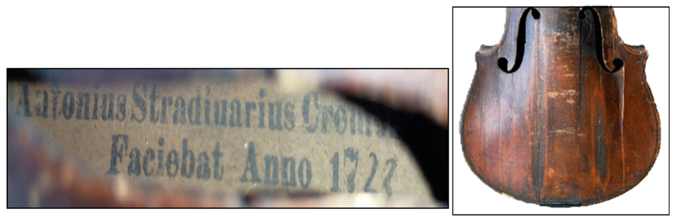
3.1. History of the Violin

The violin is part of a private collection and was purchased on the open market in 2018 from an owner in Botoșani, who received it from his Russian grandparents. It has been kept in the classic model cloth box of recent manufacture. No other data are known about how it was preserved, about other places where it had been (from whom it was previously purchased by the great-grandparents) or about who sang it and until when. Moreover, the owner does not know any other data regarding the restoration interventions, the current state of preservation and the last time it was played.

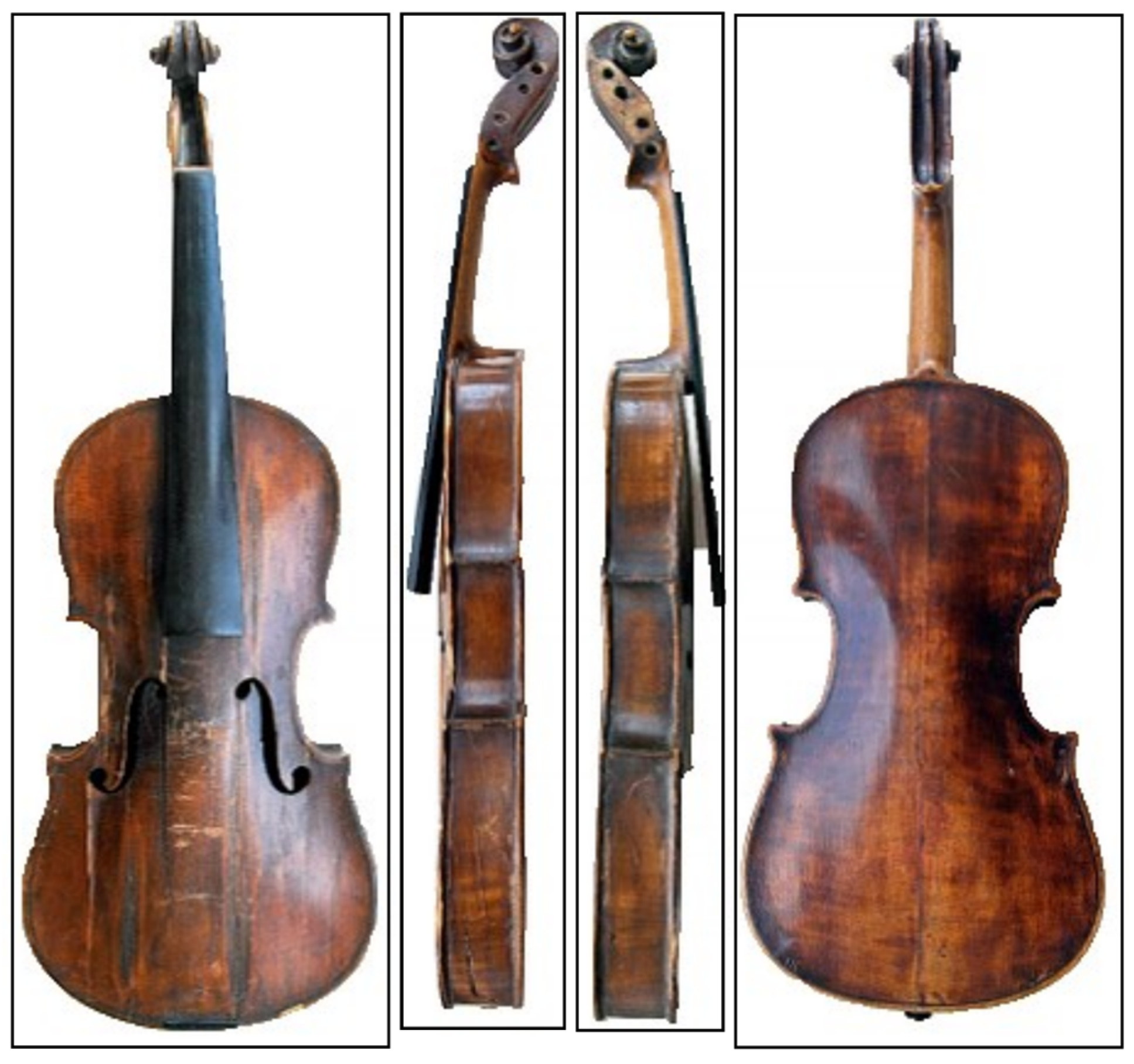
3.2. Dimensions and Identification of Model 1723, “Petite Violin”

Diameter of large loop (B): 175 mm (between fillets/black lines 170 mm); diameter of small loop (C): 140 mm (between fillets/black lines 135 mm); width of the eclipses at the tailpiece (D): 29 mm; width of neck eclipses (E): 29 mm; length of the violin body or the resonance box (A): 313 mm; length of f-holes 64 mm; diameter of the large C bout of the left/right f-hole: 10/10 mm; diameter of the small C bout of the left/right f-hole; 8/8 mm; distance between notches: 9 mm; f-holes opening: 7 mm; minimum distance at the ends of the f-holes: ≈ 1.5/1.5 mm; width of the resonance box at the f-holes level: 96 mm; thickness of the resonance box: 50 mm; fingerboard length: 230 mm (children’s model); fingerboard thickness: 4 mm; neck: 4 mm; neck height plus scroll: 207 mm; scroll width: 401 mm; scroll quadrature: 38 mm; central thickness: 38 mm; thickness of the scroll at the support plate: 18 mm; distance in the peg box to the plate: 14 mm; distance in the peg box to the scroll: 12 mm and depth of the peg box: 15 mm [24].
All sizes are found in the range of small violins, Stradivarius 1723.

3.3. Conservation Status

The violin is in a poor preservation state (Figure 3), with the elements of the resonance box cracked longitudinally (two large cracks which extend along the upper cover of the resonance box and which run through both C-bouts and four cracks with vertical displacement at the level of the f-holes). It is also marked with a lot of damage (fractures and lack of material in the marginal area of the fillets, inlays and pegs and superficial scratches on the large loop, in the area of the chinrest and the outer edge near the fillet) from poor handling, both on the upper cover (front plate) and on the lower one (back plate).
The violin has undergone devarnishing and revarnishing and restoration interventions with the opening and closing of the resonance box (removal and re-installation of the covers). On the scroll, in the upper outer area, there are two cracks—one at the level of the peg hole and two on one side—and another at the level of peg three on the other side, which resulted from handling, pressing and turning of the button. On both verso and obverse, in the bow work area, the varnish is encrusted, with extensive eroded areas. The violin is missing all structural and functional elements, retaining only the head/scroll with neck, neck and rib-centered tension knob at the end of the large loop. It is missing the four pegs/tension knobs, the bride, the chinrest, the tailpiece with the clamps and the four strings, the end button and the bow. It has been recently kept in the current cloth box, which is of more recent provenance, after 2018. The soundpost and the bass bar are missing. The upper cover is made of two tops, and the central joint area is poorly highlighted. Only the symmetry of the variation in the size of the annual rings proves the presence of the two tops. The large loop has annual rings with a thickness of 1 mm and max. 3 mm; the cover is made of spruce [25]. In the area of the two loops, the rings keep their parallelism along the entire length of the resonance box. There is a lot of use erosion in the f-hole, neck and chinrest area. The neck, ribs and back are made of maple wood. In the area of the big loop, on the right side, it has a central natural defect in the form of an irregular crack. On the left side, there are two holes resulting from xylophagic attack, which is currently inactive. The verso is strongly eroded in the center, affecting the varnish and plaster preparation layer, along with embossing from the fixing of the chinrest. In a long-ago attempt to remove the bottom cover, the neck coupling area was replaced by a well-cut mahogany-colored ornamental patch. There are heavy traces of dirt in the areas on either side of the bridge. The fillet preserves the parallelism of the lines, being made by inlay, but with many interruptions and lack of material. In the area of the chinrest or the bridge, there is mounting iridescence and embossing. The corners are fractured, as is the edge of the fillet in the area of the f-holes on the left. After damage and degradation, the violin was played before 1940 only. The pattern of the f-holes is found on the small Stradivarius violin type, which were made by luthiers from Cremona in the period 1723, according to the inner label.
The other aspects regarding the conservation state, related to the physical condition of the violin, are briefly presented in the preliminary conclusions.

3.4. Microscopic Analysis

The OM analysis performed on the wood sampled from the upper cover (Figure 4a) highlights the distribution of the wood fiber and the degree of its damage. On the varnish (Figure 4b), it illustrates a series of cracks and surface or embedded impurities.
Using optical microscopy, a series of samples were selected from the varnish, which presented a fractal stratigraphic section at the edge. In the edge, the degree of penetration of the encrusted surface deposits in the varnish, considered an archaeometric characteristic, was highlighted by means of SEM electronic microscopy, through which the age of the primary varnishing, and, thus, the age of the violin, was evaluated.

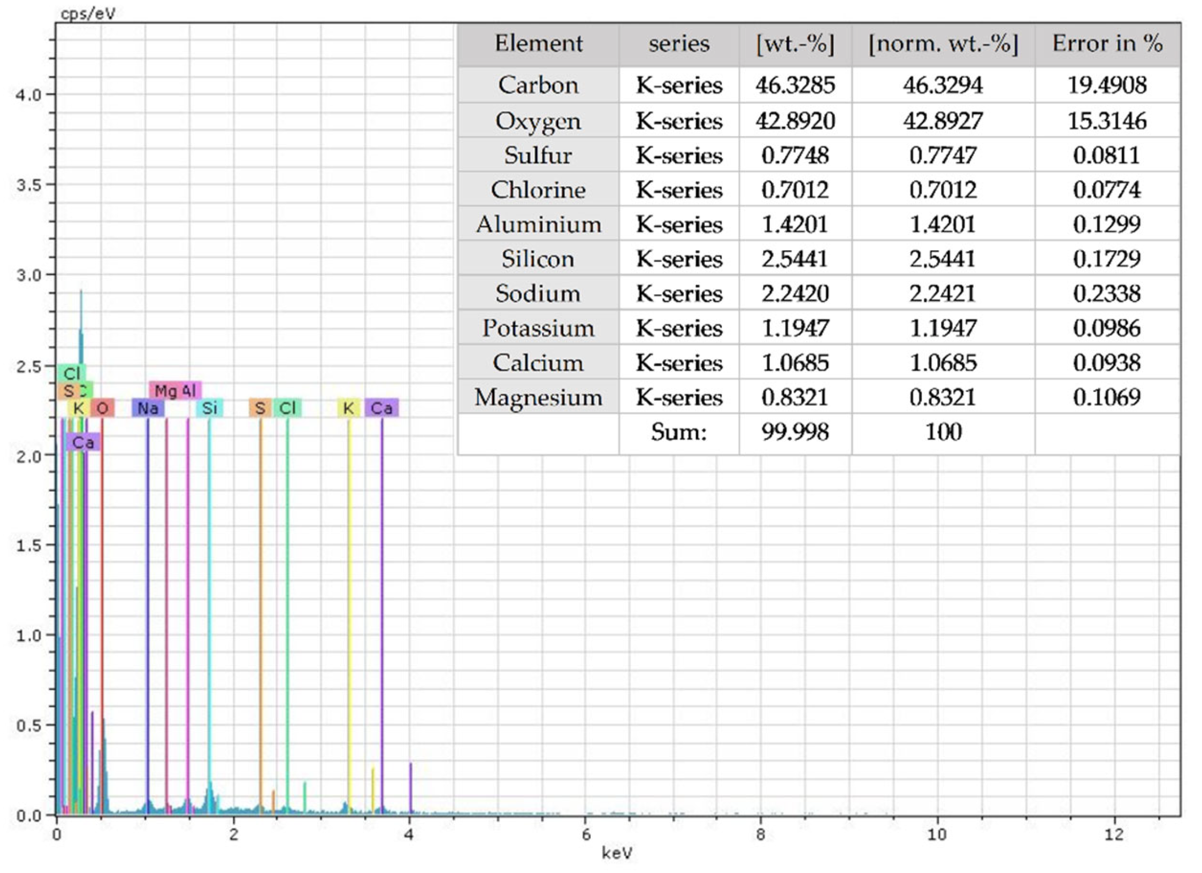
3.5. SEM–EDX Analysis

From the SEM micrographs (Figure 5a) taken on the same spruce wood sample, the degree of fiber fragility can be observed. On the analyzed layer of varnish, impurities and cracks are visible in the marginal structures with a stratigraphic profile (Figure 5b) [1,2].
Based on the EDX spectra (Figure 6), the elemental composition was determined, and the following chemical elements were identified: C, O, Al, Na, K, Ca, S and Cl.
The identified elements and their composition make it possible to appreciate the fact that the wood of the upper cover is from spruce and was treated with alum (double sulfate of aluminum and potassium), and the presence of sodium proves that the wood was treated hydrothermally and with borax (sodium tetraborate); the boron was not detected due to the technique [1,26,27,28,29].
The varnish sample was analyzed by EDX on two areas on the SEM photomicrograph (Figure 7, areas 3–4), both of which showed in the marginal stratigraphic section a strip of the penetration area of the encrusted dirt. On the zone marked 3, the following elements were identified by EDX: C, O, Si, Al and Na (Figure 8). On zone 4, the following elements were identified: C, O, Si, Al, Na, K, Ca, Mg, S and Cl (Figure 9).

3.6. Micro-FTIR Analysis

From the data of µ-FTIR spectra (Figure 10), based on the two groups of vibrations (deformation and valence), it is possible to identify the structural groups belonging to cellulose (1318, 1172, 1043 and 895 cm−1), lignin (1602, 1513, 1463, 1431, 1377 and 1234 cm−1), calcium sulfate (1121, 1089 and 676 cm−1), alum (617 cm−1) and borax (1653 cm−1) [2,30,31,32].
The peaks in the range 2906–2744 cm−1 represent the deformation vibrations of the symmetric C–H bonds in cellulose and hemicellulose [23]. The variations of the FTIR spectra around the value of 2906 cm−1 show that these absorption peaks increase with aging, the mechanism of which is based on the degradation process by oxidation and acid–base hydrolysis [33].
Instead, the increase in the intensity of the 1653 cm−1 band can be attributed to the CO group and the COO group at 1602 cm−1, respectively. Both demonstrate evolutionary oxidative and hydrolytic processes [34]. The peak at 1513 cm−1 is a marker for lignin.
Over time, the FTIR signals at 1653 and 1602 cm−1 are the most affected due to the aging processes. Furthermore, the group of absorption bands in the region 1400–1200 cm−1 corresponds to the crystalline component of cellulose [35]. Peaks with frequencies at 1431, 1377 and 1318 cm−1 are very sensitive to the degradation process of crystalline cellulose [36].
The peak at 895 cm−1 represents amorphous cellulose [36,37]. Of interest is the peak at 813 cm−1, which can vary in shape from the appearance of a shoulder to a small peak. This may be due to the presence of a substituted aromatic ring, an aliphatic C=C group or a furan group. Lignin and hemicellulose show this peak when they are aged [37].
The crystallinity index of the cellulose from the analyzed samples can be determined by calculating the ratio between the intensity of the absorbances corresponding to the bands which are characteristic of the crystalline and amorphous domains of cellulose (1377 cm−1 (C–H band), 1431 cm−1 and 895 cm−1 and 2906 cm−1). Thus, the ratio between the absorbance of the band at 1377 cm−1 and that at 2906 cm−1 corresponds to the crystallinity index, and the ratio between the absorbance of the band at 1431 cm−1 and that at 895 cm−1 can be attributed to the strength of the bonds of hydrogen [38,39]. The cellulose crystallinity index from the FTIR spectra is calculated according to the following relationship [40]: χ = A1377/A2906, where χ is the cellulose crystallinity index, and A1377 and A2906 are the characteristic absorbances of the bands from 1377 and 2906 cm−1.
The degradation rate of cellulose fibers was determined by calculating the crystallinity index (Ic), the degree of lateral ordering (GOL) and the intensity of hydrogen bonds (IlegH).
Table 1 shows the values of the crystallinity indices, the degree of ordering, the intensity of hydrogen bonds and the energy of hydrogen bonds calculated from the IR spectra for the spruce wood sample taken in the study.
The high values of the index are correlated with the higher amount of type I cellulose. According to the data in the table, it can be concluded that the crystallinity index of the sample justifies the degree of degradation due to both hydrolytic cleavage and oxidation processes of glycosidic groups.
The EDX composition correlates with the µ-FTIR spectrum (Figure 11) of the varnish sample through the vibrational and valence group peaks that belong to the structural components based on rosin (3443, 2939 and 2879 cm−1), wax (2833, 1744, 1470 and 728 cm−1) and egg white proteins (1626 and 1563 cm−1) [4,30].

3.7. Thermal Analysis

By evaluating the data from the two curves (Figure 12) of the thermal analysis in dynamic mode (TG and DTA), it is possible to highlight the three stages of mass loss (TG) and, thus, DTA, in correlation with the increase in temperature and the subsequent evolution of chemical components in the wood of the top cover. Thus, starting from the temperature of 40 °C to 180 °C, the sample loses approx. 16% of the volatile components (hygroscopic water, ethers, esters, etc.) through endothermic processes, which expand with a minimum DTA at 100 °C and evolve up to 10 min. Between 180 and 350 °C, the sample loses 72% of its weight in 30 min when exothermic combustion processes take place (with a maximum DTA at 300 °C), with the elimination of CO2 and water vapor, the stage presenting for two curves (TG and DTA), the specific effects of the combustion of the organic components in wood superficially treated with varnish and the structural changes for the inorganic components (alum, borax and calcium sulfate). Finally, after 350 °C to 700 °C, a mass loss of 12% occurs through the decomposition of metal oxides and hydroxo complexes, with the formation of residual ash from the inorganic components in a time of over 40 min. The processes is endo/exothermic with a DTA maximum at 380 °C and a DTA minimum at 520 °C.
One of the characteristics evaluated from dynamic thermal analysis, which is especially important in archaeometry, is related to the percentage of ash (remaining mass after 600 °C), which increases significantly with age [41,42].
The data of thermal analysis in dynamic mode highlight the presence in the structure of wood (from the upper cover) and other inorganic components. They come from hydrothermal treatment in borax and alum solutions, which are initially applied for hydraulic, chemical and microbiological (insectofungal) stabilization and from varnishing. The varnishing is for aesthetic and climate protection purposes, with alcoholic dispersions based on rosin and propolis applied over a very thin layer of plaster. These products are found in the remaining (residual) ash, which has a value of about 1.21%. These data correlate very well with those obtained by EDX and µ-FTIR.

3.8. Age Assessment of Violin

The age evaluation of the violin was made on the basis of the archaeometric and chemometric characteristics identified by SEM–EDX analyses for wood, varnish and thermal for spruce wood.
Based on previous experiences [3,8,9,19,43,44,45,46,47,48,49,50,51] regarding the identification of some archaeometric characteristics and, thus, the evolutionary chemometric ones with archaeometric value, in the present study, the following aspects were taken into account:
-
disposition of areas of varnish contamination and superficial dirt [52,53];
-
the depth of the presence of the archaeometric characteristics (the gradient of the penetration of oxidatively encrusted dirt in the volume phase of the varnish, the crystallinity index of the cellulose, its degree of lateral ordering and the intensity of the hydrogen bonds, and, thus, the remaining concentration of the ash of the wood sample) and the chemometric ones with archaeometric value (elemental stoichiometry ratios: C/O for wood, C/S and C/O for varnish and Si/Al for the encrusted dirt on the varnish) [54];
-
the morphology of the surfaces, highlighting the iridescence, texture and microtopographical structures [55];
-
the shape and ordering of the structures in the wood and the old, degraded varnish, in relation to the non-degraded areas of the two structural components (elemental stoichiometry ratios: C/O in the wood, C/S and C/O in the varnish and Si/Al in the encrusted dirt superficial) [56,57,58].
Both the archaeometric and chemometric characteristics place the violin as having been made around 1723.
The data presented above show a good correlation between the two archaeometric characteristics highlighted in the wood and varnish samples (the gradient of penetration of the oxidatively encrusted dirt in the volume phase of the varnish at a depth varying between 0.6 and 2.5 μm (with a rate of 5 × 10⁻³ μm/year), the cellulose crystallinity index (1.17), the degree of lateral ordering (1.12), the hydrogen bond intensity (0.94) and the ash concentration of the wood sample (1.21%). Among the evolutionary chemometric data, with archaeometric functions for the wood sample, the data for new hydro-stabilized wood were established within our collective (Table 2). The elemental ratios were identified as follows: C/O = 0.7127 and Al/K = 0.75 (from alum). For the varnish layer, the following were identified: C/S = 47 of the gypsum preparation under the varnish, whereas C/O = 1.08 for the varnish and Si/Al = 0.5582 for the dirt layer on the surface of the varnish. These have changed significantly, which indicates an age of about 300 years. For example, for the newly installed wood with the same treatments, the C/O ratio decreased from 0.801 to 0.713, with a rate of about 3 × 10⁻⁴/year. In contrast, for Al/K, it increased from 0.692 to 0.75, with a rate close to 2 × 10⁻⁴/year. The C/S ratio decreased from 51 to 47, with a rate of 1.3 × 10⁻²/year, a change attributed to oxidative fouling processes. For varnish, because over time, the surface processes have been strongly influenced by very different environmental factors throughout the use of the violin, these ratios are not subject to the same laws of variation as for wood.
Archaeometric features have an easily readable evolution rate. The values vary between 10−3 and 10−4, as determined by SEM electron microscopy, µ-FTIR spectroscopy and differential thermal analysis. The rates of evolution for the four groups confirm the age of the violin as approx. 300 years.
The first C/S chemometric characteristic is related to the increase in porosity following the embrittlement of the plaster preparation (the thin plaster layer under the varnish) and the penetration of CO2 in the presence of hygroscopic moisture (the carbonation process), the value of which decreases by 1.085× compared to the reference. Thus, the C/O ratio, the value of which decreases by approx. 1.12×, is also suitable for an age of approx. 300 years.

4. Conclusions

Based on the analyses conducted on this violin, a series of special artistic and scientific conclusions emerge.
The violin is a 1723 small Stradivarius (1/2) model which was built by an Italian luthier (possibly one of the two sons of Antonius Stradivari) and is a replica of the Stradivarius violins from 1721–1729.
The leucometric characteristics of the label and the patina inside the resonance box are the basic elements that confirm the age of approx. 300 years.
Through UV reflectography, the presence of gypsum, egg white and rosin was established in the preparation; the presence of these substances is a relevant aspect in the authentication of the violin.
It has an old paper label on which the inscription is printed in black ink on two lines: “Antonius Stradivarius Cremonensis/Faciebat Anno 1723”, without the copyright logo. The label is not in a border and has evenly cut sides. The paper support of the label has the patina of age with the degree of apparent whiteness corresponding to an age of over 300 years, and the printing ink is slightly embrittled, which demonstrates that the binder degraded slowly; it shows slight traces of cracking and expansion without traces of microbiological contamination [30,59].
The ribs, which are six in number (two for the small loop, two for the middle loop and two for the large loop), are made of maple wood and have the same width around the entire perimeter of the violin. According to the patina, it is the age of the front and back ribs board; it seems that the inner joint cord was made when the box was resealed (Interwar Period). When the two boards were mounted, blocks were used at the ends. Between them and the corners, a cord-type band, which is made of flexible wood, was fixed in the blocks and corners by splicing or gluing. All the elements are more recent than the front board, the verso and the ribs. The fillet inlays are uniform and equidistant from the edges; they have very thin and flexible veneer inlays, which are darker in color and are narrow, equidistant and 3 mm from the edge, which is similar to all Stradivarius violins (the inlay of the front headboard is of the same age as the stand). The neck and scroll were made from maple wood, and the dimensions correspond to a 1723 Stradivarius violin. The pegs are missing, as are the tailpiece and chinrest; the violin retains only the button or cord spur. The distance between the pegs corresponds to a 1723 Stradivari violin. The fingerboard is original and is made of ebony, which is similar to the tailpiece; the fingerboard is longitudinally fractured and is missing material. The catch button is original and has an authentic mother-of-pearl center ornament in the form of intarsia.
The stereoscopic analysis of the inner surface of the sound box shows an easily legible age patina. In the Interwar Period, this box was opened and cleaned. Moreover, the embedment of the counter-rib is specific to the period 1721–1729.
Analysis by UV reflectography revealed that the varnish is thick, with extensive areas of damage and degradation. The protein binder in the coating cannot be highlighted, but only the thin plaster layer on the back can be highlighted. In certain areas, there is a weak fluorescence from the binder based on egg white and rosin. The varnish, which is one from the Interwar Period, was applied over the original one. The varnish has in its composition pine rosin initially dissolved in alcohol/turpentine then dispersed in linseed oil, and the preparation was based on egg white. Instead, the new varnish contains as a diluted dye in shellac.
The SEM–EDX and µ-FTIR analyses correlate very well with those obtained by thermal analysis in dynamic mode (differential thermal analysis). The analyses highlight the presence in the wood structure (from the upper cover) of a hydrothermal treatment in borax and alum solutions for water, chemical and microbiological stabilization (insectofungal). The wood was varnished for aesthetics and climate protection. In addition, cellulose and lignin, as well as other components from the preservation treatment and varnishing, represent the strong point of the authentication.
The data on the evolution rate of the archaeometric and chemometric characteristics, with archaeometric value determined by SEM, µ-FTIR and differential thermal analysis, confirm the age of the violin as being approx. 300 years.
The violin was played only until 1940 and was kept later, after 2018, in the new case purchased by the owner. This violin is part of the Cultural Heritage group as a musical instrument, with an age of approx. 300 years. It is classified as a B category national asset of Romania. The study also had another purpose: in addition to authentication, the results will be used in the development of a preservation–restoration protocol, which the owner must keep in mind.

Author Contributions

Conceptualization, I.S., P.O.T. and V.V.; methodology, I.S., P.O.T., F.B., V.V., A.D. and V.D.; formal analysis, F.B., V.V., A.D. and V.D.; investigation, F.B., V.V., A.D. and V.D.; data curation, I.S. and V.V.; writing—original draft preparation, I.S., P.O.T. and V.V.; writing—review and editing, I.S., P.O.T., V.V. and A.D. All authors have read and agreed to the published version of the manuscript.

Funding

This research received no external funding.

Acknowledgments

This work was co-funded by the European Social Fund, through Operational Programme Human Capital 2014–2020, project number POCU/993/6/13/153322, project title “Educational and training support for PhD students and young researchers in preparation for insertion in the labor market”.

Conflicts of Interest

The authors declare no conflict of interest.

References

  1. Sandu, I.; Tanasa, P.O.; Sandu, I.C.A.; Negru, I.C.; Sandu, A.V.; Vasilache, V. Authentication of an Old Violin by Multianalytical Methods. App. Sci. 2020, 10, 306. [Google Scholar] [CrossRef] [Green Version]
  2. Stani, C.; Invernizzi, C.; Briarda, G.; Davit, P.; Vaccari, L.; Magalodi, M.; Gulmini, M.; Fiocco, G. A Nanofocused Light on Stradivari Violins: Infrared s-SNOM Reveals New Clues Behind Craftsmanship Mastery. Anal. Chem. 2022, 94, 14815–14819. [Google Scholar] [CrossRef] [PubMed]
  3. Malagodi, M.; Canevari, C.; Bonizzoni, L.; Galli, A.; Maspero, F.; Martini, M. A multi-technique chemical characterization of a Stradivari decorated violin top plate. Appl. Phys. A 2013, 112, 225–234. [Google Scholar] [CrossRef]
  4. Cortea, I.M.; Cristache, R.A.; Sandu, I. Characterization of historical violin varnishes using ATR-FTIR spectroscopy. Rom. Rep. Phys. 2016, 68, 615–622. [Google Scholar]
  5. Cristache, R.A. Study of New Archaeometric Characteristics of Old Wood Artefacts. Ph.D. Thesis, Alexandru Ioan Cuza University, Faculty of Geography and Geology, Iasi, Romania, 2015. [Google Scholar]
  6. Brandmair, B.; Greiner, S.P. Stradivari Varnish: Scientific Analysis of His Finishing Technique on Selected Instruments; Serving Audio: London, UK; Munich, Germany, 2010. [Google Scholar]
  7. Daher, C.; Paris, C.; Le Hô, A.S.; Bellot-Gurlet, L.; Echard, J.P. A joint use of Raman and infrared spectroscopies for the identification of natural organic media used in ancient varnishes. J. Raman Spectrosc. 2010, 41, 1494–1499. [Google Scholar] [CrossRef] [Green Version]
  8. Sacconi, S.F. The “Secrets” of Stradivari; Libreria del Convegno: Cremona, Italy, 1979. [Google Scholar]
  9. Weththimuni, M.L.; Canevari, C.; Legnani, A.; Licchelli, M.; Malagodi, M.; Ricca, M.; Zeffiro, A. Experimental characterization of oil-colophony varnishes: A preliminary study. Int. J. Conserv. Sci. 2016, 7, 813–826. [Google Scholar]
  10. The “Stradivarius” Violin Label: What It Means. Available online: https://www.benningviolins.com/the-stradivarius-violin-label-what-it-means.html (accessed on 3 January 2023).
  11. Mukhopadhyay, R. How Stradivari and Guarneri got their music violins. Anal. Chem. 2009, 79, 819–820. [Google Scholar] [CrossRef]
  12. Bertrand, L.; Robinet, L.; Cohen, S.X.; Sandt, C.; Le Hô, A.-S.; Soulier, B.; Lattuati-Derieux, A.; Echar, J.P. Identification of the finishing technique of an early eighteenth century musical instrument using FTIR spectromicroscopy. Anal. Bioanal. Chem. 2011, 399, 3025–3032. [Google Scholar] [CrossRef]
  13. Chen, G.F. Developments in the field of rosin chemistry and its implications in coatings. Prog. Org. Coat. 1992, 20, 139–167. [Google Scholar] [CrossRef]
  14. Echard, J.P.; Benoit, C.; Peris-Vicente, J.; Malecki, V.; Gimeno-Adelantado, J.V.; Vaiedelich, S. Gas chromatography/mass spectrometry characterization of historical varnishes of ancient Italian lutes and violin. Anal. Chim. Acta 2007, 584, 172–180. [Google Scholar] [CrossRef]
  15. Echard, J.P.; Bertrand, L.; von Bohlen, A.; le Hô, A.-S.; Paris, C.; Bellot-Gurlet, L.; Soulier, B.; Lattuati-Derieux, A.; Thao, S.; Robinet, L.; et al. The nature of the extraordinary finish of Stradivari’s instruments. Angew. Chem. Int. Ed. 2010, 49, 197–201. [Google Scholar] [CrossRef]
  16. Fioravanti, M.; Goli, G.; Carlson, B. Viscoelastic and mechano-sorptive studies appliaed to the conservation of historical violins: A case study of the Guarneri “del Gesù” violin (1743) known as the “Cannone”. J. Cult. Herit. 2013, 14, 297–303. [Google Scholar] [CrossRef]
  17. Noguera, J.R.; Galiano, F.C.B.; Lòpez, J.M.R.; Vivas, M.A.F.; Sànchez, I.M. Study of biodegradation of diterpenic varnishes used in art painting: Colophony and Venetian turpentine. Int. Biodeterior. Biodegrad. 2008, 62, 427–433. [Google Scholar] [CrossRef]
  18. Sandu, I.; van Saanen, A. Expertiza Ştiinţifică a Operelor de artă, Vol. I, Autentificarea, Stabilirea Paternităţii şi Evaluarea Patrimonială; Editura Universităţii “Alexandru Ioan Cuza”: Iași, Romania, 1998. [Google Scholar]
  19. Sandu, I.; Sandu, I.G. Aspecte Moderne Privind Conservarea Bunurilor Culturale, Vol.I. Nomenclatură, Tipologii şi Cazuistici; Editura Performantica: Iași, Romania, 2005. [Google Scholar]
  20. Scalarone, D.; Lazzari, M.; Chiantore, O. Ageing behaviour and pyrolytic characterization of diterpenic resins used as art materials: Colophony and Venice turpentine. J. Anal. Appl. Pyrolysis 2002, 64, 345–361. [Google Scholar] [CrossRef]
  21. Caruso, F.; Chillura Martino, D.F.; Saverwyns, S.; Van Bos, M.; Burgio, L.; Di Stefano, C.; Peschke, G.; Caponetti, E. Micro-analytical identification of the components of varnishes from South Italian historical musical instruments by PLM, ESEM–EDX, microFTIR, GC–MS, and Py–GC–MS. Microchem. J. 2014, 116, 31–40. [Google Scholar] [CrossRef]
  22. Echard, J.P.; Lavedrine, B. Review on the characterisation of ancient stringed musical instruments varnishes and implementation of an analytical strategy. J. Cult. Herit. 2008, 9, 420–429. [Google Scholar] [CrossRef]
  23. Fierascu, I.; Fierascu, R.C.; Stirban, A.; Panaitescu, D.M.; Nicolae, C.A.; Raditoiu, V.; Zgarciu, M.S.; Leahu, A.C. Chemical and mineral characterisation of Roumanian book paper materials (XVII-XIXth century). Microchem. J. 2020, 152, 104307. [Google Scholar] [CrossRef]
  24. Frohlich, B.; Sturm, G.; Hinton, J.; Frohlich, E. The Secrets of The Stradivari String Instruments. A Non Destructive Study of Music Instruments from The Smithsonian Institution, The Library of Congress, and Private Collections. A Pilot Study of Seven Violins Made by Antonio Stradivari in Cremona, Italy, Between 1677 and 1709; The Materialise Group: Leuven, Belgium; Materialise USA: Ann Arbor, MI, USA; Washington, DC, USA, 2009. [Google Scholar]
  25. Su, C.; Chen, S.-Y.; Chung, J.-H.; Li, G.-C.; Brandmair, B.; Huthwelker, T.; Fulton, J.L.; Borca, C.; Huang, S.-J.; Nagyvary, J.; et al. Materials Engineering of Violin Soundboards by Stradivari and Guarneri. Angw. Chem. 2021, 133, 19293–19303. [Google Scholar] [CrossRef]
  26. Bellamy, L.J.; Gerrard, W.; Lappert, M.F.; Williams, R.L. Infrared spectra of boron compounds. J. Chem. Soci. 1958, 481, 2412–2415. [Google Scholar] [CrossRef]
  27. Jun, L.; Shuping, X.; Shiyang, G. FT-IR and Raman spectroscopic study of hydrated borates. Spectrochim. Acta Part A Mol. Biomol. Spectrosc. 1995, 51, 519–532. [Google Scholar] [CrossRef]
  28. Weir, C.E. Infrared spectra of the hydrated borates. J. Res. Natl. Bur. Stand. A Phys. Chem. 1966, 70, 153–164. [Google Scholar] [CrossRef]
  29. Caldeira, F. Boron in wood preservation. A review in its physico-chemical aspects. Silva Lusit. 2010, 18, 179–196. [Google Scholar]
  30. Schwanninger, M.; Rodrigues, J.C.; Pereira, H.; Hinterstoisser, B. Effects of short-time vibratory ball milling on the shape of FT-IR spectra of wood and cellulose. Vib. Spectrosc. 2004, 36, 23–40. [Google Scholar] [CrossRef]
  31. Fackler, K.; Stevanic, J.S.; Ters, T.; Hinterstoisser, B.; Schwanninger, M.; Salmén, L. Localisation and characterisation of incipient brown-rot decay within spruce wood cell walls using FT-IR imaging microscopy. Enzym. Microb. Technol. 2010, 47, 257–267. [Google Scholar] [CrossRef] [PubMed] [Green Version]
  32. Simonović, J.; Stevanic, J.; Djikanović, D.; Salmén, L.; Radotić, K. Anisotropy of cell wall polymers in branches of hardwood and softwood: A polarized FTIR study. Cellulose 2011, 18, 1433–1440. [Google Scholar] [CrossRef]
  33. Munajad, A.; Subroto, C.; Suwarno. Fourier Transform Infrared (FTIR) Spectroscopy Analysis of Transformer Paper in Mineral Oil-Paper Composite Insulation under Accelerated Thermal Aging. Energies 2018, 11, 364. [Google Scholar] [CrossRef] [Green Version]
  34. Ciofini, D.; Osticioli, I.; Micheli, S.; Montalbano, L.; Siano, S. Laser removal of mold and foxings stains from paper artifacts: Preliminary investigation. In Fundamentals of Laser-Assisted Micro- and Nanotechnologies 2013; SPIE: Bellingham, WA, USA, 2013; Volume 9065, p. 906512. [Google Scholar] [CrossRef]
  35. Calvini, P.; Gorassini, A. The Degrading Action of Iron and Copper on Paper A FTIR-Deconvolution Analysis. Restaur. Int. J. Preserv. Libr. Arch. Mater. 2002, 23, 205–221. [Google Scholar] [CrossRef]
  36. Librando, V.; Minniti, Z.; Lorusso, S. Ancient and modern paper characterization by FTIR and Micro-Raman spectroscopy. Conserv. Sci. Cult. 2011, 11, 249–268. [Google Scholar] [CrossRef]
  37. Chiriu, D.; Ricci, P.C.; Cappellini, G.; Salis, M.; Loddo, G.; Carbonaro, C.M. Ageing of ancient paper: A kinetic model of cellulosedegradation from Raman spectra. J. Raman Spectrosc. 2018, 49, 1802–1811. [Google Scholar] [CrossRef]
  38. Baccaro, S.; Carewska, M.; Casieri, C.; Cemmi, A.; Lepore, A. Structure modifications and interaction with moisture in γ-irradiated pure cellulose by thermal analysis and infrared spectroscopy. Polym. Degrad. Stab. 2013, 98, 2005–2010. [Google Scholar] [CrossRef]
  39. Poletto, M.; Ornaghi Júnior, H.L.; Zattera, A.J. Native cellulose: Structure, characterization andthermal properties. Materials 2014, 7, 6105. [Google Scholar] [CrossRef] [PubMed] [Green Version]
  40. Măluțan, T.; Oborcea, P.P.; Pui, A. IR spectroscopy of the pulps obtained by kraft additivated delignification. Rev. Celul. Hârtie 2002, 51, 17–20. [Google Scholar]
  41. Chang, S.T.; Chang, H.T.; Huang, Y.S.; Hsu, F.L. Effects of chemical modification reagents on acoustic properties of wood. Holzforschung 2000, 54, 669–675. [Google Scholar] [CrossRef]
  42. Ahmed, S.A.; Adamopoulos, S. Acoustic properties of modified wood under different humid conditions and their relevance for musical instruments. Appl. Acoust. 2018, 140, 92–99. [Google Scholar] [CrossRef]
  43. Tanasa, P.O.; Sandu, I.; Vasilache, V.; Sandu, I.G.; Negru, I.C.; Sandu, A.V. Authentication of a Painting by Nicolae Grigorescu Using Modern Multi-Analytical Methods. Appl. Sci. 2020, 10, 3558. [Google Scholar] [CrossRef]
  44. Tănasă, P.O. Modern Methods of Authenticating Criminal Assets, Conservation Procedures during the Investigation, Saving and Reintroducing Them into the Circuit of Cultural Heritage Values. Ph.D. Thesis, Faculty of Geography and Geology, “Alexandru Ioan Cuza” University of Iași, Iași, Romania, 2022. [Google Scholar]
  45. Vasilache, V.; Sandu, I.C.A.; Pruteanu, S.; Caldeira, A.T.; Simionescu, A.E.; Sandu, I. Testing the Cleaning Effectiveness of New Ecological Aqueous Dispersions Applied on Old Icons. Appl. Surf. Sci. 2016, 367, 70–79. [Google Scholar] [CrossRef]
  46. Pruteanu, S.; Sandu, I.; Vasilache, V. Modern Procedures Used in Cleaning Old, Illegibly and Blackened Icons. Environ. Dev. Sustain. 2013, 9, 219–236. [Google Scholar] [CrossRef] [Green Version]
  47. Pruteanu, S.; Vasilache, V.; Sandu, I.C.A.; Budu, A.M.; Sandu, I. Assessment of Cleaning Effectiveness for New Ecological Systems on Ancient Tempera Icon by Complementary Microscopy Techniques. Microsc. Res. Tech. 2014, 77, 1060–1070. [Google Scholar] [CrossRef]
  48. Iurcovschi, T.C.; Vasilache, V.; Sandu, I.; Zaharia, M.; Pintilie, O.; Sandu, A.V. New Ecological Solutions Involved in the Cleaning of a 19th Century Icon. Appl. Sci. 2020, 10, 1175. [Google Scholar] [CrossRef] [Green Version]
  49. Rovetta, T.; Invernizzi, C.; Fiocco, G.; Albano, M.; Licchelli, M.; Gulmini, M.; Alf, G.; Fabbri, D.; Rombolà, A.G.; Malagodi, M. The case of Antonio Stradivari 1718 ex-San Lorenzo violin: History, restorations and conservation perspectives. J. Archaeol. Sci. Rep. 2019, 23, 443–450. [Google Scholar] [CrossRef] [Green Version]
  50. Bonizzoni, L.; Canevari, C.; Galli, A.; Gargano, M.; Ludwig, N.; Malagodi, M.; Rovetta, T. A multidisciplinary materials characterization of a Joannes Marcus viol (16th century). Herit. Sci. 2014, 2, 15. [Google Scholar] [CrossRef] [Green Version]
  51. Fichera, G.V.; Rovetta, T.; Fiocco, G.; Alberti, G.; Invernizzi, C.; Licchelli, M.; Malagodi, M. Elemental analysis as statistical preliminary study of historical musical instruments. Microchem. J. 2018, 137, 309–317. [Google Scholar] [CrossRef]
  52. Lattuati-Derieux, A.; Gomes, S.; Tirat, S.; Thao-Hey, S.; Echard, J.-P. New insights into molecular evolution of oil/colophony varnishes, towards pyrolysis-gas chromatography/mass spectrometry based quantitation. E-Preserv. Sci. 2014, 11, 53–63. [Google Scholar]
  53. Odlyha, M.; Lucejko, J.J.; Lluveras-Tenorio, A.; di Girolamo, F.; Hudziak, S.; Strange, A.; Bridarolli, A.; Bozec, L.; Colombini, M.P. Violin Varnishes: Microstructure and Nanomechanical Analysis. Molecules 2022, 27, 6378. [Google Scholar] [CrossRef]
  54. Azemard, C.; Vieillescazes, C.; Menager, M. Effect of photodegradation on the identification of natural varnishes by FT-IR spectroscopy. Microchem. J. 2014, 112, 137–149. [Google Scholar] [CrossRef]
  55. Beltran, V.; Salvadó, N.; Butí, S.; Cinque, G. Micro infrared spectroscopy discrimination capability of compounds in complex matrices of thin layers in real sample coatings from artworks. Microchem. J. 2015, 118, 115–123. [Google Scholar] [CrossRef] [Green Version]
  56. Tai, H.-C.; Li, G.-C.; Huang, S.-J.; Jhu, C.-R.; Chung, J.-H.; Wang, B.Y.; Hsu, C.-S.; Brandmair, B.; Chung, D.-T.; Chen, H.M.; et al. Chemical distinctions between Stradivari’s maple and modern tonewood. Proc. Natl. Acad. Sci. USA 2016, 114, 27–32. [Google Scholar] [CrossRef] [Green Version]
  57. Lämmlein, S.L.; Mannes, D.; van Damme, B.; Schwarze, F.W.M.R.; Burgert, I. The influence of multi-layered varnishes on moisture protection and vibrational properties of violin wood. Sci. Rep. 2019, 9, 18611. [Google Scholar] [CrossRef] [Green Version]
  58. Albano, M.; Comelli, D.; Fiocco, G.; Mattonai, M.; Lucejko, J.J.; Zoia, L.; Colombini, M.P.; Malagodi, M. Chemical modification of wood induced by the traditional making procedures of bowed string musical instruments: The effect of alkaline treatments. Herit. Sci. 2022, 10, 76. [Google Scholar] [CrossRef]
  59. Hinterstoisser, B.; Salmén, L. Two-dimensional step-scan FTIR: A tool to unravel the OH-valency-range of the spectrum of Cellulose I. Cellulose 1999, 6, 251–263. [Google Scholar] [CrossRef]
Figure 1. The body of the violin (front, sides and back).
Applsci 13 01048 g001
Figure 2. The inscriptions (label with dating, authorship and ownership) and the image with extended annual rings used for dendrochronological analysis.
Applsci 13 01048 g002
Figure 3. Details with visible elements of the preservation state of the violin.
Applsci 13 01048 g003
Figure 4. OM images of the analyzed sample: (a)—wood and (b)—varnish.
Applsci 13 01048 g004
Figure 5. SEM micrograph of the wood sample from the top cover of the violin’s resonance box (a) and varnish (b).
Applsci 13 01048 g005
Figure 6. EDX spectrum for the spruce wood sample.
Applsci 13 01048 g006
Figure 7. SEM micrograph of the varnish sample (yellow circles represent the analyzed areas).
Applsci 13 01048 g007
Figure 8. EDX spectrum for the varnish sample, zone 3.
Applsci 13 01048 g008
Figure 9. EDX spectrum for the varnish sample, zone 4.
Applsci 13 01048 g009
Figure 10. µ–FTIR spectrum of the wood sample from the upper cover.
Applsci 13 01048 g010
Figure 11. µ–FTIR spectrum of the varnish sample.
Applsci 13 01048 g011
Figure 12. TG and DTA curves of the wood sample from the upper cover.
Applsci 13 01048 g012
Table 1. Crystallinity index, Degree of lateral ordering, Intensity of hydrogen bond, which were evaluated from the IR spectra of the spruce wood sample.
Crystallinity Index (Ic)Degree of Lateral Ordering (GOL)Intensity of Hydrogen Bonds (IlegH)
A1377/A2906 = 1.17A1431/A896 = 1.12A3420/A1318 = 0.94
Table 2. Archaeometric and chemometric data for the wood sample.
CharacteristicActual ValueReference ValueEvolution Rate (/year)
ArchaeometricThe penetration gradient of the oxidatively fouled dirt in the volume phase of the varnish (μm)0.6–2.50.0–0.0~5 × 10⁻³
Cellulose crystallinity index1.171.384~7 × 10⁻⁴
Degree of lateral ordering1.121.184~2 × 10⁻⁴
The intensity of hydrogen bonds0.941.063~4 × 10⁻⁴
Ash concentration (%)1.210.58~2 × 10⁻³
ChemometricC/O for wood0.7130.801~3 × 10⁻⁴
Al/K for wooden alum0.7500.692~2 × 10⁻⁴
C/S for the preparation under the varnish47.0051.00~1.3 × 10⁻²
C/O for varnish1.080.650~1.4 × 10⁻³
Al/Si from the dirt layer on the varnish0.55820.00~1.9 × 10⁻³
Disclaimer/Publisher’s Note: The statements, opinions and data contained in all publications are solely those of the individual author(s) and contributor(s) and not of MDPI and/or the editor(s). MDPI and/or the editor(s) disclaim responsibility for any injury to people or property resulting from any ideas, methods, instructions or products referred to in the content.

