Article

Symptomatic Diastasis Rectus Abdominis in Children: Review of Current Management Options and Presentation of a Novel Minimally Invasive Epifascial Repair Technique

by
Markus Denzinger
1,*,
Domitille Stark
2,
Clemens Memmel
2,
Patricia Reis Wolfertstetter
2 and
Christian Knorr
2
1
Faculty of Medicine, University Medical Center Regensburg, 93053 Regensburg, Germany
2
Department of Pediatric Surgery, Klinik St. Hedwig, University Medical Center Regensburg, 93049 Regensburg, Germany
*
Author to whom correspondence should be addressed.
Appl. Sci. 2023, 13(17), 9841; https://doi.org/10.3390/app13179841
Submission received: 7 June 2023 / Revised: 18 August 2023 / Accepted: 30 August 2023 / Published: 31 August 2023

Abstract

:
Several surgical techniques are available for an adult patient collective with diastasis recti. Only few research papers addresses the treatment options of diastasis of the rectus abdominis in children. In this case series, we present a new technique of epifascial repair as a novel possibility in successfully repairing defects of the anterior abdominal wall using minimally invasive surgery. In this case series, we present an epifascial repair technique for patients with a diastasis recti with a dehiscence cranial of the umbilicus. Four pediatric patients with symptomatic rectus diastasis were treated with this new surgical technique. All procedures were conducted successfully, and no recurrence was observed in the follow up. All patients showed clinical regression of the rectus diastasis without any postoperative abdominal wall protrusion and good improvement of the symptoms. Intraoperative intraperitoneal air loss, postoperative scar keloid, thermal lesions due to Ultracision and one seroma/hematoma after the removal of the drain were minor complications observed during the follow-up.

1. Introduction

Diastasis of the rectus abdominis (DRA, diastasis recti, divarication of the rectus abdominis, abdominal muscle separation) describes a condition in which an abnormally large distance separates the two rectus muscles [1]. These defects can range from a few millimeters to several centimeters and are often located between the xyphoid and the umbilicus. In DRA, however, there is no fascial defect and, therefore, no hernia.
In adults, rectus diastasis occurs predominantly in two populations: peripartum middle-aged women (where the prevalence is up to 60% [2]) and older men. In women carrying a child, the occurrence of diastasis recti associated with the thinning and widening of the linea alba can be explained by an increase in intra-abdominal pressure and by the influence of the hormone relaxin. Despite a natural regression of the inter-rectus distance (IRD) in the postpartum period, physiotherapists recommend refraining from vigorous physical activity for at least 12 weeks after delivery because the abdominal wall may not have sufficient strength to withstand additional stress [3].
It has been observed that the occurrence of diastasis recti in infants is due to the immaturity of the abdominal wall. However, it can be postulated that diastasis recti may occur in children and adolescents when acute stress (recurrent cough due to pertussis, extreme exercise, etc.) is placed on a pre-weakened anterior abdominal wall (immature tissues, after significant weight loss, obstipation, etc.). More research is needed on the prevalence and incidence of DRA in children.
A deficiency of the abdominal wall maturation—especially in the Carnegie stadiums 19 to 20 (Day 46–49), where the four abdominal wall muscles develop—can lead to a congenital type of DRA [4]. This can occur with associated sequences or syndromes (such as prune belly syndrome, Beckwith–Wiedemann Syndrome, Cantrell pentalogy (thoracoabdominal syndrome), Opitz syndrome and midline defect syndrome) or through isolated autosomal dominant transmission [5].
Considering all pediatric patients, the prevalence of diastasis recti seems to be high, but there are no sources indicating the exact incidence of diastasis recti in children. The description of DRA in textbooks on the physical examination of the abdominal wall is: “Many normal infants have diastasis recti, resulting from the non-union of the two rectus muscles, which may result in an umbilical hernia.” [6], and “abdominal wall protrusions are common [in children]” [7]. This may be due to the immaturity of the abdominal wall with underdeveloped musculature and thin linea alba [8].
Considering that DRAs are frequently described in the literature and that surgical repair is not often mentioned, it must be admitted that most DRAs have a good prognosis and regress spontaneously, following a pattern similar to that of an umbilical hernia.
The fascial opening of umbilical hernias closes spontaneously as the rectus abdominis muscles continue to grow towards each other, resulting in the fusion of the peritoneal and fascial layers around the umbilicus. Spontaneous closure is to be expected in almost all children at 5 years of age. Fascial openings greater than 1.5 cm, protruding skin, increased intra-abdominal pressure (from ascites or peritoneal dialysis) and underlying medical conditions (Ehlers–Danlos, Beckwith–Wiedemann syndrome, Down syndrome, mucopolysaccharidoses, hypothyroidism or trisomy 18) may prevent spontaneous closure.
As with many aesthetic or functional pathologies, the initial course of action is conservative, and surgical or interventional treatment should be carefully considered if a progression of the pathology is observed.
Tupler [9] hypothesized that as the nervous system matures, the abdominal muscles move closer together, but in approximately 30% of all children the diastasis recti remains. She has developed a technique to reduce diastasis recti in children using an educational and playful concept called the Belly Button Program. This program can be used in children older than three years old [9]. Unfortunately, no study has been conducted to confirm the effectiveness of this program.
Recent publications on the conservative management of diastasis recti in postpartum women recommend physiotherapy, especially anterior abdominal wall strengthening, which has been shown to have a positive effect on reducing the IRD [10,11,12]. However, no generally accepted protocol has been established so far. Michalska et al. [11] list the following conservative treatment options: abdominal exercises (strengthening of the transversus abdominis muscles or rectus abdominis muscles), postural training, education and training on in proper mobility and lifting techniques, methods to strengthen the transversus abdominis muscles (Pilates, functional training, Tupler’s technique exercises with or without abdominal splinting), the Noble technique (manual approximation of the rectus abdominis muscles during a partial sit-up), manual therapy (soft tissue mobilization, myofascial release), abdominal bracing and taping, the tubigrip or a corset. A Norwegian study [10] on this matter showed that even a weekly exercise program in addition to daily home exercise did not reduce the prevalence of diastasis.
The use of minimally invasive techniques is particularly appropriate in pediatric surgery. Prolotherapy is the technique of injecting small amounts of an irritant solution into injured tissue in order to stimulate regenerative processes. Feins et al. [13] described a technique for the minimally invasive closure of (pediatric) umbilical hernias: deflux, a biodegradable compound of dextranomer microspheres in hyaluronic acid, was injected percutaneously into the border and preperitoneal space in four quadrants of the hernia defect, thereby occluding the lumen. Follow-up visits showed the closure or reduction in the size of umbilical hernias in patients immediately after surgery; and within months, 21 (84%) of 25 hernias were closed. Similar protocols have been published using injection therapy to treat umbilical hernias in children [14]. Strauchman and Morningstar [15] reported the use of prolotherapy in a postpartum female patient with DRA. The injection consisted of dextrose as the active ingredient, lidocaine and methylcobalamin and was injected every 2 weeks for a total of seven prolotherapy sessions. The diastasis recti was reduced from 2.7 cm to 0.5 cm. Based on these two studies, it is possible that the interventional reduction in DRA in children using prolotherapy could also be implemented for DRA as well. To date, there are no published studies addressing this issue.
Rectus abdominis diastasis occurs in many children and has a good prognosis as the abdominal musculature continues to develop. Even in adults, the surgical repair of DRA is controversial, but there are two indications for the surgical repair of DRA in children: a medical and a functional one. In some cases, children present with coexisting pathologies such as epigastric or persistent umbilical hernias, and where surgical intervention is required, a DRA repair in the same procedure seems opportune. In addition, DRA has been shown to be an independent risk factor for the recurrence of ventral hernia in adults [16]. In some cases, the protrusion is cosmetically and functionally disturbing, and surgical intervention should be considered. Various criteria have been proposed for operative repair in adults: Various criteria have been proposed for operative repair in adults such as the widening of the IRD above 3 cm [14] or the protrusion [14]. A Repair of DRA following after multiple pregnancies with loss of abdominal musculature has shown improvement in pulmonary and abdominal wall function [17]. Therefore, surgical repair should be discussed as a treatment for these rare individual cases. Unfortunately, few studies have addressed the possible treatment options for diastasis of the rectus abdominis in children.
The aim of this study was to present a new technique of epifascial repair as a new possibility to successfully repair hernias and defects of the anterior abdominal wall using minimally invasive surgery.

2. Materials and Methods

2.1. Data Collection

The study was approved by the local ethics committee of the medical faculty of the University of Regensburg, Germany (Project number: 23-3262-104). Data were collected retrospectively from medical records.

2.2. Patient Inclusion

Between November 2008 and October 2013, 4 patients (3 girls and 1 boy) were treated with this new technique in two different hospitals by the same surgeon. Detailed informed consent for the operation was obtained from each individual participants included in this study.
Inclusion criteria were a rectus diastasis with clinical complaints or significant progression. Two subjects underwent preoperative ultrasound examinations with accurate assessment of the maximum dehiscence prior to surgery.
Table 1 shows the patient characteristics.

2.3. Surgical Technique

All patients underwent surgery in a supine position. After anesthesia, a catheter was placed into the bladder and left in place throughout the procedure. After a skin incision at the superior border of the umbilicus (Figure 1), the fascia was exposed, the subcutaneous fat was mobilized from the anterior fascia and an epifascial cavity was prepared. A trocar was then placed in the cavity, CO2 was insufflated at 16 mmHg and one or two additional trocars (3–5 mm, depending on patient age) were placed laterally under video-assisted guidance (Figure 1). All operations were performed by a right-handed surgeon. For this reason, the access for the second trocar (if only one trocar was needed) was placed on the left side of the patient to achieve the greatest possible ergonomics during surgery. The median borders of both musculi recti abdominis were visualized (Figure 2A). A puncture incision was made caudal to the sternum for the cranial knot, and a continuous suture was placed caudally from the xiphoid process down to the end of the rectus abdominis muscle (Figure 2B,C). A drain was then inserted, and the wounds were closed with sutures and/or wound glue. There was no standardized post-op rehabilitation program for the patients.

3. Results

All procedures were performed with the same technique, and none required open conversion to open access. The mean operation time was 1 h 45 min (range 01:09–02:55 h:min). Patients were discharged after a mean of 4.5 days (range 3–6 days).

3.1. Patient 1

The first patient was a two-year-old boy with a remarkable protrusion in the upper abdomen since birth, initially mistaken as an epigastric hernia. The protrusion was clearly progressive over time, which was the reason for a medical consultation. At the initial presentation, there was a palpatory rectus diastasis of more than 5 cm, which could be quantified sonographically as 3.2 cm above the umbilicus and 1 cm below the umbilicus at rest. A 12 mm camera trocar was used as well as two additional trocars in the mid-abdomen on both sides. An Ethibond 2-0 suture was used for the fascia. The patient received an antibiotic prophylaxis with cefotiam during operation. Operation time was 2 h and 55 min and one drainage was placed. The patient could be discharged after 6 days and was treated postoperatively with an abdominal bandage for two weeks. At an examination approximately 5 months after surgery, the patient still had a sonographic dehiscence of 2.3 cm in a small area near the umbilicus, but beside this with a regular abdominal wall closure. The patient had a darker skin tone and developed scar keloids after the operations. Other than that, there were no post-operative abnormalities.

3.2. Patient 2

The second patient was also a two-year-old boy with rectus diastasis present since birth. The patient was presented to the physician for two months of severe periumbilical abdominal pain. The DRA was measured with ultrasound and was 2 cm wide and 6 cm long in total. A 10 mm camera trocar was used as well as one working trocar on the left abdomen, and a Vicryl 2-0 suture was used for the fascia. Operation time was 1 h and 9 min. The patient could be discharged after 4 days. When the surgical drape was removed after operation, two small blisters appeared, apparently due to thermal damage caused by the ultracision preparation. In the postoperative course, brand blisters as a sign of a superficial burn appeared, which healed well in the course of time. No further surgical intervention was needed. In a follow up approximately 11 months after surgery, a maximum diastasis was measured sonographically at 1.8 cm when resting and 1.2 cm when sitting up in a small area, whereas in general there was no remaining dehiscence detectable with ultrasound.

3.3. Patient 3

The third patient was an 8-year-old female with recurrent severe abdominal pain after eating or during defecation. Sonographically, the rectus diastasis was measured to be 4.4 cm at rest and 2.5 cm with contraction. A 5 mm camera trocar was used as well as a working trocar (3 mm) in the right side of the abdomen where a nevus had been excised. The midline appeared very thinned and there was an air loss due to a perforation in this thinned midline that required suturing. For further preparation, another trocar (3 mm) was placed on the left side.
A Vicryl 2-0 suture was used for the fascia and a drain was placed. The operative time was 1 h and 35 min. Postoperatively, the patient developed a seroma after the drain was removed, which required further treatment with an abdominal bandage. The patient was discharged after 5 days, and the patient did not present again due to complications or problems.

3.4. Patient 4

The last patient was a four-year-old girl who first presented to the surgical service at the age of three for an umbilical hernia with additional rectus diastasis. As the patient initially had few problems, a wait-and-see approach was advised. The indication for umbilical hernia repair with simultaneous treatment of the rectus diastasis was given after the findings had not regressed. A 5 mm camera trocar was used as well as one working trocar on the right abdomen, and a Ethibond 2-0 suture was used for the fascia. The umbilical hernia was closed in the same procedure. Operation time was 1 h and 22 min. The patient could be discharged after 3 days. The patient was advised not to exercise for 6 weeks. There were no complications.

4. Discussion

In this case series, we present a new technique of epifascial repair as a new way to successfully repair hernias and defects of the anterior abdominal wall using minimally invasive surgery. Most of the published literature to date has focused on adult surgery. Both plication-based methods and hernia repair techniques are used for adult DRA repair. Based on the current literature, no clear distinction can be made regarding the recurrence rates, postoperative complications or patient-reported outcomes.
Linea alba plication can be performed using open surgery or laparoscopic techniques. Both approaches use mesh reinforcement.
Open surgery allows for the repair of complex abdominal hernias as well as extended abdominoplasty, including resection of skin laxity. Plication can be performed on either the posterior or the anterior rectus fascia, as well as on the widened linea alba. Open surgery can be associated with very large wound areas, postoperative seroma, poor wound healing and in the worst cases, mesh infection [18,19].
In contrast, laparoscopic approaches do not have these disadvantages. Palanivelu et al. [20] described the use of the “Venetian Blinds” technique a in laparoscopic repair of diastasis recti, a technique in which they place interrupted sutures than run in and out of the widened linea alba several times, causing the midline to fold like a ‘Venetian blind’ as the sutures are tied. Thie minimal-invasive procedure provided a solid repair and reduced the incidence seroma without any recurrence of diastasis recti. This technique is also used for the treatment of ventral hernia repair in combination with botulinum toxin injections 4–6 weeks prior to surgery [21]. However, both studies used mesh reinforcement. Siddiky [22] described a laparoscopic mesh-free approach in a single case study.
Hernia-based techniques for DRAM repair are often modifications of the original Chevrel or Rives–Stoppa techniques. The modified Chevrel technique preserves the musculofascial continuity of the ventral abdominal wall by not opening the abdominal cavity or posterior rectus fascia. Instead, the anterior rectus fascia is incised and overturned to form a wider posterior rectus fascia. The widened midline is not incised [23]. In the meshless modification of the Rives–Stoppa repair, the hernia sac is excised and the posterior rectus fascia is sutured using an overlapping technique. The anterior rectus fascia is then closed [24]. Both methods are open techniques and have seroma as a major complication.
However, there are also promising recently developed hybrid minimally invasive techniques used for combined repair of DRA and small midline hernias. They offer similar advantages to the laparoscopic technique: less wound infection and pain, shorter hospital stays, good cosmetic results and rapid postoperative recovery. In addition, there is no need to enter the abdominal cavity, which eliminates the risk of adhesions and subsequent obstructive ileus, as well as the risk of organ perforation. Secondly, despite the small access, a good overview can be achieved. Thirdly, the risk of a postoperative incisional hernia is reduced because the hernia repair is not performed through a damaged linea alba.
Köckerling et al. [25] presented the endoscopic-assisted linea alba reconstruction (ELAR) plus mesh augmentation technique. This technique consists of an endoscopic extraperitoneal myofascial release followed by a shoelace repair of the rectus sheath and the insertion of an onlay mesh.
Reinpold et al. [26] described a similar technique, the mini- or less open sublay technique (e)MILOS. The technique consists of an extraperitoneal mesh repair of a ventral hernia (umbilical, epigastric or incisional). A 5 cm skin incision is made to correct the hernia and then the retro-muscular space is dissected endoscopically to place a mesh to stabilize the entire ventral abdominal wall.
Bellido Luque et al. [27] described a similar minimally invasive technique, the totally endoscopic midline plication. The plication was performed after a totally endoscopic subcutaneous approach (which differs from the other two approaches that begin with a mini-open procedure). In this study, ultrasound was used to determine inter-rectus distances and recurrences at the xiphoid, 3 cm supraumbilical and 2 cm subumbilical sites.
Medina et al. [28] and Muas et al. [29] used a suprapubic endoscopic approach called preaponeurotic endoscopic repair (REPA). This localization provides a better overview of the supra- or preaponeurotic space and the linea alba, allowing for hernia repair if necessary. Recti plication was performed with a barbed suture and reinforced with polypropylene mesh. A drainage was systematically left out.
Champault et al. [30] described a similar approach to the video-parietoscopic surgery of the abdominal wall. Using a supra-pubic or umbilical approach, depending on the location of the lesion, the aponeurotic space was released endoscopically and the pathological zones were repaired using standard endoparietal sutures or percutaneous sutures with a Reverdin needle.
The plication of diastasis may result in extensive tension, in which case it is necessary to reduce the tension by dividing the fascia of the external oblique muscle near the semilunar line, thus medializing the rectus abdominis muscle (component separation). Ramirez et al. [31] first described this technique and achieved up to 10 cm of unilateral recti advancement. The laparoscopic approach preserves the rectus abdominis perforators, supplying the overlying skin and the connection between the subcutaneous fat and the anterior rectus sheath, thereby reducing subcutaneous dead space and potentially improving the vascularity of the overlying skin flap [29,32].
Although reports of component separation in children are less common, it has been described in the treatment of children with giant omphalocele. Levy et al. [33] even used CST in the operative repair of large abdominal wall defects in infants and children, such as gastroschisis or omphalocele, and showed that CST can be a very useful technique for children with a loss of abdominal domain. Ikoma et al. [34] used CST with a double-layered biological mesh placement for neonates with large gastroschisis. Miller et al. [35] described an extended component separation that facilitated the mobilization of the rectus abdominis muscle along its costal insertion to close an upper midline defect in a child with a giant omphalocele, who had previously undergone traditional component separation. Van Eijck et al. [36] found no adverse physiological effects and a normalization of abdominal muscle thickness over time.
Since CST has been used in combination with the plication of diastasis recti and has shown no physiological effects in children, we suggest that it could be used in children with marked DRA.
To date, no other report has described the use of an epifascial approach in pediatric DRA surgery as it has been carried out in adults.
Inoue et al. [37] reported the use of the open method for an epigastric incisional hernia closure after a laparoscopic onlay mesh placement (IPOM procedure).
However, laparoscopic repair has also been reported for ventral hernia repair in children. Albanese et al. [38] described a novel laparoscopic technique for umbilical and epigastric hernias consisting of an intraperitoneal defect suturing of the fascia with non-absorbable Ethibond 2-0 and extraperitoneal knots tying to close the defect.
Other approaches include single-incision laparoscopic surgery (SILS) or single-incision pediatric endoscopic surgery (SIPES). Both have shown excellent results in pediatric hernia repair [39,40].
We did not observe any serious or life-threatening complications with our method. In our study, all subjects showed a clinical regression of rectus diastasis without abdominal wall protrusion and a good improvement of symptoms. One patient developed a mild thermal injury due to the preparation of the epifascial cavity with ultracision. At the follow-up, this burn wound has healed without significant scarring. Because the skin and subcutaneous tissue of infants is very thin, preparation with thermocoagulation devices should be performed very carefully. One patient with dark skin developed port-operative scar keloids. Keloids during childhood are not rare [41] and patients with a darker skin tone are known to show a tendency towards pronounced scarring [42]. One subject developed a postoperative seroma, so drainage should be considered and removal should not be performed too early. Since this technique does not require the use of foreign material, serious complications such as mesh infection could be excluded. There was an intraoperative intraperitoneal loss of air in one patient. Since the abdominal wall is thinner compared to adults, a perforation of the abdominal wall with intraperitoneal air loss is more likely. We therefore recommend careful preparation in the subcutaneous/epifascial cavity, especially in DRA, where the midline is often thinned. Based on our experience with the patient in this case series it seems important that the drainage should not be removed too early to avoid the development of a postoperative seroma or hematoma. In contrast to open surgery, the minimally invasive surgical approach allows only small wounds and, consequently, small scars, which is especially important for children. On the other hand, our study is limited by the small number of cases because the number of children requiring surgical repair of DRA is very small. Due to this retrospective approach, our study lacks data about a longtime follow up. Therefore, further research is needed to address the specific requirements for the surgical repair of DRA in children.

5. Conclusions

In this article, we present a new technique of epifascial repair as a new way to successfully repair hernias and defects of the anterior abdominal wall using minimally invasive surgery. This new method is feasible and has achieved promising results with only modest risks and complications. Because of its minimally invasive approach, this technique offers a new possible surgical option without the large, aesthetically unpleasing scars that can result from open surgery. Further research with a larger study population is needed to determine the benefits and limitations of this new technique.

Author Contributions

Conceptualization, M.D., D.S. and C.K.; methodology, M.D. and D.S.; formal analysis, M.D. and D.S.; investigation, M.D. and D.S.; data curation, M.D. and D.S.; writing—original draft preparation, M.D., D.S. and C.K.; writing—review and editing, C.M. and P.R.W.; visualization, M.D. and D.S.; supervision, C.K.; project administration, C.K. All authors have read and agreed to the published version of the manuscript.

Funding

This research received no external funding.

Institutional Review Board Statement

The retrospective study was conducted in accordance with the Declaration of Helsinki and approved by the Ethics Committee of the University of Regensburg (protocol code 23-3262-104, date 07.02.2023).

Informed Consent Statement

Not applicable.

Data Availability Statement

Data sharing not applicable.

Conflicts of Interest

The authors declare no conflict of interests.

References

  1. Maurice Nahabedian, M.; Brooks, D.C. Rectus Abdominis Diastasis; UpToDate: Waltham, MA, USA, 2021. [Google Scholar]
  2. Sperstad, J.B.; Tennfjord, M.K.; Hilde, G.; Ellström-Engh, M.; Bø, K. Diastasis recti abdominis during pregnancy and 12 months after childbirth: Prevalence, risk factors and report of lumbopelvic pain. Br. J. Sports Med. 2016, 50, 1092–1096. [Google Scholar] [CrossRef] [PubMed]
  3. Hsia, M.; Jones, S. Natural resolution of rectus abdominis diastasis. Two single case studies. Aust. J. Physiother. 2000, 46, 301–307. [Google Scholar] [CrossRef] [PubMed]
  4. Human Embryology. Available online: http://www.embryology.ch/anglais/mmuskel/skelett05.html (accessed on 4 October 2021).
  5. Digilio, M.C.; Capolino, R.; Dallapiccola, B. Autosomal dominant transmission of nonsyndromic diastasis recti and weakness of the linea alba. Am. J. Med. Genet. Part A 2008, 146A, 254–256. [Google Scholar] [CrossRef] [PubMed]
  6. McKee-Garrett, T.M. Assessment of the Newborn Infant; UpToDate: Waltham, MA, USA, 2021. [Google Scholar]
  7. E Drutz, J.E. The Pediatric Physical Examination: Chest and Abdomen; UpToDate: Waltham, MA, USA, 2021. [Google Scholar]
  8. Uffman, C. Surgically Correctable Hernias in Children. Available online: https://cookchildrens.org/SiteCollectionDocuments/professionals/CCPNsurgicallycorrectablehernias_nov2012.pdf (accessed on 1 April 2020).
  9. Tupler Technique Treatment for Diastasis Recti. Available online: https://diastasisrehab.com/pages/start-it-kids (accessed on 8 August 2021).
  10. Gluppe, S.L.; Hilde, G.; Tennfjord, M.K.; Engh, M.E.; Bø, K. Effect of a Postpartum Training Program on the Prevalence of Diastasis Recti Abdominis in Postpartum Primiparous Women: A Randomized Controlled Trial. Phys. Ther. 2018, 98, 260–268. [Google Scholar] [CrossRef]
  11. Michalska, A.; Rokita, W.; Wolder, D.; Pogorzelska, J.; Kaczmarczyk, K. Diastasis recti abdominis—A review of treatment methods. Ginekol. Pol. 2018, 89, 97–101. [Google Scholar] [CrossRef]
  12. Thabet, A.A.; Alshehri, M.A. Efficacy of deep core stability exercise program in postpartum women with diastasis recti abdominis: A randomised controlled trial. J. Musculoskelet. Neuronal Interact. 2019, 19, 62–68. [Google Scholar]
  13. Feins, N.R.; Dzakovic, A.; Papadakis, K. Minimally invasive closure of pediatric umbilical hernias. J. Pediatr. Surg. 2008, 43, 127–130. [Google Scholar] [CrossRef]
  14. Hartenstein, H.J. On injection therapy of umbilical hernia in childhood. Kinderarztl. Prax. 1959, 27, 361–365. [Google Scholar]
  15. Strauchman, M.; Morningstar, M. Prolotherapy Injections for Diastasis Recti: A Case Report. Case Rep. Clin. Med. 2016, 5, 342–346. [Google Scholar] [CrossRef]
  16. Köhler, G.; Luketina, R.R.; Emmanuel, K. Sutured repair of primary small umbilical and epigastric hernias: Concomitant rectus diastasis is a significant risk factor for recurrence. World J. Surg. 2015, 39, 121–126; discussion 127. [Google Scholar] [CrossRef]
  17. Helal, O.F.; Alshehri, M.A.; Alayat, M.S.; Alhasan, H.; Tobaigy, A. The effectiveness of short-term high-intensity exercise on ventilatory function, in adults with a high risk of chronic obstructive pulmonary disease. J. Phys. Ther. Sci. 2017, 29, 927–930. [Google Scholar] [CrossRef]
  18. Mavros, M.N.; Athanasiou, S.; Alexiou, V.G.; Mitsikostas, P.K.; Peppas, G.; Falagas, M.E. Risk factors for mesh-related infections after hernia repair surgery: A meta-analysis of cohort studies. World J. Surg. 2011, 35, 2389–2398. [Google Scholar] [CrossRef] [PubMed]
  19. Kao, A.M.; Arnold, M.R.; Augenstein, V.A.; Heniford, B.T. Prevention and Treatment Strategies for Mesh Infection in Abdominal Wall Reconstruction. Plast. Reconstr. Surg. 2018, 142, 149s–155s. [Google Scholar] [CrossRef] [PubMed]
  20. Palanivelu, C.; Rangarajan, M.; Jategaonkar, P.A.; Amar, V.; Gokul, K.S.; Srikanth, B. Laparoscopic repair of diastasis recti using the ’Venetian blinds’ technique of plication with prosthetic reinforcement: A retrospective study. Hernia J. Hernias Abdom. Wall Surg. 2009, 13, 287–292. [Google Scholar] [CrossRef] [PubMed]
  21. Chan, D.L.; Ravindran, P.; Fan, H.S.; Elstner, K.E.; Jacombs, A.S.W.; Ibrahim, N.; Talbot, M.L. Minimally invasive Venetian blinds ventral hernia repair with botulinum toxin chemical component separation. ANZ J. Surg. 2020, 90, 67–71. [Google Scholar] [CrossRef] [PubMed]
  22. Siddiky, A.H.; Kapadia, C.R. Laparoscopic plication of the linea alba as a repair for diastasis recti—A mesh free approach. J. Surg. Case Rep. 2010, 2010, 3. [Google Scholar] [CrossRef]
  23. Ismail, M.; Shalaby, R. Single instrument intracorporeal knot tying during single port laparoscopic hernia repair in children: A new simplified technique. J. Pediatr. Surg. 2014, 49, 1044–1048. [Google Scholar] [CrossRef]
  24. Gireev, G.I.; Zagirov, U.Z.; Shakhnazarov, A.M. Treatment of linea alba hernia and diastasis of rectus abdominis. Khirurgiia 1997, 7, 58–61. [Google Scholar]
  25. Köckerling, F.; Botsinis, M.D.; Rohde, C.; Reinpold, W. Endoscopic-Assisted Linea Alba Reconstruction plus Mesh Augmentation for Treatment of Umbilical and/or Epigastric Hernias and Rectus Abdominis Diastasis—Early Results. Front. Surg. 2016, 3, 27. [Google Scholar] [CrossRef]
  26. Reinpold, W.; Schröder, M.; Berger, C.; Stoltenberg, W.; Köckerling, F. MILOS and EMILOS repair of primary umbilical and epigastric hernias. Hernia J. Hernias Abdom. Wall Surg. 2019, 23, 935–944. [Google Scholar] [CrossRef]
  27. Bellido Luque, J.; Bellido Luque, A.; Valdivia, J.; Suarez Gráu, J.M.; Gomez Menchero, J.; García Moreno, J.; Guadalajara Jurado, J. Totally endoscopic surgery on diastasis recti associated with midline hernias. The advantages of a minimally invasive approach. Prospective cohort study. Hernia J. Hernias Abdom. Wall Surg. 2015, 19, 493–501. [Google Scholar] [CrossRef] [PubMed]
  28. Medina, P.; Busnelli, G.L.; Nardi, W. Diastasis Recti and Other Midline Defects: Totally Subcutaneous Endoscopic Approach. New Horiz. Laparosc. Surg. 2018, 13. [Google Scholar] [CrossRef]
  29. Juárez Muas, D.M. Preaponeurotic endoscopic repair (REPA) of diastasis recti associated or not to midline hernias. Surg. Endosc. 2019, 33, 1777–1782. [Google Scholar] [CrossRef] [PubMed]
  30. Champault, G.; Catheline, J.M.; Barrat, C. Video-parietoscopic surgery of the abdominal wall. A study of 15 cases. Chir. Mem. De L’academie De Chir. 1998, 123, 474–477. [Google Scholar] [CrossRef]
  31. Ramirez, O.M. Abdominoplasty and abdominal wall rehabilitation: A comprehensive approach. Plast. Reconstr. Surg. 2000, 105, 425–435. [Google Scholar] [CrossRef]
  32. Scheuerlein, H.; Thiessen, A.; Schug-Pass, C.; Köckerling, F. What Do We Know About Component Separation Techniques for Abdominal Wall Hernia Repair? Front. Surg. 2018, 5, 24. [Google Scholar] [CrossRef]
  33. Levy, S.; Tsao, K.; Cox, C.S., Jr.; Phatak, U.R.; Lally, K.P.; Andrassy, R.J. Component separation for complex congenital abdominal wall defects: Not just for adults anymore. J. Pediatr. Surg. 2013, 48, 2525–2529. [Google Scholar] [CrossRef]
  34. Ikoma, N.; Chen, L.; Andrassy, R.J. Technical note: Component separation technique with double-layered biologic mesh placement for neonate with large gastroschisis. J. Plast. Reconstr. Aesthetic Surg. JPRAS 2014, 67, e230–e231. [Google Scholar] [CrossRef]
  35. Miller, E.A.; Goldin, A.; Tse, G.N.; Tse, R. Extended Component Separation for Repair of High Ventral Hernia in Pediatric Omphalocele. Plast. Reconstr. Surg. Glob. Open 2015, 3, e503. [Google Scholar] [CrossRef]
  36. van Eijck, F.C.; van Vlimmeren, L.A.; Wijnen, R.M.; Klein, W.; Kruijen, I.; Pillen, S.; Nijhuis-van der Sanden, M.W. Functional, motor developmental, and long-term outcome after the component separation technique in children with giant omphalocele: A case control study. J. Pediatr. Surg. 2013, 48, 525–532. [Google Scholar] [CrossRef]
  37. Inoue, M.; Aoi, S.; Taniguchi, A.; Sakai, K.; Higashi, M.; Fumino, S.; Furukawa, T.; Tajiri, T. Laparoscopic intraperitoneal onlay mesh for pediatric incisional hernia—A case report. Surg. Case Rep. 2017, 3, 122. [Google Scholar] [CrossRef] [PubMed]
  38. Albanese, C.T.; Rengal, S.; Bermudez, D. A novel laparoscopic technique for the repair of pediatric umbilical and epigastric hernias. J. Pediatr. Surg. 2006, 41, 859–862. [Google Scholar] [CrossRef] [PubMed]
  39. Jun, Z.; Na, J.; Zhen, C.; Xuan, Y.; Yan-Dong, W.; Shu-Li, L.; Long, L. Single-incision laparoscopic approach for linea alba hernia in children. J. Minim. Access. Surg. 2019, 15, 42–45. [Google Scholar] [CrossRef] [PubMed]
  40. Babsail, A.A.; Abelson, J.S.; Liska, D.; Muensterer, O.J. Single-incision pediatric endosurgical epigastric hernia repair. Hernia J. Hernias Abdom. Wall Surg. 2014, 18, 357–360. [Google Scholar] [CrossRef]
  41. Michael, A.I.; Ademola, S.A.; Olawoye, O.A.; Iyun, A.O.; Adebayo, W.; Oluwatosin, O.M. Pediatric keloids: A 6-year retrospective review. Pediatr. Dermatol. 2017, 34, 673–676. [Google Scholar] [CrossRef]
  42. Czerkasij, V. Skin of color: A basic outline of unique differences. Nurse Pract. 2013, 38, 34–40. [Google Scholar] [CrossRef]
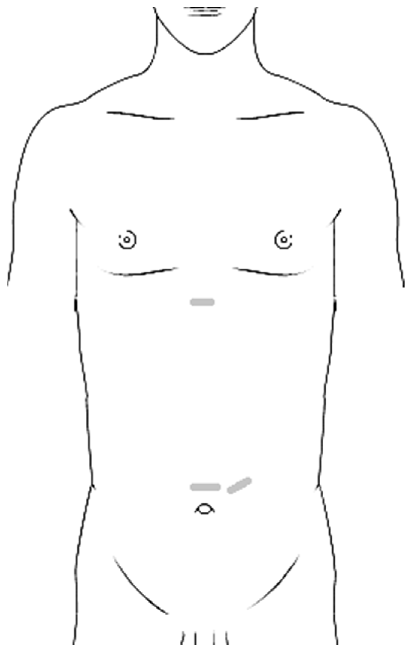
Figure 1. Schema of surgical approach: cranial incision for the first knot and caudal incisions for trocars and the last knot (grey short lines). The figure was created with smart.servier.com.
Applsci 13 09841 g001
Figure 2. Different steps of surgical preparation: (A) Preparation of an epifascial cavity. (B,C) Continuous suture of the rectus abdominis.
Applsci 13 09841 g002
Table 1. The characteristics of the subjects.
Male/Female1/3
Age (mean, range)4 (2–8) years
Dehiscence (maximum, n = 2)4 and 2 cm measured with sonography
Secondary diagnosisUmbilical hernia (1×)
Clinical complaintsProgredience of the rectus diastasis
(8 mm within 7 months) with significant prolapse of intrabdominal structures, periumbilical pain, postprandial pain, pain on passing stool
Disclaimer/Publisher’s Note: The statements, opinions and data contained in all publications are solely those of the individual author(s) and contributor(s) and not of MDPI and/or the editor(s). MDPI and/or the editor(s) disclaim responsibility for any injury to people or property resulting from any ideas, methods, instructions or products referred to in the content.