Article

Gender Dimorphism in Maxillary Permanent Canine Odontometrics Based on a Three-Dimensional Digital Method and Discriminant Function Analysis in the Saudi Population

by
Yousef Majed Almugla
1,*,†,
Guna Shekhar Madiraju
2,†,
Rohini Mohan
3 and
Sajith Abraham
2
1
Faculty in Orthodontics, Department of Preventive Dental Sciences, College of Dentistry, King Faisal University, Al Ahsa 31982, Saudi Arabia
2
Department of Preventive Dental Sciences, College of Dentistry, King Faisal University, Al Ahsa 31982, Saudi Arabia
3
Community Dental Services, Port Talbot Research Centre, Swansea Bay University Health Board, Port Talbot SA12 7BJ, UK
*
Author to whom correspondence should be addressed.
These authors contributed equally to this work.
Appl. Sci. 2023, 13(16), 9326; https://doi.org/10.3390/app13169326
Submission received: 5 June 2023 / Revised: 11 August 2023 / Accepted: 12 August 2023 / Published: 17 August 2023
(This article belongs to the Special Issue Applications of Three-Dimensional Technology in Health Care Sciences)

Abstract

:
The present study aimed to analyze the gender dimorphism in odontometrics of permanent maxillary canines using a three-dimensional digital method and to test the accuracy in gender estimation using discriminant function analysis in a sample of the Saudi population. A total of 120 diagnostic dental casts of patients aged 16–29 years were used in the present study. Plaster models of their maxillary dentition obtained from the archives were scanned and digitally measured using a three-dimensional digital method. The mesiodistal width of the right and left maxillary canines and intercanine distance were measured. Gender dimorphism was established using the Garn method. Data were statistically analyzed using descriptive statistics, the Mann–Whitney U test and discriminant analyses. Males showed larger mean dimensions of canines than females with regard to both mesiodistal width and intercanine distance, and the difference was statistically significant (p < 0.05). The right maxillary canine mesiodistal width showed a higher percentage of gender dimorphism (3.5%). Discriminant function analysis showed that the overall accuracy of gender prediction was 80.5% for the multivariate analysis. The univariate discriminant function equation revealed that intercanine distance was the most accurate predictor of gender (78%), followed by the right canine mesiodistal width (76.3%). The use of three-dimensional technology for odontometrics presents a promising method, and permanent maxillary canine parameters can be used as an acceptable ancillary tool for gender estimation in forensic science.

1. Introduction

Odontometrics has become a valuable tool for age and gender estimation in forensic investigation. Whilst biochemical analysis such as DNA profiling, fingerprints, and anthropometric data are among the standard methods employed for human identification in forensic science, odontometric measurements also can provide comparatively reliable results [1]. Gender dimorphism has been reported using various anthropometric parameters including foot and hand dimensions [2]. Moreover, studies suggest that it can also be explored through the analysis of teeth based on the pattern of dental development and eruption, the expression of amelogenin protein, dental morphology, and tooth dimensions [3].
Quantitative analyses, such as discriminant function analysis, have been reported to have a crucial role in forensic applications for gender differentiation. Moreover, any method which depends on the least number of predictors possible to improve the percentage of correct classification would be a significant addition to the forensic anthropology [4]. Gender estimation using the permanent teeth dimensions has been the subject of previous studies, and a variety of measuring methods and equations have been developed in different populations. Recent advancements have enabled the digitization of patient dental casts by capturing the surface data of these casts using holography and photo-optical or laser-optical scanning [5]. The advantage of digital models is their ability to analyze tooth characteristics in a three-dimensional aspect ensuring clinically acceptable and reproducible results. Odontometric assessment using canine dimensions has been proven to be valuable in gender identification owing to their ability to withstand vulnerable conditions. Mesiodistal and buccolingual widths and the canine index of permanent teeth were the most commonly used parameters in predicting an individual’s gender based on dental measurements [6]. Permanent canines are given importance during orthodontic treatment as they play a vital role in achieving an ideal occlusion and aesthetics owing to their anatomical and functional value. Moreover, tooth size discrepancies or variations assessed using odontometrics might aid in customized orthodontic management in individuals. Odontometric values vary between different population groups and also within the groups [7], and hence the use of external databases does not seem advisable even though they come from genetically or geographically related people [8]. Odontometric standards for estimation of gender in the Saudi population are scarce, and no population-specific standard is available in the literature. Hence, the present study aimed to analyze the gender dimorphism in the mesiodistal width and intercanine distance of the permanent maxillary canines using a three-dimensional (3D) digital technique and to test the accuracy in gender estimation using discriminant analysis in a sample of the Saudi population from Al Ahsa in the Eastern region of Saudi Arabia.

2. Materials and Methods

2.1. Study Sample

This descriptive study utilized the diagnostic dental casts of 120 subjects (60 males and 60 females) aged 16–29 years who had visited the dental clinical complex during the period from September 2019 to April 2022 for various diagnostic and/or orthodontic purposes. Dental casts which met the inclusion criteria were selected from the archives of the university dental setting based on the convenience sampling technique. This study had used only the maxillary arch dental casts for analysis. The inclusion criteria were dental casts of subjects aged 16–29 years with fully erupted teeth from the first molar on the right side to the first molar on the left side, with no evidence of developmental shape anomalies and occlusal abnormalities involving the canines. Subjects with parafunctional habits, mouth breathing, periodontal disease, crowding, malaligned teeth, proximal caries, large restorations, fixed or temporary crowns on canines that might interfere with accuracy of tooth measurement, abrasions or attrition, previous history of or ongoing orthodontic treatment, abnormal morphology, or congenitally missing teeth were excluded from the study sample. The study age group was selected as the dimensional changes due to abrasion and attrition are minimal, and the intercanine distance is fixed by the age of 12 years [9]. The study protocol was reviewed and approved by the Institutional Ethical and Review Board (Ref: Kfu-Rec-2022-Oct-Ethics215), and the study was conducted in accordance with the ethical standards set out in the 1964 Declaration of Helsinki.

2.2. Three-Dimensional Digitization Method

The dental casts were scanned, and 3D digital models were obtained through the CAD/CAM (Computer-Aided Design and Computer-Aided Manufacturing) system (Kavo Arctica, Kavo, Germany). This desktop scanner uses a combination of striped light scanning technology and highly sensitive 3D sensors for scanning and has a built-in blue filter to scan dental casts to an accuracy of 20 μm. The generated data were stored in STL (Standard Triangle Language) format, which utilizes small triangles to describe the surface of digitally captured objects and is a standard format used by many CAD systems. The STL format files were imported to 3Shape® Ortho Analyzer™ software version 2020 (3Shape, Copenhagen, Denmark) for digital measurements. A single experienced and precalibrated investigator who was blinded to the gender selection of casts conducted all the measurements, including scanning and marking of the study casts to avoid interexaminer error. Each parameter was measured three times, and the average value was recorded as the final result. The same investigator repeated the measurements at a two-week interval on a total of 30 randomly selected digital casts to assess the intraexaminer variability using intraclass correlation coefficient (ICC) measurement agreement. Measurements on the dental casts included the mesiodistal crown (MD) width of maxillary canines and the intercanine distance (ICD). The greatest MD width of each maxillary canine was measured between the mesial and distal anatomical contact points. In the absence of contact points, the greatest MD width of the canine was measured and recorded. ICD referred to the linear distance between the cusp tips of both the maxillary canines or in the center of their wear facets should they be present (Figure 1a,b).
The maxillary canine index (MCI) and standard canine index (SCI) were calculated using the formula given by Muller et al. [10].
Maxillary Canine Index = mesiodistal crown width of maxillary canine/maxillary intercanine width.
Standard canine index = (mean male MCI − SD) + (mean female MCI + SD)/2
Gender was then predicted based on the observed maxillary canine index and standard canine index for the Saudi population of the Eastern province. If the observed MCI was more than SCI, then the individual was considered to be male, and if the observed MCI was less than SCI, then the individual was considered to be female [11].
The percentage of gender dimorphism was assessed using the Garn et al. [12] formula:
Gender Dimorphism = [Xm/Xf] − 1 × 100
where:
  • Xm = mean value of tooth dimensions for males;
  • Xf = mean value of tooth dimensions for females.

2.3. Statistical Analysis

Data were subjected to statistical analysis using the Statistical Package for the Social Sciences (version 20.0 IBM Corp., Armonk, NY, USA). Descriptive statistics and frequencies were ascertained. Variables were evaluated for normality using the Shapiro–Wilk test. Since the data showed non-normal distribution, nonparametric tests were carried out using the Mann–Whitney U test to identify gender differences among the samples. The mean difference values of variables between the right and the left side within the males and females were tested for significance using the Wilcoxon signed-ranks test. The level of significance was set as p < 0.05. The discriminant function analysis was conducted, and discriminant functions for each variable were generated in order to determine the best possible gender-differentiation predictor. For gender identification, the unstandardized discriminant function procedure was employed. The ability of this function to identify males and females is indicated as the percentage of individuals correctly classified from the sample that generated the function. Cross-validation was used to improve the performance of the predictive statistical method in this study.

3. Results

3.1. Descriptive Statistics

A total of 118 out of 120 dental casts were included for final analysis. Data related to two dental casts (1 male and 1 female) were excluded from the final sample due to corrupted files during analysis. The mean age in years for males and females were 23.3 ± 3.22 (Mean ± SD) and 21.45 ± 2.46, respectively, and 22.3 ± 3.01 for the total study sample. The intraclass correlation coefficient for MD width and ICD of permanent maxillary canines displayed by intraexaminer agreement was between 0.956 and 0.974 (excellent; 95% CI: 0.910–0.981). Males had significantly larger mean dimensions of both the right and left maxillary permanent canines compared to females. The mean MD width of the maxillary canine for males and females on the right side was 7.65 and 7.39 mm, respectively, while on the left side it was 7.55 mm in males and 7.33 mm in females. The difference in the mean MD width of the maxillary canine between males and females was statistically significant on both the right (p < 0.001) and the left (p < 0.001) sides. The mean MD width of the right canine was greater than that of the left canine in both males and females. The mean maxillary ICD was significantly higher in males than that seen in females (p < 0.001). The 95% confidence interval of maxillary canine MD width and ICD for the study sample shows that there is a probability of 95% that the mean width of the right and left maxillary canines is greater than 7.53 and 7.43 mm, respectively, in males and lower than 7.46 and 7.40 mm, respectively, in females. The mean values of MCI calculated for both the right and left maxillary canines showed statistically significant differences between the genders (p < 0.05) (Table 1).

3.2. Percentage of Gender Dimorphism

The gender dimorphism percentages for the right and left MD width of maxillary canines were 3.5% and 3%, respectively, whilst for maxillary ICD it was found to be 3.14%. However, the percentage of gender dimorphism for both the right and the left MCI was found to be 0.94. The right maxillary canine MD width exhibited the greatest percentage of gender dimorphism (3.5%) (Figure 2).
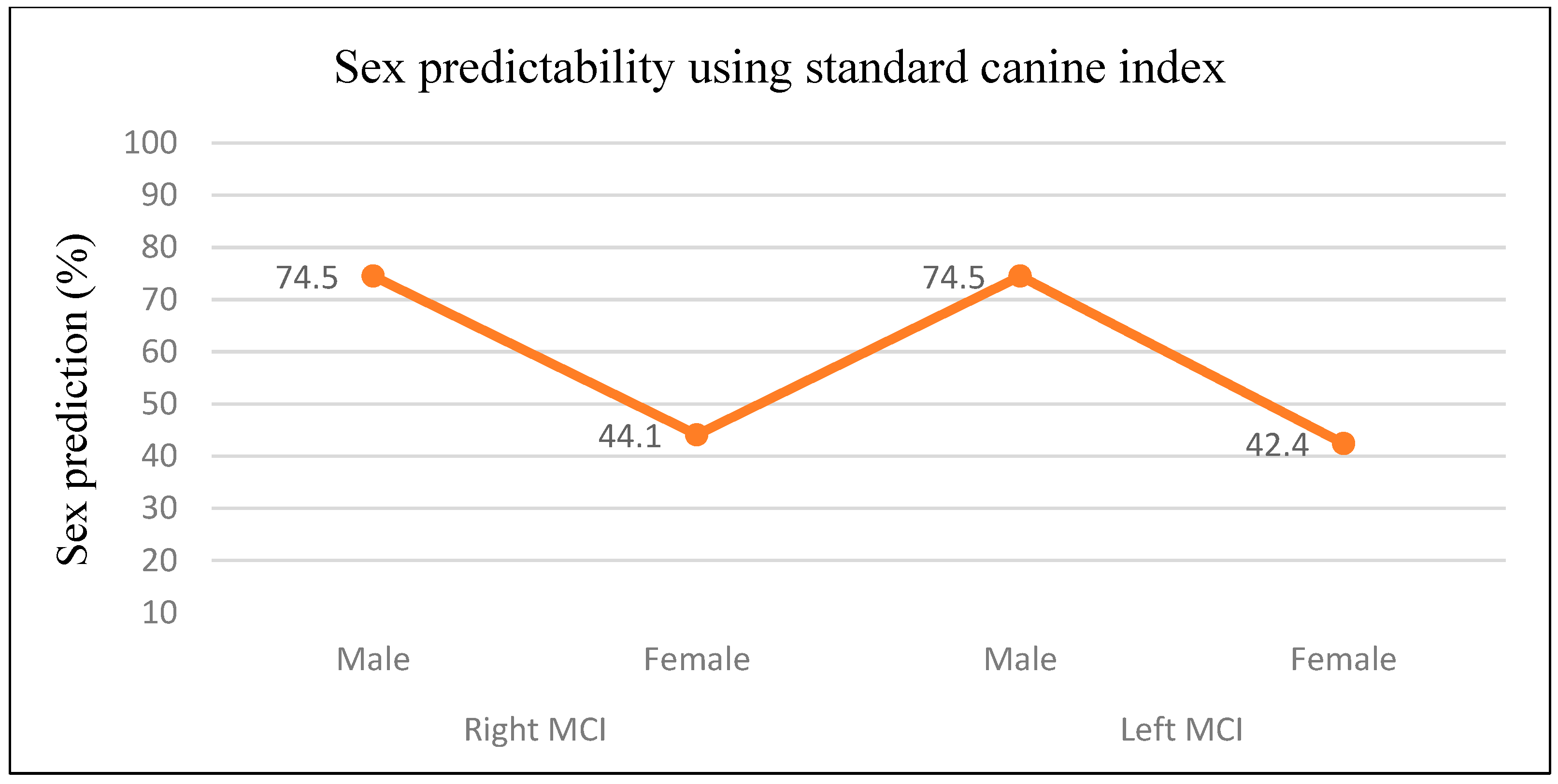
The standard canine index measurements for the right and left maxillary canines were 0.213 and 0.211, respectively. The prediction of gender using the standard canine index for both the right and left canines showed an overall accuracy of 59.3% and 58.4%, respectively, with males being predicted more accurately (74.5%) than the females (Figure 3).

3.3. Discriminant Analysis

Discriminant function analysis was conducted using the right canine MD width, left canine MD width, and ICD as predictor variables. The multivariate discriminant function equation showed an overall gender prediction accuracy of 80.5%. The unstandardized coefficients and constants were used to generate the discriminant function equation. The cutting point of the discriminant function calculated from the average summation of the group centroid value was “zero”. Hence, a discriminant function score greater than the cutting point classifies the individual as male, and a score less than the cutting point classifies the individual as “zero”. Hence, a discriminant function score greater than the cutting point classifies the individual as male, and a score less than the cutting point classifies the individual as female. The overall tested accuracy of gender determination for univariate discriminant function equations ranged from 66.9% to 78%. The accuracy of gender determination ranged from 74.6% to 83.1% for females and 59.3% to 76.3% for males. Function 4, which included ICD, was the most accurate overall (78%), and Function 3, which included tooth 23, showed the least accuracy rate (66.9%) (Table 2).
DFA showed that females were classified with better accuracy than the males. Discriminant function equations calculated for the study population are shown in Table 3. Cross-validation accuracy was found to be similar to the original classification accuracy in all the cases.

4. Discussion

Gender dimorphism of permanent teeth has been well established in the literature. Several studies including recent systematic reviews on tooth crown mesiodistal measurements for estimating sexual dimorphism across different population groups had documented that canine dimensions provide the most effective indicator of gender dimorphism, whilst few other studies have reported dimorphism in the premolars and molars, and in the mean maxillary arch and palatal depths [8,13,14,15]. Canines are considered to be among the teeth least affected by developmental abnormalities in relation to the tooth size and morphology, and they are the most stable teeth in the arch. Moreover, the permanent maxillary canine was found to be the most reliable in the assessment of gender, and this view seems to be reflected in several previous studies [13,16,17,18]. Other authors, however, had reported mandibular canines to exhibit the most effective gender dimorphism [2,19]. Most studies conducted on gender dimorphism in the Middle East region have not included Saudi Arabia, despite having the largest population compared to other ethnic groups, and the literature contains very few studies on the measurement of tooth dimensions in the Saudi population [16,20,21,22]. The present study attempted to estimate gender dimorphism using digitized dental casts by measuring the mesiodistal width and ICD of maxillary permanent canines and verify the accuracy for gender estimation in a sample of the native Saudi population from the Eastern region of Saudi Arabia.
Measurements of linear dimensions of teeth have been reported to be simple, less time-consuming, reliable, noninvasive, and easy to perform when compared to other forensic techniques. Several authors had concluded the MD dimension to be a better predictor of gender than the buccolingual diameter [23,24]. Odontometric studies evaluating gender dimorphism in teeth dimensions had performed measurements conventionally using an electronic digital caliper, either intraorally or from the dental casts using an indirect method, radiographs, or using Raman spectra of teeth [8,25]. Plaster or dental casts have been traditionally used for diagnostic purposes as they are economical, but they present drawbacks such as storage problems and risk of damage and fracture due to their brittle nature. Digital study models offer an alternative to overcome these limitations as they present comparatively better accuracy in measurements, ease of storage and transfer in a computer, facilitate reproducibility of information, and have no risk of physical deterioration that may occur with plaster casts [26]. The present study used a 3D scanning method for measuring odontometric data owing to its advantages in obtaining reliable and high-precision measurements. Previous studies evaluating the accuracy of digital models compared to plaster models had reported significant differences in tooth width measurements between the two methods [27]. Other authors reported that digital models were time-saving and yielded reliable and clinically acceptable measurements compared to a digital caliper [5,28]. This study used only maxillary canine odontometrics to establish gender dimorphism. This may be particularly useful for predicting gender in situations where only a part of the skull with maxilla is available for forensic investigations [29].
The MD width of both the right and the left maxillary canines was significantly greater in males than in females in the present study. This finding corroborates with that of previous studies conducted among the Saudi population [20,21,22] and other studies in the literature [17,18,30]. Contrary to this, a previous study among the Saudi population had reported no statistically significant differences in the mean MD width of right and left maxillary canines between the genders [16]. Another study in Nigeria, however, had reported that the MD width of the left maxillary canine was significantly higher in males than in females [31].
Intercanine distance has been reported to be a useful parameter in gender differentiation, as the eruption of canines and growth in the width of both the jaws, including the width of the dental arches, are completed before the adolescent growth changes, and the intercanine distance does not increase after 12 years of age [9]. In the present study, it was observed that the males had a tendency to have statistically significant higher mean values of ICD compared to the female counterparts (p < 0.001). This finding was consistent with those of other authors [16,24,30]. However, Shetty et al. [32] and Alanazi et al. [33], in their studies, reported no statistically significant differences in the mean maxillary ICD between males and females. Previous studies in the central region of Saudi Arabia reported smaller dimensions of maxillary ICD compared to that seen in this study on a sample of the Eastern Saudi Arabian population [16,20].
This study revealed significant gender dimorphism in the MD width and ICD of the maxillary canines, with males showing larger corresponding mean values than females, which was in accordance with previous studies [24,30,31]. The results of gender dimorphism of the maxillary canine in the present study showed a greater dimorphism in the MD width of the right canine (3.5%), followed by ICD (3.14%). A recent study on the native Saudi Arabian population found reverse gender dimorphism, as the mean MD width of the maxillary canine was comparatively lesser in males than the females [33]. Gupta et al. [30] reported a higher percentage of gender dimorphism with respect to maxillary ICD and found that the right canine was more dimorphic compared to the left canine. Liu et al. (2021), in a study on the Chinese population, revealed a slightly higher percentage (4.26%) of gender dimorphism for MD width of the left canine compared to the right canine (4.18%) [24]. In contrast to the present findings, a previous study on the Saudi Arabian population found a lower magnitude of gender dimorphism for maxillary canines [16]. Variations in the degree of gender dimorphism have been noted in different population groups, and the specific reason is still unclear. Cultural, environmental, and genetic factors have been known to influence tooth morphology. Furthermore, differences in study criteria, measurement methods employed, and racial and ethnic differences in different studies might contribute to varying results. As a result, population-specific data are needed for establishing reference data and prediction models for gender estimation.
The method of gender prediction by means of canine indices is beneficial as it is economical, does not require complex equipment, involves simple mathematical calculation, and is convenient for the analysis of large samples [34]. Gender prediction using the standard canine index in the present study was more accurate in males compared to female counterparts; however, an overall accuracy of prediction was closer to 59% for both the right and left canines. Hence, the accuracy of gender prediction using SCI was considered poor in the present study. This study revealed that the difference in the mean values of right and left MCI between males and females was significant (p < 0.05). Within the gender, the mean difference values of MCI between the right and left sides were found to be statistically significant both in males (p < 0.001; Wilcoxon Z = −6.695) and females (p < 0.001; Wilcoxon Z = −5.615). On the contrary, Gupta et al. [30] found no significant difference in the mean maxillary canine index of the right and left sides between the two genders or between two sides within the same gender.
Discriminant analysis has been reported to be a reliable method to ascertain gender discriminant functions, whilst multivariate discriminant analyses generate higher discrimination percentages compared to the univariate method [4]. The multivariate discriminant function analysis with maxillary canine MD width and ICD, reported by Al-Rifaiy et al. [16] on the Central Saudi population, permitted correct classification of 66.67% of males and 64.29% of females, with an overall accuracy of 65.48%. However, contrary to their finding, this study found that females (81.4%) were predicted slightly better than males (79.7%), and with a higher overall accuracy rate (80.5%). Another odontometric study on dental cast models found a high gender prediction accuracy rate of 99.8% in the population using stepwise discriminant functions [35]. Univariate discriminant analysis has been reported to be useful in circumstances where a limited number of teeth are available for forensic investigations. Peckmann et al. [18], in their study on the Chilean population, found that the univariate discriminant functions of the right and left maxillary canines showed overall accuracy rates of 65% and 64%, respectively, which are comparatively lower than those in the current study. Al-Rifaiy et al. [16] further concluded that ICD was more reliable than MD width in the correct classification of gender. Similarly, in the present study, discriminant function analysis showed that ICD was the most accurate predictor of gender (78%), followed by the right canine MD width (76.3%), whilst the left canine MD width showed 66.9% overall accuracy. Recent advances in the forensic and dental research fields have enhanced the gender determination methods with the development of newer computer-aided techniques. Application of machine learning methods in the age and gender estimation has been increasingly gaining attention as they are more accurate, less time-consuming, and cost-efficient compared to other methods [36,37].
One limitation of the present study was that the sample used for evaluation of maxillary canines was selected from patients who sought dental care at a university dental clinical complex in Al-Ahsa, in the Eastern region of Saudi Arabia, and therefore generalization of results derived from this study in the Saudi population is limited. Further investigations involving larger samples from different ethnic populations should be conducted. Whilst plaster casts were scanned and digitized into 3D digital models for measurement and analysis of data in the present study, the possibility of minor deformities in the resultant plaster casts pertaining to impression material manipulation and cast pouring due to human errors such as voids or bubbles at critical regions of the impression warrants consideration. Another limitation might include the technical errors during scanning and computer processing of the images, which could result in distortions in the 3D digital models [38].

5. Conclusions

The findings of the present study indicate that there are gender-specific differences in linear dimensions of maxillary permanent canine teeth in the study population. When the MD width of maxillary canines was greater than 7.43 mm, there was 95% probability of accurate male gender prediction. The maxillary canine index may not be useful in gender prediction due to low accuracy. This study showed that multivariate discriminant functions provided a higher level of accuracy (80.5%), and univariate analysis using ICD and right MD width demonstrated accuracy rates of more than 75% for gender prediction. Hence, discriminant functions derived from the present study could be of value in gender determination of the Saudi population from the Eastern region. This also emphasizes the need for population-specific odontometric data for forensic odontology, treatment planning, and patient care. The use of 3D digital models might be a promising alternative to plaster models for odontometry with clinically acceptable accuracy and reliability of tooth dimensions.

Author Contributions

Conceptualization, Y.M.A.; methodology, G.S.M. and Y.M.A.; software, Y.M.A. and R.M.; validation, Y.M.A. and G.S.M.; formal analysis, G.S.M.; investigation, Y.M.A.; resources, S.A.; data curation, G.S.M. and R.M.; writing—original draft preparation, G.S.M. and R.M.; writing—review and editing, G.S.M., Y.M.A. and S.A.; project administration, G.S.M. and S.A.; funding acquisition, Y.M.A. All authors have read and agreed to the published version of the manuscript.

Funding

This work was supported through the Annual Funding track by the Deanship of Scientific Research, Vice Presidency for Graduate Studies and Scientific Research, King Faisal University, Al Ahsa, Saudi Arabia (Ref. Grant 3336).

Institutional Review Board Statement

This study was conducted at the College of Dentistry, King Faisal University, Al Ahsa, Saudi Arabia, following the “Helsinki Declaration”, and approved by the Institutional Ethical and Review Board, Deanship of Scientific Research (Ref. Kfu-Rec-2022-Oct-Ethics215). The study was retrospective and noninterventional; therefore, patients did not undergo any treatment for the study.

Informed Consent Statement

Informed consent was obtained from all subjects involved in the study. The patients sign a general consent before any treatment or investigation is rendered, which includes a consent to use the findings in future retrospective studies without any personal identification.

Data Availability Statement

The data presented in this study are available on reasonable request from the corresponding author.

Acknowledgments

The authors want to thank the Deanship of Scientific Research, King Faisal University, Al Ahsa, Saudi Arabia, for supporting this study.

Conflicts of Interest

The authors declare no conflict of interest.

References

  1. Prabhu, R.V.; Dinkar, A.D.; Prabhu, V.D.; Rao, P.K. Cheiloscopy: Revisited. J. Forensic Dent. Sci. 2012, 4, 47–52. [Google Scholar] [CrossRef] [PubMed]
  2. Sivakumar, N.; Bansal, D.; Narwal, A.; Kamboj, M.; Devi, A. Gender determination analysis using anthropometrical dimensions of 2D:4D, foot index and mandibular canine index. J. Oral Maxillofac. Pathol. 2020, 24, 510–516. [Google Scholar] [CrossRef] [PubMed]
  3. Moreno-Gómez, F. Gender dimorphism in human teeth from dental morphology and dimensions: A dental anthropology viewpoint. In Gender Dimorphism [Internet]; Moriyama, H., Ed.; IntechOpen: London, UK, 2013; pp. 97–124. [Google Scholar] [CrossRef]
  4. Jiménez-Arenas, J.M.; Esquivel, J.A. Comparing two methods of univariate discriminant analysis for gender discrimination in an Iberian population. Forensic Sci. Int. 2013, 228, 175e1–175e4. [Google Scholar] [CrossRef] [PubMed]
  5. Fleming, P.S.; Marinho, V.; Johal, A. Orthodontic measurements on digital study models compared with plaster models: A systematic review. Orthod. Craniofac. Res. 2011, 14, 1–16. [Google Scholar] [CrossRef] [PubMed]
  6. Tardivo, D.; Sastre, J.; Catherine, J.H.; Leonetti, G.; Adalian, P.; Foti, B. Gender determination of adult individuals by three-dimensional modeling of canines. J. Forensic Sci. 2015, 60, 1341–1345. [Google Scholar] [CrossRef]
  7. Al-Gunaid, T.; Yamaki, M.; Saito, I. Mesiodistal tooth width and tooth size discrepancies of Yemeni Arabians: A pilot study. J. Orthod. Sci. 2012, 1, 40–45. [Google Scholar] [CrossRef] [PubMed]
  8. Beschiu, L.M.; Ardelean, L.C.; Tigmeanu, C.V.; Rusu, L.-C. Cranial and Odontological Methods for Gender Estimation—A Scoping Review. Medicina 2022, 58, 1273. [Google Scholar] [CrossRef]
  9. Proffit, M.R.; Field, H.W., Jr.; Ackerman, J.L.; Thompson, P.M.; Tullock, S.A. Contemporary Orthodontics; CV Mosby Co.: St. Louis, MO, USA, 1984; pp. 84–89. [Google Scholar]
  10. Muller, M.; Lupi-Pegurier, L.; Quatrehomme, G.; Bolla, M. Odontometrical method useful in determining gender and dental alignment. Forensic Sci. Int. 2001, 121, 194–197. [Google Scholar] [CrossRef]
  11. Bakkannavar, S.M.; Monteiro, F.N.; Arun, M.; Pradeep Kumar, G. Mesiodistal width of canines: A tool for gender determination. Med. Sci. Law 2012, 52, 22–26. [Google Scholar] [CrossRef]
  12. Garn, S.M.; Lewis, A.B.; Kerewsky, R.S. Buccolingual size asymmetry and its developmental meaning. Angle Orthod. 1967, 37, 186–193. [Google Scholar] [CrossRef]
  13. Neves, J.A.; Antunes-Ferreira, N.; Machado, V.; Botelho, J.; Proença, L.; Quintas, A.; Mendes, J.J.; Delgado, A.S. Gender Prediction Based on Mesiodistal Width Data in the Portuguese Population. Appl. Sci. 2020, 10, 4156. [Google Scholar] [CrossRef]
  14. Da Silva, P.R.; Lopes, M.C.; Martins-Filho, I.E.; Haye Biazevic, M.G.; Michel-Crosato, E. Tooth Crown Mesiodistal Measurements for the Determination of Sexual Dimorphism across a Range of Populations: A Systematic Review and Meta-analysis. J. Forensic Odontostomatol. 2019, 37, 2–19. Available online: https://ojs.iofos.eu/index.php/Journal/article/view/1034 (accessed on 10 August 2023). [PubMed]
  15. Ajmal, M.A.; Roberts, T.S.; Beshtawi, K.R.; Raj, A.C.; Sandeepa, N.C. Sexual dimorphism in odontometric parameters using cone beam CT: A systematic review. Head Face Med. 2023, 19, 6. [Google Scholar] [CrossRef] [PubMed]
  16. Al-Rifaiy, M.Q.; Abdullah, M.A.; Ashraf, I.; Khan, N. Dimorphism of mandibular and maxillary canine teeth in establishing identity. Saudi Dent. J. 1997, 9, 17–20. [Google Scholar]
  17. Filipovic, G.; Radojicic, J.; Stosic, M.; Janosevic, P.; Ajdukovic, Z. Odontometric analysis of permanent canines in gender determination. Arch. Biol. Sci. 2013, 65, 1279–1283. [Google Scholar] [CrossRef]
  18. Peckmann, T.R.; Logar, C.; Garrido-Varas, C.E.; Meek, S.; Pinto, X.T. Gender determination using the mesio-distal dimension of permanent maxillary incisors and canines in a modern Chilean population. Sci. Justice 2016, 56, 84–89. [Google Scholar] [CrossRef]
  19. Abaid, S.; Zafar, S.; Kruger, E.; Tennant, M. Mesiodistal dimensions and gender dimorphism of teeth of contemporary Western Australian adolescents. J. Oral Sci. 2021, 63, 247–251. [Google Scholar] [CrossRef]
  20. Hashim, H.A.; Murshid, Z.A. Mesiodistal tooth width. A comparison between Saudi males and females. Part 1. Egypt. Dent. J. 1993, 39, 343–346. [Google Scholar]
  21. Alkofide, E.; Hashim, H. Intermaxillary tooth size discrepancies among different malocclusion classes: A comparative study. J. Clin. Pediatr. Dent. 2002, 26, 383–387. [Google Scholar] [CrossRef]
  22. Asiry, M.; Hashim, H. Tooth Size Ratios in Saudi Subjects with Class II, Division 1 Malocclusion. J. Int. Oral Health 2012, 4, 29–34. Available online: http://www.ispcd.org/journal-of-international-oral-health.html (accessed on 7 May 2023).
  23. Ramakrishnan, K.; Sharma, S.; Sreeja, C.; Pratima, D.B.; Aesha, I.; Vijayabanu, B. Gender determination in forensic odontology: A review. J. Pharm. Bioallied Sci. 2015, 7 (Suppl. S2), S398–S402. [Google Scholar] [CrossRef] [PubMed]
  24. Liu, J.; Liu, Y.; Wang, J.; Ge, S.; Zhang, Y.; Wang, X.; Du, L.; He, H. Permanent Maxillary Odontometrics for Gender Estimation Based on a 3-Dimensional Digital Method. Med. Sci. Monit. 2021, 27, e933450. [Google Scholar] [CrossRef]
  25. Gamulin, O.; Skrabic, M.; Serec, K.; Par, M.; Bakovic, M.; Krajacic, M.; Babic, S.D.; Segedin, N.; Osmani, A.; Vodanovic, M. Possibility of human gender recognition using Raman spectra of teeth. Molecules 2021, 26, 3983. [Google Scholar] [CrossRef] [PubMed]
  26. Lippold, C.; Kirschneck, C.; Schreiber, K.; Abukiress, S.; Tahvildari, A.; Moiseenko, T.; Danesh, G. Methodological accuracy of digital and manual model analysis in orthodontics—A retrospective clinical study. Comput. Biol. Med. 2015, 62, 103–109. [Google Scholar] [CrossRef]
  27. Grunheid, T.; Patel, N.; De Felippe, N.L.; Wey, A.; Gaillard, P.R.; Larson, B.E. Accuracy, reproducibility, and time efficiency of dental measurements using different technologies. Am. J. Orthod. Dentofac. Orthop. 2014, 145, 157–164. [Google Scholar] [CrossRef] [PubMed]
  28. Amuk, N.G.; Karsli, E.; Kurt, G. Comparison of dental measurements between conventional plaster models, digital models obtained by impression scanning and plaster model scanning. Int. Orthod. 2019, 17, 151–158. [Google Scholar] [CrossRef]
  29. Phulari, R.G.S.; Rathore, R.; Talegaon, T.; Jariwala, P. Comparative assessment of maxillary canine index and maxillary first molar dimensions for gender determination in forensic odontology. J. Forensic Dent. Sci. 2017, 9, 110. [Google Scholar] [CrossRef]
  30. Gupta, S.; Chandra, A.; Gupta, O.P.; Verma, Y.; Srivastava, S. Establishment of gender dimorphism in North Indian population by odontometric study of permanent maxillary canine. J. Forensic Res. 2014, 5, 224. [Google Scholar] [CrossRef]
  31. Nuhu, S.; Dalori, B.M.; Adamu, L.H.; Buba, M.A. Establishment of gender dimorphism using maxillary canine of the university of maiduguri students, Nigeria. Int. J. Forensic Odontol. 2019, 4, 68–72. [Google Scholar] [CrossRef]
  32. Shetty, S.J.; Ratnaparkhi, I.; Pereira, T.; Acharya, S.; Gotmare, S.; Kamath, P. Odontometric analysis of canines to establish gender dimorphism in an urban population. Indian J. Dent. Res. 2019, 30, 855–859. [Google Scholar] [CrossRef]
  33. Alanazi, A.A.; Almutair, A.M.; Alhubayshi, A.; Almalki, A.; Naqvi, Z.A.; Alassaf, A.; Almulhim, B.; Alghamdi, S.A.; Mallineni, S.K. Morphometric Analysis of Permanent Canines: Preliminary Findings on Odontometric Gender Dimorphism. Int. J. Environ. Res. Public Health 2022, 19, 2109. [Google Scholar] [CrossRef] [PubMed]
  34. Patel, R.A.; Chaudhary, A.R.; Dudhia, B.B.; Macwan, Z.S.; Patel, P.S.; Jani, Y.V. Mandibular canine index: A study for gender determination in Gandhinagar population. J. Forensic Dent. Sci. 2017, 9, 135–143. [Google Scholar] [CrossRef] [PubMed]
  35. Litha; Girish, H.C.; Murgod, S.; Savita, J.K. Gender determination by odontometric method. J. Forensic Dent. Sci. 2017, 9, 44. [Google Scholar] [CrossRef] [PubMed]
  36. Bertsatos, A.; Chovalopoulou, M.E.; Bružek, J.; Bejdová, Š. Advanced procedures for skull gender estimation using genderly dimorphic morphometric features. Int. J. Legal Med. 2020, 134, 1927–1937. [Google Scholar] [CrossRef] [PubMed]
  37. Uabundit, N.; Chaiyamoon, A.; Iamsaard, S.; Yurasakpong, L.; Nantasenamat, C.; Suwannakhan, A.; Phunchago, N. Classification and morphometric features of pterion in Thai population with potential gender prediction. Medicina 2021, 57, 1282. [Google Scholar] [CrossRef]
  38. Amornvit, P.; Rokaya, D.; Sanohkan, S. Comparison of Accuracy of Current Ten Intraoral Scanners. Biomed. Res. Int. 2021, 2021, 2673040. [Google Scholar] [CrossRef]
Figure 1. Measurements on mesiodistal width (a) and intercanine distance (b) of maxillary canines on three-dimensional digital models.
Applsci 13 09326 g001
Figure 2. Percentage of gender dimorphism in maxillary parameters.
Applsci 13 09326 g002
Figure 3. Gender predictability using standard canine index.
Applsci 13 09326 g003
Table 1. Comparison of right and left maxillary canine dimensions of study sample.
VariableToothGenderNMean ± SD (mm)95% Confidence Intervalp-Value *
LowerUpper
Mesiodistal Width 13Male597.65 ± 0.4537.537.760.000
(Z = −5.034)
Female597.39 ± 0.2667.327.46
23Male597.55 ± 0.4487.437.660.000
(Z = −4.035)
Female597.33 ± 0.2647.267.40
Intercanine Distance Male5935.28 ± 0.67935.1035.450.000
(Z = −6.731)
Female5934.21 ± 0.69234.0334.39
Maxillary Canine Index13Male590.216 ± 0.0120.21290.21940.045
(Z = −2.005)
Female590.214 ± 0.0070.21160.2158
23Male590.214 ± 0.0120.21090.21730.030
(Z = −2.167)
Female590.212 ± 0.0080.20960.2137
* Mann–Whitney U test; SD: standard deviation; N: number of subjects; Tooth numbering in FDI system.
Table 2. Discriminant function analysis for gender estimation.
FunctionVariableUnstandardized
Coefficient
ConstantWilks’s LambdaCentroidsClassification Accuracy
MaleFemaleMale
n (%)
Female
n (%)
Overall
n (%)
Multivariate discriminant function
1Right canine (13) MD7.356−46.1450.5710.860−0.86047 (79.7)48 (81.4)95 (80.5)
Left canine (23) MD−7.037
ICD1.243
Univariate discriminant function
2Right canine (13) MD2.687−20.2100.8900.348−0.35841 (69.5)49 (83.1)90 (76.3)
3Left canine (23) MD2.718−20.2270.9210.291−0.29135 (59.3)44 (74.6)79 (66.9)
4ICD1.457−50.6300.6200.777−0.77745 (76.3)47 (79.7)92 (78)
MD: mesiodistal width; ICD: intercanine distance.
Table 3. Discriminant function equations of the study sample.
FunctionEquation (Discriminant Function)
1y = −46.145 + (7.356)(Tooth 13) + (−7.037)(Tooth 23) + (1.243)(ICD)
2y = −20.210 + (2.687)(Tooth 13)
3y = −20.227 + (2.718)(Tooth 23)
4y = −50.630 + (1.457)(ICD)
y is the discriminant score; cutting point = 0; If y-value ≥ cutting point, the individual is classified as male.
Disclaimer/Publisher’s Note: The statements, opinions and data contained in all publications are solely those of the individual author(s) and contributor(s) and not of MDPI and/or the editor(s). MDPI and/or the editor(s) disclaim responsibility for any injury to people or property resulting from any ideas, methods, instructions or products referred to in the content.