Unsupervised Feature Extraction for Various Computer-Aided Diagnosis Using Multiple Convolutional Autoencoders and 2.5-Dimensional Local Image Analysis
Abstract
Featured Application
Abstract
1. Introduction
2. Materials and Methods
2.1. Proposed Feature Extraction Method
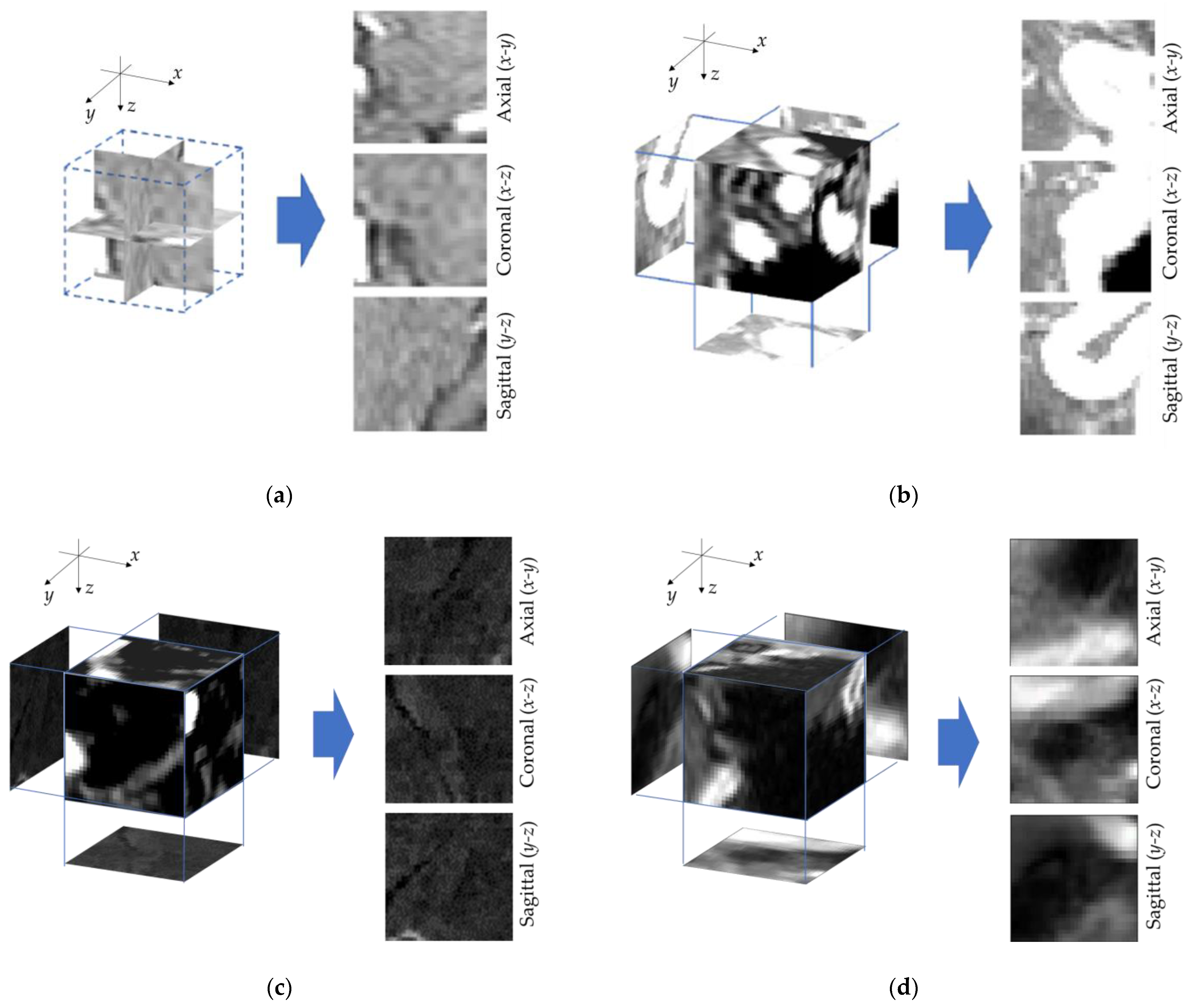
2.1.1. The 2.5D Image Transformation
2.1.2. Latent Variable Extraction Using CAE
2.1.3. Feature Extraction
- (a)
- The latent variables.
- (b)
- The Mahalanobis distance to the normal dataset on the latent variable space.
- (c)
- Features based on errors in 2.5D image reconstruction from the latent variables.
2.2. Proposed Method Application for Evaluation with Clinical Data
2.2.1. Detection of Cerebral Aneurysms in Head MRA Images with Proposed Feature Extraction
2.2.2. Detection of Lung Nodules in Chest CT Images with Proposed Feature Extraction
2.2.3. Evaluation of Lesion Candidate Classification
3. Results
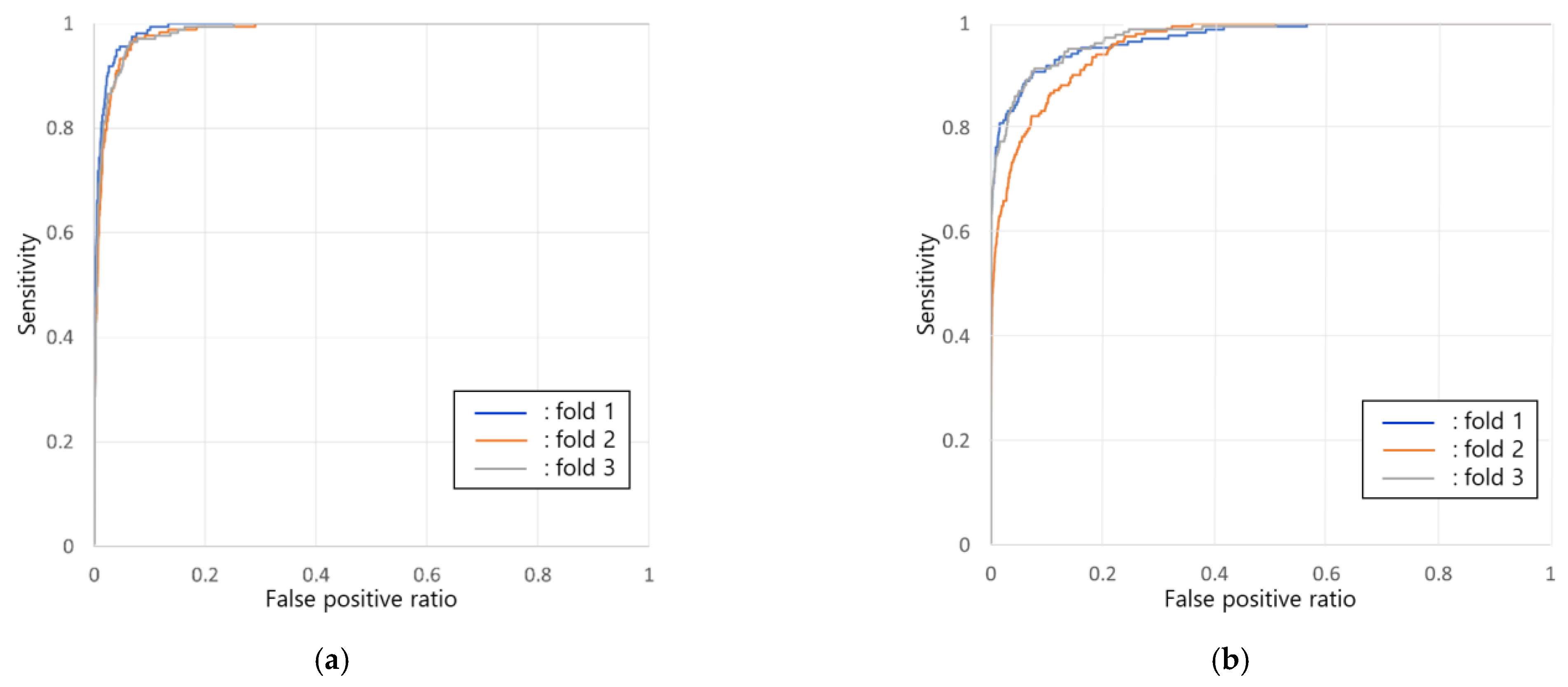
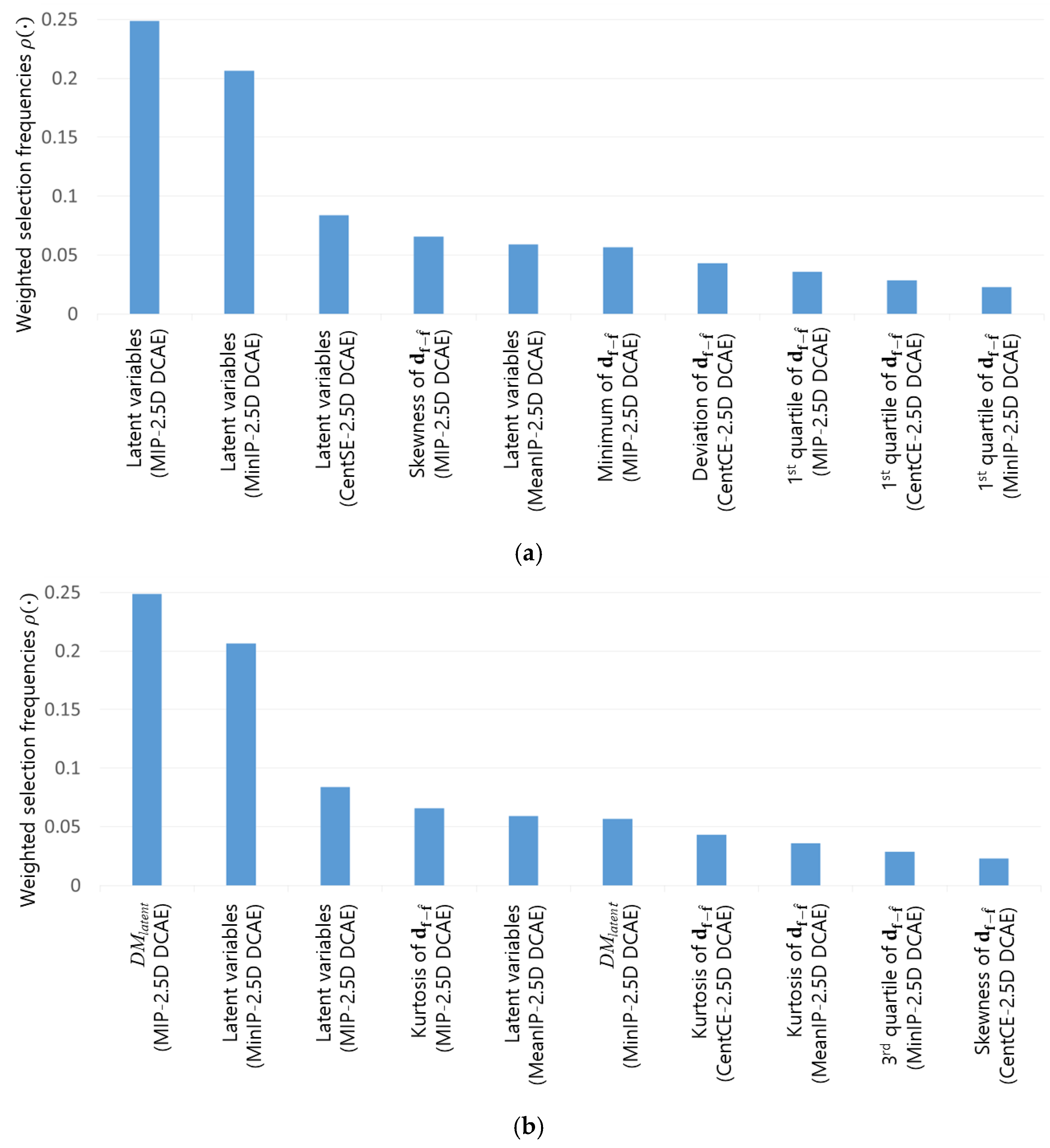
3.1. Results of Cerebral Aneurysm Detection in Head MRA Images
3.2. Results of Pulmonary Nodule Detection in Chest CT Images
4. Discussion
5. Conclusions
Author Contributions
Funding
Institutional Review Board Statement
Informed Consent Statement
Data Availability Statement
Acknowledgments
Conflicts of Interest
References
- Rubin, G.D. Data Explosion: The Challenge of Multidetector-Row CT. Eur. J. Radiol. 2000, 36, 74–80. [Google Scholar] [CrossRef] [PubMed]
- Li, F.; Sone, S.; Abe, H.; MacMahon, H.; Armato, S.G.; Doi, K. Lung Cancers Missed at Low-Dose Helical CT Screening in a General Population: Comparison of Clinical, Histopathologic, and Imaging Findings. Radiology 2002, 225, 673–683. [Google Scholar] [CrossRef] [PubMed]
- Doi, K. Current Status and Future Potential of Computer-Aided Diagnosis in Medical Imaging. Br. J. Radiol. 2005, 78, s3–s19. [Google Scholar] [CrossRef] [PubMed]
- Kozuka, T.; Matsukubo, Y.; Kadoba, T.; Oda, T.; Suzuki, A.; Hyodo, T.; Im, S.; Kaida, H.; Yagyu, Y.; Tsurusaki, M.; et al. Efficiency of a Computer-Aided Diagnosis (CAD) System with Deep Learning in Detection of Pulmonary Nodules on 1-Mm-Thick Images of Computed Tomography. Jpn. J. Radiol. 2020, 38, 1052–1061. [Google Scholar] [CrossRef] [PubMed]
- Li, F.; Arimura, H.; Suzuki, K.; Shiraishi, J.; Li, Q.; Abe, H.; Engelmann, R.; Sone, S.; MacMahon, H.; Doi, K. Computer-Aided Detection of Peripheral Lung Cancers Missed at CT: ROC Analyses without and with Localization. Radiology 2005, 237, 684–690. [Google Scholar] [CrossRef] [PubMed]
- Hirai, T.; Korogi, Y.; Arimura, H.; Katsuragawa, S.; Kitajima, M.; Yamura, M.; Yamashita, Y.; Doi, K. Intracranial Aneurysms at MR Angiography: Effect of Computer-Aided Diagnosis on Radiologists’ Detection Performance. Radiology 2005, 237, 605–610. [Google Scholar] [CrossRef] [PubMed]
- Pacilè, S.; Lopez, J.; Chone, P.; Bertinotti, T.; Grouin, J.M.; Fillard, P. Improving Breast Cancer Detection Accuracy of Mammography with the Concurrent Use of an Artificial Intelligence Tool. Radiol. Artif. Intell. 2020, 2, e190208. [Google Scholar] [CrossRef] [PubMed]
- van Ginneken, B.; Schaefer-Prokop, C.M.; Prokop, M. Computer-Aided Diagnosis: How to Move from the Laboratory to the Clinic. Radiology 2011, 261, 719–732. [Google Scholar] [CrossRef] [PubMed]
- Litjens, G.; Kooi, T.; Bejnordi, B.E.; Setio, A.A.A.; Ciompi, F.; Ghafoorian, M.; van der Laak, J.A.W.M.; van Ginneken, B.; Sánchez, C.I. A Survey on Deep Learning in Medical Image Analysis. Med. Image Anal. 2017, 42, 60–88. [Google Scholar] [CrossRef] [PubMed]
- Cheplygina, V.; de Bruijne, M.; Pluim, J.P.W. Not-so-Supervised: A Survey of Semi-Supervised, Multi-Instance, and Transfer Learning in Medical Image Analysis. Med. Image Anal. 2019, 54, 280–296. [Google Scholar] [CrossRef] [PubMed]
- Armato III, S.G.; McLennan, G.; Bidaut, L.; McNitt-Gray, M.F.; Meyer, C.R.; Reeves, A.P.; Zhao, B.; Aberle, D.R.; Henschke, C.I.; Hoffman, E.A.; et al. The Lung Image Database Consortium (LIDC) and Image Database Resource Initiative (IDRI): A Completed Reference Database of Lung Nodules on CT Scans. Med. Phys. 2011, 38, 915–931. [Google Scholar] [CrossRef] [PubMed]
- Roth, H.R.; Lu, L.; Seff, A.; Cherry, K.M.; Hoffman, J.; Wang, S.; Liu, J.; Turkbey, E.; Summers, R.M. A New 2.5D Representation for Lymph Node Detection Using Random Sets of Deep Convolutional Neural Network Observations. In Proceedings of the Medical Image Computing and Computer-Assisted Intervention—MICCAI 2014, Boston, MA, USA, 14–18 September 2014; Golland, P., Hata, N., Barillot, C., Hornegger, J., Howe, R., Eds.; Springer International Publishing: Cham, Switzerland, 2014; pp. 520–527. [Google Scholar]
- Nakao, T.; Hanaoka, S.; Nomura, Y.; Sato, I.; Nemoto, M.; Miki, S.; Maeda, E.; Yoshikawa, T.; Hayashi, N.; Abe, O. Deep Neural Network-Based Computer-Assisted Detection of Cerebral Aneurysms in MR Angiography. J. Magn. Reson. Imaging 2018, 47, 948–953. [Google Scholar] [CrossRef] [PubMed]
- Masci, J.; Meier, U.; Cireşan, D.; Schmidhuber, J. Stacked Convolutional Auto-Encoders for Hierarchical Feature Extraction. In Proceedings of the Artificial Neural Networks and Machine Learning—ICANN 2011, Espoo, Finland, 14–17 June 2011; Honkela, T., Duch, W., Girolami, M., Kaski, S., Eds.; Springer: Berlin/Heidelberg, Germany, 2011; pp. 52–59. [Google Scholar]
- Keogh, E.; Mueen, A. Curse of Dimensionality. In Encyclopedia of Machine Learning and Data Mining; Sammut, C., Webb, G.I., Eds.; Springer: Boston, MA, USA, 2017; pp. 314–315. ISBN 978-1-4899-7687-1. [Google Scholar]
- Viola, P.; Jones, M. Rapid Object Detection Using a Boosted Cascade of Simple Features. In Proceedings of the 2001 IEEE Computer Society Conference on Computer Vision and Pattern Recognition, CVPR 2001, Kauai, HI, USA, 8–14 December 2001; Volume 1, p. I. [Google Scholar]
- Sun, Y.; Kamel, M.S.; Wong, A.K.C.; Wang, Y. Cost-Sensitive Boosting for Classification of Imbalanced Data. Pattern Recognit. 2007, 40, 3358–3378. [Google Scholar] [CrossRef]
- Nomura, Y.; Nemoto, M.; Masutani, Y.; Hanaoka, S.; Yoshikawa, T.; Miki, S.; Maeda, E.; Hayashi, N.; Yoshioka, N.; Ohtomo, K. Reduction of False Positives at Vessel Bifurcations in Computerized Detection of Lung Nodules. J. Biomed. Graph. Comput. 2014, 4, p36. [Google Scholar] [CrossRef]
- Van Ginneken, B.; Armato, S.G.; De Hoop, B.; Van Amelsvoort-van De Vorst, S.; Duindam, T.; Niemeijer, M.; Murphy, K.; Schilham, A.; Retico, A.; Fantacci, M.E.; et al. Comparing and Combining Algorithms for Computer-Aided Detection of Pulmonary Nodules in Computed Tomography Scans: The ANODE09 Study. Med. Image Anal. 2010, 14, 707–722. [Google Scholar] [CrossRef] [PubMed]
- Nomura, Y.; Masutani, Y.; Miki, S.; Nemoto, M.; Hanaoka, S.; Yoshikawa, T.; Hayashi, N.; Ohtomo, K. Performance Improvement in Computerized Detection of Cerebral Aneurysms by Retraining Classifier Using Feedback Data Collected in Routine Reading Environment. J. Biomed. Graph. Comput. 2014, 4, 12. [Google Scholar] [CrossRef]
- Xie, H.; Yang, D.; Sun, N.; Chen, Z.; Zhang, Y. Automated Pulmonary Nodule Detection in CT Images Using Deep Convolutional Neural Networks. Pattern Recognit. 2019, 85, 109–119. [Google Scholar] [CrossRef]
- Jaiswal, A.; Babu, A.R.; Zadeh, M.Z.; Banerjee, D.; Makedon, F. A Survey on Contrastive Self-Supervised Learning. Technologies 2021, 9, 2. [Google Scholar] [CrossRef]


| Layer | Kernel Size | Stride | Output Size |
|---|---|---|---|
| 2.5D Input | − | − | 32 × 32 × 3 ch |
| Conv + BN + ReLU | 3 × 3 | 1 | 32 × 32 × 6 ch |
| Max Pooling | 2 × 2 | 2 | 16 × 16 × 6 ch |
| Conv + BN + ReLU | 3 × 3 | 1 | 16 × 16 × 9 ch |
| Max Pooling | 2 × 2 | 2 | 8 × 8 × 9 ch |
| Conv + BN + ReLU | 3 × 3 | 1 | 8 × 8 × 12 ch |
| Full Connection | − | − | n |
| Dataset | Diseased (for Evaluation) | Normal (for Training) |
|---|---|---|
| N cases (male:female) | 378 (208:170) | 252 (131:120) |
| Ages, average ± deviation (min, max) | 61.9 ± 11.0 (34, 85) | 55.6 ± 11.6 (34, 90) |
| N lesions | 434 | 0 |
| Lesion diameter (mm), average ± deviation (min, max) | 3.08 ± 1.28 (2, 9) | – |
| Scanners | GE Signa HDxt 3.0T or GE DiscoveryMR750 3.0T | |
| Original pixel size (mm) | 0.468 × 0.468 | |
| Original slice thickness (mm) | 0.6 | |
| Scanning date | Nov. 2006~May 2013 | |
| Institute | The University of Tokyo Hospital | |
| Dataset | Diseased (for Evaluation) | Normal (for Training) |
|---|---|---|
| N cases (male:female) | 450 (281:169) | 300 (194:106) |
| Ages, average ± deviation (min, max) | 60.2 ± 11.3 (40, 90) | 51.5 ± 9.09 (40, 81) |
| N lesions | 582 | 0 |
| Lesion diameter (mm), average ± deviation (min, max) | 7.61 ± 3.37 (5, 31) | – |
| Scanners | GE LightSpeed CT scanner | |
| Original pixel size (mm) | 0.781 × 0.781 | |
| Original slice thickness (mm) | 1.25 | |
| Scanning date | January 2007~March 2016 | |
| Institute | The University of Tokyo Hospital | |
| Num. FPs/Case | Proposed | Nomura et al. [20] | Nakao et al. [13] |
|---|---|---|---|
| 2.9 | 70.0 % | – | 94.2% |
| 5.0 | 83.3 % | 93.5% | – |
| 9.0 | 91.4 % | 95.2% | – |
| Num. FPs/Case | Proposed | Nomura et al. [18] | Xie et al. [21] |
|---|---|---|---|
| 4.8 | 46.3% | 80.0% | – |
| 14.1 | 57.2% | 90.0% | – |
| ANODE score | 0.218 | – | 0.790 |
Disclaimer/Publisher’s Note: The statements, opinions and data contained in all publications are solely those of the individual author(s) and contributor(s) and not of MDPI and/or the editor(s). MDPI and/or the editor(s) disclaim responsibility for any injury to people or property resulting from any ideas, methods, instructions or products referred to in the content. |
© 2023 by the authors. Licensee MDPI, Basel, Switzerland. This article is an open access article distributed under the terms and conditions of the Creative Commons Attribution (CC BY) license (https://creativecommons.org/licenses/by/4.0/).
Share and Cite
Nemoto, M.; Ushifusa, K.; Kimura, Y.; Nagaoka, T.; Yamada, T.; Yoshikawa, T. Unsupervised Feature Extraction for Various Computer-Aided Diagnosis Using Multiple Convolutional Autoencoders and 2.5-Dimensional Local Image Analysis. Appl. Sci. 2023, 13, 8330. https://doi.org/10.3390/app13148330
Nemoto M, Ushifusa K, Kimura Y, Nagaoka T, Yamada T, Yoshikawa T. Unsupervised Feature Extraction for Various Computer-Aided Diagnosis Using Multiple Convolutional Autoencoders and 2.5-Dimensional Local Image Analysis. Applied Sciences. 2023; 13(14):8330. https://doi.org/10.3390/app13148330
Chicago/Turabian StyleNemoto, Mitsutaka, Kazuyuki Ushifusa, Yuichi Kimura, Takashi Nagaoka, Takahiro Yamada, and Takeharu Yoshikawa. 2023. "Unsupervised Feature Extraction for Various Computer-Aided Diagnosis Using Multiple Convolutional Autoencoders and 2.5-Dimensional Local Image Analysis" Applied Sciences 13, no. 14: 8330. https://doi.org/10.3390/app13148330
APA StyleNemoto, M., Ushifusa, K., Kimura, Y., Nagaoka, T., Yamada, T., & Yoshikawa, T. (2023). Unsupervised Feature Extraction for Various Computer-Aided Diagnosis Using Multiple Convolutional Autoencoders and 2.5-Dimensional Local Image Analysis. Applied Sciences, 13(14), 8330. https://doi.org/10.3390/app13148330


