Article

Neuroprotective Effects of High-Intensity Interval Training through Neuroplastic Changes in a Restraint Stress-Induced Depression Model

1
Department of Exercise Biochemistry, Korea National Sport University, Seoul 05541, Republic of Korea
2
Department of Sports Medicine, Konyang University, Nonsan 32992, Republic of Korea
*
Authors to whom correspondence should be addressed.
Appl. Sci. 2023, 13(13), 7680; https://doi.org/10.3390/app13137680
Submission received: 30 May 2023 / Revised: 18 June 2023 / Accepted: 25 June 2023 / Published: 29 June 2023

Abstract

:
This study aimed to analyze the neuroprotective effects of various exercise intensities in a mouse model of depressive behavior disorders. Seven-week-old male C57BL/6 mice were divided into a control group, depressive disorder group (RST), moderate-intensity sustained exercise group (RST_MICT), high-intensity sustained exercise group (RST_HICT), and high-intensity interval exercise group (RST_HIIT). The animal model was established by applying restraint stress (RST) at 2 h/day for 14 days. Behavioral function was better in all exercise groups, especially in the RST_HIIT group, than in the RST group. Factors related to brain-derived neurotrophic factor showed higher levels in the exercise groups than in the RST group. The levels of 4-hydroxynonenal, an oxidative stress index, were significantly lower in the exercise groups than in the RST group. Malondialdehyde levels were lower in the exercise groups than in the RST group, but the difference was not significant. The analysis of serotonin and corticosterone, indicators of depression, revealed positive results in the exercise groups. The neuroplasticity-related variables c-fos and glial fibrillary acidic protein were more positive in the RST_HIIT group than in the RST group. Thus, HIIT improved neuroplasticity, oxidative stress, and neurotrophic factors in the depressive disorder model, indicating its potential for preventing and treating depression.

1. Introduction

Depression is a common mental illness clinically characterized by persistent feelings of lethargy, difficulty concentrating, and anxiety; it is a serious social problem because of its potential risk factor for high suicide rates [1,2]. Among the various causes of depression, externally derived stress reportedly causes degenerative changes in the central nervous system (CNS) [3,4].
Chemical imbalances in neurotransmitters, such as serotonin, dopamine, and norepinephrine, and complex feedback dysregulation of the hypothalamic–pituitary–adrenal (HPA) axis, a key component of the neuroendocrine system that responds to stress, are strongly associated with depression [5,6]. A longitudinal study on the clinical symptoms and brain biopsy findings of patients found a close relationship between early depression and the risk of developing dementia, suggesting that cognitive deficits contribute to depressive disorders [7,8,9].
Many studies have attempted to identify the role of neurogenesis as a risk factor for the development and progression of CNS disorders, including depression [5,10]. Furthermore, precursor cells in the dentate gyrus (DG) are involved in a cascade of neurogenic processes leading to neuronal proliferation and survival, and defects in neurogenesis and the subsequent decrease of neurogenesis contribute to depressive behavioral disorders [5]. Several antidepressants designed to promote neurogenesis in the hippocampus partly improve the mood, emotion, and cognitive function of patients with depressive disorders [11]. In addition, neurons with receptors that are stimulated by serotonin, a neurotransmitter involved in regulating mood and emotion, have been reported to promote the release of neurotrophic factors such as brain-derived neurotrophic factor (BDNF) and insulin-like growth factor-1 (IGF-1), which are implicated in depression [12,13]. Therefore, cognitive improvements caused by the promotion of neurogenesis in neurogenic regions of the brain have been proposed to alleviate some clinical symptoms of depressive disorders.
Researchers are proposing exercise as a noninvasive intervention to induce structural and functional changes throughout the CNS. Exercise reportedly contributes to neuroplasticity by improving the chemical imbalance of neurotransmitters, increasing the secretion of nerve growth factors, and activating neurogenesis. In addition, it increases neurogenesis in the hippocampus, which governs learning and memory formation, and prevents various forms of CNS disorders, including depression [13,14]. Furthermore, exercise restores normal levels of endocrine system responses, nerve growth factors, and neural stem cells through the feedback regulation of the HPA axis, a mechanism that promotes neurogenesis [15,16].
In terms of hormonal effects, sustained low- to moderate-intensity exercise improves brain function by decreasing the levels of adrenocorticotropic hormone (ACTH) and blood lactate secretion [17]. High-intensity interval training (HIIT), a combination of short bursts of high- and low-intensity exercises, has received much attention in recent years ever since exercise scientists have emphasized the need for high-intensity exercise as part of the recommended physical activity guidelines for adults [18,19]. In terms of physiology, the direct effects of HIIT include increasing metabolic capacity throughout the cardiorespiratory system; improving structural and functional properties in localized areas, such as the skeletal muscle; and enhancing CNS functions. Some researchers also suggested the potential of HIIT to promote neurogenesis [20,21]. Previous studies have reported that HIIT induces cognitive improvements and upregulates the expression of neurotrophic factors (e.g., BDNF, IGF-1, and VEGF) in the blood and brain, which are potential neurogenesis enhancers [22,23,24].
Given all of these benefits, exercise has been proposed as a strategy to prevent and treat clinical depressive disorders, but the efficacy of timed and intensity-controlled exercise in patients with depression remains to be validated. Therefore, this study aimed to analyze the effects of different exercise intensities, including HIIT, on brain neural system plasticity in an animal model of depressive behavioral disorders.

2. Materials and Methods

2.1. Laboratory Animals

Five-week-old male C57BL/6 mice were purchased from Laon Bio (Gyeonggi-do, Republic of Korea) and housed in the animal house at K University (temperature 22 ± 2 °C, humidity 50 ± 5%, 12 h light/dark cycle). According to the experimental plan, the animals were divided into five groups: control group (Con, n = 12), depressive disorder group (RST, n = 12), medium-intensity continuous exercise group (RST_MICT, n = 12), high-intensity continuous exercise group (RST_HICT, n = 12), and high-intensity interval exercise group (RST_HIIT, n = 12). This study was approved by the Institutional Animal Care and Use Committee of K University (KNSU-IACUC-2020-05).

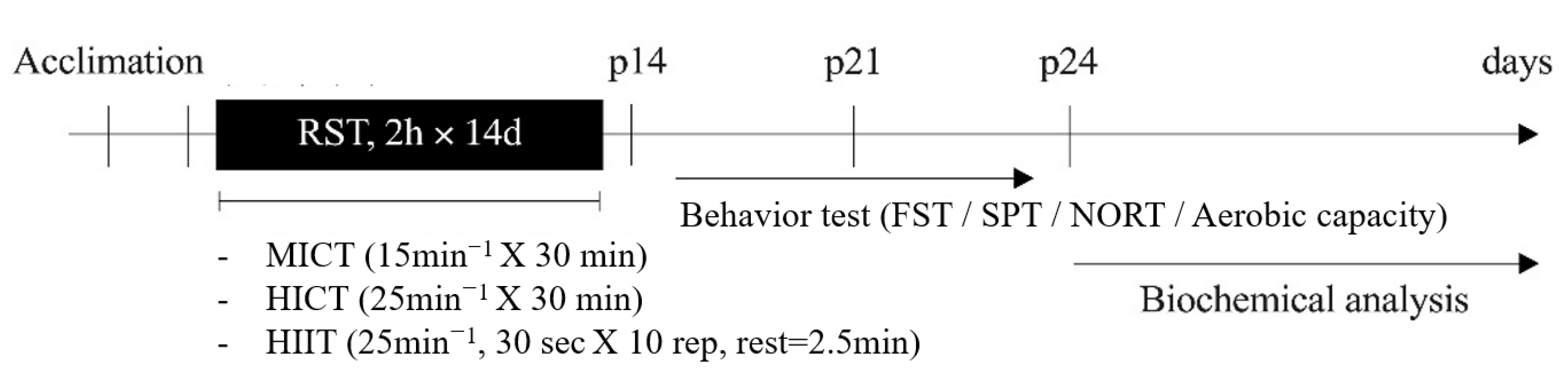
2.2. Animal Models of Depressive Disorders

For the animal model of depressive disorders, immobilization was maintained by placing each individual in a 50 mL polyethylene tube modified to provide good ventilation, following the previous work of Seo [25], and restraint stress (RST, 2 h/day) was applied for 14 days. The animals were transferred to dedicated cages in the animal laboratory at a temperature of 23 ± 3 °C, humidity of 50 ± 5%, and a 12 h light/dark cycle and provided free access to food and water. The experimental plan and group divisions are shown in Figure 1.

2.3. Exercise Method

The animals from the RST_MICT, RST_HICT, and RST_HIIT groups were placed on an animal treadmill (eight lanes, Daemyung Scientific Co., Ltd., Daejeon, Republic of Korea) for the different exercises. The intensity was adjusted based on the lactate threshold (LT) after learning to drive, 5 days a week for 2 weeks.
MICT, including warm-up and cool-down, was performed for 40 min per day. HICT was performed as 18 min of fixed-load continuous treadmill exercise at 25 m/min to match the daily exercise volume (450 m) of MICT. HIIT consisted of sprinting (25 m/min, 30 s) and resting low-intensity walking (8 m/min, 2.5 min) [17] and was performed for the same amount of time as MICT. The specific exercise protocols are listed in Table 1.

2.4. Forced Swim Test (FST)

The forced swim test is an experimental method used to observe depression-related behaviors. Following the methodology described by Slattery [26], the animals were immersed in a cylinder of water with a temperature of 23 °C for 15 min as a preliminary test, and the same procedure was repeated 24 h later for 5 min. During this 5 min test, the immobility time of the animals was measured. Immobility was defined as floating upright and motionless with minimal movement to keep only the head above the water.

2.5. Sucrose Preference Test (SPT)

The sugar preference test is an experimental method used to observe depression-related behaviors as markers of unpleasant responses to a stimulus. To acclimatize the animals to 1% sucrose solution before testing, sucrose solution was added to their water bowls 48 h prior to testing. The animals were watered and fasted for 24 h prior to the test, and the change in sucrose intake over a 3 h period on the day of the test was determined by the change in the weight of the water bottle.

2.6. Novel Object Recognition Test (NORT)

A novelty detection test was performed to measure attention and visual recognition memory. The equipment used in this experiment was an acrylic box (32 × 32 × 32 cm) without a lid, and the animals were placed in the acrylic box for 5 min, one day before the test, to familiarize them with the environment. On the first day of the experiment, two identical objects were placed in the box for 5 min, and the animal’s curious behavior toward the two objects was measured in seconds. After 20 s, the animal was replaced with the next one, and the same experiment was performed. On the second day of the experiment, a new object of a similar size but different shape was placed in front of the previous day’s object, and the same behavior was measured for 1 min. After each test, the acrylic box and objects were disinfected and dried with 70% alcohol to limit external stimuli as much as possible. The results are expressed as the percentage of time spent exploring the novel object for each group.
R e c o g n i t i o n   i n d e x % = N o v e l   o b j e c t   r e c o g n i t i o n   t i m e E x i s t i n g   o b j e c t   r e c o g n i t i o n   t i m e T o t a l   o b j e c t   r e c o g n i t i o n   t i m e

2.7. Aerobic Capacity

The aerobic performance of the animals was measured by performing an incremental exercise test, starting at an initial treadmill speed of 5 m/min and increasing the speed by 2.5 m/min every 3 min until exhaustion, which was defined as the inability to maintain pace at the treadmill speed or remain at the rear of the treadmill machine for more than 30 s despite soft contact. The measurement item was total work by substituting the weight, exercise speed, and time of the experimental animal into the formula (body mass (kg) × gravity (9.81 m/s2) × vertical speed (m/s × angle) × time (s)) designed in previous studies.

2.8. Administration and Detection of Bromodeoxyuridine (BrdU)

To assess the level of neurogenesis in the DG of the hippocampus of the experimental animals, we intraperitoneally injected a BrdU solution diluted to 50 mg/kg during the first 5 days of exercise.

2.9. Tissue Preparation

After performing the treadmill exercise and motor function tests, seven animals per group were selected for western blot, and five animals per group were used for immunohistochemical staining. The rats for western blot were anesthetized by inhalation of CO2 gas using an animal chamber, and then their brain tissue was harvested and stored in a deep freezer (Deep Freezer, SANYO, Tokyo, Japan) at −80 °C until analysis. The rats for immunohistochemical staining were anesthetized by inhalation of CO2 gas using an animal chamber. The thoracic cavity was opened, and 50 mM phosphate buffer saline (PBS) was injected through the left ventricle for 5 min, followed by perfusion with 4% paraformaldehyde (PFA) fixative dissolved in 0.1 M phosphate buffer for 10 min. Then, the brain and skeletal muscle were harvested, embedded in 4% PFA fixative, and then fixed by precipitation for 12 h at 4 °C. The tissues were precipitated in 30% sucrose solution for 5 days, and serial coronal sections of 40 μm thickness were prepared using a freezing microtome (Leica, Nussloch, Germany).

2.10. Spectrophotometry

Levels of blood corticosterone (CORT), an indicator of stress response, serotonin, a neurotransmitter present in the hypothalamic center of the brain, and malondialdehyde (MDA), an indicator of oxidative stress, were measured using ELISA assay kits (CORT, Abcam, Cambridge, UK, ab108821; Serotonin, Abcam, ab133053; MDA, Elabsience, Houston, TX, USA, E-BC-K025-S) in accordance with the manufacturer’s instructions.

2.11. Western Blot

The harvested brain tissue was homogenized using a homogenizer with lysis buffer and then centrifuged at 13,000× g for 15 min at 4 °C, and the supernatant was quantified for total protein by using the Bradford (1976) method. Proteins were electrophoresed (120 min) on a 10% SDS-polyacrylamide gel at 30 μg, transferred to a membrane, blocked, and then incubated with primary antibodies (BDNF, cAMP-response element-binding protein (CREB), tropomyosin-related kinase B receptor (TrkB), and 4-hydroxynonenal (4HNE)). The next day, the membrane was reacted with the secondary antibody for 1 h at room temperature, placed in WBLR solution (western blotting Luminol Reagent SC-2048, Santa Cruz Biotechnology, Dallas, TX, USA), and developed for 1 min. The obtained membrane was scanned using an image analysis system (Molecular Imager ChemiDoc XRS System, Bio-Rad, Hercules, CA, USA) to calculate the protein amount using Quantity One 1-D Analysis Software (Bio-Rad).

2.12. Immunohistochemistry

Brain tissues for immunohistochemical staining were embedded in an OCT compound, sectioned at 35–30 μm, and then stored in a storage solution. The sections were selected on three sheets for each animal. The selected sections were then washed thrice with PBS for 5 min, blocked with 3% serum at 37 °C for 40 min, and incubated with primary antibodies (c-fos and glial fibrillary acidic protein (GFAP)) at 4 °C overnight. The next day, the sections were incubated with the secondary antibody at room temperature for 60 min, counterstained with diaminobenzidine (DAB, Vector, Torrance, CA, USA), and observed under a light microscope (Leica, Nussloch, Germany). All observed sections were analyzed using ImageJ 1.53a (Image Processing and Analysis in JAVA), and the number of proteins (c-fos and GFAP) stained in the dorsal dentate gyrus of the hippocampus in each section was counted. The counted proteins were averaged for comparison and analysis. The number of apoptotic cells was counted using LAS AF Lite 4.0 analysis software.

2.13. Statistical Analyses

Statistical analysis of the collected data was performed using SPSS Statistics 23.0, and descriptive statistics for each variable were calculated as the mean ± standard deviation (mean ± SD). One-way analysis of variance (ANOVA) was performed to check the differences in variables between groups and time periods, and Bonferroni’s post-hoc test was used to specifically check the differences. The statistical significance level for all tests was set at α = 0.05.

3. Results

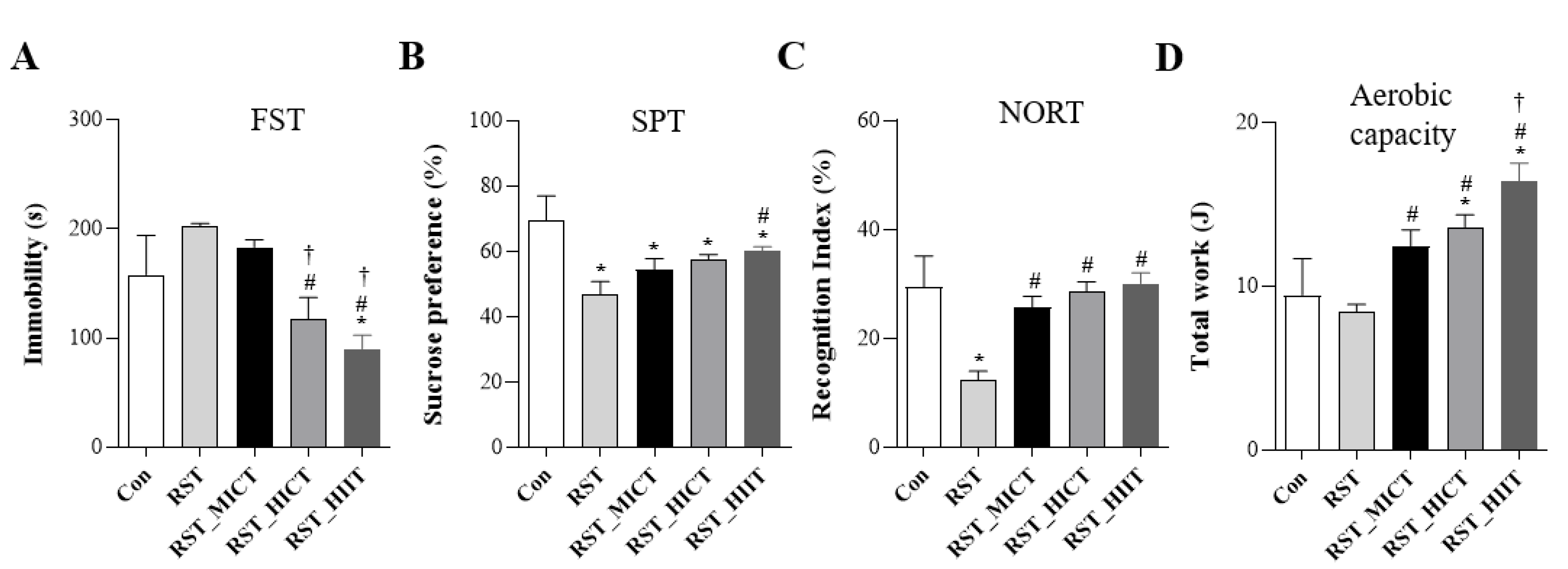
3.1. Changes in Behavioral Function with Treadmill Exercise Intensity

To determine the protective effects of various exercise intensities against depressive disorders, we analyzed the changes in behavioral functions during the 2 weeks of RST (Figure 2) and found significant differences in the results of all behavioral function tests (FST: F = 15.556, p = 0.001; SPT: F = 15.138, p = 0.001; NORT: F = 13.548, p = 0.001; aerobic capacity: F = 9.794, p = 0.001). FST results showed that the immobility time was significantly shorter in the RST_HIIT group than in the CON group and in the RST_HICT group than in the RST and RST_HIIT groups. The immobility time was also significantly shorter in the RST_HIIT group than in the RST_MICT group. SPT results showed that the sucrose preference was significantly lower in the RST, RST_MICT, and RST_HICT groups than in the CON group. However, the sucrose preference was significantly higher in the RST_HIIT group than that in the RST group. NORT results showed that the recognition index was significantly lower in the RST group than in the CON group. By contrast, this index was significantly higher in the RST_MICT, RST_HICT, and RST_HIIT groups than in the RST group. Aerobic capacity measurement results showed that the total work was significantly higher in the RST_HICT and RST_HIIT groups than in the CON group; in the RST_MICT, RST_HICT, and RST_HIIT groups than in the RST group; and in the RST_HIIT group than in the RST_MICT group.

3.2. Changes in Neurogenesis-Related Variables with Treadmill Exercise Intensity

To determine the protective effects of the different exercise intensities against depressive disorders, we analyzed the changes in BDNF-related variables during the 2 weeks of RST (Figure 3). Significant differences were found in the expression levels of all BDNF-related variables (BDNF: F = 4.035, p = 0.002; CREB1: F = 7.058, p = 0.001; TrkB: F = 4.862, p = 0.004). BDNF levels were significantly higher in the RST_MICT, RST_HICT, and RST_HIIT groups than in the RST group. CREB1 levels were significantly higher in the RST_HICT and RST_HIIT groups than in the RST group. TrkB levels were significantly lower in the RST group than in the CON group and significantly higher in the RST_MICT, RST_HICT, and RST_HIIT groups than in the RST group.

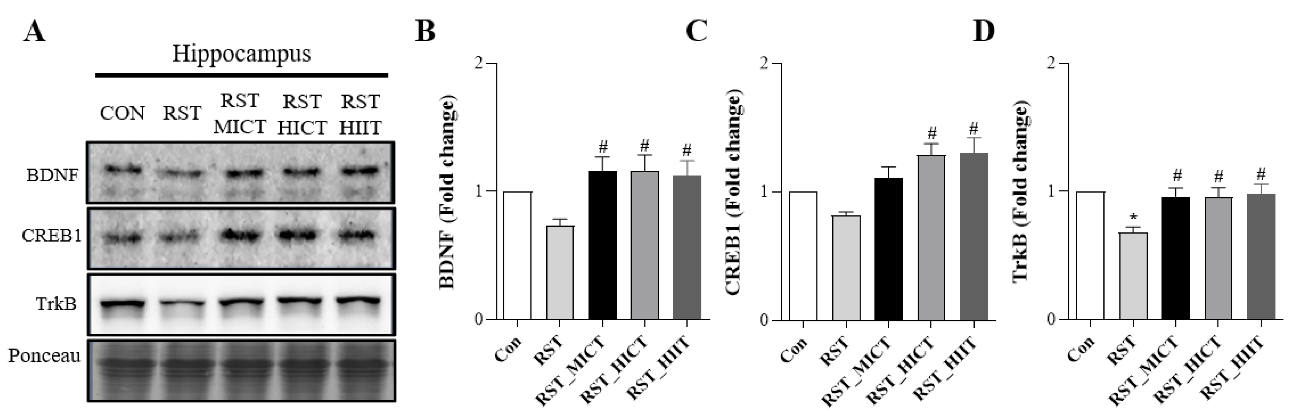
3.3. Changes in Neuroplasticity-Related Metrics with Treadmill Exercise Intensity

To determine the protective effects of the various exercise intensities against depressive disorders, we analyzed the changes in c-fos and GFAP levels during the 2 weeks of RST (Figure 4) and found significant differences in the levels of all variables (c-fos: F = 155.670, p = 0.001; GFAP: F = 127.640155.670, p = 0.001). c-Fos levels were significantly higher in all groups compared with the CON group. By contrast, c-fos levels were significantly lower in the RST_HCIT and RST_HIIT groups than in the RST group and in the RST_HIIT group than in the RST_MICT group. Meanwhile, GFAP levels were significantly higher in all groups compared with the CON group. By contrast, GFAP levels were significantly lower in the RST_MCIT and RST_HIIT groups than in the RST group.

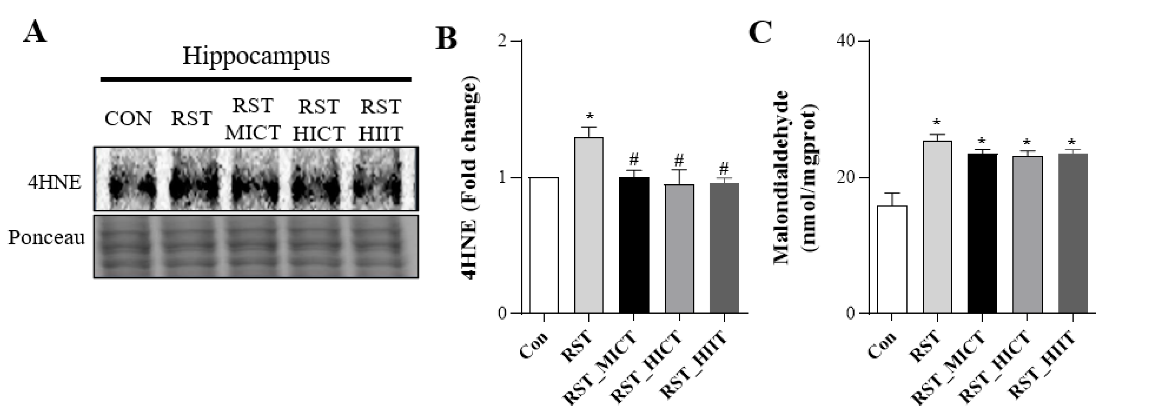
3.4. Changes in Oxidative Stress with Treadmill Intensity

To determine the protective effects of the different exercise intensities against depressive disorders, we analyzed the changes in oxidative stress during the 2 weeks of RST (Figure 5) and found significant differences in the levels of all variables (4HNE: F = 5.534, p = 0.002; MDA: F = 31.531, p = 0.001). 4HNE levels were significantly higher in the RST group than in the CON group and in the RST_MICT group than in the RST, RST_HICT, and RST_HIIT groups. Meanwhile, MDA levels were significantly higher in all groups compared with the CON group.

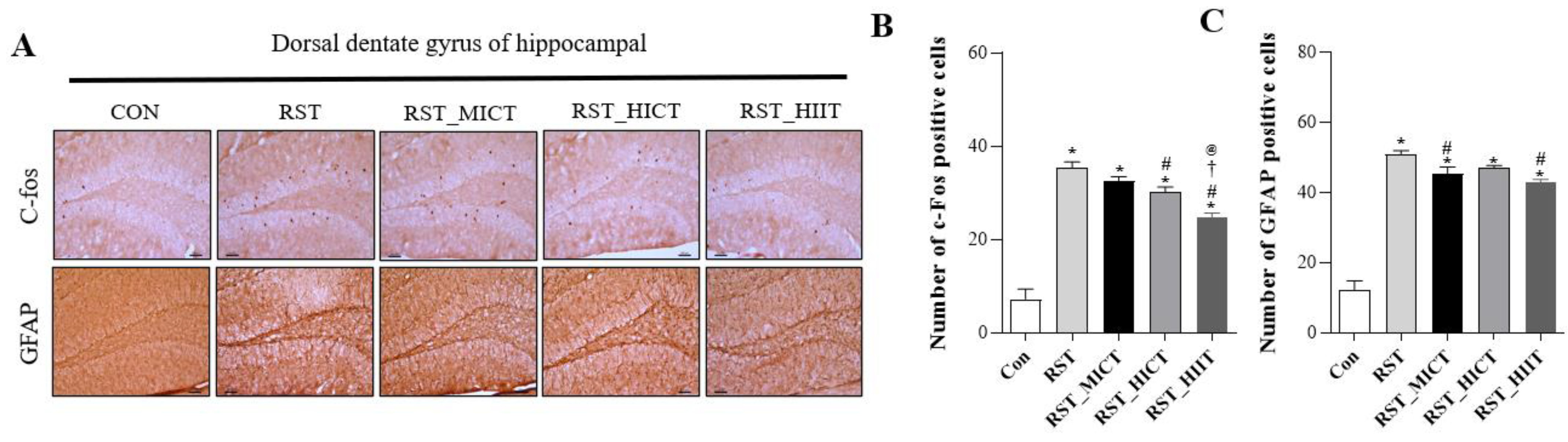
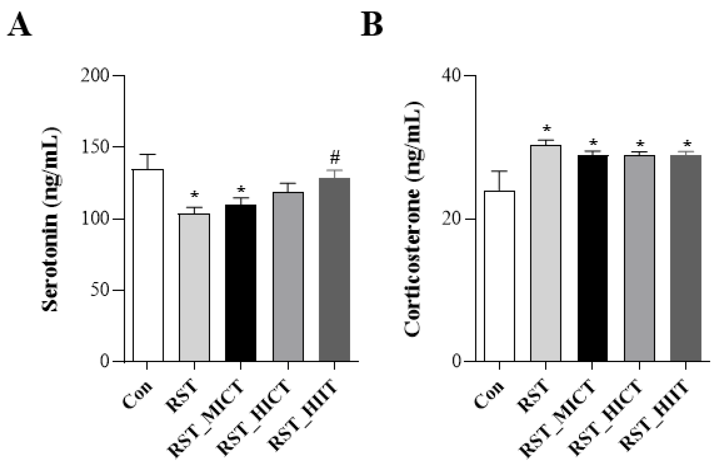
3.5. Changes in Stress Response Markers with Treadmill Exercise Intensity

To determine the protective effects of different exercise intensities against depressive disorders, we analyzed the changes in oxidative stress during the 2 weeks of RST (Figure 6) and found significant differences in the levels of all variables (Serotonin: F = 15.079, p = 0.001; CORT: F = 6.939, p = 0.001). Serotonin levels were significantly lower in the RST and RST_MICT groups than in the CON group but significantly higher in the RST_HIIT group than in the RST group. CORT levels were significantly higher in all groups compared with the CON group.

4. Discussion

Exercise affects the sensitivity of stress-related hormones; modulates neurotransmitters, such as serotonin and dopamine; produces mood-enhancing effects, such as reduced depressive behavior; and restores normal levels of endocrine system responses, nerve growth factors, and neural stem cells through feedback regulation of the HPA axis [15,16,27,28]. Intense exercise enhances protective mechanisms against depression by increasing neurite outgrowth, neuronal proliferation, and survival in response to acute stress and certain levels of CORT treatment [29,30,31]. Therefore, the present study aimed to investigate the neuroprotective effects of different exercise intensities, including HIIT, via brain neurogenesis in an animal model of RST-induced depressive behavioral disorders.
Recently, many researchers have attempted to induce depressive disorders by applying various forms of stress, such as RST, which restricts movement for 2 h a day for 14 d and promotes modifications in the brain’s neural networks; such phenomena cause behavioral hopelessness and anhedonia, which is reported to be the closest to depression-like behavior [32,33,34].
In the present study, FST and SPT were conducted to analyze the effects of different exercise intensities on anxiety and depression-like behaviors caused by RST. NORT was performed to analyze the learning and memory abilities of the experimental animals, and their aerobic capacity was determined according to the exercise intensity. In all behavioral tests, the RST group showed increased anxiety and depression-like behaviors, decreased cognitive function, and decreased aerobic capacity. These results suggest that 14 days of RST for 2 h daily is physiologically appropriate to induce anxiety and depression-like behaviors. Two weeks of treadmill exercise combined with RST produced positive results in all tests, with the HIIT group showing more positive results than the moderate- and high-intensity exercise groups. This is consistent with the findings of Masrour [35] and Sohroforouzani [36]. Exercise is thought to improve behavioral functioning not only because of its antidepressant effects but also because it improves the metabolic capacity of the cardiorespiratory system and the functioning of the CNS [20,21].
Various neuroplastic pathways play important roles in the pathophysiology and treatment of depression [37,38,39]. Among the various neuroplastic pathways in depression, attention has been focused on the BDNF, TrkB, and CREB pathways [38,40]. Specifically, BDNF is involved in cognitive enhancement by improving brain plasticity and enhancing learning and memory functions in the hippocampus [41]. Increased BDNF synthesis in the brain is associated with increased BDNF signaling, resulting in improved mood, cognitive function, and memory [42]. In the present study, the BDNF, CREB1, and TrkB levels were reduced in the RST group. Stress reduces the expression of BDNF in the hippocampus and prefrontal cortex, which consequently reduces TrkB signaling possibly via the cAMP-CREB1 signaling pathway [43,44]. The behavioral effects of antidepressants have been linked to their ability to increase BDNF expression and activate TrkB signaling in the hippocampus and prefrontal cortex, which are mediated in part by CREB1 [40,45]. In the present study, the BDNF, CREB1, and TrkB levels were significantly higher in the MICT, HICT, and HIIT groups than in the RST group. Moderate-to-vigorous aerobic exercises, such as running and swimming, increase BDNF biosynthesis, resulting in up to a threefold increase in plasma and brain BDNF [46,47]. These results suggest that exercise enhances the effectiveness of antidepressants by acting through neurochemical pathways similar to those of BDNF [48].
Astrocytes are glial cells that support the survival and function of neurons and maintain nervous system stability. GFAP, a marker of astrocyte reactivity, is expressed in the CNS and is neurotoxic when accumulated by stress and disease [49]. As a proto-oncogene, c-fos is an immediate early gene that plays a role in cell division and differentiation and mediates cellular responses. The results of this study showed that c-fos and GFAP levels increased with RST and decreased with exercise. This is consistent with the finding that 12 weeks of treadmill exercise increased GFAP levels in the hippocampus of animals. However, previous studies have reported that c-fos levels increase with increasing exercise intensity in the DG of animal brains, which is inconsistent with our results. c-Fos levels increased until day 7 of exercise and then began to decline [50]. The c-fos gene product, Fos protein, is activated and increased in certain areas of the brain in response to brain injury or stress, and its expression is reduced when depression is improved by antidepressant medications [51,52]. One possible explanation for these results is that the exercise itself is perceived as stressful.
Collectively, astrocytes regulate neurogenesis and neuroplasticity by providing neurotrophic factors, such as BDNF, to neighboring neuronal cell bodies [53], which consequently inhibit cell death by protecting neurons from oxidative stress [54]. Oxidative stress is closely associated with neurogenesis and neuroplasticity in depressive disorders. The levels of 4HNE and MDA, markers of lipid peroxidation, are elevated in depressive disorders, and increased lipid peroxidation damages proteins and DNA, leading to cell membrane permeability and the dysfunction of enzymes and receptors [55]. Exercise inhibits oxidative stress by effectively increasing the activities of antioxidant enzymes. The present study demonstrated that treadmill exercise of varying intensities suppressed 4HNE and MDA levels in RST-induced depressive disorders. However, no significant differences in the effects of exercise type or intensity were found. Moradi-Kor [56] reported that 15 days of voluntary wheel exercise in an animal model of depressive disorder alleviated depressive behaviors and reduced the levels of oxidative stress markers, such as MDA and lipid peroxidation. In addition, exercise reduces oxidative stress by modulating the balance of antioxidant enzymes in depressive disorders [57,58].
RST-induced depressive disorders decrease the levels of serotonin, a neurotransmitter involved in mood and emotion regulation, and increase the levels of CORT [59]. In the present study, serotonin levels were higher in the MICT, HICT, and HIIT groups than in the RST group. In particular, serotonin levels in the HIIT group significantly increased, similar to those in the CON group. CORT levels were higher in the RST group than in the CON group but lower in the exercise groups, although the difference was not significant. Previous clinical studies have reported that serotonin levels tend to increase numerically [60]. In addition, high-intensity exercise increases the amount of serotonin in the CNS because trytophan is displaced by lipolysis from its attachment to albumin, a free fatty acid. The increased concentration of tryptophan in the blood allows it to cross the blood–brain barrier in the central nervous system, and exercise enhances feedback mechanisms to increase serotonin synthesis [61]. Furthermore, the levels of CORT, which is constantly elevated by RST, is reduced by exercise, showing positive anti-anxiety and anti-depressant effects.
Taken together, these results suggest that varying exercise intensity may positively influence factors related to neuroplasticity in the CNS in an RST-induced depressive disorder model, leading to a reduction in oxidative stress and improvement in depression-like behavior and cognitive function. Further studies on the various pathways involved in brain neuroplasticity are required to elucidate the mechanisms by which exercise intensity prevents depressive disorders.

5. Conclusions

This study analyzed the effects of different exercise intensities, including HIIT, on CNS neuroplasticity in an animal model of depressive behavior disorders induced by RST. Two weeks of varying-intensity treadmill exercise combined with the application of restraint stress resulted in, first, improvements in anxiety and depression-like behaviors (FST and SPT), cognitive function (NORT), and aerobic capacity. Second, factors related to neurogenesis (BDNF, CREB1, and TrkB) improved. Third, factors related to neuroplasticity (c-fos and GFAP) were reduced. Fourth, factors related to oxidative stress (4HNE and MDA) were reduced. Finally, the stress response hormone serotonin increased and CORT decreased. Therefore, we believe that exercise can be an effective intervention to contribute to the improvement of depressive behavioral disorders. Future studies should explore the efficacy of different exercise intensities and densities in improving and preventing depressive disorders.

Author Contributions

Experimental design, performance, and analysis, D.-H.C. and D.-J.H.; experimental design and interpretation, H.-S.U., D.-H.C. and J.-Y.C.; writing, D.-H.C., D.-J.H. and J.-Y.C. All authors acknowledge responsibility for the full content of the submitted manuscript and approved its submission. All authors have read and agreed to the published version of the manuscript.

Funding

This study was supported by the Ministry of Education of the Republic of Korea and the National Research Foundation of Korea (grant number NRF-2020S1A5A2A01047758).

Institutional Review Board Statement

This study was approved by the Institutional Animal Care and Use Committee (IACUC) of Korea National Sports University (KNSU-IACUC-2020-05).

Informed Consent Statement

Not applicable.

Data Availability Statement

All data are contained within the article.

Conflicts of Interest

The authors declare no conflict of interest.

References

  1. Smith, K. Mental health: A world of depression. Nature 2014, 515, 181. [Google Scholar] [CrossRef] [Green Version]
  2. Sher, L.; Grunebaum, M.F.; Burke, A.K.; Chaudhury, S.; Mann, J.J.; Oquendo, M.A. Depressed Multiple-Suicide-Attempters—A High-Risk Phenotype. Crisis 2017, 38, 367–375. [Google Scholar] [CrossRef] [PubMed]
  3. Yang, L.; Zhao, Y.; Wang, Y.; Liu, L.; Zhang, X.; Li, B.; Cui, R. The Effects of Psychological Stress on Depression. Curr. Neuropharmacol. 2015, 13, 494–504. [Google Scholar] [CrossRef] [PubMed] [Green Version]
  4. Ménard, C.; Hodes, G.E.; Russo, S.J. Pathogenesis of depression: Insights from human and rodent studies. Neuroscience 2016, 321, 138–162. [Google Scholar] [CrossRef] [PubMed] [Green Version]
  5. Mahar, I.; Bambico, F.R.; Mechawar, N.; Nobrega, J.N. Stress, serotonin, and hippocampal neurogenesis in relation to depression and antidepressant effects. Neurosci. Biobehav. Rev. 2014, 38, 173–192. [Google Scholar] [CrossRef]
  6. Blier, P. Neurobiology of depression and mechanism of action of depression treatments. J. Clin. Psychiatry 2016, 77, e319. [Google Scholar] [CrossRef]
  7. Rajkowska, G. Depression: What we can learn from postmortem studies. Neuroscientist 2003, 9, 273–284. [Google Scholar] [CrossRef]
  8. Nihonmatsu-Kikuchi, N.; Hayashi, Y.; Yu, X.J.; Tatebayashi, Y. Depression and Alzheimer’s disease: Novel postmortem brain studies reveal a possible common mechanism. J. Alzheimers Dis. 2013, 37, 611–621. [Google Scholar] [CrossRef]
  9. Tan, E.Y.L.; Köhler, S.; Hamel, R.E.G.; Muñoz-Sánchez, J.L.; Verhey, F.R.J.; Ramakers, I. Depressive Symptoms in Mild Cognitive Impairment and the Risk of Dementia: A Systematic Review and Comparative Meta-Analysis of Clinical and Community-Based Studies. J. Alzheimers Dis. 2019, 67, 1319–1329. [Google Scholar] [CrossRef]
  10. Peng, L.; Bonaguidi, M.A. Function and Dysfunction of Adult Hippocampal Neurogenesis in Regeneration and Disease. Am. J. Pathol. 2018, 188, 23–28. [Google Scholar] [CrossRef] [Green Version]
  11. Rosenblat, J.D.; Kakar, R.; McIntyre, R.S. The Cognitive Effects of Antidepressants in Major Depressive Disorder: A Systematic Review and Meta-Analysis of Randomized Clinical Trials. Int. J. Neuropsychopharmacol. 2015, 19, pyv082. [Google Scholar] [CrossRef] [Green Version]
  12. Popova, N.K.; Ilchibaeva, T.V.; Naumenko, V.S. Neurotrophic Factors (BDNF and GDNF) and the Serotonergic System of the Brain. Biochemistry 2017, 82, 308–317. [Google Scholar] [CrossRef]
  13. Kondo, M.; Nakamura, Y.; Ishida, Y.; Shimada, S. The 5-HT3 receptor is essential for exercise-induced hippocampal neurogenesis and antidepressant effects. Mol. Psychiatry 2015, 20, 1428–1437. [Google Scholar] [CrossRef] [PubMed] [Green Version]
  14. Krogh, J.; Speyer, H.; Gluud, C.; Nordentoft, M. Exercise for patients with major depression: A protocol for a systematic review with meta-analysis and trial sequential analysis. Syst. Rev. 2015, 4, 40. [Google Scholar] [CrossRef] [PubMed] [Green Version]
  15. Vivar, C.; Potter, M.C.; van Praag, H. All about running: Synaptic plasticity, growth factors and adult hippocampal neurogenesis. Curr. Top. Behav. Neurosci. 2013, 15, 189–210. [Google Scholar] [CrossRef] [Green Version]
  16. Nishii, A.; Amemiya, S.; Kubota, N.; Nishijima, T.; Kita, I. Adaptive Changes in the Sensitivity of the Dorsal Raphe and Hypothalamic Paraventricular Nuclei to Acute Exercise, and Hippocampal Neurogenesis May Contribute to the Antidepressant Effect of Regular Treadmill Running in Rats. Front. Behav. Neurosci. 2017, 11, 235. [Google Scholar] [CrossRef] [PubMed] [Green Version]
  17. Okamoto, M.; Yamamura, Y.; Liu, Y.F.; Min-Chul, L.; Matsui, T.; Shima, T.; Soya, M.; Takahashi, K.; Soya, S.; McEwen, B.S.; et al. Hormetic effects by exercise on hippocampal neurogenesis with glucocorticoid signaling. Brain Plast. 2015, 1, 149–158. [Google Scholar] [CrossRef] [Green Version]
  18. Haskell, W.L.; Lee, I.M.; Pate, R.R.; Powell, K.E.; Blair, S.N.; Franklin, B.A.; Macera, C.A.; Heath, G.W.; Thompson, P.D.; Bauman, A. Physical activity and public health: Updated recommendation for adults from the American College of Sports Medicine and the American Heart Association. Med. Sci. Sports Exerc. 2007, 39, 1423–1434. [Google Scholar] [CrossRef] [Green Version]
  19. Borrega-Mouquinho, Y.; Sánchez-Gómez, J.; Fuentes-García, J.P.; Collado-Mateo, D.; Villafaina, S. Effects of High-Intensity Interval Training and Moderate-Intensity Training on Stress, Depression, Anxiety, and Resilience in Healthy Adults During Coronavirus Disease 2019 Confinement: A Randomized Controlled Trial. Front. Psychol. 2021, 12, 643069. [Google Scholar] [CrossRef]
  20. Astorino, T.A.; Edmunds, R.M.; Clark, A.; King, L.; Gallant, R.A.; Namm, S.; Fischer, A.; Wood, K.M. High-Intensity Interval Training Increases Cardiac Output and VO2max. Med. Sci. Sports Exerc. 2017, 49, 265–273. [Google Scholar] [CrossRef]
  21. Crozier, J.; Roig, M.; Eng, J.J.; MacKay-Lyons, M.; Fung, J.; Ploughman, M.; Bailey, D.M.; Sweet, S.N.; Giacomantonio, N.; Thiel, A.; et al. High-Intensity Interval Training After Stroke: An Opportunity to Promote Functional Recovery, Cardiovascular Health, and Neuroplasticity. Neurorehabilit. Neural Repair 2018, 32, 543–556. [Google Scholar] [CrossRef] [PubMed] [Green Version]
  22. Pereira, A.C.; Huddleston, D.E.; Brickman, A.M.; Sosunov, A.A.; Hen, R.; McKhann, G.M.; Sloan, R.; Gage, F.H.; Brown, T.R.; Small, S.A. An in vivo correlate of exercise-induced neurogenesis in the adult dentate gyrus. Proc. Natl. Acad. Sci. USA 2007, 104, 5638–5643. [Google Scholar] [CrossRef] [PubMed] [Green Version]
  23. Jiménez-Maldonado, A.; Rentería, I.; García-Suárez, P.C.; Moncada-Jiménez, J.; Freire-Royes, L.F. The Impact of High-Intensity Interval Training on Brain Derived Neurotrophic Factor in Brain: A Mini-Review. Front. Neurosci. 2018, 12, 839. [Google Scholar] [CrossRef] [PubMed]
  24. Kondo, M.; Koyama, Y.; Nakamura, Y.; Shimada, S. A novel 5HT3 receptor-IGF1 mechanism distinct from SSRI-induced antidepressant effects. Mol. Psychiatry 2018, 23, 833–842. [Google Scholar] [CrossRef]
  25. Seo, J.S.; Park, J.Y.; Choi, J.; Kim, T.K.; Shin, J.H.; Lee, J.K.; Han, P.L. NADPH oxidase mediates depressive behavior induced by chronic stress in mice. J. Neurosci. 2012, 32, 9690–9699. [Google Scholar] [CrossRef] [Green Version]
  26. Slattery, D.A.; Cryan, J.F. Using the rat forced swim test to assess antidepressant-like activity in rodents. Nat. Protoc. 2012, 7, 1009–1014. [Google Scholar] [CrossRef]
  27. Josefsson, T.; Lindwall, M.; Archer, T. Physical exercise intervention in depressive disorders: Meta-analysis and systematic review. Scand. J. Med. Sci. Sports 2014, 24, 259–272. [Google Scholar] [CrossRef]
  28. Ranjbar, E.; Memari, A.H.; Hafizi, S.; Shayestehfar, M.; Mirfazeli, F.S.; Eshghi, M.A. Depression and Exercise: A Clinical Review and Management Guideline. Asian J. Sports Med. 2015, 6, e24055. [Google Scholar] [CrossRef] [Green Version]
  29. Kirby, E.D.; Muroy, S.E.; Sun, W.G.; Covarrubias, D.; Leong, M.J.; Barchas, L.A.; Kaufer, D. Acute stress enhances adult rat hippocampal neurogenesis and activation of newborn neurons via secreted astrocytic FGF2. eLife 2013, 2, e00362. [Google Scholar] [CrossRef]
  30. Luo, L.; Li, C.; Deng, Y.; Wang, Y.; Meng, P.; Wang, Q. High-Intensity Interval Training on Neuroplasticity, Balance between Brain-Derived Neurotrophic Factor and Precursor Brain-Derived Neurotrophic Factor in Poststroke Depression Rats. J. Stroke Cerebrovasc. Dis. 2019, 28, 672–682. [Google Scholar] [CrossRef]
  31. Viana, R.B.; Gentil, P.; Naves, J.P.A.; Rebelo, A.C.S.; Santos, D.A.T.; Braga, M.A.O.; de Lira, C.A.B. Interval Training Improves Depressive Symptoms But Not Anxious Symptoms in Healthy Women. Front. Psychiatry 2019, 10, 661. [Google Scholar] [CrossRef] [PubMed] [Green Version]
  32. Voorhees, J.L.; Tarr, A.J.; Wohleb, E.S.; Godbout, J.P.; Mo, X.; Sheridan, J.F.; Eubank, T.D.; Marsh, C.B. Prolonged restraint stress increases IL-6, reduces IL-10, and causes persistent depressive-like behavior that is reversed by recombinant IL-10. PLoS ONE 2013, 8, e58488. [Google Scholar] [CrossRef] [PubMed] [Green Version]
  33. Chu, X.; Zhou, Y.; Hu, Z.; Lou, J.; Song, W.; Li, J.; Liang, X.; Chen, C.; Wang, S.; Yang, B.; et al. 24-hour-restraint stress induces long-term depressive-like phenotypes in mice. Sci. Rep. 2016, 6, 32935. [Google Scholar] [CrossRef] [Green Version]
  34. Tsuchimine, S.; Matsuno, H.; O’Hashi, K.; Chiba, S.; Yoshimura, A.; Kunugi, H.; Sohya, K. Comparison of physiological and behavioral responses to chronic restraint stress between C57BL/6J and BALB/c mice. Biochem. Biophys. Res. Commun. 2020, 525, 33–38. [Google Scholar] [CrossRef] [PubMed]
  35. Masrour, F.F.; Peeri, M.; Azarbayjani, M.A.; Hosseini, M.J. Voluntary Exercise During Adolescence Mitigated Negative the Effects of Maternal Separation Stress on the Depressive-Like Behaviors of Adult Male Rats: Role of NMDA Receptors. Neurochem. Res. 2018, 43, 1067–1074. [Google Scholar] [CrossRef] [PubMed]
  36. Sohroforouzani, A.M.; Shakerian, S.; Ghanbarzadeh, M.; Alaei, H. Effect of forced treadmill exercise on stimulation of BDNF expression, depression symptoms, tactile memory and working memory in LPS-treated rats. Behav. Brain Res. 2022, 418, 113645. [Google Scholar] [CrossRef]
  37. Castrén, E.; Võikar, V.; Rantamäki, T. Role of neurotrophic factors in depression. Curr. Opin. Pharmacol. 2007, 7, 18–21. [Google Scholar] [CrossRef]
  38. Duman, R.S.; Monteggia, L.M. A neurotrophic model for stress-related mood disorders. Biol. Psychiatry 2006, 59, 1116–1127. [Google Scholar] [CrossRef]
  39. Carlson, P.J.; Singh, J.B.; Zarate, C.A., Jr.; Drevets, W.C.; Manji, H.K. Neural circuitry and neuroplasticity in mood disorders: Insights for novel therapeutic targets. NeuroRx 2006, 3, 22–41. [Google Scholar] [CrossRef] [Green Version]
  40. Kozisek, M.E.; Middlemas, D.; Bylund, D.B. Brain-derived neurotrophic factor and its receptor tropomyosin-related kinase B in the mechanism of action of antidepressant therapies. Pharmacol. Ther. 2008, 117, 30–51. [Google Scholar] [CrossRef]
  41. Yamada, K.; Nabeshima, T. Brain-derived neurotrophic factor/TrkB signaling in memory processes. J. Pharmacol. Sci. 2003, 91, 267–270. [Google Scholar] [CrossRef] [Green Version]
  42. Zhong, L.; Yan, C.H.; Lu, C.Q.; Xu, J.; Huang, H.; Shen, X.M. Calmodulin activation is required for the enhancement of hippocampal neurogenesis following environmental enrichment. Neurol. Res. 2009, 31, 707–713. [Google Scholar] [CrossRef]
  43. Gomez-Pinilla, F.; Hillman, C. The influence of exercise on cognitive abilities. Compr. Physiol. 2013, 3, 403–428. [Google Scholar] [CrossRef] [Green Version]
  44. Schuch, F.B.; Vancampfort, D.; Rosenbaum, S.; Richards, J.; Ward, P.B.; Stubbs, B. Exercise improves physical and psychological quality of life in people with depression: A meta-analysis including the evaluation of control group response. Psychiatry Res. 2016, 241, 47–54. [Google Scholar] [CrossRef] [Green Version]
  45. Nair, A.; Vaidya, V.A. Cyclic AMP response element binding protein and brain-derived neurotrophic factor: Molecules that modulate our mood? J. Biosci. 2006, 31, 423–434. [Google Scholar] [CrossRef] [Green Version]
  46. Denham, J.; Marques, F.Z.; O’Brien, B.J.; Charchar, F.J. Exercise: Putting action into our epigenome. Sports Med. 2014, 44, 189–209. [Google Scholar] [CrossRef] [PubMed]
  47. Phillips, C.; Baktir, M.A.; Srivatsan, M.; Salehi, A. Neuroprotective effects of physical activity on the brain: A closer look at trophic factor signaling. Front. Cell. Neurosci. 2014, 8, 170. [Google Scholar] [CrossRef] [PubMed] [Green Version]
  48. Duman, C.H.; Schlesinger, L.; Russell, D.S.; Duman, R.S. Voluntary exercise produces antidepressant and anxiolytic behavioral effects in mice. Brain Res. 2008, 1199, 148–158. [Google Scholar] [CrossRef] [PubMed] [Green Version]
  49. Smith, M.E.; Eng, L.F. Glial fibrillary acidic protein in chronic relapsing experimental allergic encephalomyelitis in SJL/J mice. J. Neurosci. Res. 1987, 18, 203–208. [Google Scholar] [CrossRef]
  50. Lee, T.H.; Jang, M.H.; Shin, M.C.; Lim, B.V.; Kim, Y.P.; Kim, H.; Choi, H.H.; Lee, K.S.; Kim, E.H.; Kim, C.J. Dependence of rat hippocampal c-Fos expression on intensity and duration of exercise. Life Sci. 2003, 72, 1421–1436. [Google Scholar] [CrossRef]
  51. Doucet, J.P.; Squinto, S.P.; Bazan, N.G. Fos-jun and the primary genomic response in the nervous system. Possible physiological role and pathophysiological significance. Mol. Neurobiol. 1990, 4, 27–55. [Google Scholar] [CrossRef] [PubMed]
  52. Dragunow, M.; Robertson, H.A.; Robertson, G.S. Amygdala kindling and c-fos protein(s). Exp. Neurol. 1988, 102, 261–263. [Google Scholar] [CrossRef] [PubMed]
  53. Bergami, M.; Santi, S.; Formaggio, E.; Cagnoli, C.; Verderio, C.; Blum, R.; Berninger, B.; Matteoli, M.; Canossa, M. Uptake and recycling of pro-BDNF for transmitter-induced secretion by cortical astrocytes. J. Cell Biol. 2008, 183, 213–221. [Google Scholar] [CrossRef] [PubMed]
  54. Dringen, R. Metabolism and functions of glutathione in brain. Prog. Neurobiol. 2000, 62, 649–671. [Google Scholar] [CrossRef]
  55. Ayala, A.; Muñoz, M.F.; Argüelles, S. Lipid peroxidation: Production, metabolism, and signaling mechanisms of malondialdehyde and 4-hydroxy-2-nonenal. Oxidative Med. Cell. Longev. 2014, 2014, 360438. [Google Scholar] [CrossRef] [Green Version]
  56. Moradi-Kor, N.; Dadkhah, M.; Ghanbari, A.; Rashidipour, H.; Bandegi, A.R.; Barati, M.; Kokhaei, P.; Rashidy-Pour, A. Protective Effects of Spirulina platensis, Voluntary Exercise and Environmental Interventions Against Adolescent Stress-Induced Anxiety and Depressive-Like Symptoms, Oxidative Stress and Alterations of BDNF and 5HT-3 Receptors of the Prefrontal Cortex in Female Rats. Neuropsychiatr. Dis. Treat. 2020, 16, 1777–1794. [Google Scholar] [CrossRef]
  57. Schuch, F.B.; Vasconcelos-Moreno, M.P.; Borowsky, C.; Zimmermann, A.B.; Wollenhaupt-Aguiar, B.; Ferrari, P.; de Almeida Fleck, M.P. The effects of exercise on oxidative stress (TBARS) and BDNF in severely depressed inpatients. Eur. Arch. Psychiatry Clin. Neurosci. 2014, 264, 605–613. [Google Scholar] [CrossRef]
  58. Silva, L.A.D.; Tortelli, L.; Motta, J.; Menguer, L.; Mariano, S.; Tasca, G.; Silveira, G.B.; Pinho, R.A.; Silveira, P.C.L. Effects of aquatic exercise on mental health, functional autonomy and oxidative stress in depressed elderly individuals: A randomized clinical trial. Clinics 2019, 74, e322. [Google Scholar] [CrossRef]
  59. Hamon, M.; Blier, P. Monoamine neurocircuitry in depression and strategies for new treatments. Prog. Neuropsychopharmacol. Biol. Psychiatry 2013, 45, 54–63. [Google Scholar] [CrossRef]
  60. Kim, M.Y.; Kim, J.W. The Effect of Regular Exercise on Neurotransmitter Concentration in Elderly Woman. Korea Sport Res. 2006, 17, 143–152. [Google Scholar]
  61. Strüder, H.K.; Weicker, H. Physiology and pathophysiology of the serotonergic system and its implications on mental and physical performance. Part II. Int. J. Sports Med. 2001, 22, 482–497. [Google Scholar] [CrossRef] [PubMed]
Figure 1. Study design. RST: Restraint stress, FST: Forced swim test, SPT: Sucrose preference test, NORT: Novel object recognition test, MICT: medium-intensity continuous training, HICT: high-intensity continuous training, HIIT: high-intensity interval training.
Applsci 13 07680 g001
Figure 2. The effects of treadmill exercise on FST, SPT, NORT, and aerobic capacity in mice with restraint stress-induced depressive disorder. (A) Immobility time in FST. (B) Sucrose preference (%) in SPT. (C) Recognition index (%) in NORT. (D) Total work (J) in aerobic capacity. Bonferroni post hoc test after ANOVA (n = 12). Values are the mean ± SD. * p < 0.05 vs. CON; # p < 0.05 vs. RST; † p < 0.05 vs. RST_MICT.
Applsci 13 07680 g002
Figure 3. The effects of treadmill exercise on BDNF, CREB1, and TrkB in the hippocampus of mice with restraint stress-induced depressive disorder. (A) Representative western blot image of BDNF signaling (n = seven per group). (BD) Quantification of BDNF, CREB1, and TrkB levels. Bonferroni post hoc test after ANOVA. Values are the mean ± SD. * p < 0.05 vs. CON; # p < 0.05 vs. RST.
Applsci 13 07680 g003
Figure 4. The effects of treadmill exercise on c-fos and GFAP in the hippocampus (DG) of mice with restraint stress-induced depressive disorder. (AC) Immunofluorescence staining of c-fos and GFAP in the hippocampus (DG) (Scale bar: 200 μm, n = 5 per groups). Bonferroni post hoc test after ANOVA. Values are the mean ± SD. * p < 0.05 vs. CON; # p < 0.05 vs. RST; † p < 0.05 vs. RST_MICT; @ p < 0.05 vs. RST_HICT.
Applsci 13 07680 g004
Figure 5. The effects of treadmill exercise on 4HNE and MDA in the hippocampus of mice with restraint stress-induced depressive disorder. (A) Representative western blot image of 4HNE signaling (n = seven per group). (B) Quantification of 4HNE levels. (C) MDA levels based on ELISA analysis (n = twelve per group). Bonferroni post hoc test after ANOVA. Values are the mean ± SD. * p < 0.05 vs. CON; # p < 0.05 vs. RST.
Applsci 13 07680 g005
Figure 6. The effects of treadmill exercise on serotonin and corticosterone in the serum of mice with restraint stress-induced depressive disorder. (A,B) Serotonin and corticosterone levels based on ELISA analysis (n = twelve per group). Bonferroni post hoc test after ANOVA. Values are the mean ± SD. * p < 0.05 vs. CON; # p < 0.05 vs. RST.
Applsci 13 07680 g006
Table 1. Exercise protocol.
MICTHICTHIIT
Exercise protocol15 m/min
× 30 min
25 m/min
× 18 min
25 m/min, 30 s × 10 rep
(rest = 2.5 min)
IntensityModerate (Sub-LT)High (Supra-LT)High (Supra-LT)
Training volume
(Daily running distance)
450 m450 m125 m
Weekly exercise duration
(Warm-up/main exercise/cool-down)
200 min140 min200 min
MICT: medium-intensity continuous training, HICT: high-intensity continuous training, HIIT: high-intensity interval training.
Disclaimer/Publisher’s Note: The statements, opinions and data contained in all publications are solely those of the individual author(s) and contributor(s) and not of MDPI and/or the editor(s). MDPI and/or the editor(s) disclaim responsibility for any injury to people or property resulting from any ideas, methods, instructions or products referred to in the content.

