Article

MIDOM—A DICOM-Based Medical Image Communication System

1
Department of Control and Computer Engineering, Faculty of Electrical Engineering and Computing, University of Zagreb, 10000 Zagreb, Croatia
2
Information Support Centre, Faculty of Electrical Engineering and Computing, University of Zagreb, 10000 Zagreb, Croatia
*
Author to whom correspondence should be addressed.
Appl. Sci. 2023, 13(10), 6075; https://doi.org/10.3390/app13106075
Submission received: 7 April 2023 / Revised: 8 May 2023 / Accepted: 9 May 2023 / Published: 15 May 2023
(This article belongs to the Special Issue eHealth Innovative Approaches and Applications)

Abstract

:
Despite the existing medical infrastructure being limited in terms of interoperability, the amount of medical multimedia transferred over the network and shared through various channels increases rapidly. In search of consultations with colleagues, medical professionals with the consent of their patients, usually exchange medical multimedia, mainly in the form of images, by using standard instant messaging services which utilize lossy compression algorithms. That consultation paradigm can easily lead to losses in image representation that can be misinterpreted and lead to the wrong diagnosis. This paper presents MIDOM—Medical Imaging and Diagnostics on the Move, a DICOM-based medical image communication system enhanced with a couple of variants of our previously developed custom lossless Classification and Blending Predictor Coder (CBPC) compression method. The system generally exploits the idea that end devices used by the general population and medical professionals alike are satisfactorily performant and energy-efficient, up to a point to support custom and complex compression methods successfully. The system has been implemented and appropriately integrated with Orthanc, a lightweight DICOM server, and a medical images storing PACS server. We benchmarked the system thoroughly with five real-world anonymized medical image sets in terms of compression ratios and latency reduction, aiming to simulate scenarios in which the availability of the medical services might be hardly reachable or in other ways limited. The results clearly show that our system enhanced with the compression methods in the question pays off in nearly every testing scenario by lowering the network latency to at least 60% of the latency required to send raw and uncompressed image sets and 25% in the best-case, while maintaining the perfect reconstruction of medical images and, thus, providing a more suitable environment for healthcare applications.

1. Introduction

The volume of medical data (mostly multi-dimensional, such as images, videos, and volumes) in modern hospital information systems (HIS) can easily exceed dozens of terabytes. Utilizing efficient compression techniques in these environments brings benefits from both a financial and functional perspective. In addition to reducing storage costs, exchanging medical data in distributed environments of connected and mobile devices is also required to increase efficiency and reduce communication costs (bandwidth, latency), battery consumption, user experience, etc. Lossless compression techniques are used in medical applications even though they result in a modest compression ratio compared to lossy compression. However, the reversibility of lossless compression is vital in remote diagnostic consultancy and diagnostic data archival since it does not introduce artifacts into stored data and provides perfect reconstruction of medical images when acquired from the underlying PACS (Picture Archiving and Communication System).
The European Union’s General Data Protection Regulation (GDPR) came into effect on 25 May 2018, as a piece of data protection legislation within the EU and had much impact on the global technology development [1]. It is important to stress that GDPR considers health data as a particular category for data protection, together with its US counterpart, named the Health Insurance Portability and Accountability Act (HIPAA). These data protection laws even inspired some researchers to work on de-identification techniques to additionally protect the identity of subjects [2]. As GDPR applies only to EU health data and HIPAA covers the USA, patients’ health data are not protected worldwide. Results of a study by Moris et al. [3] analyze the use of instant messaging apps showing these are still in use. One example is Botswana, where doctors use instant messaging apps such as WhatsApp for telepathology [4] even though WhatsApp does not comply with GDPR, which is further confirmed by the EU’s second-largest GDPR fine of EUR 225m directly to WhatsApp [5].
As DICOM (Digital Imaging and Communications in Medicine) contains health data, its access is usually limited only inside the institution’s network. There are several ways to make these data remotely available, such as the use of virtual private networks (VPN), sharing the images using CDs or online services, or using cloud PACS. There are several issues with these; a VPN requires configuration or even software installation on computers that users use to connect to the PACS; sharing the images using the CD or public online services usually does not provide enough privacy protection. Cloud PACS stores the private health data on other companies’ servers which may be contrary to the laws such as GDPR or HIPAA.
Privacy is not the only issue when accessing the DICOM files remotely. Downloading a 1GB file which a modern CT scanner produces using a typical download speed for 2G (EDGE) of 0.1Mbit/s will take more than 22 h; using a 3G (HSPA+) at 4Mbit/s will take 44 min; using a 4G (LTE cat 4) at 15 Mbit/s will take 3 min; while on 5G at 150 MBit/s will take less than one minute. With no size reduction in such files and using a slow network speed, such as a 2G or 3G network, it will take too long to download them for fast analysis. Such network types are still the most common in some relatively underdeveloped parts of the world, where doctors use instant messaging apps for data sharing [6]. As instant messaging apps automatically use lossy image compression to decrease upload and download time, shared images are losing information, which may result in an incorrect diagnosis.
This paper demonstrates MIDOM, Medical Imaging and Diagnostics on the Move, a complex system used for the efficient and secure exchange of medical images over a broad spectrum of connection profiles and devices. The system consists of a server component used as the hub for storing diagnostic data and providing functionalities to create studies (batches of diagnostic data), consultation requests, and responses. The system features two versions of a Classification and Blending Predictor Coder (CBPC) lossless compression method that we developed as part of our previous research [7,8]. Depending on the client-side parameters, such as connection bandwidth, display size, and storage capacity, the exchange of consultation requests can be completed with or without the CBPC compression. The contributions of the paper are as follows:
  • A framework for exchange of DICOM-based medical images between remote medical professionals in heterogeneous environments with closed and isolated PACS systems and varying connectivity;
  • Reduction of transmission latency and cost through integration of our customized lossless image compression method (CBPC) into the system in question;
  • A system for the increase in the availability of medical care in underdeveloped parts of the world, dependent on low-grade mobile networks.
The rest of the paper is organized as follows. First, in Section 2 we provide the background needed to understand the paper completely, through theoretical concepts and related work. In Section 3, we describe the implementation of the MIDOM system, together with the integration of our custom compression method. Next, in Section 4 we describe the testing methodology and benchmarks, and present the results in terms of the compression ratio and latency reduction in the selected network environments. Section 5 finally concludes the paper.

2. Background

This section provides necessary technical background needed for the full understanding of the text. It also provides an overview of the compression methods used for medical images, and state of the art in the field of communication in health systems.

2.1. DICOM

Medical Imaging and Technology Alliance, a division of the National Electrical Manufacturers Association (NEMA), specifies the DICOM standard. It defines the communication and management of medical imaging information and related data. It establishes a set of protocols for network communication; commands’ syntax and semantics for data exchange; file formats and folder structure for images and other information data storage; and information supplied for given DICOM standard. NEMA published version 1.0 of the DICOM in 1985, while it introduced the latest changes in 2019. DICOM standards led to high diagnostic criteria, such as:
  • Universal standard of digital medicine—for its data and image transmission and storage.
  • High image quality—up to 16 bits monochrome images.
  • Support for various image-acquisition parameters—much information about the image is also stored, such as size, orientation, and number of layers.
  • Encoding of medical data—it defines more than 2000 standardized attributes, such as patients name, age, or diagnosis.
  • Well described imaging devices—it precisely describes the device and its functionality.
The structure of the DICOM is an object defined with standardized attributes—DICOM Information Object Definitions (IOD). It represents the patient’s name, sex, height, name of the study, physician, and other similar attributes. There are 27 types of values, such as date, time, or age.
The speed of changes in the DICOM standard may not follow the pace of changes in devices. Therefore, DICOM allows the use of the Private Data Dictionaries as reserved values that manufacturers may use for their internal use to overstep this limitation. The problem is that manufacturers are not required to publish their Private Data Dictionaries, and the data are not available to other medical devices.

2.2. CBPC

Data compression is a process defined by storing information in a more compact form with an ultimate goal of lowering necessary space needed to store the data, or lowering time needed to transfer the data over a communication channel. CBPC [7,8] is a lossless image compression method based on error prediction coding. A phenomena that the aforementioned coder is based on is the fact that in natural images neighbouring image elements correlate, which alternatively means that the difference between two neighbouring elements is relatively small. If we turn this fact around, we can freely conclude that the value of a single element can be predicted by values of a few of the previous elements. The CBPC method, instead of encoding actual values of image elements, first tries to predict their values, followed by encoding the difference between the real and predicted value in entropy coder. By approaching the problem in such a manner, and assuming relatively high quality of prediction, model of the prediction errors will have smaller entropy than image elements values model whose larger entropy is mostly caused by the irregular distribution of their values. With fairly good prediction, most of the prediction errors will converge towards zero (0), with the small amount of cases with large prediction errors, e.g., borders between two different regions in image. Such a distribution is generally considered better for entropy coding, as entropy coders encode the most frequent values with the shortest code words, and the least frequent values with the longest code words. Out of that follows that such distribution of prediction errors yields a better compression level.
CBPC has also been implemented for heterogeneous systems featuring graphics processing units, as presented in [9]. The method itself can be divided in two steps:
  • Predictive model,
  • Entropy coder.

2.2.1. Predictive Model

Prediction error is being calculated for each image element sequentially, starting from the leftmost element in the first row, to the last one, which is the rightmost element in the last row. To predict values of the current image element, a small number of neighbouring elements called prediction context is being used. An example of such context can be observed in Figure 1. The current element whose value is being predicted is marked with I ( x , y ) .
CBPC compression methods defines and implements multiple prediction functions, ranging from simpler to more complex ones [7]. Simpler functions usually return one of the element from the context:
  • N—returns value of the image element positioned north (up) from the current element.
  • W—returns value of the image element positioned west (left) from the current element.
  • NW—returns value of the image element positioned northwest (up-left) from the current element.
  • NE—returns value of the image element positioned northeast (up-right) from the current element.
On the contrary, more complex functions return non-trivial values calculated by using individual values from the context:
  • GradW—returns result of calculating 2 N N N .
  • GradN—returns result of calculating 2 W W W .
  • L-JPEG4—returns result of calculating N + W N W .
  • MED—returns result according to the function displayed in Equation (1).
I ^ ( x , y ) = m i n ( N , W ) if N W m a x ( N , W ) m i n ( N , W ) if N W m i n ( N , W ) N + W + N W otherwise
  • GAP—chooses one of the previously mentioned functions based on the horizontal and vertical gradient around the current element.
  • FPG—combines neighbouring elements based on the approximation of the gradients from the context.
These functions are used in a complex process of image elements classification and correction of the predicted values. Final prediction error is acquired at the end of the modelling, finally being sent to the Golomb–Rice entropy coder.

2.2.2. Entropy Coder

The CBPC compression method implements more entropy coding methods, namely Huffman, Arithmetic, and Golomb–Rice. The latter is being used as a primary method as it provides satisfactory performance while retaining ease of implementation. This method encodes only positive values, implying the necessity to mirror negative elements to the positive part of the set. Every code word produced by the Golomb–Rice coder consists of two parts separated by the separator. The first or unary part is a series of bits with length equal to the result of integer division of the value being coded and parameter m. In the current implementation, the first part is a series of logical 1 s, making 0 the separator. The second or binary part of the code word is a binary representation of the modulo of the aforementioned integer division, with length k. Prior to the coding process, parameters m and k need to be determined, such that the relation (2) is satisfied.
m = 2 k
While the length of the binary part is constant, the length of the unary part is variable and is growing with the increase in the value being coded. This implies that the most frequent values will be coded with the shortest coding word, thus making it particularly suitable for coding prediction errors which converge towards zero.
The original value is decoded by counting the number of 1 s prior to the separator, multiplying it by m and adding decade value of the next k bits. If n denotes the original value, u number of 1s in the unary part, b decade value of the binary part, original value reconstruction can be achieved by the relation (3).
n = u × m + b

2.3. CBPC2

CBPC2 is an upgrade of the CBPC lossless image compression method. It uses the same procedure as the original version, but implements Run-Length Encoding (RLE) method in addition to Golomb-Rice coding to provide additional compression. Although the procedure is the same, the encoder is more complex and therefore it consumes more computing power.
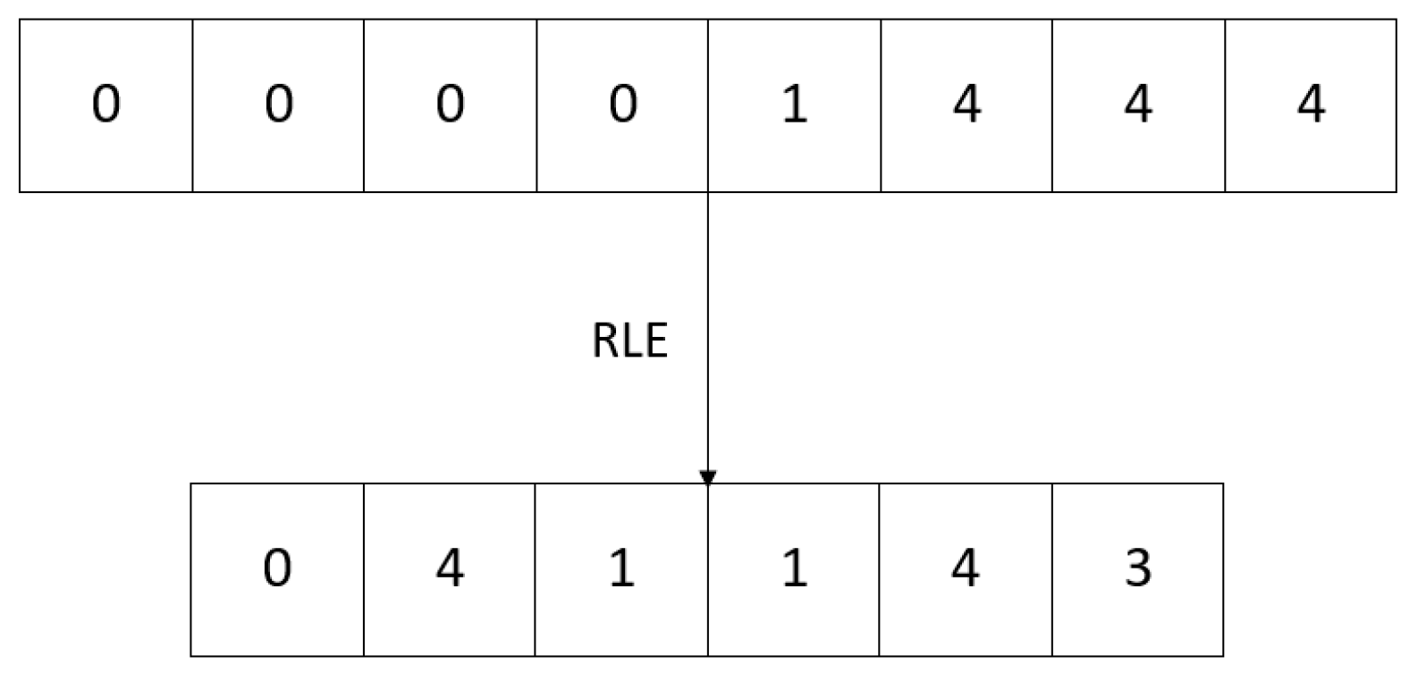
RLE is a simple lossless data compression method. The core idea is to count the successively repeating elements of the original series and write them as a pair (element, consecutive repetition number). An example is shown in Figure 2 where the first four 0s are encoded as a pair (0, 4), then a single 1 as (1, 1) and, finally, three 4 s as (4, 3).
This method is suitable only for series with a high number of successively repeating values since, on the contrary, it can lead to the expansion of the original series. Considering that medical images have a lot of large areas of uniform colour, with a fairly good prediction most of the values converge towards zero which makes RLE a suitable extension for CBPC.
Because of the aforementioned properties of the RLE, it is usually used as an auxiliary rather than a primary encoding method. It is activated when a region with a high number of successively repeating elements is found. The minimum number of element repetitions for activating the RLE method should be high enough to avoid expansion of the series and make the cost of switching the encoding methods acceptable. In this context, switching to RLE is called run-mode and is a part of the LOCO-I algorithm, which is accepted as a fundamental part of the JPEG-LS standard.

2.4. Related Work

Many modern medical imaging devices, such as CT scanners, MRIs, or X-rays, use DICOM for information sharing and standardized output. As Liu et al. [10] describe, the size of a typical CT exam had about 12 MB in the early 1990s. In 2017, a similar exam on a modern CT scanner increased the output size of the file between 600 MB and 1 GB. The size of a standard DICOM file may require higher bandwidth and storage, which is an issue even during the data migration [11].
Compression techniques for images stored in DICOM files are one approach to lowering the file size and bandwidth required. There are two main types of image compression, lossless, which retains complete image information, and lossy, which removes some information from the image. As lossy compression removes some data from the picture, it is questionable if that image is still valid for further analysis. Authors, such as García-Vílchez et al. in their paper [12], analyze the impact of lossy compression on hyperspectral image classification.
Authors, such as Li Z. et al. in their work [13], describe an optimized JPEG-XT method (OPT_JPEG_XT) that better compresses 16-bit medical images than JPEG-XT and JPEG 2000. They continue their efforts to increase the compression ratio and publish their upgraded work [14] in 2023.
Other authors, such as Nassef et al. [15], describe a lossy algorithm inspired by translating DNA sequences into protein sequences to store grayscale images, which they represent as promising compared to JPEG lossy image compression. The output file size is not the only limitation—another is computing power and running memory. Zhang et al. describe their approach [16] to lossy JPEG image compression. They use a new image compression algorithm based on a non-uniform partition and U-system designed for such devices. In their work [17], Kumar et al. propose lossless compression of CT images by an improved prediction scheme using the least square algorithm. Their implementation on Raspberry Pi shows a significant improvement compared to other algorithms, such as JPEG, JPEG lossless, BCOT, SPIHT, and Maxshift. In another work, Kumar et al. [18] propose Gaussian Hemite polynomial-based lossless medical image compression, which produces better results when compared with the Legendre polynomial-based compression and JPEG lossless compression. Authors, such as Aldemir et al. [19], suggest two new chain code approaches as solutions to the large data size required to store in PACS over time.
Some authors, such as Latha et al. [20], even suggest combining data compression with encryption for biomedical images to achieve better data security in telemedicine. They achieve lossless compression using the hybrid of Huffman coding, linear predictive coding, and discrete wavelet. Some, such as de Aguiar et al. in their work [21], suggest implementing a blockchain token mechanism stored in DICOM files to track access to the shared medical data.
PACS are generally used to store and access DICOM files. These systems are usually accessible only from inside medical institutions. There were several attempts to create mobile or web applications to facilitate peer-review data entry. In their work, Lewis et al. [22] analyze their responsive web application to support the logging of cases and conclude that it took much fewer clicks and screen taps than previous formalized data entry using an electronic spreadsheet.
O’Sullivan et al., in their work [23], researched the use of WhatsApp in health in 2017. They discovered that all health professionals in University Hospital Limerick (UHL), Ireland, who responded to their questionnaire (41/51) used WhatsApp. All of them participated in the group chat for clinical medicine at UHL.
In 2017, Mars et al. [6] examined the future of telehealth implementation in two cases. First is the usage of smartphones in rural areas of South Africa, where doctors take photographs of skin lesions and send them to dermatologists in regional hospitals using WhatsApp. This approach significantly reduced the travel time for many patients (an estimated 70%). The problem was that many doctors then contacted dermatologists directly by phone after they replied to a message. The second case was the support for burn victims, where doctors also used WhatsApp to transfer images. In both cases, the issue was that doctors contacted specialists during specialists’ busiest times and their vacations or while they were on conferences.
Giansanti et al. [24] further improved on the work of Mars et al. They created software that uploads a video file from the PACS to the Google Drive cloud and sends links to cardiologists using WhatsApp Web. Experts responded with high grades for ease of use (3.8) and graded AVI quality with 3.1 out of 5 points in their feedback. As the authors note, this approach still requires work on compliance with adequate regulations.
Other authors, such as Pongkunakorn et al. in their work [25], use DICOM images to create a Keynote presentation on 6.5-inch iPhone to superimpose a preoperative template for hip replacement. They describe this method as accurate and reproducible for predicting the implant size of cementless THA. Experience in using iPads as Radiological Workstations and their creation of user interfaces was examined by Costa et al. in their work [26] in 2016. They concluded that the device and application were well accepted.
Some authors, such as Wattanapisit et al. [27], analyze the challenges of implementing a mHealth application. Even with no access to PACS data or other direct access to the medical data, they concluded that users mainly used the application for medical references (77.0%) and medical calculations (78.3%). It was less used to track health information (22.1%) and patient education (14.3%).
Authors, such as Ziegler et al. in their work [28], introduce Open Health Imagins Foundation Viewer—an Extensible Open-Source Framework for Building Web-Based Imaging Applications. Their work suggests that the viewer only uses a desktop browser, such as Chrome, Firefox, or Internet Explorer. Authors, such as Bai et al. in their work [29], analyze the 3D visualization of data using only web technologies as they recognize that most existing visualizations are desktop applications that may be challenging to use and maintain as they often rely on specific operating systems.

3. MIDOM

MIDOM is a software system mainly developed at the University of Zagreb, Faculty of Electrical Engineering and Computing, which aims to deliver a novel approach in medical diagnostics by allowing collaboration in distributed and mobile environments.

3.1. Overview

The system primarily targets medical specialists and enables inter-specialist consultations with colleagues from other institutions or locations, with focus on delivering healthcare to places which lack a wider range of medical specialists and to generally underdeveloped parts of the world. The novelty of MIDOM is the usage of mobile technologies to bridge the gap between a diagnosing physician and a specialist who provides the required expertise in the interpretation of the diagnostic medical data in the form of a medical image. Exchanged data are in the form of studies and consultation requests, which can be consisted of various types of multimedia content.
To completely describe MIDOM as a system, its components, and functionalities, we introduce the following keywords:
  • Study—A basic set of diagnostic data created with the purpose of exchange in the system;
  • Consultation Request (CR)—A request for medical advice, opinion, or feedback for the existing study;
  • Administrator—A system user tasked with user administration, management of system settings, and application-specific tasks;
  • Study Provider (SP)—A physician or other medical professional who is in charge of creating medical studies and issuing consultation requests for medical specialists;
  • Medical Specialist (MS)—A physician or domain expert who can accept or reject consultation requests and provide feedback to the requesting study provider.
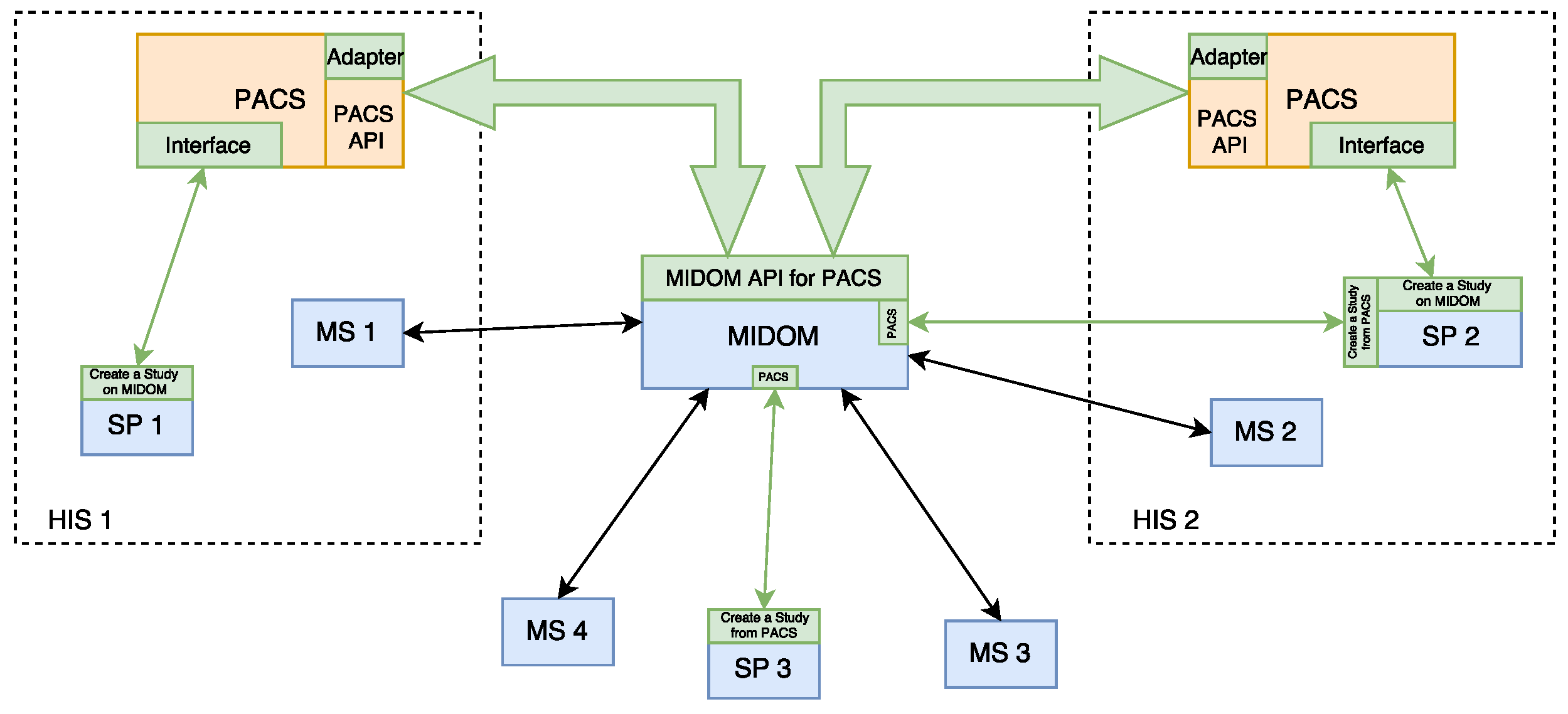
Block diagram in Figure 3 presents a high-level overview of MIDOM. Different Hospital Information Systems, HIS 1 and HIS 2, are connected to MIDOM, each with an independent PACS 1 and PACS 2 subsystems, and disjunct medical personnel, SP, MS 1, and MS 2. Medical professional outside HIS can still access MIDOM, and are depicted under acronyms MS 3, MS 4, and SP 3. Parts of the figure that are shaded in blue represent MIDOM, while orange-shaded parts represent PACS components. Green-shaded components and arrows are implemented as part of this work.
In order to provide and support good performance and user experience, the system emphasizes these three aspects:
  • On-demand server-side compression of medical images of ultra-high-resolution—Algorithms for lossless compression of one or series of images comprising a study, adaptation of such algorithms for multicore and many-core systems, the performance of such algorithms for different compression levels;
  • Client-side decompression of medical images of ultra-high-resolution—Capabilities of Android mobile platform for high storage and computing requirements posed by the application;
  • Adaptation to current environmental characteristics—Strategies and techniques for different communication channel throughput and client platform capabilities. Adaptation mechanisms as a specific feature of MIDOM consider three main challenges, user experience, data transmission cost (in particular for clients connected through mobile networks), and throughput capabilities of client devices.
A collaborative environment has been realized in the form of a MIDOM application server and several client applications in a Web interface and mobile application for the Android platform.

3.2. MIDOM Components

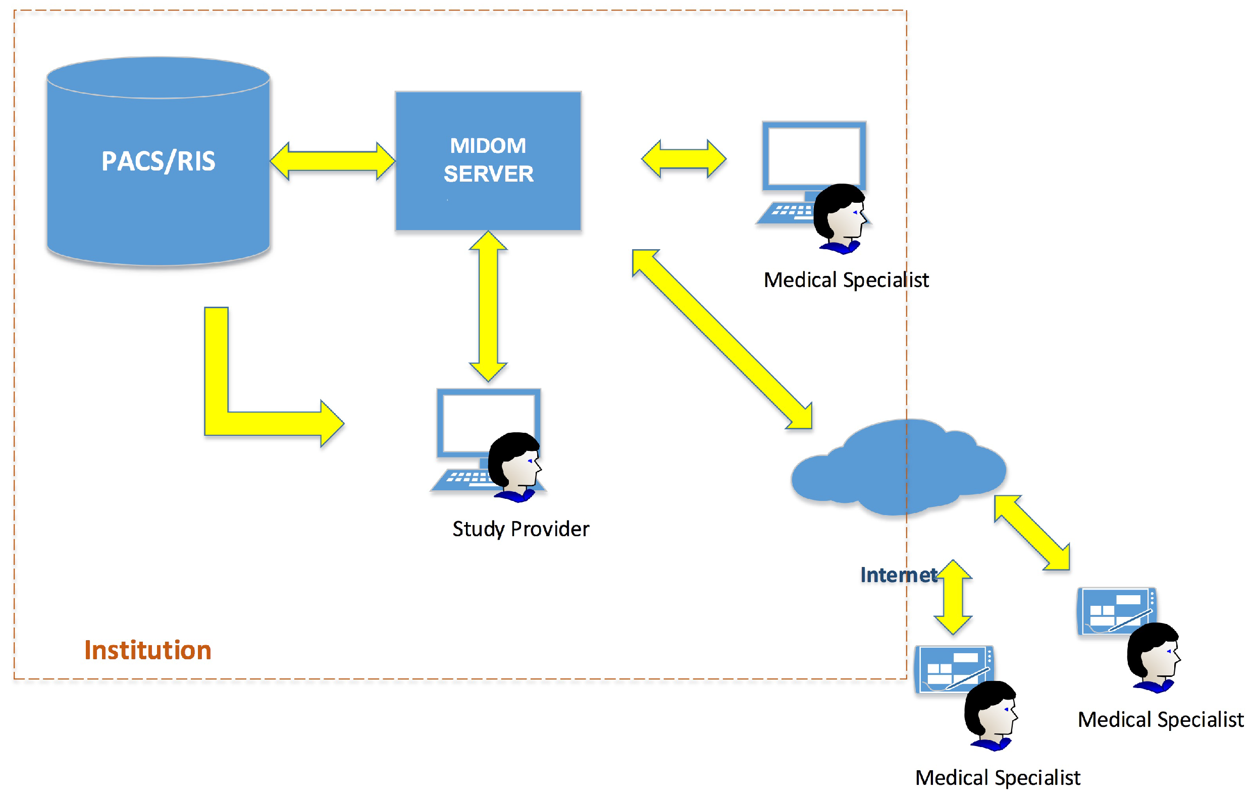
A simple usage of the MIDOM in a medical institution can be observed in Figure 4. Four main components can be easily distinguished, namely:
  • MIDOM server—The main component which facilitates the collaboration of medical specialists on sets of diagnostic procedure results, i.e., studies. This component hosts studies and related multimedia data, mainly in high-resolution images, and keeps track of the statuses of each study and consultation request. It also provides methods for system administration and performance monitoring, and performs computationally and storage-intensive operations in order to adjust to communication and processing power characteristics of the medical specialist and devices transparently to the system users;
  • PACS server—This component, originally not an integrated part of MIDOM, is used for storage purposes, for efficient medical data accessing and fetching. By attaching PACS component with MIDOM, fast access to a large number of medical images and their import for usage was enabled;
  • Study Provider—A component used by medical professionals seeking consultations with other colleagues. It allows inspection, selection, and extraction of diagnostic imaging data from the PACS server to form a study. It also allows for requests for consultations and the collection of feedback returned by medical specialists. The study provider component can manage the study and associated consultation requests’ lifecycle. The main goals of this work have been developed through this component by enabling the study provider to access the PACS server and allowing it to create a study using image data from that server;
  • Medical Provider—This component is usually implemented as a mobile part of the system. A medical specialist offers consultation services to the study provider in a standard flow. The study provider assigns consultation requests to a medical specialist then they seek further consultations about a particular study. A single study usually consists of medical images paired with their description and further explanation of the problem. A medical specialist can accept or reject a consultation request. If the consultation request is accepted, a medical specialist can inspect image data from the study for which the consultation has been requested and provide either textual or audio feedback. Medical specialists’ end devices can be connected through various types of networks, which can cause significant delays in terms of latency or time needed to pass studies through network channels. This aspect is thoroughly addressed in MIDOM by appropriate tooling and required logic for compressing exchanged imaging data to optimize user experience, throughput, and data communication costs associated with mobile networks.

4. Evaluation

This section describes the evaluation we performed to assess if the integrations mentioned above benefit the end-users, mainly in terms of faster and, thus, cheaper data transfers.

4.1. Benchmarks and Methodology

All of the experiments were conducted in a controlled environment, measuring latency, i.e., the time needed to transfer medical images from MIDOM server to medical specialist’s client application running on a mobile device with the Android operating system. We additionally measured and presented the size of the data that we worked with, in order to calculate appropriate compression rates. Although one could argue that file size and transfer latency are tightly coupled, i.e., proportional, we opted to present both the latency and compression rates, as the implied proportionality does not necessarily hold. Data transfers in conventional networks usually impose a certain amount of packet losses that incur re-transfers and thus further charges, as Internet service providers usually charge for the organic data traffic and not for the successful transfer of data granulated as data packages. Furthermore, we wanted to stress the latency reduction as it presents a major benefit for the users of our system.
The experiments were conducted in different simulated network environments nowadays, widely available. Those environments are 3G, Wi-Fi, and LTE whose throughputs are available in Table 1.
We opted for the environments mentioned earlier to test our system in various environments. 3G was included not only because it is still widely used as a fallback network around the globe, but because one of the aims of our system is to widen medical care to places which lack a range of medical specialists, which includes some underdeveloped parts of the world. By that, we wanted to express the necessity for high-quality worldwide medical care to be as reachable as possible. Wi-Fi and LTE environments are de facto standards, and 5G was not included as its usage is still limited to the relatively developed parts of the world which directly counters our aim of delivering a globally applicable system.
As much as we wanted to conduct the experiments in a real-world environment, we opted for a simulated one due to the expenses related to the on-field testing, which further influenced our possibility to consider fading models. However, we argue that that does not present a shortcoming, as additionally incurred re-transmissions when considering fading effects would even more stress the benefit of using our customized compression methods. The measuring environment consisted of a personal computer running the Android emulator (manufactured by Google, Mountain View, USA) with Intel x86 Emulator Accelerator (manufactured by Intel, Santa Clara, USA) emulating a Google Nexus device running the Android operating system. Network Link Conditioner, a tool for simulating different network environments, was used to probe our system with different parameters by limiting download and upload bandwidths and delays.
The experiments have been conducted on medical studies in each of the aforementioned network environments and using CBPC2, CBPC, and ZIP compressions, paired with raw image experiments, i.e., without compression. To obtain results as reliable as possible, each measurement was conducted five times for faster networks (Wi-Fi and LTE) and three times for the slower, 3G network. Within each measurement we compressed and transferred each image from the respective datasets, calculated averages, and, finally, after all measurements were completed, we once again calculated average of every measurement. The results for ZIP, CBPC, and CBPC2 in both subsequent Section 4.2 and Section 4.3 are displayed as relative percentages to raw data size and latencies measured when transferring raw images. The displayed result was rounded to two decimal places prior to multiplying with 100 % .
Regarding compression methods, we primarily used CBPC2 and CBPC, both described in Section 2.2, as those two methods present a backbone of our work in general and key to delivering high-quality medical content to end devices. Although ZIP is not adapted and used for image compression, we used it as it was simple to implement in our solution, it is widely available, popular, and most importantly, lossless. We thus satisfied the need to add at least one more compression method.
Benchmarking sets, or studies, consist of multiple monochromatic images, namely:
  • Angio—One image, DICOM modality: XA, 1252 × 1200 pixels in size, 1.43 MB large;
  • X-ray—Two images, DICOM modality: PX, 2372 × 2372 and 2880 × 2372 pixels in size, total of 11.88 MB large;
  • MR-1—383 images, DICOM modality: MR, 512 × 512 pixels in size, total of 95.82 MB large;
  • CT—307 images, DICOM modality: CT, 512 × 512 pixels in size, total of 76.81 MB large;
  • MR-2—2534 images, DICOM modality: MR, 320 × 320 pixels in size, total of 486.83 MB large.

4.2. Compression Ratio

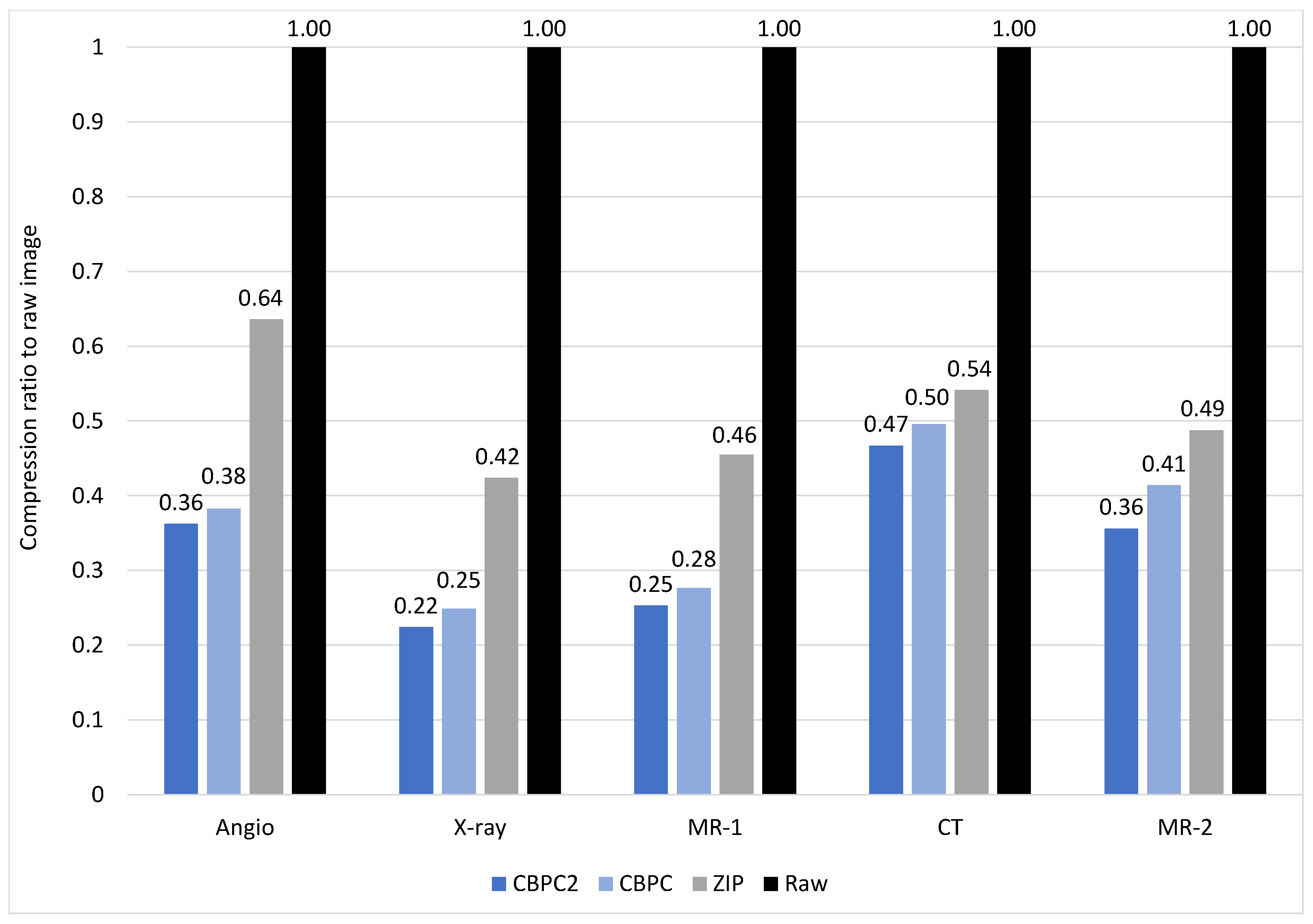
The first set of results we display in this section is compression ratios acquired by comparing compressed files’ data sizes with uncompressed raw files’ sizes. The results can be seen in Table 2 as sizes in Megabytes, and in Figure 5, grouped by individual benchmarks with each bar, but the black one, representing one compression method. Blue-shaded bars represent CBPC and CBPC2 compressions, while the grey-shaded bar represents the ZIP compression ratio. The black bar is fixed to 1 representing the raw and uncompressed image size. Both CBPC and CBPC2 compression methods significantly reduce uncompressed images, with compression levels ranging from 22 % and 25 % for the X-ray benchmark to 47 % and 50 % for CT benchmarks. When assessing compression ratios of CBPC and CBPC2 compression methods with respect to ZIP compression, results vary between more considerable reductions for Angio, X-ray, and MR-1 benchmarks and relatively minor reductions for CT and MR-2 benchmarks. When assessing the benefits of improvements of the CBPC2 compression method concerning CBPC, every benchmark shows an improvement of at least 2 percentage points, with MR-2 being an example of an improvement of 6 percentage points.

4.3. Latency Reduction

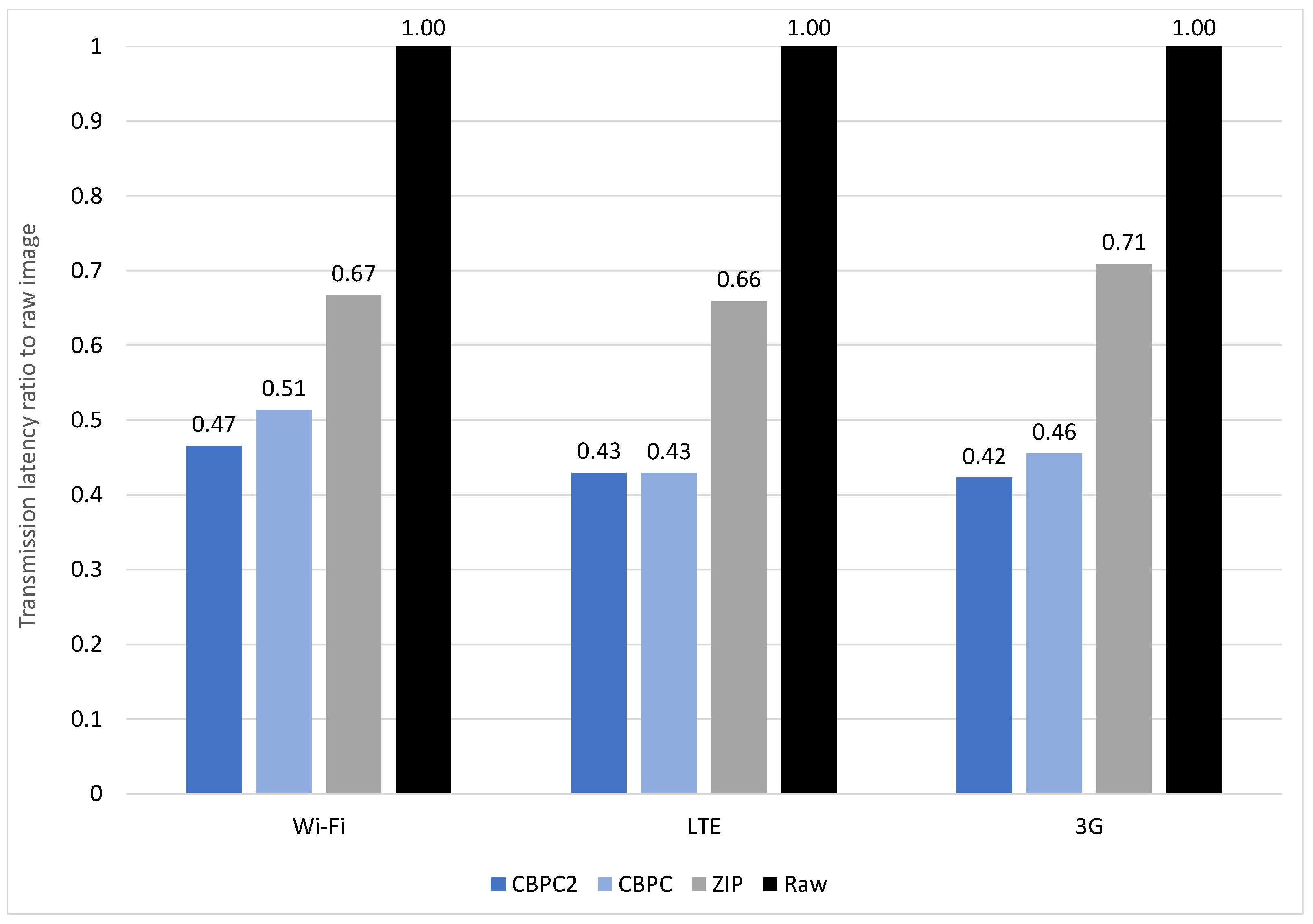
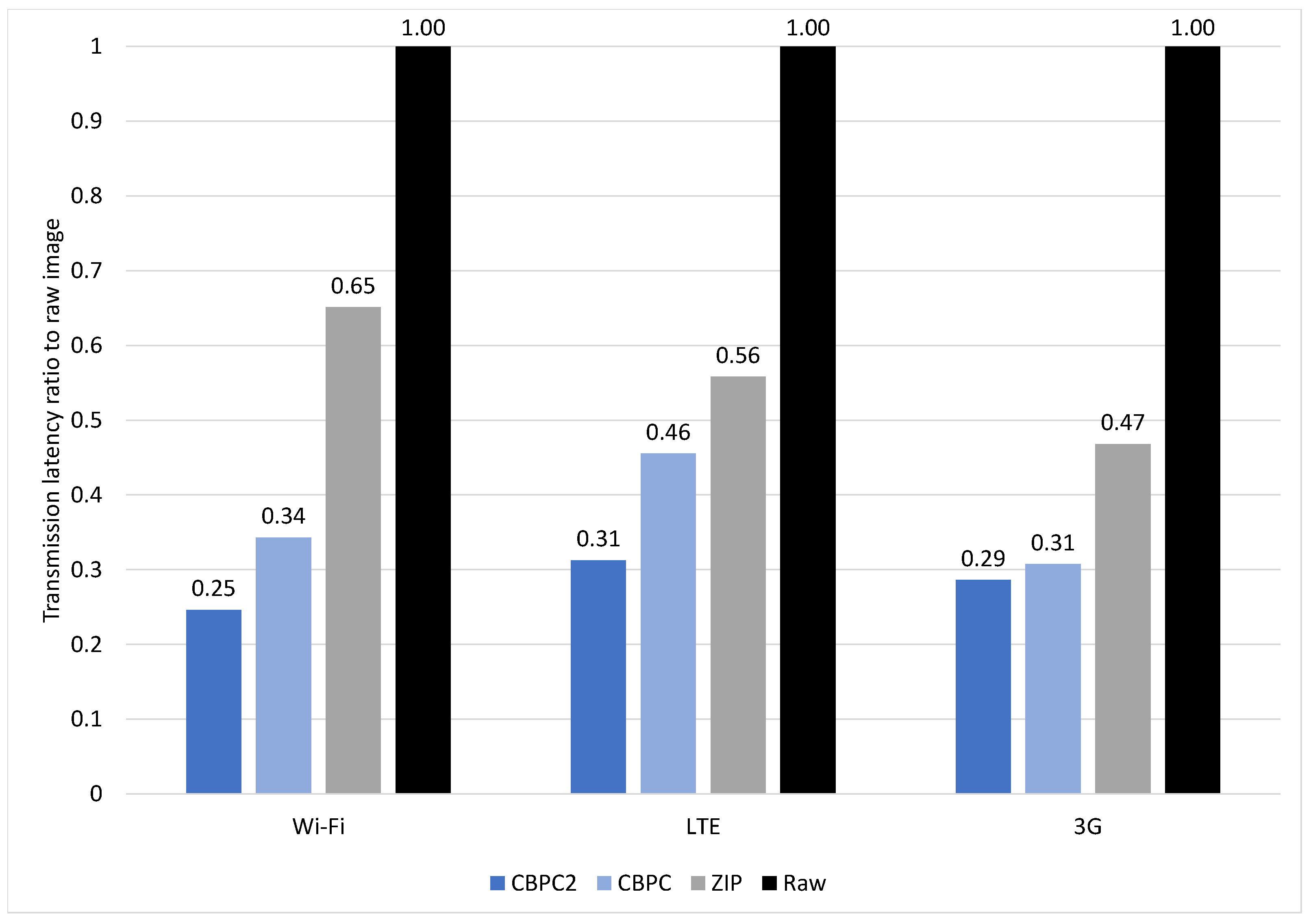
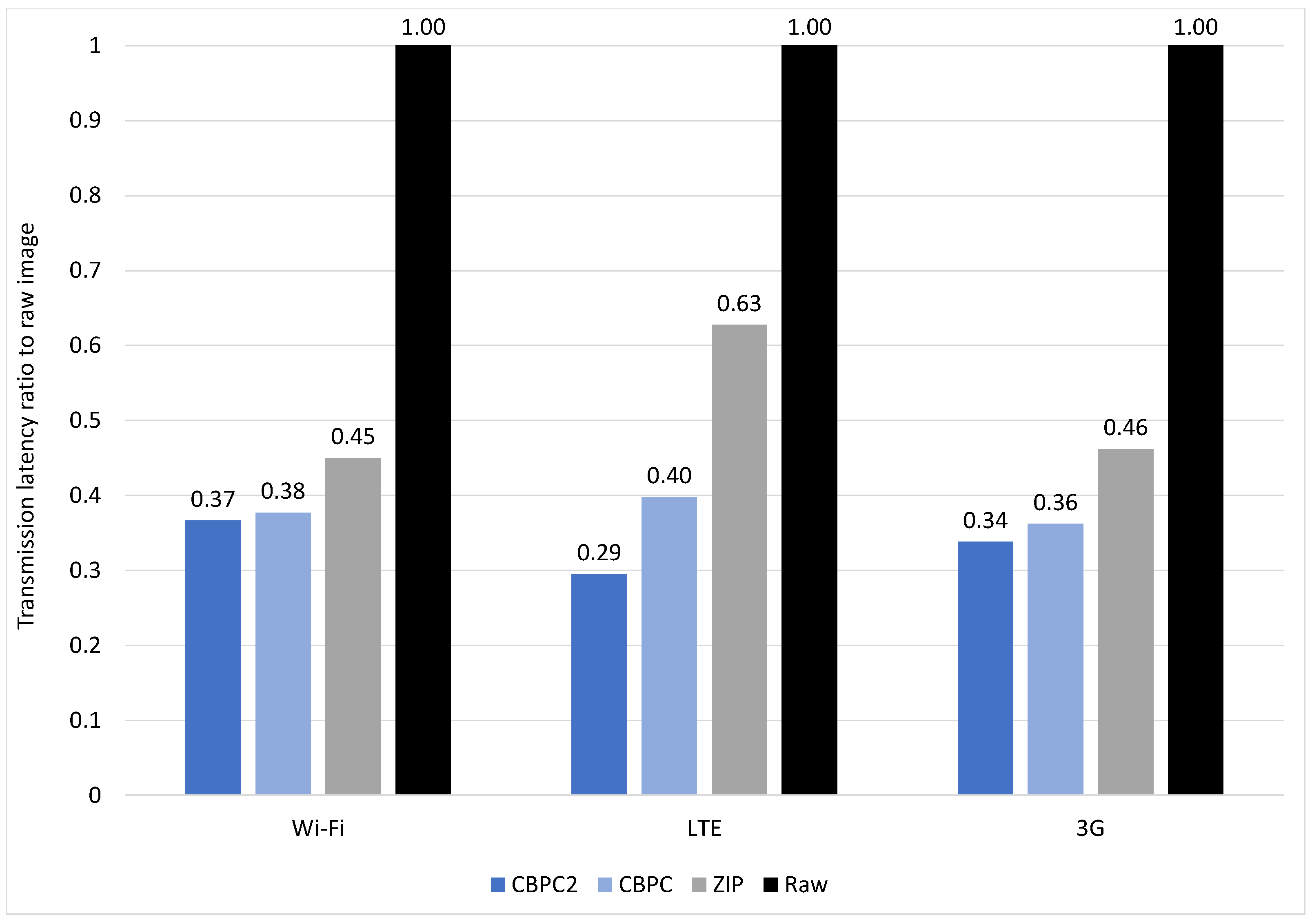
In this subsection, we display the second set of results, the network latency reduction when transmitting medical images over the various types of networks. The results are presented as transmission time ratios of the transmission time when using different data compression methods to the transmission time of raw data for different network environments. The results are grouped by the network type and each bar represents a different compression method. As in the previous result representation, the black-shaded bars are fixed to 1, representing raw and uncompressed data transmission time, while blue-shaded bars represent CBPC and CBPC2 compression, and grey-shaded ones represent ZIP compression. Additionally, raw data in terms of latencies in seconds are presented in Table 3.

4.3.1. Angio

Figure 6 displays latency reduction results for Angio study. As expected, CBPC2 and CBPC compression methods achieve the best results for all three simulated network environments. For LTE, CBPC2, and CBPC results are identical, thus rendering CBPC2 unnecessary, as it lacks any benefits in terms of latency reduction while being more computationally expensive. The best result is achieved in the 3G environment for the CBPC2 method, with latency lowered down to 42 % of the latency needed to transfer the raw study, which is particularly important as 3G is the network environment with the relatively smallest throughput used due to its wide applicability. In the context of the 3G network, a couple of percentage points gained by using CBPC2 make a significant difference. At the same time, analogously, that benefit can be disregarded in the context of a Wi-Fi network. Therefore, it can be concluded that, due to its added computational cost, CBPC2 pays off for the 3G network only. ZIP compression yields results close to 70 % for all networks. Compared to ZIP, both CBPC2 and CBPC yield significantly better results and quickly pay off for every network environment.

4.3.2. X-ray

Unlike the previously described Angio benchmark, the X-ray benchmark fully displays the plausibility of using the CBPC2 compression method, as it achieved significantly better results than its simpler counterpart. This is depicted on Figure 7. For Wi-Fi and LTE, CBPC2 achieved better results for 9 and 15 percentage points, respectively, than CBPC. For 3G, that margin is smaller but can still be considered significant, as relatively low speeds of the 3G network could use each increase in latency reduction. When comparing our custom compression methods with ZIP compression, using CBPC and CBPC2 led to better results regarding latency reductions. The margin was the smallest on the LTE network and larger on Wi-Fi and 3G networks. For this benchmark, latency reduction varies greatly for ZIP compression, ranging from 65 % for a Wi-Fi network to 47 % for a 3G network. The largest margin is achieved on the Wi-Fi network as the CBPC2 compression method reduces latency to just 25 % , relative to raw image transfer, and for 40 percentage points with respect to ZIP compression.

4.3.3. MR-1

When it comes to the MR-1 benchmark, results can be seen in Figure 8. Again, both CBPC2 and CBPC cause significant reductions in latency, with CBPC2 paying off better for Wi-Fi, due to a large increase in latency reduction, and for 3G, due to a minor yet not insignificant reduction in the context of this network type. CBPC2 gains the expected result in sense of additional latency reduction for LTE network. For each network profile, latency is reduced for approximately 25 % relative to raw image transfer. ZIP compression is steady at around 50 % for each network type but is still significantly worse than CPBC2 and CBPC for each network profile, again rendering them plausible for use.

4.3.4. CT

When putting all of the benchmarks performed in this paper in perspective, it cannot be avoided to state that this benchmark yielded the worst results for latency reduction. As can be observed in Figure 9, the significant difference between CBPC2 and CBPC compression methods can be observed for Wi-Fi and 3G, with the latter being significant due to relatively low throughput, which makes every percentage point essential to spare. CBPC2 compression method yields at most 47 % reduction in latency for the 3G environment, 49 % for the LTE environment, and 52 % for the Wi-Fi environment. For the CBPC compression method, every environment yields up to 50 % reduction in latency. When comparing reductions with ZIP compression, both CBPC2 and CBPC do not achieve significant reductions, especially for the 3G network with ZIP-reducing latency to 55 % , CBPC to 50 % , and CBPC2 to 47 % of the raw image transfer latency, meaning that CBPC2 and CBPC are only 8 and 5 percentage points better than ZIP compression method. The margin between ZIP and CBPC2 compression methods is at least 18 percentage points for other benchmarks. Here, we again observe cases in which our compression methods do not achieve at least 50 % of reduction in latency (CBPC2 and CBPC in Wi-FI environment, CBPC in LTE environment), which, in the context of other benchmarks, was observed only for Angio benchmark (CBPC in Wi-Fi environment).

4.3.5. MR-2

The latency reduction results for the MR-2 benchmark are available in Figure 10. The difference between CBPC2 and CBPC is negligible for the Wi-Fi network environment and significant for LTE and 3G environments, with particular significance in LTE environments with a gain in latency reduction of 11 percentage points. Compared to the ZIP compression method, CBPC2 and CBPC methods generate the best addition in the reduction for LTE and 3G networks. ZIP produces 45 % reduction in latency regarding raw image transfer for the Wi-Fi network environment, which, paired with a relatively weak result of CBPC2 and CBPC compressions, renders them unplausible to use.

4.4. Discussion

MIDOM as a system for the exchange of DICOM-based medical images between remote medical professionals in heterogeneous environments with isolated PACS systems envisioned for underdeveloped parts of the world with limited connectivity, truly is one of a kind which makes it hard to find a system that is similar enough to compare with. We aimed to compare our work with the one found in Ziegler et al. [28], but their approach differs from ours since mobile phones are neither their primary target nor the lossless compression of the files to achieve smaller file sizes and latency, as in MIDOM.
On the other hand, Bai et al. [29] are focused primarily on 3D visualization and not on compression of the files for smaller file sizes and latency with the goal of developing a system for underdeveloped parts of the world, as in MIDOM.

5. Conclusions

This section briefly discusses the outcomes of the research presented through this paper, evaluates the limitations, and proposes further work in this area.

5.1. Summary

In this paper, we proposed MIDOM, an e-health platform whose main aim is to increase the availability of medical care in underdeveloped parts of the world and, generally, in places with limited access to medical professionals. It does so mainly by increasing interoperability by acting as a man in the middle between different medical actors, devices, medical professionals, and inherently standards in electronic health. MIDOM enables the increase in communication reliability, confidence, and confidentiality by providing secure communication packed with a lossless compression method, which significantly reduces sizes, implying a reduction in network latencies and data savings. Implicitly, it provides mitigation mechanisms between various standards in electronic health systems, which are heavily limited in terms of interoperability.
In addition to MIDOM’s implementation and integration with broader systems like PACS server, one of the main focuses of this paper is integration of the aforementioned custom compression method (CBPC), and its additional implementation (CBPC2) which features run-length coding enhancement. Higher compression rates with lossless compression methods are crucial in achieving the availability of electronic health systems, which is especially important for low throughput networks. The latter kind of network is still widely used as failover networks in developed parts of the world and as main networks in underdeveloped parts.
We demonstrate significant data reductions through experiments by using our custom compression methods, which implied latency reductions in the tested network. We performed five benchmarks on three different networks, namely Wi-Fi, LTE, and 3G, demonstrating that our compression methods pay off in nearly every testing scenario by lowering the network latency to at least 60% of the original latency measured when transferring equivalent uncompressed dataset. The best result is achieved by lowering network latency to mere 25% of uncompressed transfer latency for MR-1 benchmark on Wi-Fi and LTE networks and X-ray benchmark on the Wi-Fi network environment.

5.2. Limitations

We acknowledge the limitations regarding our simulation environment and commit to extend the benchmarks with on-field testing that will simulate real-world environment and provide the so much needed measurements of the signal fading effects. Furthermore, we were limited with the amount of the available medical data, mostly due to GDPR. An increased amount of data would further increase the reliability of the study. The implemented system was used in laboratory environment and may require additional improvements in terms of scalability and stability. The study does not consider the security aspect of the data transfer as we assume the use of the standard security protocols.

5.3. Suggestions for Future Research

As electronic health services keep developing, we see promising aspects for future work. Conceptually, medical imaging and consultation with a medical professional could vastly profit from 3D modelling. It is now possible to transfer complex 3D models by dissecting a model to multiple layers, encoding that layers as images, and transferring them accordingly. Future work in the field of compression will include the development of such an algorithm that explores and exploits space similarities to different layers with the ultimate purpose of increasing the compression ratios of such a model. By developing such an algorithm, the compression ratio of a complete model would be larger than the sum of disjunct compression ratios of each independent layer. We will further continue developing our system on a system level by increasing stability and reliability on low-throughput networks. We will additionally work on further expanding platforms that the system can target.

Author Contributions

Conceptualization, B.P. and J.K.; Funding acquisition, J.K.; Investigation, B.P. and S.T.; Methodology, B.P.; Resources, J.K.; Software, J.K.; Supervision, J.K.; Validation, B.P. and S.T.; Visualization, H.I.; Writing—original draft, B.P., S.T. and H.I.; Writing—review and editing, B.P. and J.K. All authors have read and agreed to the published version of the manuscript.

Funding

This research received no external funding.

Institutional Review Board Statement

Not applicable.

Informed Consent Statement

Not applicable.

Data Availability Statement

The availability of the images used in this work can be discussed with the corresponding author, as well as the data collected by simulating the described system in testing scenarios.

Acknowledgments

The authors would like to thank Eugen Druzin for implementing parts of the system and conducting the experiments while working on their Master’s degree thesis at the University of Zagreb Faculty of Electrical Engineering and Computing.

Conflicts of Interest

The authors hereby declare that no interest that could be perceived as conflicting exist.

Abbreviations

The following abbreviations are used in this manuscript:
APIApplication Programming Interface
CBPCClassification and Blending Predictor Coder
CRConsultation Request
CTComputed Tomography
DICOMDigital Imaging and Communications in Medicine
GDPRGeneral Data Protection Regulation
HIPAAHealth Insurance Portability and Accountability Act
HISHospital Information System
IODInformation Object Definitions
MIDOMMedical Imaging and Diagnostics on the Move
MRMagnetic Resonance
MSMedical Specialist
NEMANational Electrical Manufacturers Association
OPT_JPEG_XTOptimized JPEG-XT
PACSPicture Archiving and Communication System
RLERun-Length Encoding
SPStudy Provider
UHLUniversity Hospital Limerick
VPNVirtual Private Network

References

  1. Li, H.; Yu, L.; He, W. The Impact of GDPR on Global Technology Development. J. Glob. Inf. Technol. Manag. 2019, 22, 1–6. [Google Scholar] [CrossRef]
  2. Jeong, Y.U.; Yoo, S.; Kim, Y.H.; Shim, W.H. De-identification of facial features in magnetic resonance images: Software development using deep learning technology. J. Med. Internet Res. 2020, 22, e22739. [Google Scholar] [CrossRef] [PubMed]
  3. Morris, C.; Scott, R.E.; Mars, M. Whatsapp in clinical practice—The challenges of record keeping and storage. A scoping review. Int. J. Environ. Res. Public Health 2021, 18, 13426. [Google Scholar] [CrossRef]
  4. Ncube, B.; Mars, M.; Scott, R.E. The need for a telemedicine strategy for Botswana? A scoping review and situational assessment. BMC Health Serv. Res. 2020, 20, 794. [Google Scholar] [CrossRef]
  5. BBC News. WhatsApp Issued Second-Largest GDPR Fine of €225m. Available online: https://www.bbc.com/news/technology-58422465 (accessed on 8 May 2023).
  6. Mars, M.; Scott, R.E. Being Spontaneous: The Future of Telehealth Implementation? Telemed. J. Health Off. J. Am. Telemed. Assoc. 2017, 23, 766–772. [Google Scholar] [CrossRef] [PubMed]
  7. Knezovic, J. Kompresija Slika Bez Gubitaka Metodom Predvidjanja; Faculty of Electrical Engineering and Computing, University of Zagreb: Zagreb, Croatia, 2005. [Google Scholar]
  8. Knezovic, J.; Kovac, M.; Mlinaric, H. Classification and Blending Prediction for Lossless Image Compression. In Proceedings of the MELECON 2006—2006 IEEE Mediterranean Electrotechnical Conference, Malaga, Spain, 16–19 May 2006; IEEE: Piscataway, NJ, USA, 2006; Volume 2006, pp. 486–489. [Google Scholar] [CrossRef]
  9. Strizic, L.; Knezovic, J. Optimization of losless image compression method for GPGPU. In Proceedings of the 2016 18th Mediterranean Electrotechnical Conference (MELECON), Lemesos, Cyprus, 18–20 April 2016; IEEE: Piscataway, NJ, USA, 2016. number April. pp. 1–6. [Google Scholar] [CrossRef]
  10. Liu, F.; Hernandez-Cabronero, M.; Sanchez, V.; Marcellin, M.W.; Bilgin, A. The current role of image compression standards in medical imaging. Information 2017, 8, 131. [Google Scholar] [CrossRef]
  11. van Ooijen, P.M.; Aryanto, K.Y.; Broekema, A.; Horii, S. DICOM data migration for PACS transition: Procedure and pitfalls. Int. J. Comput. Assist. Radiol. Surg. 2015, 10, 1055–1064. [Google Scholar] [CrossRef]
  12. García-Vílchez, F.; Muñoz-Marí, J.; Zortea, M.; Blanes, I.; González-Ruiz, V.; Camps-Valls, G.; Plaza, A.; Serra-Sagristà, J. On the impact of lossy compression on hyperspectral image classification and unmixing. IEEE Geosci. Remote Sens. Lett. 2011, 8, 253–257. [Google Scholar] [CrossRef]
  13. Li, Z.; Ramos, A.; Li, Z.; Osborn, M.L.; Li, X.; Li, Y.; Yao, S.; Xu, J. An optimized JPEG-XT-based algorithm for the lossy and lossless compression of 16-bit depth medical image. Biomed. Signal Process. Control 2021, 64, 102306. [Google Scholar] [CrossRef]
  14. Li, Z.; Ramos, A.; Li, Z.; Osborn, M.L.; Zaid, W.; Li, X.; Li, Y.; Xu, J. Nearly-lossless-to-lossy medical image compression by the optimized JPEGXT and JPEG algorithms through the anatomical regions of interest. Biomed. Signal Process. Control 2023, 83, 104711. [Google Scholar] [CrossRef]
  15. Nassef, M.; Alkinani, M.H. A Novel Multilevel Lossy Compression Algorithm for Grayscale Images Inspired by the Synthesization of Biological Protein Sequences. IEEE Access 2021, 9, 149657–149680. [Google Scholar] [CrossRef]
  16. Zhang, Y.; Cai, Z.; Xiong, G. A New Image Compression Algorithm Based on Non-Uniform Partition and U-System. IEEE Trans. Multimed. 2021, 23, 1069–1082. [Google Scholar] [CrossRef]
  17. Kumar, S.N.; Fred, A.L.; Kumar, H.A.; Varghese, P.S. Lossless Compression of CT Images by an Improved Prediction Scheme Using Least Square Algorithm. Circuits Syst. Signal Process. 2020, 39, 522–542. [Google Scholar] [CrossRef]
  18. Kumar, S.N.; Ahilan, A.; Haridhas, A.K.; Sebastian, J. Gaussian Hermite polynomial based lossless medical image compression. Multimed. Syst. 2021, 27, 15–31. [Google Scholar] [CrossRef]
  19. Aldemir, E.; Dueñas, O.A.T.; Kavur, A.E.; Tohumoglu, G.; Sánchez-Cruz, H.; Selver, M.A. Chain code strategy for lossless storage and transfer of segmented binary medical data. Expert Syst. Appl. 2023, 216, 119449. [Google Scholar] [CrossRef]
  20. Latha, H.R.; Ramaprasath, A. HWCD: A hybrid approach for image compression using wavelet, encryption using confusion, and decryption using diffusion scheme. J. Intell. Syst. 2023, 32, 20229056. [Google Scholar] [CrossRef]
  21. de Aguiar, E.J.; dos Santos, A.J.; Meneguette, R.I.; Grande, R.E.D.; Ueyama, J. A blockchain-based protocol for tracking user access to shared medical imaging. Future Gener. Comput. Syst. 2022, 134, 348–360. [Google Scholar] [CrossRef]
  22. Lewis, T.; Berkowitz, S.; Weinstein, J. Abstract No. 565 Smartphone and web-based interventional radiology case logging system to facilitate recording of procedures performed by interventional radiology residents. J. Vasc. Interv. Radiol. 2021, 32, S157. [Google Scholar] [CrossRef]
  23. O’Sullivan, D.M.; O’Sullivan, E.; O’Connor, M.; Lyons, D.; McManus, J. WhatsApp Doc? BMJ Innov. 2017, 3, 238–239. [Google Scholar] [CrossRef]
  24. Giansanti, D.; Cosentino, L. WhatsApp in mHealth: Design and evaluation of an mHealth tool to share dynamic images in hemodynamics. mHealth 2021, 7, 1–7. [Google Scholar] [CrossRef]
  25. Pongkunakorn, A.; Aksornthung, C.; Sritumpinit, N. Accuracy of a New Digital Templating Method for Total Hip Arthroplasty Using Picture Archiving and Communication System (PACS) and iPhone Technology: Comparison with Acetate Templating on Digital Radiography. J. Arthroplast. 2021, 36, 2204–2210. [Google Scholar] [CrossRef] [PubMed]
  26. Costa, P.; von Wangenheim, A.; von Wangenheim, C.; Inácio, A.; de Macedo, D. Usability Engineering of a Radiological Workstation for Mobile Devices: An Experience Report. Soc. Bras. Comput. SBC 2016, 7, 2471–2480. [Google Scholar] [CrossRef]
  27. Wattanapisit, A.; Amaek, W.; Wattanapisit, S.; Tuangratananon, T.; Wongsiri, S.; Pengkaew, P. Challenges of implementing an mhealth application for personalized physical activity counselling in primary health care: A qualitative study. Int. J. Gen. Med. 2021, 14, 3821–3831. [Google Scholar] [CrossRef] [PubMed]
  28. Ziegler, E.; Urban, T.; Brown, D.; Petts, J.; Pieper, S.D.; Lewis, R.; Hafey, C.; Harris, G.J. Open Health Imaging Foundation Viewer: An Extensible Open-Source Framework for Building Web-Based Imaging Applications to Support Cancer Research. JCO Clin. Cancer Inform. 2020, 4, 336–345. [Google Scholar] [CrossRef]
  29. Bai, S.; Ma, C.; Wang, X.; Zhou, S.; Jiang, H.; Ma, L.; Jiang, H. Application of Medical Image 3D Visualization Web Platform in Auxiliary Diagnosis and Preoperative Planning. J. Image Graph. 2023, 11, 32–39. [Google Scholar] [CrossRef]
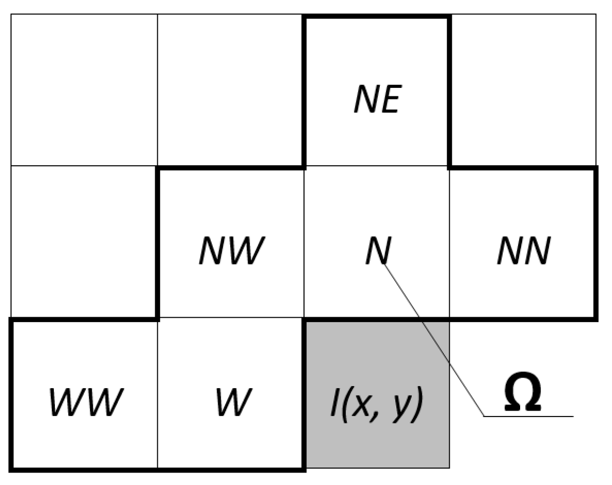
Figure 1. An example of the prediction context—processed element shaded gray.
Applsci 13 06075 g001
Figure 2. An example of run-length encoding.
Applsci 13 06075 g002
Figure 3. MIDOM in use.
Applsci 13 06075 g003
Figure 4. MIDOM components.
Applsci 13 06075 g004
Figure 5. Reduction in terms of data size relative to raw image.
Applsci 13 06075 g005
Figure 6. Latency reduction for Angio benchmark.
Applsci 13 06075 g006
Figure 7. Latency reduction for the X-ray benchmark.
Applsci 13 06075 g007
Figure 8. Latency reduction for MR-1 benchmark.
Applsci 13 06075 g008
Figure 9. Latency reduction for CT benchmark.
Applsci 13 06075 g009
Figure 10. Latency reduction for MR-2 benchmark.
Applsci 13 06075 g010
Table 1. Network link conditioner parameters.
ProfileBandwidthDelay
DownloadUploadDownloadUpload
3G780 Kbps330 Kbps100 ms100 ms
Wi-Fi40 Mbps30 Mbps1 ms1 ms
LTE50 Mbps10 Mbps50 ms50 ms
Table 2. Raw sizes of the original and compressed data.
Data Size [MB]
OriginalCBPC2CBPCZIP
Angio1.430.520.550.91
X-ray11.882.662.955.03
MR-195.8224.2326.4843.62
CT76.8135.8738.0741.56
MR-2486.83173.41201.52237.37
Table 3. Raw latencies in miliseconds.
Latency [s]
AngioX-rayMR-1CTMR-2
3GOriginal28.41146.571108.39879.666197.09
CBPC212.0141.97286.48415.872095.19
CBPC12.9445.06312.58443.332242.86
ZIP20.1568.61506.64483.022861.06
LTEOriginal3.2810.4658.2943.58532.27
CBPC21.413.2714.7821.41156.95
CBPC1.414.7618.0023.57211.59
ZIP2.175.8430.3226.73334.01
Wi-FiOriginal1.065.9436.2428.58224.94
CBPC20.491.468.8914.7582.40
CBPC0.552.0412.7317.1784.78
ZIP0.713.8718.7719.87101.17
Disclaimer/Publisher’s Note: The statements, opinions and data contained in all publications are solely those of the individual author(s) and contributor(s) and not of MDPI and/or the editor(s). MDPI and/or the editor(s) disclaim responsibility for any injury to people or property resulting from any ideas, methods, instructions or products referred to in the content.