Article

Phytochemical Profiling of the Ethanolic Extract of Zaleya pentandra L. Jaffery and Its Biological Activities by In-Vitro Assays and In-Silico Molecular Docking

1
Department of Pharmaceutical Chemistry, Faculty of Pharmacy, The Islamia University of Bahawalpur, Bahawalpur 63100, Pakistan
2
Department of Pharmacognosy, College of Pharmacy, King Saud University, Riyadh 11495, Saudi Arabia
3
Department of Pharmacognosy, College of Pharmacy, Prince Sattam bin Abdul Aziz University, Alkharj 11942, Saudi Arabia
4
Department of Pharmacognosy, Faculty of Pharmacy, Mansoura University, Mansoura 35516, Egypt
5
Department of Pharmaceutical Chemistry, Faculty of Pharmaceutical Sciences, Prince of Songkla University, Hat Yai 90112, Songkla, Thailand
6
College of Pharmacy, Shenzhen Technology University, Shenzhen 518060, China
7
State Key Laboratory of Food Science and Technology, Nanchang University, Nanchang 330047, China
8
Department of Food Development and Food Quality, Institute of Food Science and Human Nutrition, Gottfried Wilhelm Leibniz University Hannover, Am Kleinen Felde 30, 30167 Hannover, Germany
9
Department of Food Science, Faculty of Agriculture, Zagazig University, Zagazig 44519, Egypt
10
School of Food Science and Engineering, South China University of Technology, Guangzhou 510641, China
*
Authors to whom correspondence should be addressed.
Appl. Sci. 2023, 13(1), 584; https://doi.org/10.3390/app13010584
Submission received: 5 December 2022 / Revised: 23 December 2022 / Accepted: 26 December 2022 / Published: 31 December 2022
(This article belongs to the Special Issue Recent Advances in Applied Microbiology and Food Sciences, Volume II)

Abstract

:
Zaleya pentandra L. jaffery is the only species of the genus Zaleya that grows in the Cholistan desert, Pakistan. It is a Xero-halophyte plant with high phenolic and flavonoid content. The present research was designed to investigate the phytochemical composition, biological activities, and in silico molecular docking of the ethanolic extract of Z. pentandra. The phytochemical evaluation was done through preliminary phytochemical testing, estimation of total bioactive content, and gas chromatography–mass spectrometry (GC–MS) analysis for the identification of volatile compounds. For the evaluation of biological activities, antioxidants, and enzyme inhibition (α-glucosidase, cholinesterase, and tyrosinase), antibacterial and antiviral assays were performed. GC–MS analysis revealed the presence of 29 tentative volatile compounds. The ethanolic extract of Z. pentandra contains high phenolic content (119.6 ± 0.12 mg GAE/g extract) and flavonoid content (45.5 ± 0.19 mg QE/g extract), which correlates with the strong DPPH, FRAP, and enzyme inhibition results. The ethanolic extract of Z. pentandra also showed dose-dependent antibacterial activity. Micrococcus luteus and Pseudomonas aeruginosa were found to be most susceptible, with 16 mm and 17 mm zone of inhibitions at a maximum dose of 20 mg/mL. Antiviral results showed that the ethanol extract has excellent activity against H9, IBV, and NDV viral strains. Additionally, in silico molecular docking was performed in order to determine the interaction and binding affinity between the enzymes and compounds identified by GC–MS. α-glucosidase, cholinesterase, and tyrosinase showed the highest binding affinity toward 1,2-benzenedicarboxylic acid, 2-hydroxy-n-(2-phenylethyl) benzamide, γ-sitosterol, and lactose. These findings can serve as a benchmark for anti-diabetic-, neuro-, and skin-protective uses of this plant and can be used for the isolation of pure bioactive compounds in the future.

1. Introduction

Recently, pharmaceutical and nutrition sciences have detected a boom in the scientific literature geared toward the utilization of medicinal plants due to their diverse health benefits and therapeutic potential [1]. During the last few decades, the complexity of secondary metabolites in traditionally used medicinal plants was exploited and thus being the mainstay for the discovery of novel lead compounds [2]. Medicinal flora has great importance as they contain novel entities possessing multiple therapeutic and pharmacological activities [1,3,4,5]. According to the World Health Organization, the rate of use of phytomedicines across the world has increased three times that of conventional medicine, and nearly 80% of developing countries’ populations rely on medicinal plants for primary health care. Due to the diverse therapeutic potential of medicinal plants, the governments of some countries encourage and initiate botanical drug investigation programs to explore phytopharmaceuticals [6]. Phytotherapy is based on the bioactive compounds present in medicinal plants as they are a rich source of biochemical compounds such as anthocyanins, carotenoids, lycopene, glucosinolates, omega-3 fatty acids, phytoestrogens, and polyphenols. These bioactive compounds are responsible for anti-oxidant, antimutagenic, anticarcinogenic, anti-aging, anti-inflammatory, and anti-microbial effects that might be advantageous in avoiding illnesses and maintaining genome integrity [7,8]. By virtue of these characteristics, around more than 20,000 plants have been investigated for medicinal purposes [1]. Plants are rich sources of antioxidant agents, and there is huge interest in the exploitation of natural antioxidants for treating major diseases such as diabetes, cancer, cardiovascular, neurodegenerative, and skin diseases [9]. All of these diseases are associated with reactive oxygen species (ROS) and can be treated with strong antioxidant agents. Conclusively, polyphenols and other phytoconstituents have the ability to inhibit α-glucosidase, acetylcholinesterase, and tyrosinase. Therefore, natural compounds or products containing phenols with strong ROS scavenging activity are important approaches for treating diabetes, neurodegenerative disease, hyperpigmentation, and skin cancer [10].
Halophytes have a strong ability to withstand severe environmental conditions and have toxic ROS due to the presence of a strong antioxidant system comprising both enzymatic and non-enzymatic systems [11]. Plants are rich sources of antioxidant agents, preventing cellular damage by inhibiting the initiation or proliferation of oxidative chain reactions or oxidation processes. In recent times, the strong biological activity, cost-effectiveness, and safety of medicinal plants surpassing synthetic antioxidant components hold huge interest for the exploitation of natural antioxidants in treating major diseases such as diabetes, cancer, cardiovascular, neurodegenerative, and skin diseases [9]. Inhibition of α-glucosidase is one of the main targets in treating diabetes, which prevents the breakdown of polysaccharides to monosaccharides. Acetylcholinesterase inhibitors enhance cholinergic transmission and can be used to treat Alzheimer’s disease (and other neurodegenerative diseases) [10]. Moreover, the inhibition of tyrosinase protects the skin from ultraviolet radiation, which is majorly responsible for skin cancer [12]. All of these diseases are associated with ROS and can be treated with strong antioxidant agents. Conclusively, polyphenols have the ability to inhibit α-glucosidase, acetylcholinesterase, and tyrosinase.
The Cholistan desert in Pakistan has been blessed with a wide variety of traditional medicinal plants in terms of multiple biological pyramids, and natives utilize these traditional plants as an alternative to treat major or minor health alignments [13]. Zaleya pentandra (Z. pentandra) belongs to the family Aizoaceae and has 1170 species and 128 genera. It is a Xero-halophyte perennial plant, grown near coastal sandy areas of African and Asian countries such as Pakistan, India, South America, Africa, and Iran. Only one species, Z. pentandra, is found in the Cholistan desert of Pakistan [14]. Traditionally, this plant has been used to treat different diseases such as respiratory tract infections, gonorrhea, coughs, and stomach diseases, and also acts as an astringent in snake bites [15]. A number of ethnopharmacological uses of this genus have been reported in the literature, such as Trianthema decandra and Trianthema portulacastrum, which have anti-microbial, anti-diabetic, anti-oxidant, antipyretic, anti-inflammatory, anti-cancer, anti-fungal, anti-helminthic, analgesic, and hepatoprotective activity [14]. The previously reported literature revealed that the methanolic extract of Z. pentandra contains bioactive compounds responsible for the following pharmacological activities, including anti-cancer, anti-bacterial, anti-oxidant, and anti-inflammatory activity [15,16,17]. They are also useful in hyperlipidemia, hyperpigmentation, and fungal infection, as well as in neurodegenerative diseases. To the best of our knowledge, no comprehensive scientific study was conducted on the ethanolic extract of Z. pentandra previously.
Scientific investigations on traditional herbal plants are necessary to explore their pharmacological potential with the help of advanced experimental tools and techniques. This research work has been designed to investigate the biochemical and in silico characteristics of the ethanolic extract of Z. pentandra. Phytochemical evaluation of the plant was performed by qualitative phytochemical testing, total bioactive content, and by gas chromatography–mass spectrometry (GC–MS) analysis. Moreover, biological activities such as anti-oxidant, anti-bacterial, and enzyme inhibitory assays on selected enzymes were also conducted. The in silico molecular docking studies of major phytometabolites identified by GC–MS were performed in order to highlight the underlying mechanism for these activities.

2. Materials and Methods

2.1. Collection and Preparation of Plant Extract

The whole plant was collected from the Cholistan desert, Bahawalpur, Pakistan in November 2020. The sample of the whole plant was submitted with specimen reference number 164/Botany to the Herbarium of the Islamia University of Bahawalpur, Pakistan. The plant was washed, then dried for two weeks at ambient room temperature in a well-ventilated room, ground, and placed in a glass jar [18]. The ground plant (800 g) was subjected to maceration in 80% ethanol solution at room temperature with occasional shaking and was initially filtered using muslin cloth followed by a Whatman filter paper No. 1. The extract was dried using a rotary evaporator (at 45 °C and 70 rpm) and after that, the semi-solid mass obtained (136 g) was labeled and stored at 4 °C in an airtight container for future use [13].

2.2. Phytochemical Analysis

2.2.1. Phytochemical Screening

The presence of primary and secondary metabolites, for example, amino acids, carbohydrates, tannins, alkaloids, and phenolic compounds, including flavonoids and saponins in ethanol extract of Z. pentandra, were assessed through previously used methods (Table 1) [19] with little modifications.

2.2.2. Determination of Total Flavonoid and Total Phenolic Content

Total flavonoid content (TFC) and total phenolic content (TPC) in the ethanol extract of Z. pentandra were determined according to the methods reported in the literature, with some modifications [20]. The sample was prepared by dissolving about 10 mg plant extract in 20 mL ethanol and 0.1 mL aliquot was placed in the test tube. To analyze the TFC, colorimetric method (aluminum chloride) was used and expressed as the milligram equivalent of quercetin per gram of dry extract, while the TPC is presented as the milligram equivalent of gallic acid per gram of dry extract. Tests were performed thrice and expressed as mean ± standard deviation.

2.2.3. Gas Chromatography–Mass Spectrometry (GC–MS) Analysis

Volatile compounds present in the ethanolic extract of Z. pentandra were identified with GC–MS analysis by using a previously reported method [21], with some changes. The equipment used was a combination of the Agilent 6890 series (Agilent Technologies, Santa Clara, CA, USA) and the 5973 series Hewlett Packard mass detector. An HP-5MS column (30 m length, 250 µL diameter, and 0.25 µm film thickness; Agilent) was used for separation. The sample (1.5 µL) was diluted with ethanol and in a splitless mode at 250 °C. Helium gas was used as a carrier at a constant flow of 1.05 mL/min. The initial temperature was 50–140 °C, which increased at 3 °C/min, and the retention time of each temperature was 8 min. The final temperature was 280 °C at 8 °C/min. The detected peaks were identified by comparing them with the data of the NIST library (NIST, 2011).

2.3. Biological Assay

2.3.1. Antioxidant Activity

The antioxidant capability (DPPH scavenging activity and ferric reducing antioxidant power (FRAP)) of the ethanolic extract of Z. pentandra was determined by following the previously established method with slight modifications [20]. The sample was prepared by dissolving about 10 mg plant extract in 20 mL ethanol and 100 µL aliquot was taken in the test tube. Ascorbic acid was used as a standard and the results were calculated by assessing the IC50 of the standard and the extract. Tests were performed thrice and expressed as mean ± standard deviation.

2.3.2. Antibacterial Activity

The agar diffusion method was used to analyze antibacterial activity against five gram-positive (B. subtilis, B. pumilus, S. aureus, S. epidermidis, and M. luteus) and three gram-negative (E. coli, P. aeruginosa, and B. bronchiseptica) strains of bacteria obtained from Drug Testing Laboratory Bahawalpur, Punjab, Pakistan. Amoxicillin/clavulanic acid was used as a standard drug [1].

2.3.3. Antiviral Activity

Z. pentandra extract was tested against three viral strains, namely NDV (Lasoota strain of Newcastle disease virus), H9 (H9N2 strain of Influenza virus), and IBV (H120 strain of Avian infectious bronchitis virus) through a method reported by [22] with some changes. SPF (specific-pathogen-free) embryonated eggs (7 days old) were used for inoculation of virus and before inoculation all the eggs were washed and disinfected with 70% alcohol. A hole in the eggs was made with the help of a sterile needle and the virus was inserted with the help of a 5 mL syringe. After injection of the virus, the eggs were sealed with melted wax and incubated at 37 °C for 2 days. Post-incubation periods, Haemagglutination (HA) test, also known as Hemagglutination titration, was performed using a 96-well microtiter plate. HA test was performed by preparing red blood cells in 1% solution. In a test tube, Alsever’s solution was mixed with 5 mL fresh chicken blood and centrifuged at 5000 rpm for 10 min. The supernatant was discarded, 1 mL phosphate buffer solution (pH 7.4) was taken in an Eppendorf tube and to it 10 µL of these packed RBCs were mixed. In each well of the microtiter plate, 50 µL phosphate buffer solution was added, and 50 µL of the sample was added to the first column of the microtiter plate. This was mixed properly and 50 µL of this mixture was added to 2nd well of the microtiter plate to prepare a dilution. This process was continued up to the 11th well and 12th well was labeled as a control, only containing phosphate buffer solution. RBC solution (50 µL) prepared earlier was added to each well and incubated at 37 °C for one hour. Red dots at the bottom indicated negative results, whereas a consistent red color (clumps) represented positive results. RBCs interact with HA protein of the virus and form a lattice, which is dispersed as a clump instead of a red dot. HA titer represents the highest dilution of virus-containing samples at which clumps are observed.

2.3.4. Enzyme Inhibition Assay

The enzyme inhibition assays of the ethanolic extract of Z. pentandra were determined by the modified previously used spectrophotometric procedures [23,24,25]. Cholinesterase-, α-glucosidase-, and tyrosinase-inhibitory potential of plant extract was determined by using Eserine, Acarbose, and Kojic acid, respectively, as standards, and % inhibition as well as IC50 was calculated. The % Inhibition of enzymatic assays was calculated by the following equation. Tests were performed thrice and expressed as mean ± standard deviation.
Percentage   Inhibition   ( % ) = A b s .   c o n t r o l A b s .   s a m p l e A b s .   c o n t r o l ×   100

2.4. Computational Method

2.4.1. Molecular Docking Studies

Enzyme inhibition potential of Z. pentandra was also analyzed virtually by molecular docking of tentative compounds in the extract against selected enzymes α-glucosidase, acetylcholinesterase, and tyrosinase. These enzymes were downloaded from the protein data bank (PBD) and prepared from BIOVIA discovery studio (2.10.1, 2021) (from Dassault Systemes, Vélizy-Villacoublay France). The compounds identified from GC–MS were used as ligands and downloaded in 3D SDF format from PubChem along with the standard and prepared with Open Babel. Both the ligand and receptor were uploaded to Vina in PyRx and binding energies were calculated by adjusting the grid. The 3D structures of the end product were prepared by the BIOVIA discovery studio 2021 client.

2.4.2. ADMET Studies

On 8 November 2022, an online tool, http://www.swissadme.ch/ (SwissADME), was accessed to determine the ADME characteristics of the docked compounds [26]. The toxicity of the docked compounds was checked through https://tox-new.charite.de/ (PROTOX II), an online tool accessed on 8 November 2022 [27].

2.5. Statistical Analysis

All tests were performed three times and IBM SPSS (IBM SPSS Statistics, Version 22.0. Armonk, NY, USA) was used to perform ANOVA (one-way analysis of variance) and post hoc tests. Significant values were considered as p < 0.05.

3. Results

3.1. Phytochemical Analysis

3.1.1. Primarily Phytochemical Screening

The present study qualitatively evaluates the phytochemicals present in the ethanolic extract of Z. pentandra. The results are summarized in Table 1.

3.1.2. Total Phenolic and Flavonoid Content

There were high amounts of total phenolic and flavonoid content (TPC and TFC), of 119.6 ± 0.12 mg GAE/g extract and 45.5 ± 0.19 mg QE/g, in the ethanolic extract of Z. pentandra, respectively (Table 2). The high value of bioactive compounds in the plant extract correlates with strong bioactivity and predicts various potential biological activities.

3.1.3. Gas Chromatography–Mass Spectrometry (GC–MS) Analysis

The GC–MS analysis of volatile compounds of the Z. pentandra ethanolic extract revealed a total of 29 compounds. Most compounds were steroids, saturated and unsaturated fatty acids, and phenols. 1,2-benzenedicarboxylic acid, 2-hydroxy-n-(2-phenylethyl) benzamide, γ-sitosterol, and lactose were among the tentatively identified compounds.

3.2. Biological Activities

3.2.1. Antioxidant Activity

The IC50 was calculated as it is the most widely used measure of drugs’ efficacy and represents the quantity of drug required to stop the biological process by half. Hence, in pharmacological research, it provides the strength of an antagonist drug [28]. Free radical scavenging activity of DPPH showed dose-dependent activity; at the highest concentration (0.5 mg/mL), the maximum scavenging activity (67.38%) was observed with IC50 0.356 mg/mL. The ferric-reducing antioxidant power (FRAP) of the ethanolic extract of Z. pentandra showed concentration-dependent results. At the highest concentration (1 mg/mL) of plant extract, maximum activity was observed, as shown in Table 2.

3.2.2. Antibacterial Activity

Eight different strains of gram-negative and gram-positive bacteria were used for the evaluation of the antibacterial potential of the ethanolic extract of Z. pentandra. Amoxicillin–clavulanic acid (mg/mL) was used as a standard. The results of the antibacterial study are depicted in Table 3. The ethanolic extract of Z. pantandra has a strong activity on Pseudomonas aeruginosa and Micrococcus luteus at a dose of 20 mg/mL and weak activity was observed against Bacillus subtilis at all doses (5, 10, and 20 mg/mL); however, no activity was observed against E. coli and S. epidermidis. These outcomes also suggested that plant extract showed dose-dependent antibacterial activity. It is the least active at the lowest dose (5 mg/mL) and, mostly, it does not show any result against the tested bacterial strains.

3.2.3. Antiviral Activity

Hemagglutination (HA) assay was employed in order to assess the antiviral potential of Z. pentandra; viral strains including NDV, H9, and IBV were used. Acyclovir was used as the standard and the potential shown by Z. pentandra is in close proximity to the standard drug (Acyclovir), as shown in Table 4.

3.2.4. Enzyme Inhibition Assays

The IC50 values were obtained by inhibiting α-glucosidase from the ethanolic extract of Z. pentandra. A higher inhibitory value of the extract was observed by a lower IC50 value (<50 μg/mL) [29]. From the results, it is concluded that the ethanolic extract of Z. pentandra exhibited promising antidiabetic activity with a 10.0 ± 0.08 µg/mL IC50 value as compared to standard Acarbose (5.87 ± 0.01 µg/mL).
Eserine was used as a standard to determine acetylcholinesterase inhibition activity. The results showed that the ethanol extract of Z. pentandra exhibits marvelous acetylcholinesterase activity with an IC50 of 38.3 ± 0.08 µg/mL as compared to the standard Eserine IC50 (1.21 ± 0.02 µg/mL).
The ethanol extract of Z. pentandra showed excellent tyrosinase inhibitory effect with an IC50 of 20.67 ± 0.07 µg/mL compared with the standard, Kojic acid (IC50 1.04 ± 0.02 µg/mL; Table 5).

3.3. Molecular Docking Studies

3.3.1. Molecular Docking for the Enzyme α-Glucosidase

In silico molecular docking was performed for the α-glucosidase receptor to identify the binding energy, binding affinity, and binding interaction at active sites of all the tentative compounds identified by GC–MS, along with the standard Acarbose.
The docking results of α-glucosidase revealed that 2-hydroxy-n-(2-phenylethyl) benzamide and lactose were the best-docked compounds. The binding affinity of 2-hydroxy-n-(2-phenylethyl) benzamide (−8.1 kcal/mol) is equal to standard Acarbose (−8.1 kcal/mol), while lactose showed 7.1 kcal/mol. Table 6 shows the docking results (binding interactions, energies, and amino acids) of best-docked compounds, and the sites of interactions between the enzyme and ligands are shown in Figure 1.

3.3.2. Molecular Docking for the Enzyme Acetylcholinesterase

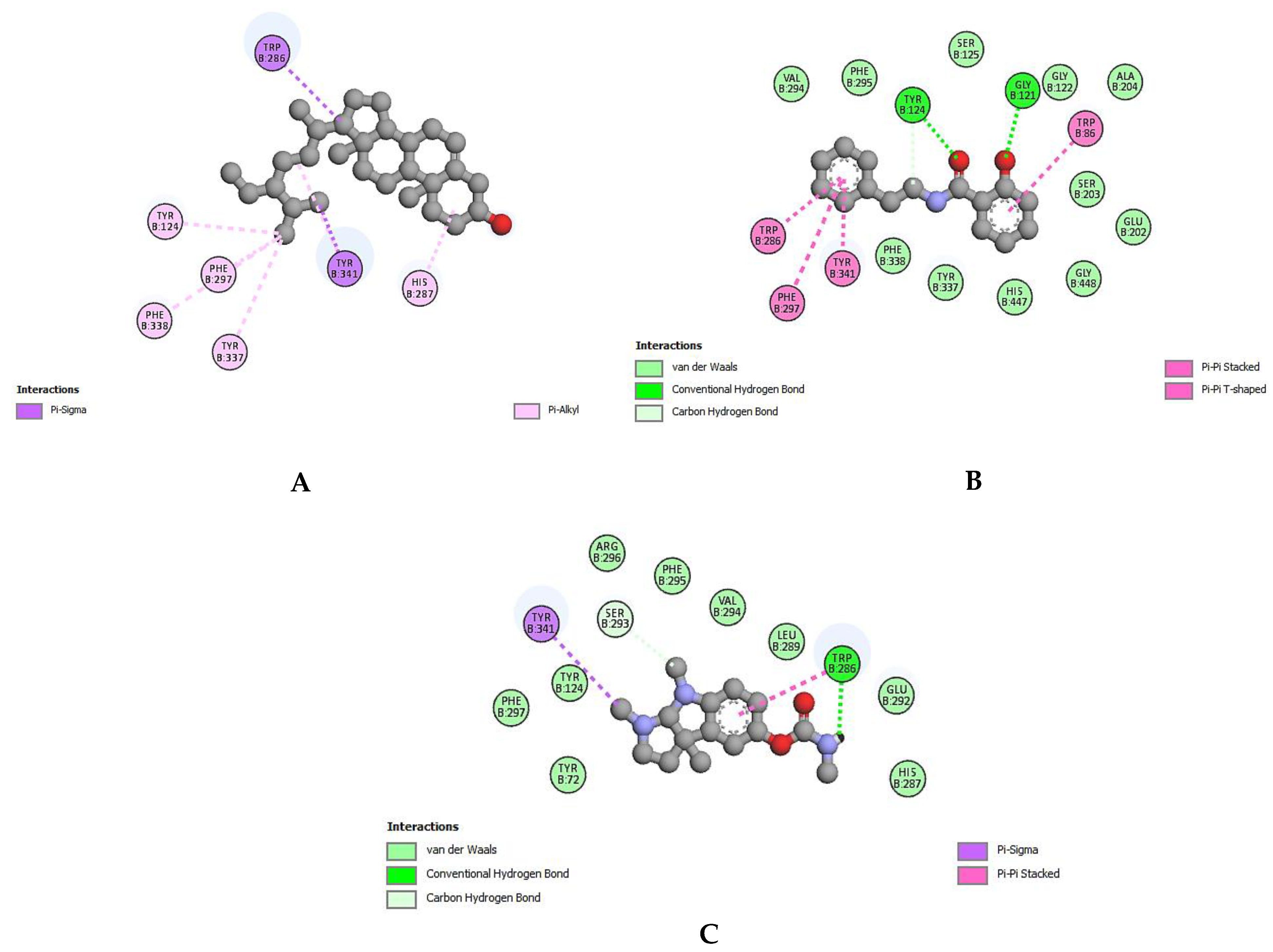
Molecular docking was performed for acetylcholinesterase with the compounds identified by GC–MS of ethanolic extract of Z. pentandra and the standard compound Eserine (physostigmine). The results showed [1,3] diazepan-2,4-dione, 1,2-benzenedicarboxylic acid, 2-hydroxy-n-(2-phenylethyl) benzamide, phthalic acid, lactose, dioctyl phthalate, and γ-sitosterol are the best-docked compounds with strong binding affinity scores. The binding energy of the standard Eserin was calculated at −8.4 kcal/mol, which is equal to phthalic acid and [1,3] diazepan-2,4-dione; γ-sitosterol, 1,2-benzenedicarboxylic acid, 2-hydroxy-n-(2-phenylethyl)benzamide, and [1,3] diazepan-2,4-dione have −11.5 kcal/mol, −9.5 kcal/mol, −9.4 kcal/mol, and −9.4 kcal/mol binding affinity scores, respectively, and showed better results than the standard compound (Eserine). Table 7 and Figure 2 show the binding energy and interactions between enzyme and ligands.

3.3.3. Molecular Docking for the Enzyme Tyrosinase

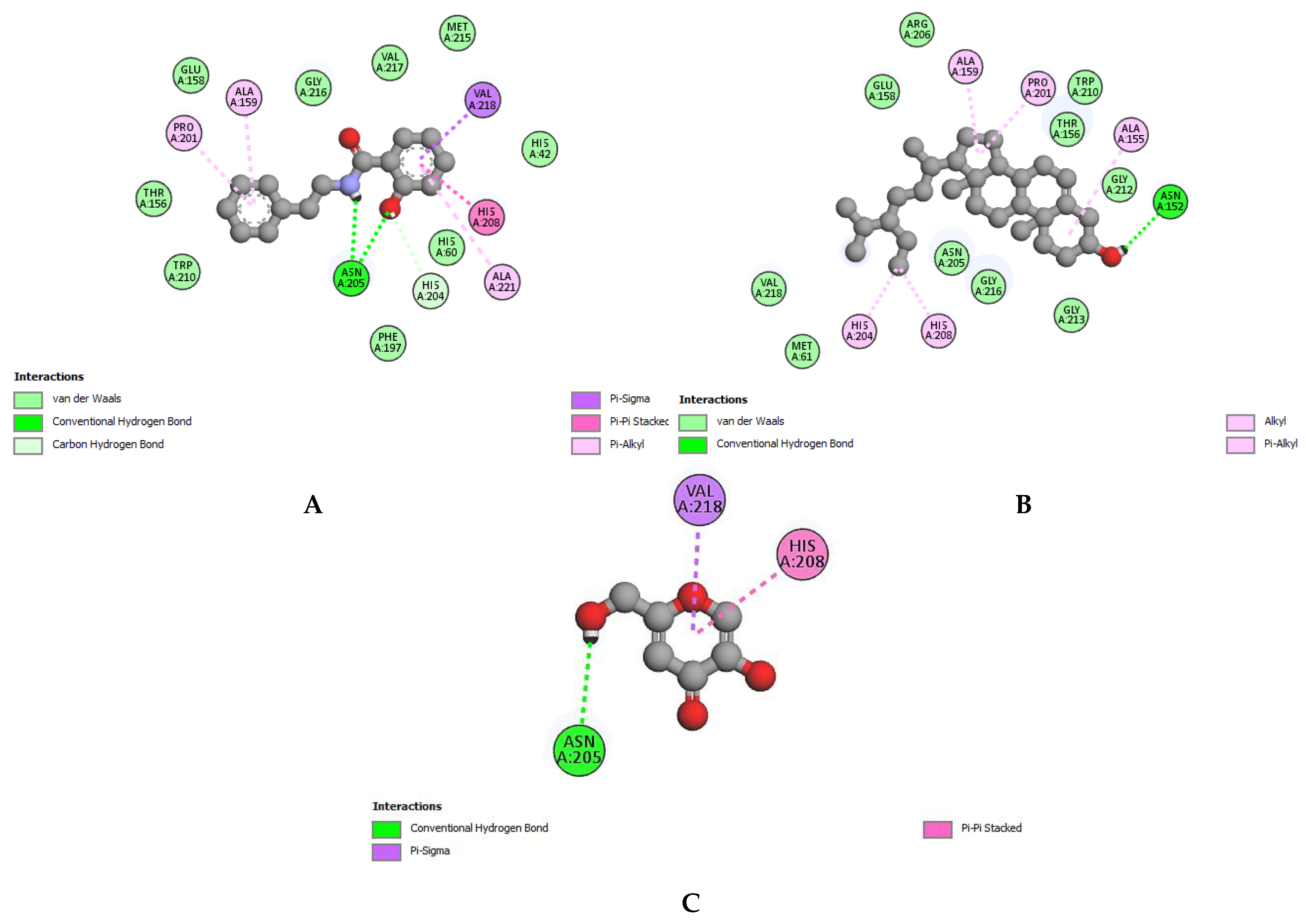
Molecular docking for tyrosinase revealed that all the tentative compounds identified from GC–MS of the ethanolic extract of Z. pentandra could interact with the enzyme tyrosinase. However, 2-hydroxy-n-(2-phenylethyl) benzamide, 1,2-benzenedicarboxylic acid, lactose, [1,3] diazepan-2,4-dione, cis-(−)-carvone-5,6-oxide, phthalic acid, and bis-7-methyloctyl ester showed a higher docking score than the standard Kojic acid. The results showed that 2-hydroxy-n-(2-phenylethyl) benzamide has the strongest binding affinity (−7.9 kcal/mol), which is two-thirds-times greater than the standard Kojic acid (−5.6 kcal/mol). Table 8 and Figure 3 show the detailed results of best-docked compounds along with the bond number and binding affinity against tyrosinase.

3.4. ADMET Studies

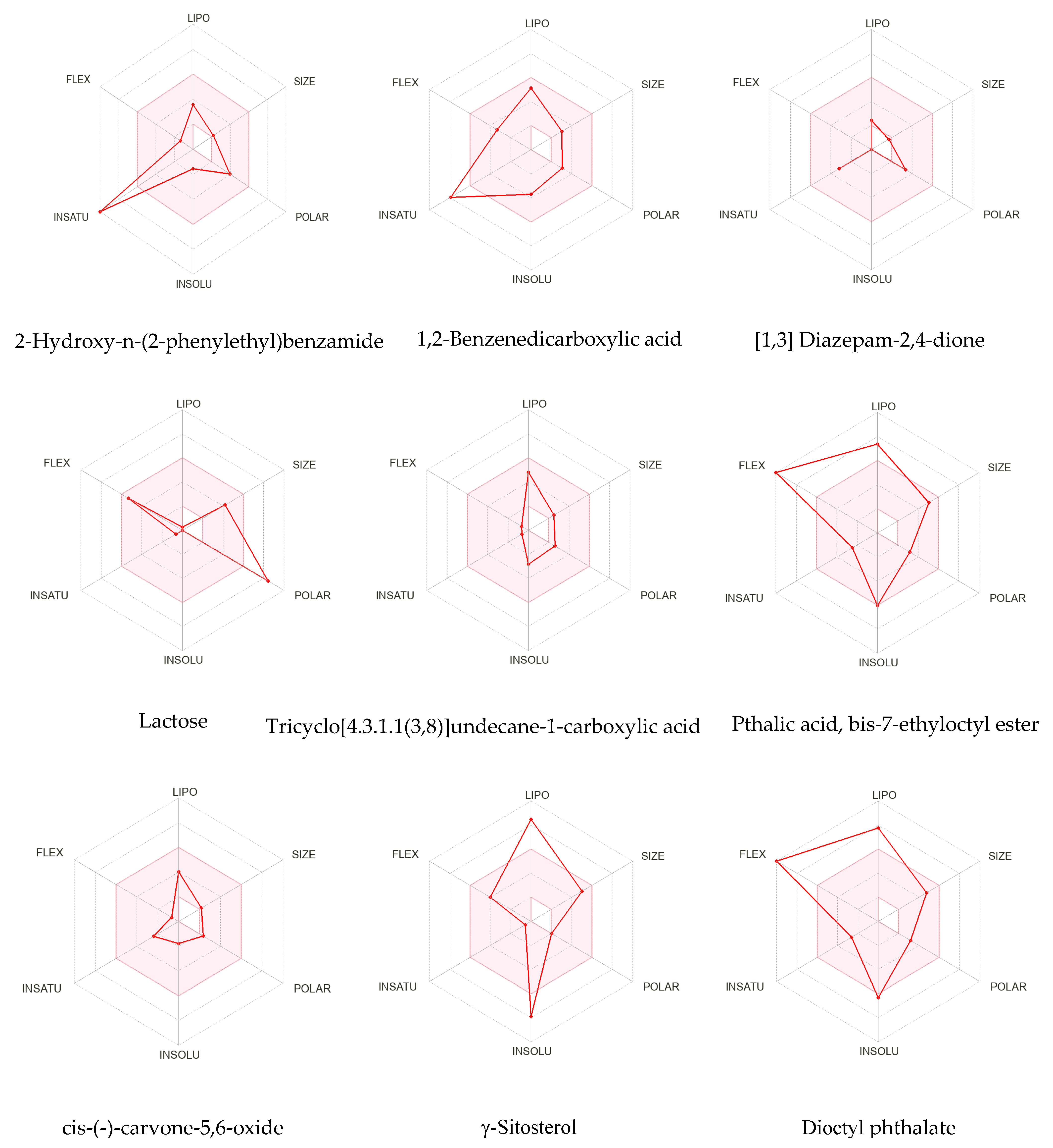
The SwissADME tool provides information about the physicochemical characteristics, drug-likeness, and pharmacokinetics of a compound, which was accessed to investigate the best-docked compounds. The results showed that 2-hydroxy-n-(2-phenylethyl)benzamide, 1,2-benzenedicarboxylic acid, [1,3] diazepan-2,4-dione, tricyclo [4.3.1.1(3,8)]undecane-1-carboxylic acid, and cis-(−)-carvone-5,6-oxide did not violate any of Lipinski’s rule of five criteria; however, phthalic acid, bis-7-methyloctyl ester, dioctyl phthalate, and γ-sitosterol broke one rule and lactose broke two rules. 1, 2-benzenedicarboxylic acid and cis-(−)-carvone-5,6-oxide had blood–brain penetration and gastrointestinal absorption. Characteristics, including the number of H-bond donors and acceptors, Lipinski’s rule, molecular weight, lipophilicity, etc. of compounds are represented in Table 9. Bioavailability radars are shown in Figure 4. Pharmacokinetic properties are tabulated in Table S1. The results of toxicity studies are given in Table S2.

4. Discussion

In this study, a solvent system that comprised ethanol 80% and water 20% was applied for the extraction. Such kinds of solvent systems bear a high polarity index, which results in high yield and constituents with diverse polarity [30,31]. Moreover, the hydroethanolic solvent systems are recommended for pharmacological assays due to their safe, non-toxic nature, and cost-effectiveness [32,33]. Qualitative phytochemical analysis revealed the therapeutic and physiological potential of the ethanolic extract of Z. pentandra. The investigation showed major chemical constituents such as carbohydrates, saponins, phenols, steroids, tannins, and lipids. These phytoconstituents are responsible for biological activities such as tannins and flavonoids contributing to anti-inflammatory, cytotoxic, free radical scavenging, and anti-microbial activity [34]. Compounds belonging to class lipids such as 2-Hydroxyhexadecanoic acid, 14-Methyl Pentadecanoic acid, and γ-Sitosterol possess anti-oxidant, anti-diabetic, anti-cancer, anti-viral, anti-bacterial, and neuroprotective activities [35,36,37]. Phenolic compounds have anti-apoptosis, cytotoxic, and anti-aging potentials as well as inhibit angiogenesis and development of the endothelial function. Similarly, saponins possess anti-cancer, anti-inflammatory, and anti-diabetic activity [38].
Total phenolic and flavonoid contents are the major secondary metabolites that correlate with the bioactivities of the plant, such as compounds rich with phenolic content that are responsible for the highest antioxidant activity [39,40]. Phenols present in the plants possess anti-oxidant, anti-inflammatory, anti-cancer, and antimicrobial activity, and they provide protection to halophytes against high damages such as DNA mutation, protein degradation, and lipid membrane peroxidation caused by reactive oxygen species [41,42,43]. The literature revealed that the methanolic extract of Z. pentandra exhibited the highest total phenolic content and antioxidant activity as compared to dichloromethane extract [15]. Therefore, our study showed that the ethanolic extract of Z. pentandra has 119.6 ± 0.12 (mg GAE/g) phenolic content and 45.5 ± 0.19 (mg QE/g) flavonoid content. It is observed that the results of our study are five times higher than the previously reported study on methanolic extract, the variation in results is due to a change in solvent for the extraction or probably due to the sample collection method or sample preparation [39]. The higher phenolic content may be attributed to the higher antioxidant activity. Moreover, compounds such as 2-hydroxyhexadecanoic acids [35], 14-Methyl Pentadecanoic acid [44], 1,2-benzenedicarboxylic acid [37], γ-Sitosterol [45], and lactose [46] also contribute to antioxidant activity. Our results showed concentration-dependent free radical scavenging and ferric-reducing antioxidant activity. The maximum activity was shown at the highest concentration.
In vitro bioassay evaluation of plant extract serves as a rapid and simple bioassay method, it serves as an initial step to identify interesting biological activities. This technique is helpful to direct all the efforts toward drug discovery and development. To further explore Z. pentandra, in vitro bioassay techniques were employed to assess the ability of the ethanolic extract of Z. pentandra as an antibacterial, antiviral, and enzyme inhibitor. The anti-bacterial and anti-viral activity may be due to the presence of bioactive compounds identified by GC–MS, such as D-threitol [47], 2-methoxy-4-vinylphenol [48], γ-sitosterol [45,49], thiodiglycol [50], 4-fluoroaniline [51], lactose [52,53], 14-methylpentadecanoic acid [54], D-4-C-methyl-myo-inosit [55], 1,2-benzenedicarboxylic acid [56,57], and 2-hydroxyhexadecanoic acids [58]. The ethanolic extract of Z. pentandra showed a dose-dependent effect and significant zone of inhibition (average greater than 8 mm) against gram-negative and gram-positive strains of bacteria. Previously, methanolic extract was tested for antibacterial activity against E. coli, S. typhi, B. spizizenii, S. aureus, and S. epidermidis [16].
The inhibition of the carbohydrate digestion enzyme (α-glucosidase) is helpful in reducing the cleavage of complex carbohydrates (from oligosaccharides to monosaccharides), thus preventing hyperglycemia [29,59]. A total of eight bioactive compounds were identified from the ethanolic extract of Z. pentandra by GC–MS with strong antidiabetic potential. These compounds were reported in Z. pentandra for the first time; however, previously, compounds such as γ-sitosterol [36], 1,2-benzenedicarboxylic acid [60], dioctyl phthalate [61], 14-methylpentadecanoic acid [62], 4-fluoroaniline [63], 2-methoxy-4-vinylphenol [64], and d-threitol [65,66] were reported in other studies. Our investigation revealed an extremely high potential of α-glucosidase inhibition of ethanolic extract of Z. pentandra as compared to standard (Acarbose) with IC50 10.0 ± 0.08 μg/mL and 5.87 ± 0.01 μg/mL, respectively.
The biosynthesis of melanin can be regulated by tyrosinase, which will lead to melasma and age spots. Tyrosinase inhibitors have a crucial role in skin protection, prevent hyperpigmentation, and are used in skin whitening products [67]. Natural tyrosinase inhibitors are considered safer and more economical, with high therapeutic activity and good skin penetration ability. Out of twenty-nine tentative compounds identified by GC–MS analysis, nine have been reported previously in other plants with potential tyrosinase inhibitory effects. The literature revealed that cyclohexane carboxamide [60], γ-sitosterol [68], 1,2-benzenedicarboxylic acid [69], lactose [70], dioctyl phthalate [71], 4-fluoroaniline [72], 2-methoxy-4-vinylphenol [73], and d-threitol [74] have tyrosinase inhibition activity due to strong binding with the tyrosinase enzyme. The result of our study supports the literature by showing exceptional tyrosinase inhibitory activity with IC50 20.7 ± 0.07 μg/mL as compared to Kojic acid (used as a standard) with IC50 1.04 ± 0.02 μg/mL.
Alzheimer’s disease and Parkinson’s disease are the two major neurodegenerative diseases with high incident rates [75]. It is estimated that by the end of 2040, neurodegenerative diseases will become the second most cause of death even surpassing cancer in aging patients. In this regard, various natural and synthetic cholinesterase inhibitors are exploited for the control and management of neurodegenerative diseases. There is substantial evidence that shows that polyphenol or phenolic contact plays a diverse/crucial role to decrease the rate of neurological disorders [76,77]. Our study was conducted to reveal the in vitro cholinesterase inhibition potential of the ethanolic extract of Z. pentandra. Furthermore, five compounds reported in the literature to have neuroprotective potential were also identified by GC–MS, such as γ-sitosterol [78], benzaldehyde 4-methoxy- [79], 4-fluoroaniline [80], lactose [81], and dioctyl phthalate [82], which might be responsible for the acetylcholinesterase inhibition activity of the extract.
To validate the enzymatic potential, each compound should be tested by in vitro analysis; however, this could not be possible due to the very limited concentration of these compounds in the plant extract and the commercial unavailability of some compounds. For this reason, these compounds can be evaluated virtually by using in silico molecular docking studies, which will predict the bioactive compounds responsible for enzymatic activities [83]. Nowadays, computational methods such as molecular docking have an important role in drug discovery as they help to predict and recognize low binding energy and high affinity, as well as better frameworks of the protein–ligand interactions. In our study, in silico molecular docking was performed in which hydrogen bonds, in addition to other interactions such as alkyl, pi-alkyl, pi-sigma, amide-pi, etc., play an important role in protein–ligand interactions and give stable binding of ligands and proteins [84]. A total of 29 compounds identified from GC–MS were docked against the α-glucosidase (PDB: 3wy1), tyrosinase (PDB: 3nq1), and acetylcholinesterase (PDB: 1gqr) based on binding affinity and the energy level.
All 29 compounds from GC–MS analysis along with the standards (Acarbose, Eserine, and Kojic acid) were docked against α-glucosidase, acetylcholinesterase, and tyrosinase enzymes, respectively. The results showed that 1,2-benzenedicarboxylic acid, 2-hydroxy-n-(2-phenylethyl) benzamide, γ-sitosterol, and lactose were the best-docked compounds with the least binding energy as compared to standards. Among all of these compounds, 1,2-benzenedicarboxylic acid showed the best α-glucosidase inhibition activity due to the presence of hydrogen, alkyl, pi-pi, and pi-alkyl interactions with a −8.3 kcal/mol binding affinity as compared to Acarbose (−8.1 kcal/mol).
For acetylcholinesterase inhibition, γ-sitosterol was the best-docked compound among all with −11.6 kcal/mol and pi-sigma, pi-pi, and pi-alkyl bonds, while no hydrogen bond was present. The maximum hydrogen bond was observed by lactose with seven hydrogen bonds and −7.9 kcal/mol binding affinity as compared to the standard Eserin, having −8.1 kcal/mol binding affinity with only one H-bond. In the case of tyrosinase inhibition, 2-hydroxy-n-(2-phenylethyl) benzamide showed the lowest binding affinity (−7.9 kcal/mol) with hydrogen, amide-pi, pi-alkyl, pi-pi, and pi-sigma interactions; lactose has the highest number of hydrogen bonds (five), with −7.1 kcal/mol binding affinity as compared to standard Kojic acid (6.5 kcal/mol). These best-docked compounds were further subjected to ADMET studies to predict their drug-like behavior and toxicity.
The SwissADME tool was used to investigate the physiochemical properties, drug-like behavior, and pharmacokinetics of the best-docked compounds [26]. These compounds were checked whether they follow the five rules described by Lipinski. If a compound follows the rules it can be considered a therapeutic agent and if a compound does not follow more than one rule it is considered an orally unavailable drug [85]. Overall, lactose showed two violations while all the others showed one and even zero violations, rendering them as orally available drugs. A compound is said to have high bioavailability, absorption, and distribution if it has a lower molecular weight, lower hydrogen bond capacity, and lipophilicity. All compounds had high GIT absorption except Phthalic acid, bis-7-methyloctyl ester, lactose, and γ-sitosterol; however, tricyclo[4.3.1.1(3,8)]undecane-1-carboxylic acid, 1,2-benzenedicarboxylic, and cis-(−)-carvone-5,6-oxide possessed blood–brain barrier penetration. Lipophilicity is measured in terms of LogP and its optimum value is between −0.7 to +5.0, and the optimum molecular weight ranges from 150 to 500 g/mol. The TPSA represents the size and polarity of compounds with an optimum range (20 to 130 A2). LogS (ESOL) ranges from 0 to 6 and measures solubility. In order to predict the toxic behavior of the compounds, the PROTOX II tool compares the chemical structures of the compounds under study with other chemicals of known toxicities and predicts toxic behavior [27]. In silico toxicity studies revealed that all the best-docked compounds had low toxicity as they belong to Class IV and Class V, whereas lactose was non-toxic as it belonged to Class VI. Owing to the above outcomes, it is recommended to conduct research on the isolation and purification of the above-mentioned compounds to establish these compounds as leads for new and potential therapeutic agents.

5. Conclusions

The ethanolic extract of Z. pentandra possesses anti-oxidant, anti-bacterial, anti-viral, and enzyme (α-glucosidase, cholinesterase, and tyrosinase) inhibition activities. To verify our study theoretically, the possible binding interaction and binding affinity of all these molecules (ligands) to inhibit the enzymes were elucidated by in silico molecular docking studies and the results showed that 1,2-benzenedicarboxylic acid, 2-hydroxy-n-(2-phenylethyl) benzamide, γ-sitosterol, and lactose exhibited strong anti-enzymatic potential. Results established from in vitro, in silico docking, and ADMET studies suggest that further work should be conducted to isolate, purify, reveal the chemical structures, and perform clinical studies of these compounds. Conclusively, the outcomes of the current work could help scientists and researchers in developing new and cost-effective drugs capable of managing bacterial infections, diabetes, neurodegenerative diseases, and skin disorders.

Supplementary Materials

The following supporting information can be downloaded at: https://www.mdpi.com/article/10.3390/app13010584/s1, Table S1: Pharmacokinetics of best docked compounds; Table S2: Toxicity studies (PROTOX II) results of the best docked compounds.

Author Contributions

Conceptualization, A.S., H.R. and K.u.R.K.; methodology, A.S., S.A.K. and T.E.; software, H.Y.A. and A.E.S.; validation, A.S., H.R., K.u.R.K., S.A.K. and T.E.; formal analysis, A.S., H.R., K.u.R.K., S.A.K. and T.E.; investigation, A.S., H.R., K.u.R.K., S.A.K. and T.E.; resources, M.U., D.A.K., S.A.K. and T.E.; data curation, A.S., H.R., K.u.R.K., S.A.K. and T.E.; writing—original draft preparation, A.S., H.R. and K.u.R.K.; writing—review and editing, A.M., M.U., A.B., S.A.K., D.A.K. and T.E.; visualization, A.S., H.R., K.u.R.K., S.A.K. and T.E.; supervision, A.S., H.R., K.u.R.K., S.A.K. and T.E.; project administration, H.R., K.u.R.K., S.A.K. and T.E.; funding acquisition, K.u.R.K., S.A.K. and T.E. All authors have read and agreed to the published version of the manuscript.

Funding

The authors are thankful to King Saud University, Riyadh, Saudia Arabia, Researchers Supporting Project number (RSP2022R504).

Institutional Review Board Statement

Not applicable.

Informed Consent Statement

Not applicable.

Data Availability Statement

All the valid databases are available and accessible online.

Acknowledgments

The authors are thankful to King Saud University, Riyadh, Saudia Arabia, Researchers Supporting Project number (RSP2022R504).

Conflicts of Interest

The authors declare no conflict of interest.

References

  1. Aziz, M.; Ahmad, S.; Iqbal, M.N.; Khurshid, U.; Saleem, H.; Alamri, A.; Anwar, S.; Alamri, A.S.; Chohan, T.A. Phytochemical, pharmacological, and In-silico molecular docking studies of Strobilanthes glutinosus Nees: An unexplored source of bioactive compounds. S. Afr. J. Bot. 2022, 147, 618–627. [Google Scholar] [CrossRef]
  2. Aumeeruddy, M.Z.; Mahomoodally, M.F. Combating breast cancer using combination therapy with 3 phytochemicals: Piperine, sulforaphane, and thymoquinone. Cancer 2019, 125, 1600–1611. [Google Scholar] [CrossRef] [PubMed]
  3. Djordjevic, S.M. From medicinal plant raw material to herbal remedies. In Aromatic and Medicinal Plants: Back to Nature; BoD—Books on Demand: Norderstedt, Germany, 2017; pp. 269–288. [Google Scholar]
  4. Savithramma, N.; Rao, M.L.; Suhrulatha, D. Screening of medicinal plants for secondary metabolites. Middle-East J. Sci. Res. 2011, 8, 579–584. [Google Scholar]
  5. Kumar, D.; Bajaj, S.; Mehrotra, R. Knowledge, attitude and practice of complementary and alternative medicines for diabetes. Public Health 2006, 120, 705–711. [Google Scholar] [CrossRef] [PubMed]
  6. Ahn, K. The worldwide trend of using botanical drugs and strategies for developing global drugs. BMB Rep. 2017, 50, 111. [Google Scholar] [CrossRef] [Green Version]
  7. Singh, B.; Bhat, T.K.; Singh, B. Potential therapeutic applications of some antinutritional plant secondary metabolites. J. Agric. Food Chem. 2003, 51, 5579–5597. [Google Scholar] [CrossRef]
  8. Seca, A.M.; Pinto, D.C. Biological potential and medical use of secondary metabolites. Medicines 2019, 6, 66. [Google Scholar] [CrossRef] [Green Version]
  9. Sadaf, H.M.; Bibi, Y.; Ishaque, M.; Nisa, S.; Qayyum, A.; Safdar, N.; Shah, Z.H.; Alsamadany, H.; Chung, G. Determination of ROS Scavenging, Antibacterial and Antifungal Potential of Methanolic Extract of Otostegia limbata (Benth.) Boiss. Plants 2021, 10, 2360. [Google Scholar] [CrossRef]
  10. Bingol, Z.; Kızıltaş, H.; Gören, A.C.; Kose, L.P.; Topal, M.; Durmaz, L.; Alwasel, S.H.; Gulcin, İ. Antidiabetic, anticholinergic and antioxidant activities of aerial parts of shaggy bindweed (Convulvulus betonicifolia Miller subsp.)–profiling of phenolic compounds by LC-HRMS. Heliyon 2021, 7, e06986. [Google Scholar] [CrossRef]
  11. Saleem, H.; Khurshid, U.; Sarfraz, M.; Tousif, M.I.; Alamri, A.; Anwar, S.; Alamri, A.; Ahmad, I.; Abdallah, H.H.; Mahomoodally, F.M. A comprehensive phytochemical, biological, toxicological and molecular docking evaluation of Suaeda fruticosa (L.) Forssk.: An edible halophyte medicinal plant. Food Chem. Toxicol. 2021, 154, 112348. [Google Scholar] [CrossRef] [PubMed]
  12. Mukherjee, P.K.; Biswas, R.; Sharma, A.; Banerjee, S.; Biswas, S.; Katiyar, C. Validation of medicinal herbs for anti-tyrosinase potential. J. Herb. Med. 2018, 14, 1–16. [Google Scholar] [CrossRef]
  13. Sajid-Ur-Rehman, M.; Ishtiaq, S.; Khan, M.A.; Alshamrani, M.; Younus, M.; Shaheen, G.; Abdullah, M.; Sarwar, G.; Khan, M.S.; Javed, F. Phytochemical profiling, in vitro and in vivo anti-inflammatory, analgesic and antipyretic potential of Sesuvium sesuvioides (Fenzl) Verdc.(Aizoaceae). Inflammopharmacology 2021, 29, 789–800. [Google Scholar] [CrossRef] [PubMed]
  14. Afzal, S.; Chaudhary, B.; Uzair, M.; Afzal, K.; Bokhari, T. Isolation of pentandraone from methanolic extract of aerial part of Zaleya pentandra. Int. Res. J. Pharm. 2013, 4, 2–23. [Google Scholar]
  15. Saleem, H.; Zengin, G.; Locatelli, M.; Ahmad, I.; Khaliq, S.; Mahomoodally, M.F.; Hussain, R.; Rengasamy, K.R.; Mollica, A.; Abidin, S.A.Z. Pharmacological, phytochemical and in-vivo toxicological perspectives of a xero-halophyte medicinal plant: Zaleya pentandra (L.) Jeffrey. Food Chem. Toxicol. 2019, 131, 110535. [Google Scholar] [CrossRef] [PubMed]
  16. Afzal, S.; Chaudhry, B.A.; Saeed, J.; Afzal, K.; Ahmed, B.; Qadir, M.I. Antibacterial and antioxidant activity of methanolic extract of Zaleya pentandra. Acta Pol. Pharm. 2016, 73, 147–151. [Google Scholar]
  17. Mughal, T.A.; Aslam, F.; Yousaf, Z.; Nisar, N.; Leung, P.C. In vitro cytotoxic activity of Zaleya pentandra L. Extracts against the breast cancer adenocarcinoma cell line MCF-7. JPMA 2020, 2019. [Google Scholar] [CrossRef]
  18. Kareti, S.R.; Subash, P. In silico exploration of anti-Alzheimer’s compounds present in methanolic extract of Neolamarckia cadamba bark using GC–MS/MS. Arab. J. Chem. 2020, 13, 6246–6255. [Google Scholar] [CrossRef]
  19. Harborne, A. Phytochemical Methods a Guide to Modern Techniques of Plant Analysis; Springer Science & Business Media: Berlin/Heidelberg, Germany, 1998. [Google Scholar]
  20. Sembiring, E.N.; Elya, B.; Sauriasari, R. Phytochemical screening, total flavonoid and total phenolic content and antioxidant activity of different parts of Caesalpinia bonduc (L.) Roxb. Pharmacogn. J. 2018, 10, 123–127. [Google Scholar] [CrossRef] [Green Version]
  21. Hayat, M.M.; Uzair, M. Biological potential and GC-MS analysis of phytochemicals of Farsetia hamiltonii (Royle). Biomed. Res. 2019, 30, 609–616. [Google Scholar] [CrossRef] [Green Version]
  22. Aati, H.Y.; Anwar, M.; Al-Qahtani, J.; Al-Taweel, A.; Khan, K.-u.-R.; Aati, S.; Usman, F.; Ghalloo, B.A.; Asif, H.M.; Shirazi, J.H. Phytochemical profiling, in vitro biological activities, and in-silico studies of Ficus vasta Forssk.: An unexplored plant. Antibiotics 2022, 11, 1155. [Google Scholar] [CrossRef] [PubMed]
  23. Ellman, G.L.; Courtney, K.D.; Andres Jr, V.; Featherstone, R.M. A new and rapid colorimetric determination of acetylcholinesterase activity. Biochem. Pharmacol. 1961, 7, 88–95. [Google Scholar] [CrossRef] [PubMed]
  24. Palanisamy, U.D.; Ling, L.T.; Manaharan, T.; Appleton, D. Rapid isolation of geraniin from Nephelium lappaceum rind waste and its anti-hyperglycemic activity. Food Chem. 2011, 127, 21–27. [Google Scholar] [CrossRef]
  25. Orhan, I.E.; Senol, F.S.; Gulpinar, A.R.; Sekeroglu, N.; Kartal, M.; Sener, B. Neuroprotective potential of some terebinth coffee brands and the unprocessed fruits of Pistacia terebinthus L. and their fatty and essential oil analyses. Food Chem. 2012, 130, 882–888. [Google Scholar] [CrossRef]
  26. Daina, A.; Michielin, O.; Zoete, V. SwissADME: A free web tool to evaluate pharmacokinetics, drug-likeness and medicinal chemistry friendliness of small molecules. Sci. Rep. 2017, 7, 42717. [Google Scholar] [CrossRef] [PubMed] [Green Version]
  27. Banerjee, P.; Eckert, A.O.; Schrey, A.K.; Preissner, R. ProTox-II: A webserver for the prediction of toxicity of chemicals. Nucleic Acids Res. 2018, 46, W257–W263. [Google Scholar] [CrossRef] [PubMed] [Green Version]
  28. Aykul, S.; Martinez-Hackert, E. Determination of half-maximal inhibitory concentration using biosensor-based protein interaction analysis. Anal. Biochem. 2016, 508, 97–103. [Google Scholar] [CrossRef] [PubMed] [Green Version]
  29. Murugesu, S.; Ibrahim, Z.; Ahmed, Q.U.; Uzir, B.F.; Yusoff, N.I.N.; Perumal, V.; Abas, F.; Shaari, K.; Khatib, A. Identification of α-glucosidase inhibitors from Clinacanthus nutans leaf extract using liquid chromatography-mass spectrometry-based metabolomics and protein-ligand interaction with molecular docking. J. Pharm. Anal. 2019, 9, 91–99. [Google Scholar] [CrossRef]
  30. Moreau, R.A.; Powell, M.J.; Singh, V. Pressurized liquid extraction of polar and nonpolar lipids in corn and oats with hexane, methylene chloride, isopropanol, and ethanol. J. Am. Oil Chem. Soc. 2003, 80, 1063–1067. [Google Scholar] [CrossRef]
  31. Sari, R.; Wahyuningrum, M.; Rafi, M.; Wientarsih, I. Effect of ethanol polarity on extraction yield, antioxidant, and sunscreen activities of phytochemicals from Gyrinops versteegii leaves. IOP Conf. Ser. Mater. Sci. Eng. 2020, 925, 012038. [Google Scholar]
  32. Zhou, K.; Yu, L. Effects of extraction solvent on wheat bran antioxidant activity estimation. LWT-Food Sci. Technol. 2004, 37, 717–721. [Google Scholar] [CrossRef]
  33. Phrompittayarat, W.; Putalun, W.; Tanaka, H.; Jetiyanon, K.; Wittaya-Areekul, S.; Ingkaninan, K. Comparison of various extraction methods of Bacopa monnieri. Naresuan Univ. J. Sci. Technol. (NUJST) 2013, 15, 29–34. [Google Scholar]
  34. Basit, A.; Ahmad, S.; Sherif, A.E.; Aati, H.Y.; Ovatlarnporn, C.; Khan, M.A.; Rao, H.; Ahmad, I.; Shahzad, M.N.; Ghalloo, B.A. New mechanistic insights on Justicia vahlii Roth: UPLC-Q-TOF-MS and GC–MS based metabolomics, in-vivo, in-silico toxicological, antioxidant based anti-inflammatory and enzyme inhibition evaluation. Arab. J. Chem. 2022, 15, 104135. [Google Scholar] [CrossRef]
  35. Rodrigues, M.J.; Custódio, L.; Mecha, D.; Zengin, G.; Cziáky, Z.; Sotkó, G.; Pereira, C.G. Nutritional and Phyto-Therapeutic Value of the Halophyte Cladium mariscus L.(Pohl.): A Special Focus on Seeds. Plants 2022, 11, 2910. [Google Scholar] [CrossRef]
  36. Balamurugan, R.; Duraipandiyan, V.; Ignacimuthu, S. Antidiabetic activity of γ-sitosterol isolated from Lippia nodiflora L. in streptozotocin induced diabetic rats. Eur. J. Pharmacol. 2011, 667, 410–418. [Google Scholar] [CrossRef]
  37. Jung Choi, S.; Kim, M.J.; Jin Heo, H.; Kim, J.K.; Jin Jun, W.; Kim, H.K.; Kim, E.-K.; Ok Kim, M.; Yon Cho, H.; Hwang, H.-J. Ameliorative effect of 1, 2-benzenedicarboxylic acid dinonyl ester against amyloid beta peptide-induced neurotoxicity. Amyloid 2009, 16, 15–24. [Google Scholar] [CrossRef]
  38. Ghalloo, B.A.; Khan, K.-u.-R.; Ahmad, S.; Aati, H.Y.; Al-Qahtani, J.H.; Ali, B.; Mukhtar, I.; Hussain, M.; Shahzad, M.N.; Ahmed, I. Phytochemical Profiling, In Vitro Biological Activities, and In Silico Molecular Docking Studies of Dracaena reflexa. Molecules 2022, 27, 913. [Google Scholar] [CrossRef]
  39. Saravanakumar, K.; Park, S.; Sathiyaseelan, A.; Kim, K.-N.; Cho, S.-H.; Mariadoss, A.V.A.; Wang, M.-H. Metabolite profiling of methanolic extract of Gardenia jaminoides by LC-MS/MS and GC-MS and its anti-diabetic, and anti-oxidant activities. Pharmaceuticals 2021, 14, 102. [Google Scholar] [CrossRef]
  40. Karakaya, S.; Bingol, Z.; Koca, M.; Dagoglu, S.; Pınar, N.M.; Demirci, B.; Gulcin, İ.; Brestic, M.; Sytar, O. Identification of non-alkaloid natural compounds of Angelica purpurascens (Avé-Lall.) Gilli. (Apiaceae) with cholinesterase and carbonic anhydrase inhibition potential. Saudi Pharm. J. 2020, 28, 1–14. [Google Scholar] [CrossRef]
  41. Gutiérrez-Grijalva, E.P.; Picos-Salas, M.A.; Leyva-López, N.; Criollo-Mendoza, M.S.; Vazquez-Olivo, G.; Heredia, J.B. Flavonoids and phenolic acids from oregano: Occurrence, biological activity and health benefits. Plants 2017, 7, 2. [Google Scholar] [CrossRef] [Green Version]
  42. Servili, M.; Sordini, B.; Esposto, S.; Urbani, S.; Veneziani, G.; Maio, I.D.; Selvaggini, R.; Taticchi, A. Biological activities of phenolic compounds of extra virgin olive oil. Antioxidants 2013, 3, 1–23. [Google Scholar] [CrossRef] [PubMed] [Green Version]
  43. Basit, A.; Ahmad, S.; Naeem, A.; Usman, M.; Ahmed, I.; Shahzad, M.N. Chemical profiling of Justicia vahlii Roth.(Acanthaceae) using UPLC-QTOF-MS and GC-MS analysis and evaluation of acute oral toxicity, antineuropathic and antioxidant activities. J. Ethnopharmacol. 2022, 287, 114942. [Google Scholar] [CrossRef] [PubMed]
  44. Widiyarti, G.; Fitrianingsih, W. Phytochemical constituents and free radical scavenging activity of Madang Gatal (Schima wallichii) Choisy stem bark. Pharmacogn. J. 2019, 11. [Google Scholar] [CrossRef] [Green Version]
  45. Karthikeyan, S.C.; Velmurugan, S.; Donio, M.B.S.; Michaelbabu, M.; Citarasu, T. Studies on the antimicrobial potential and structural characterization of fatty acids extracted from Sydney rock oyster Saccostrea glomerata. Ann. Clin. Microbiol. Antimicrob. 2014, 13, 332. [Google Scholar] [CrossRef] [Green Version]
  46. Yáñez, D.A.C.; Gagneten, M.; Leiva, G.E.; Malec, L.S. Antioxidant activity developed at the different stages of Maillard reaction with milk proteins. LWT 2018, 89, 344–349. [Google Scholar] [CrossRef]
  47. Price, N.P.; Bischoff, K.M.; Leathers, T.D.; Cossé, A.A.; Manitchotpisit, P. Polyols, not sugars, determine the structural diversity of anti-streptococcal liamocins produced by Aureobasidium pullulans strain NRRL 50380. J. Antibiot. 2017, 70, 136–141. [Google Scholar] [CrossRef] [PubMed]
  48. Rubab, M.; Chelliah, R.; Saravanakumar, K.; Barathikannan, K.; Wei, S.; Kim, J.-R.; Yoo, D.; Wang, M.-H.; Oh, D.-H. Bioactive Potential of 2-Methoxy-4-vinylphenol and Benzofuran from Brassica oleracea L. var. capitate f, rubra (Red Cabbage) on Oxidative and Microbiological Stability of Beef Meat. Foods 2020, 9, 568. [Google Scholar] [CrossRef]
  49. Abu-Lafi, S.; Rayan, M.; Masalha, M.; Abu-Farich, B.; Al-Jaas, H.; Abu-Lafi, M.; Rayan, A. Phytochemical composition and biological activities of wild Scolymus maculatus L. Medicines 2019, 6, 53. [Google Scholar] [CrossRef] [Green Version]
  50. Kiruthiga, B.; Kumar, P.S. Potential impacts of various coastal locales on the phytochemical landscape in sand dune flora calotropis giganteawhite across the coleroon valley. Plant Arch. 2019, 19, 2173–2187. [Google Scholar]
  51. Solankee, A.; Prajapati, Y. An efficient synthesis of some new fluorine containing acetyl pyrazoline and isoxazole derivatives and their antibacterial activity. Rasayan J. Chem. 2009, 2, 23–27. [Google Scholar]
  52. Singh, B.R. Antibacterial activity of glycerol, lactose, maltose, mannitol, raffinose and xylose. Noto-Are Med. 2014, 17223318. [Google Scholar]
  53. Singla, P.; Luxami, V.; Paul, K. Synthesis, in vitro antitumor activity, dihydrofolate reductase inhibition, DNA intercalation and structure–activity relationship studies of 1, 3, 5-triazine analogues. Bioorg. Med. Chem. Lett. 2016, 26, 518–523. [Google Scholar] [CrossRef] [PubMed]
  54. Carballeira, N.M.; Reyes, E.D.; Sostre, A.; Rodríguez, A.D.; Rodríguez, J.L.; González, F.A. Identification of the novel antimicrobial fatty acid (5 Z, 9 Z)-14-methyl-5, 9-pentadecadienoic acid in Eunicea succinea. J. Nat. Prod. 1997, 60, 502–504. [Google Scholar] [CrossRef]
  55. Larbie, C. Tetrapleura tetraptera of Ghanaian origin: Phytochemistry, antioxidant and antimicrobial activity of extracts of plant parts. J. Pharm. Res. Int. 2020, 32, 78–96. [Google Scholar]
  56. Fadipe, L.A.; Haruna, A.; Mohammed, I. Antibacterial activity of 1, 2-benzenedicarboxylic acid, dioctyl ester isolated from the ethyl acetate soluble sub-portion of the unripe fruits of Nauclea latifolia. 2014. [Google Scholar]
  57. Boadu, A.; Nlooto, M.; Karpoormath, R. Spondias mombin: In Silico Screening of 1, 2-Benzenedicarboxylic Acid, Butyl 2-Methylpropyl Ester (Fragment of Geraniin) as Anti-Marburg virus agent. Authorea Prepr. 2022. [Google Scholar]
  58. Harper, D.; Gilbert, R.; O’Connor, T.; Kinchington, D.; Mahmood, N.; Mcllhinney, R.; Jeffries, D. Antiviral activity of 2-hydroxy fatty acids. Antivir. Chem. Chemother. 1996, 7, 138–141. [Google Scholar] [CrossRef] [Green Version]
  59. Vijayakumar, S.; Divya, M.; Vaseeharan, B.; Chen, J.; Biruntha, M.; Silva, L.P.; Durán-Lara, E.F.; Shreema, K.; Ranjan, S.; Dasgupta, N. Biological Compound Capping of Silver Nanoparticle with the Seed Extracts of Blackcumin (Nigella sativa): A Potential Antibacterial, Antidiabetic, Anti-inflammatory, and Antioxidant. J. Inorg. Organomet. Polym. Mater. 2021, 31, 624–635. [Google Scholar] [CrossRef]
  60. Al-Hajj, N.Q.M.; Sharif, H.R.; Aboshora, W.; Wang, H. In vitro and in vivo evaluation of antidiabetic activity of leaf essential oil of Pulicaria inuloides-Asteraceae. 2016. [Google Scholar] [CrossRef]
  61. Bu, T.; Liu, M.; Zheng, L.; Guo, Y.; Lin, X. α-glucosidase inhibition and the in vivo hypoglycemic effect of butyl-isobutyl-phthalate derived from the Laminaria japonica rhizoid. Phytother. Res. 2010, 24, 1588–1591. [Google Scholar] [CrossRef] [PubMed]
  62. Emmanuel, O.; Uche, M.E.; Dike, E.D.; Etumnu, L.R.; Ugbogu, O.C.; Ugbogu, E.A. A review on Garcinia kola Heckel: Traditional uses, phytochemistry, pharmacological activities, and toxicology. Biomarkers 2021, 27, 101–117. [Google Scholar] [CrossRef]
  63. Reddy, G.A. Synthesis and Evaluation of Newer Quinoline Derivatives of Thiazolidinediones For Their Antidiabetic Activity l. Srikanth, n. Raghunandan1, p. Srinivas2 and g. Reddy. Available online: https://citeseerx.ist.psu.edu/document?repid=rep1&type=pdf&doi=3d403a7e4ab106153263a65f51de7597ad26a796 (accessed on 17 November 2022).
  64. Abbirami, E.; Selvakumar, M.; Dinesh Kumar, L.; Guna, R.; Sivasudha, T. Identification of novel drug-like compounds from Momordica cymbalaria as PPAR-γ agonists: A molecular docking study. AJEAT 2019, 8, 71–74. [Google Scholar] [CrossRef]
  65. Wang, J.C.; Hu, S.H.; Wang, J.T.; Chen, K.S.; Chia, Y.C. Hypoglycemic effect of extract of Hericium erinaceus. J. Sci. Food Agric. 2005, 85, 641–646. [Google Scholar] [CrossRef]
  66. Bharti, S.K.; Krishnan, S.; Kumar, A.; Kumar, A. Antidiabetic phytoconstituents and their mode of action on metabolic pathways. Ther. Adv. Endocrinol. Metab. 2018, 9, 81–100. [Google Scholar] [CrossRef] [PubMed]
  67. Zuo, A.-R.; Dong, H.-H.; Yu, Y.-Y.; Shu, Q.-L.; Zheng, L.-X.; Yu, X.-Y.; Cao, S.-W. The antityrosinase and antioxidant activities of flavonoids dominated by the number and location of phenolic hydroxyl groups. Chin. Med. 2018, 13, 51. [Google Scholar] [CrossRef] [PubMed] [Green Version]
  68. Younis, M.M.; Ayoub, I.M.; Mostafa, N.M.; El Hassab, M.A.; Eldehna, W.M.; Al-Rashood, S.T.; Eldahshan, O.A. GC/MS Profiling, Anti-Collagenase, Anti-Elastase, Anti-Tyrosinase and Anti-Hyaluronidase Activities of a Stenocarpus sinuatus Leaves Extract. Plants 2022, 11, 918. [Google Scholar] [CrossRef] [PubMed]
  69. Khurshid, U.; Ahmad, S.; Rehman, T.; Arshad, M.A.; Pervaiz, I.; Saba, S. GC-MS analysis, DPPH & enzyme inhibition assays of Trianthema triquetra Rottl. and Willd. growing in Pakistan. Lat. Am. J. Pharm. 2019, 38, 1181–1187. [Google Scholar]
  70. Saeki, H.; Oikawa, A. Effects of pH and type of sugar in the medium on tyrosinase activity in cultured melanoma cells. J. Cell. Physiol. 1978, 94, 139–145. [Google Scholar] [CrossRef]
  71. Nguyen, D.; Nguyen, D.H.; Hwa-La, L.; Lee, H.-B.; Shin, J.-H.; Kim, E.-K. Inhibition of melanogenesis by dioctyl phthalate isolated from Nigella glandulifera Freyn. J. Microbiol. Biotechnol. 2007, 17, 1585–1590. [Google Scholar]
  72. Barker, A.J.; Gibson, K.H.; Grundy, W.; Godfrey, A.A.; Barlow, J.J.; Healy, M.P.; Woodburn, J.R.; Ashton, S.E.; Curry, B.J.; Scarlett, L. Studies leading to the identification of ZD1839 (Iressa™): An orally active, selective epidermal growth factor receptor tyrosine kinase inhibitor targeted to the treatment of cancer. Bioorg. Med. Chem. Lett. 2001, 11, 1911–1914. [Google Scholar] [CrossRef]
  73. Fukai, S.; Tanimoto, S.; Maeda, A.; Fukuda, H.; Okada, Y.; Nomura, M. Pharmacological activity of compounds extracted from persimmon peel (Diospyros kaki THUNB.). J. Oleo Sci. 2009, 58, 213–219. [Google Scholar] [CrossRef] [Green Version]
  74. Titan, S.M.; Venturini, G.; Padilha, K.; Goulart, A.C.; Lotufo, P.A.; Bensenor, I.J.; Krieger, J.E.; Thadhani, R.I.; Rhee, E.P.; Pereira, A.C. Metabolomics biomarkers and the risk of overall mortality and ESRD in CKD: Results from the Progredir Cohort. PLoS ONE 2019, 14, e0213764. [Google Scholar] [CrossRef] [Green Version]
  75. Monroy, A.; Lithgow, G.J.; Alavez, S. Curcumin and neurodegenerative diseases. Biofactors 2013, 39, 122–132. [Google Scholar] [CrossRef] [PubMed]
  76. Barbosa, M.; Valentão, P.; Andrade, P.B. Bioactive compounds from macroalgae in the new millennium: Implications for neurodegenerative diseases. Mar. Drugs 2014, 12, 4934–4972. [Google Scholar] [CrossRef] [PubMed]
  77. Figueiredo-González, M.; Reboredo-Rodríguez, P.; González-Barreiro, C.; Carrasco-Pancorbo, A.; Simal-Gándara, J.; Cancho-Grande, B. Nutraceutical potential of phenolics from ‘brava’ and ‘mansa’ extra-virgin olive oils on the inhibition of enzymes associated to neurodegenerative disorders in comparison with those of ‘picual’ and ‘cornicabra’. Molecules 2018, 23, 722. [Google Scholar] [CrossRef] [PubMed] [Green Version]
  78. Mah, S.H.; Teh, S.S.; Ee, G.C.L. Anti-inflammatory, anti-cholinergic and cytotoxic effects of Sida rhombifolia. Pharm. Biol. 2017, 55, 920–928. [Google Scholar] [CrossRef] [Green Version]
  79. Kamireddy, K.; Chinnu, S.; Priyanka, P.; Rajini, P.; Giridhar, P. Neuroprotective effect of Decalepis hamiltonii aqueous root extract and purified 2-hydroxy-4-methoxy benzaldehyde on 6-OHDA induced neurotoxicity in Caenorhabditis elegans. Biomed. Pharmacother. 2018, 105, 997–1005. [Google Scholar] [CrossRef] [PubMed]
  80. Maqbool, M.; Manral, A.; Jameel, E.; Kumar, J.; Saini, V.; Shandilya, A.; Tiwari, M.; Hoda, N.; Jayaram, B. Development of cyanopyridine–triazine hybrids as lead multitarget anti-Alzheimer agents. Bioorg. Med. Chem. 2016, 24, 2777–2788. [Google Scholar] [CrossRef] [PubMed]
  81. Cho, Y.-R.; Chang, J.-Y.; Chang, H.-C. Production of γ-Aminobutyric Acid (GABA) by Lactobacillus buchneri isolated from Kimchi and its neuroprotective effect on neuronal cells. J. Microbiol. Biotechnol. 2007, 17, 104–109. [Google Scholar]
  82. Feng, W.; Wu, X.; Mao, G.; Zhao, T.; Wang, W.; Chen, Y.; Zhang, M.; Yang, L.; Wu, X. Neurological effects of subchronic exposure to dioctyl phthalate (DOP), lead, and arsenic, individual and mixtures, in immature mice. Environ. Sci. Pollut. Res. 2020, 27, 9247–9260. [Google Scholar] [CrossRef]
  83. Nipun, T.S.; Khatib, A.; Ibrahim, Z.; Ahmed, Q.U.; Redzwan, I.E.; Primaharinastiti, R.; Saiman, M.Z.; Fairuza, R.; Widyaningsih, T.D.; AlAjmi, M.F. GC-MS-and NMR-Based Metabolomics and Molecular Docking Reveal the Potential Alpha-Glucosidase Inhibitors from Psychotria malayana Jack Leaves. Pharmaceuticals 2021, 14, 978. [Google Scholar] [CrossRef] [PubMed]
  84. Chen, D.; Oezguen, N.; Urvil, P.; Ferguson, C.; Dann, S.M.; Savidge, T.C. Regulation of protein-ligand binding affinity by hydrogen bond pairing. Sci. Adv. 2016, 2, e1501240. [Google Scholar] [CrossRef] [Green Version]
  85. Lipinski, C.A. Lead-and drug-like compounds: The rule-of-five revolution. Drug Discov. Today Technol. 2004, 1, 337–341. [Google Scholar] [CrossRef] [PubMed]
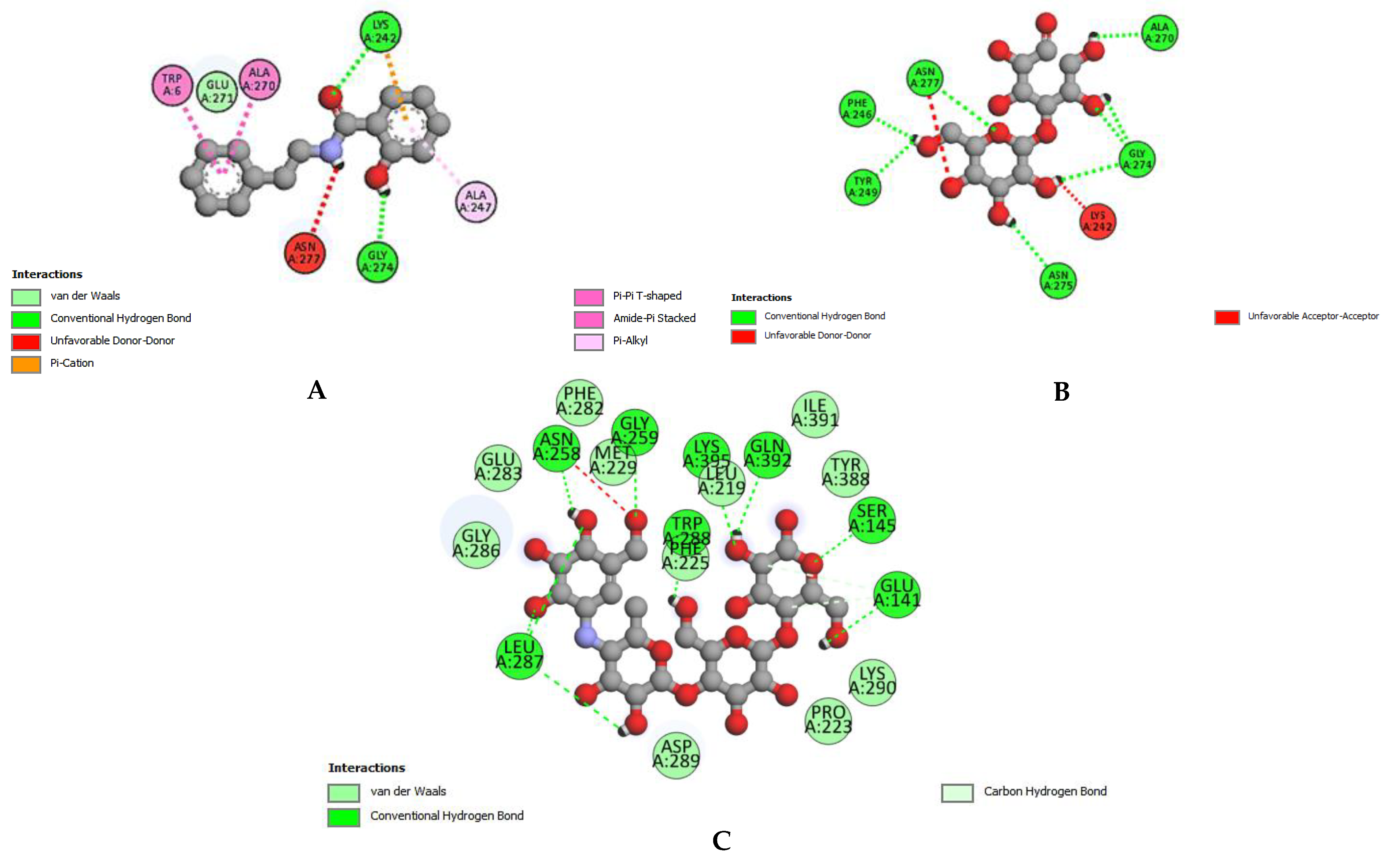
Figure 1. Interaction between α-glucosidase and ligands. (A) 2-hydroxy-n-(2-phenylethyl) benzamide, (B) lactose, and (C) Acarbose.
Applsci 13 00584 g001
Figure 2. Interaction between acetylcholinesterase and ligands. (A) γ-sitosterol, (B) 2-hydroxy-n-(2-phenylethyl) benzamide, and (C) Eserin (standard).
Applsci 13 00584 g002
Figure 3. Interaction between tyrosinase and ligand. (A) 2-hydroxy-n-(2-phenylethyl) benzamide, (B) γ-sitosterol, and (C) Kojic acid.
Applsci 13 00584 g003
Figure 4. Bioavailability radars, the pink area on the radar shows oral bioavailability. FLEX: Flexibility, LIP: lipophilicity, POLAR: polarity, and INSATU: saturation.
Applsci 13 00584 g004
Table 1. Classes of compounds detected in the ethanolic extract of Z. pentandra in preliminary phytochemical screening.
Phytochemical ClassTest Carried Out [19]Results
Primary
CarbohydratesMolisch’s test+++
Amino acidsNinhydrin test+++
Secondary
AlkaloidsDragendroff’s test++
TanninsLead acetate test+++
PolyphenolsFerric chloride test+++
FlavonoidsAlkaline reagent test+++
SaponinsFroth test++
+++: present in high quantity (positive within 5 min); ++: present in moderate quantity (positive within 10 min).
Table 2. Percentage yield of extract, polyphenolic content, and antioxidant activity of Z. pentandra.
Bioactive ContentAntioxidant Activity (IC50)
TPC (mg GAE/g)TFC (mg QE/g)DPPH (mg/mL)FRAP (mg/mL)
119.6 ± 0.1245.5 ± 0.190.356 ± 0.020.234 ± 0.03
TPC: Total phenolic content, TFC: total flavonoid content, DPPH: 1,1-diphenyl-2-picrylhydrazyl, FRAP: ferric-reducing antioxidant power.
Table 3. Zone of inhibition against different bacterial strains of ethanolic extract of Z. pentandra.
Tested StrainsTested Extract
Conc. (mg/mL)
Tested Extract
Zone of Inhibition (mm)
Standard:
Amoxicillin + Clavulanic Acid
(1 mg/mL) Zone of Inhibition (mm)
Gram-Positive
Bacillus subtilis5422
107
209
Bacillus pumilus5N/A23
107
2014
Staphylococcus aureus5N/A22
1012
2016
Staphylococcus epidermidis5N/A24
10N/A
20N/A
Micrococcus luteus5524
109
2016
Pseudomonas aeruginosa56NA
1010
2017
Gram-Negative
Escherichia coli5N/A23
10N/A
20N/A
Bordetella bronchiseptica5N/A24
107
2012
Conc.: concentration; N/A: not observed.
Table 4. Antiviral activity of Z. pentandra.
StrainsHemagglutination Titer Count
ControlStandard (Acyclovir)Z. pentandra
H9102400
IBV102402
NDV102404
HA titer 0–8: Excellent activity, 16–32: good activity, 64–128: moderate activity, 256–1024: no activity, Control: containing no extract or drug.
Table 5. IC50 values of enzyme inhibition of ethanolic extract of Z. pentandra (µg/mL).
α-Glucosidase InhibitionAcetylcholinesterase InhibitionTyrosinase Inhibition
Z. pentandraStandard (Acarbose)Z. pentandraStandard (Eserine)Z. pentandraStandard (Kojic Acid)
10.0 ± 0.085.87 ± 0.0138.3 ± 0.081.21 ± 0.0220.7 ± 0.071.04 ± 0.02
All tests were performed thrice and results are calculated as mean ± standard deviation.
Table 6. The binding affinity of ligands, bond interaction, and bond number of GC–MS identified compounds against diabetes with α-glucosidase.
Sr No.CompoundsH-Bond Interacting Amino AcidsBinding Affinity (kcal/mol)
12-hydroxy-n-(2-phenylethyl) benzamideLYS A:242, GLY A:274−8.1
2LactoseALA A:270, ASN A: 275, ASN A:277, GLY A:274, PHE A:246, TYR A:249−7.1
3Acarbose (standard)GLN A:392, LYS A:395, THR A:448, PHE A:282, SER A:145−8.1
Table 7. The binding affinity of GC–MS compounds and bond interaction with acetylcholinesterase.
Sr No.CompoundsH-Bond Interacting Amino AcidsBinding Affinity (kcal/mol)
1γ-sitosterolN/A−11.5
22-hydroxy-n-(2 phenylethyl)benzamideGLY B:121, TYR B:124−9.4
3Eserin (standard)TRP B:286−8.4
N/A: not observed.
Table 8. Binding affinity of GC–MS compounds and bond interaction with tyrosinase.
Sr No.CompoundsH-Bond Interacting Amino AcidsBinding Affinity (kcal/mol)
12-hydroxy-n-(2-phenylethyl)benzamideASN A:205−7.2
2γ-sitosterolN/A−6.9
3Kojic acid (standard)GLU A:195, HIS A:60−5.4
N/A: not observed.
Table 9. Solubility and Lipinski’s rule criteria of the compounds.
Sr No.Best-Docked CompoundsLipinski’s RuleSolubility
HBDHBAMWTLipophilicityM.RLRESOL ClassAli ClassSilicos-IT Class
12-hydroxy-n-(2-phenylethyl)benzamide24166.130.8440.360Very solubleVery solubleSoluble
21,2-benzenedicarboxylic acid22241.292.7170.760SolubleModerately solubleModerately soluble
3[1,3] diazepan-2,4-dione22128.13−0.3538.260Highly solubleHighly solubleSoluble
4Lactose811342.3−3.8469.352Highly solubleHighly solubleSoluble
5Tricyclo[4.3.1.1(3,8)]undecane-1-carboxylic acid12194.272.5154.970SolubleSolubleSoluble
6cis-(−)-carvone-5,6-oxide02166.221.8446.80Very solubleVery solubleSoluble
7Phthalic acid, bis-7-methyloctyl ester04418.616.7125.911Poorly solublePoorly solublePoorly soluble
8γ-sitosterol11414.717.19133.231Poorly solublePoorly solublePoorly soluble
9Dioctyl phthalate04390.566.3116.31Poorly solublePoorly solublePoorly soluble
MWT: Molecular weight, HBA: Hydrogen bond acceptor, HBD: Hydrogen bond donor, LR: Lipinski’s rule, M.R: molar refractivity.
Disclaimer/Publisher’s Note: The statements, opinions and data contained in all publications are solely those of the individual author(s) and contributor(s) and not of MDPI and/or the editor(s). MDPI and/or the editor(s) disclaim responsibility for any injury to people or property resulting from any ideas, methods, instructions or products referred to in the content.