Article

Evaluation of Cerium-Doped Lanthanum Bromide (LaBr3:Ce) Single-Crystal Scintillator’s Luminescence Properties under X-ray Radiographic Conditions

1
Department of Biomedical Engineering, Radiation Physics, Materials Technology and Biomedical Imaging Laboratory, University of West Attica, Ag. Spyridonos, 12210 Athens, Greece
2
Department of Biomedical Sciences, University of West Attica, Ag. Spyridonos, 12210 Athens, Greece
*
Author to whom correspondence should be addressed.
Appl. Sci. 2023, 13(1), 419; https://doi.org/10.3390/app13010419
Submission received: 12 December 2022 / Revised: 23 December 2022 / Accepted: 26 December 2022 / Published: 28 December 2022
(This article belongs to the Section Applied Physics General)

Abstract

:
In the present study, the response of the crystalline scintillator LaBr3:Ce when excited with X-rays at tube voltages from 50 kVp to 150 kVp was investigated, for possible use in hybrid medical-imaging systems. A single crystal (10 × 10 × 10 mm3) was irradiated by X-rays within the aforementioned tube-voltage range, and the absolute efficiency (AE), as well as the detective quantum efficiency for zero spatial-frequency (DQE(0)), were measured. The energy-absorption efficiency (EAE), the quantum-detection efficiency (QDE) and the spectral compatibility with various optical photodetectors were also calculated. The results were compared with the published data for the LaCl3:Ce, Bi4Ge3O12 (BGO), Lu2SiO5:Ce (LSO), and CdWO4 single crystals of equal dimensions. The AE values of the examined crystal were found to be higher than those of the compared crystals across the whole X-ray tube-voltage range. Regarding the EAE, LaBr3:Ce demonstrated a comparatively better performance than the LaCl3:Ce crystal. The emitted-light spectrum of LaBr3:Ce was found to be compatible with various types of photocathodes and silicon photomultipliers. Moreover, the LaBr3:Ce crystal exhibited excellent performance concerning its DQE(0). Considering these properties, the LaBr3:Ce crystal could be considered as a radiation-detector option for hybrid medical-imaging modalities, such as PET/CT and SPECT/CT.

1. Introduction

The role of scintillators in medical-imaging systems using either X-rays or gamma rays is crucial [1]. An appropriate choice of scintillator should reduce the radiation dose received by the patient without reducing the quality of the obtained image and therefore its diagnostic value, as guided by the ALARA principle [2]. In addition, due to the variety of imaging modalities, as well as the constantly evolving imaging techniques, new scintillators with improved properties are required [3,4]. Today, hybrid SPECT/CT and PET/CT systems are used in medical imaging. A suitable scintillator with good response over the whole range of energies employed in these systems would enable the use of a common detector, with obvious advantages in economic and operational terms.
A result of the research activity in this direction was the discovery in 2001 of LaBr3:Ce crystal, which combines excellent characteristics, such as a high light-yield, very good energy resolution and short decay-time [5]. The light yield of LaBr3:Ce for γ-ray absorption at 662 KeV (137Cs), has been calculated from 60,000 photons/MeV to approximately 81,000 photons/MeV, depending on the measurement conditions, the Ce concentration and the crystal size [5,6,7,8,9,10,11,12,13]. Energy resolution for the 662 KeV full-energy-absorption peak has been measured at between 2.8% and 3.9%, and is considered the best value measured for a crystal scintillator [5,6,8,10,11,12,13,14,15,16]. LaBr3:Ce is a fast scintillator with a decay-time of between 35 ns and 25 ns, which drops to 15 ns with the increase in the concentration of Ce ions [5,6,7,8,9,11,17]. The optical emission spectrum of LaBr3:Ce at room temperature presents an emission band of between 325 nm and 425 nm, with peaks at 360 nm and 380 nm. These peaks are due to Ce3+ ions, and appear in all crystal spectra, regardless of the Ce3+ concentration [6,7,9,12,18]. LaBr3 crystals have a UCl3-type lattice in the P63/m space group, with a lattice constant of 0.6196 and anisotropic properties [6,7,10,18]. The density is 5.2 g/cm3, the melting point 1116 °C, and the effective atomic number 46.9 [16,18,19].
The disadvantages of LaBr3:Ce are its hygroscopicity and its intrinsic radioactivity, due to the presence of a very small fraction of the natural radioisotope 138La. However, the latter is important only for certain applications, such as low-radioactivity measurements [15,19,20], and is not considered a major drawback in medical imaging, where higher exposures are used.
Based on the above characteristics, the crystal can be used in several applications, such as nuclear physics, medical imaging, oil exploration, X-ray diffraction, etc. In the field of medical imaging, the suitability of the crystal has been studied for use in SPECT nuclear-medicine-imaging systems, and especially in PET scanners based on time-of-flight (TOF) [8,16,17,20,21].
The aim of this study is to experimentally investigate the response of LaBr3:Ce, when excited with X-rays in tube voltages from 50 kVp to 150 kVp. The response was examined via (a) the absolute efficiency (AE), describing the light output-power per incident exposure rate, (b) the spectral-matching factor (SMF) and the overall efficiency (EE), investigating the suitability of various photon sensors attached to LaBr3:Ce, (c) the radiation-absorption properties of the crystal, and d) the signal-to-noise-ratio-squared (SNR)2 transfer properties of the scintillator in the zero spatial-frequency, as described by the detective quantum efficiency (DQE(0)) [22]. DQE(0) can act as a complementary efficiency index for AE, since it also incorporates noise transfer. Generally, a high DQE(0) value asserts a high SNR transfer and a resulting image of higher quality. In the published literature, the response of LaBr3:Ce under X-ray excitation has been carried out using different X-ray sources and in a different energy range than the one used in the current work. Specifically, the energy resolution of the crystal was calculated with X-rays at 17.4 KeV from the 241Am/Mo source [5]. X-ray measurements of the crystal were performed at energies between 9 KeV and 100 KeV at the Hamburger Synchrotron (HASYLAB synchrotron radiation facility) [23,24]. Additionally, the emission spectrum of LaBr3:Ce for various concentrations of Ce was measured by exciting the crystal with X-rays from the copper target of a Philips X-ray tube at 30–35 KV and 15–25 mA [6,7,12]. Our investigation is complementary to the aforementioned works in the sense that the LaBr3:Ce response is studied in an higher X-ray tube-voltage range, and properties such as effective efficiency and DQE(0) examined.
Our results were compared with results for Bi4Ge3O12 (BGO), Lu2SiO5:Ce (LSO), CdWO4 and LaCl3:Ce single crystals that are commonly employed in several imaging systems [25,26,27]. In Table 1, some of the properties of the aforementioned crystals are presented.

2. Materials and Methods

The LaBr3:Ce single cubic-shaped crystal examined in this study was supplied encapsulated in a thin aluminum protective layer from Advatech UK Limited, with dimensions of 10 × 10 × 10 mm3 and with all its surfaces polished [18]. The part of the crystal used as the output was covered with a fused-silica-glass window [31]. The crystal was evaluated by determining, experimentally or with the use of mathematical formulas [22,25,26,32,33,34], the following parameters: the energy-absorption efficiency (EAE), the quantum detection efficiency (QDE), the AE, the SMF, the effective efficiency (EE) and the DQE(0). The crystal was irradiated using an X-ray tube with 1.5 mm Al filter, coupled with a CPI, series CMP 200DR 50kW generator. The X-ray tube-voltages ranged from 50 to 150 kVp, keeping the current-time product constant at 63 mAs, with an irradiation time of 1s. An additional 20mm Al filtration was added to the exit of the X-ray tube [25,26,32,33].
QDE is defined as the fraction of the incident X-rays interacting with the scintillator [34], whereas the fraction of the incident X-ray energy absorbed in the crystal is described through EAE [34]. QDE and EAE can be calculated as [26,34]:
EAE ( E ) =   0 E 0 Φ 0   ( E ) E ( μ en   ( E ) / ρ μ att ( E ) / ρ ) ( 1 e ( μ att   ( E ) / ρ ) ρ Τ ) dE   0 E 0 Φ 0   ( E ) EdE  
and
QDE =   0 E 0 Φ 0   ( E ) ( 1 e ( μ att   ( E ) / ρ ) ρ Τ ) dE   0 E 0 Φ 0   ( E ) dE  
where Φ0(E) is the incident X-ray photon fluence on the scintillator, E is the photon energy, μatt(E)/ρ is the radiation-photon total mass-attenuation-coefficient, and μen(E)/ρ is the corresponding total mass-energy-absorption-coefficient. Τ is the detector thickness and ρ is its density expressed in g/cm3 [26,34]. The μatt(E)/ρ and μen(E)/ρ were taken from XMudat software [35] and the X-ray fluence from TASMIP Spectra Calculator [36].
AE is defined as the ratio of the energy flux, Ψ ˙ λ (units μW∙m−2), of the light photons emitted by a stimulated crystal, over the incident exposure-rate ,   X ˙ (units mR∙s−1) [37]. The experimental setup for the energy-flux measurement comprised a light integrating-sphere (Oriel 70451) coupled to a photomultiplier tube (PMT) (EMI 9798B,) which was connected to a Cary 400 vibrating-reed electrometer. The exposure rate was measured separately, with an RTI Piranha P100B dosimeter [38]. The experimental setup is shown in Figure 1.
According to its definition, AE is equal to [25,33]:
A E = Ψ ˙ λ /   X ˙
The units of AE are known as efficiency units: (EU) where 1 EU = 1 µW∙m−2/(mR∙s−1).
Crystal scintillators are always used in combination with optical photon sensors. Their compatibility is expressed by the spectral-matching factor, SMF, and it can be defined as [26]:
SMF =   S p   ( λ ) S D ( λ ) d λ   S p   ( λ ) d λ
where Sp(λ) is the light-spectrum emitted by the scintillator, obtained manually from the website of the vendor [18], SD(λ) is the spectral sensitivity of the photodetector coupled to the scintillator, and λ is the wavelength of the light emitted [39]. The spectral sensitivities of various photodetectors were obtained from the manufacturers’ data and the literature [33,40,41,42,43].
The overall efficiency corresponding to a specific scintillator–photodetector combination has been expressed by the effective efficiency (EE) [44], and is calculated as [33,45]:
EE = AE∙SMF
The detective quantum efficiency expresses the efficiency of a system in transmitting the information it receives, and therefore can be used to evaluate and compare medical-imaging systems. DQE is defined as [22]:
DQE = SNR O 2 SNR I 2
where SNRO and SNRI refer to the system output and input, respectively.
The detective quantum efficiency is a function of the scintillator’s thickness (T) and spatial frequency (u). Relation (6) transforms into the relation [22]:
DQE ( u , E , T ) =   Φ ¯ L ( Q , 0 , T ) M p   2 ( u ,   T ) Q ¯   [ m ¯ o   g L ¯   ( T ) M p   2 ( u ,   T ) + 1 ]
where Φ ¯ L is the mean photon fluence of the emitted light at zero spatial-frequency, and Q ¯ is the fluence of incident X-ray photons on the scintillator. Mp (u,T) corresponds to the modulation transfer function of the scintillator, m ¯ o   is the mean number of generated optical photons in the scintillator per absorbed X-ray quantum, g L ¯ is the mean light-transmission-efficiency representing the probability that a photon exits from the crystal volume, and thus the product, m ¯ o   g L ¯ , shows the number of the light photons escaping per absorbed X-ray photon.
The detective quantum efficiency can also be defined for zero spatial-frequency DQE(0). In this case, Mp(0,T) equals 1 and Relation (7) transforms into the following relation:
DQE ( 0 , E , T ) =   Φ ¯ L   ( Q , E , T ) Q · ¯   m ¯ o   g L ¯ + Q ¯
The product, m ¯ o   g L ¯ , can be calculated from the ratio ,   Φ ¯ L ,   to the absorbed fraction of the incident X-ray photon fluence. The number of the absorbed X-ray photons can be calculated from the product QDE· Q ¯ :
m ¯ o   g L ¯   = Φ ¯ L ( Q , E , T ) Q ¯ · Q D E  
Therefore, Relation (8) transforms into relation
DQE ( 0 , E , T ) = Φ ¯ L ( Q , E , T ) · Q D E Φ ¯ L ( Q , E , T ) + Q ¯ · Q D E
The calculation of Φ ¯ L   is carried out by experimentally measuring the energy flux of the emitted light, Ψ ˙ λ. Ψ ˙ λ can be calculated by multiplying the AE by the exposure rate, X ˙ :
  Ψ ˙ λ = AE   ·   X ˙  
The energy light flux can be converted into a fluence of visible photons, Φ ¯ L :
Φ ¯ L ( Q , E , T ) =   Ψ ˙ λ h c λ = A E ·   X ˙ h c λ  
Finally, from Relations (11) and (12), the Relation (10) transforms into the following relation:
DQE ( 0 , E , T ) = A E ( E , T ) ·   X ˙   · Q D E ( E , T ) A E ( E , T ) ·   X ˙ + Q ¯ · Q D E ( E , T ) · h c λ  
In order to calculate Q ¯ , the spectrum Q(E) was obtained through the TASMIP Spectra Calculator [36] for each voltage of the X-ray tube, and then the corresponding exposure, X(mR)T, was calculated, based on the relation [46,47]:
X ( mR ) T = 1.83 × 10 6 0 E o Q ( E ) · E · ( μ e n ρ ) a i r ( E ) d E
where E is in keV and (μen/ρ)air is the air mass-energy-absorption coefficient (cm2/gr), obtained from the XMudat software [35]. From the ratio of the exposure calculated and that measured experimentally, X(mR) = X ˙ · t , were t was taken as equal to the irradiation time (1 s). The fluence, Q ¯ , corresponding to each voltage, was then calculated as:
Q ¯   = X ( m R ) 1.83 × 10 6 0 E o Q ( E )   ·   E   ·   ( μ e n ρ ) a i r ( E ) d E

3. Results

The LaBr3:Ce crystal’s EAE values across the examined energy range, compared with calculated data for the BGO, LSO, CdWO4 and LaCl3:ce crystals are presented in Figure 2. As can be seen, the values of the LaBr3:Ce crystal were higher than those of LaCl3:Ce crystal at all energies. At low energies (50–80 kVp), the EAE values of the LaBr3:Ce crystal were lower than those of the BGO, LSO, and CdWO4 crystals, due to the high density of the BGO, LSO, and CdWO4 crystals, which were 7.13 g/cm3, 7.4 g/cm3 and 7.9 g/cm3 respectively [26,29,30]. From 70 to 150 kVp, the EAE of the LaBr3:Ce crystal increases gradually, surpassing the EAE values of the LSO and CdWO4 crystals after 100 kVp, reaching the maximum value at 150 kVp (0.647), but remaining lower than the corresponding values of the BGO crystal. The performance of the LaBr3:Ce crystal at higher energies demonstrates its suitability for usage in radiographic applications such as computed tomography.
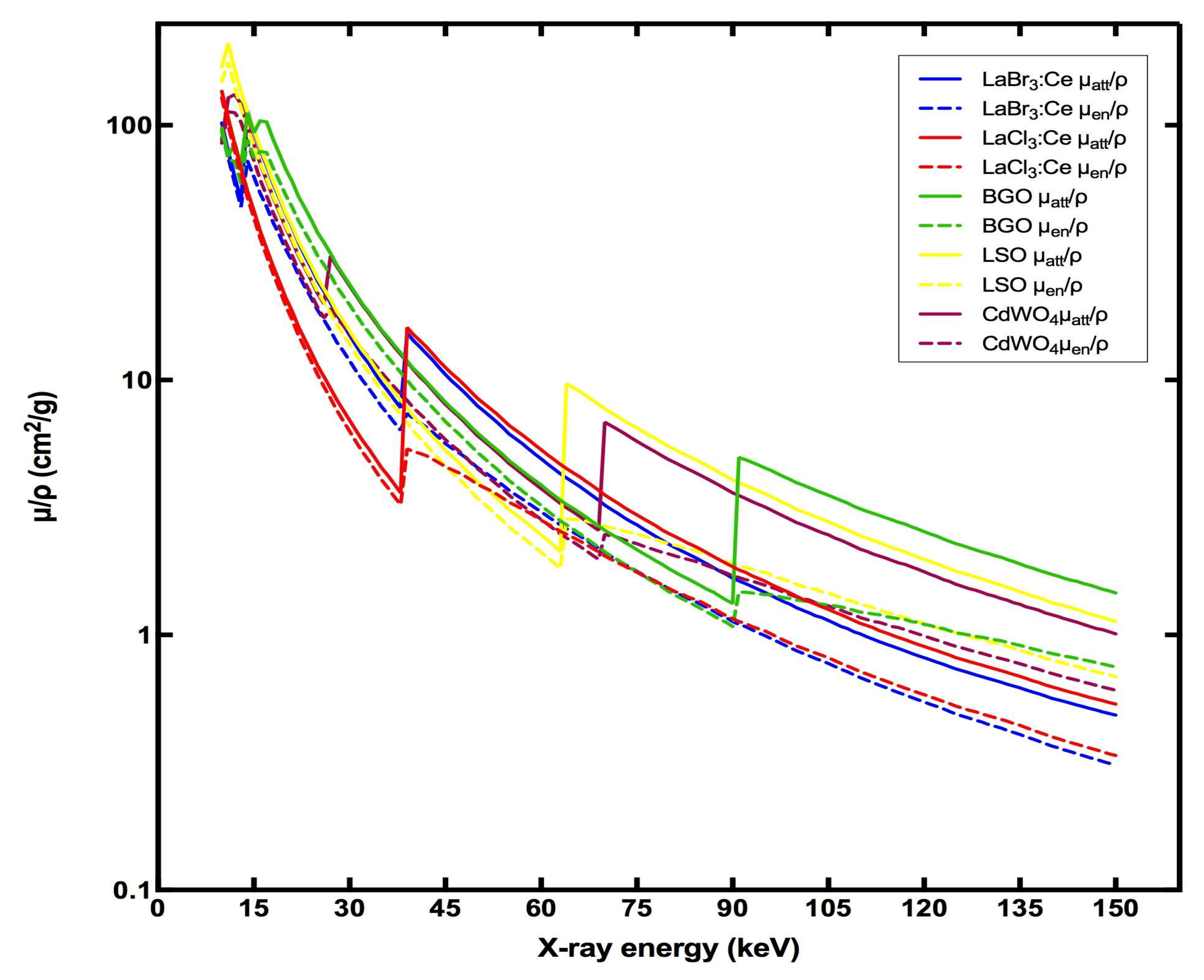
Figure 3 exhibits the graph of μatt in terms of energy, as obtained from the XmuDAT software, featuring the characteristic saw-tooth shape in the energy range where absorption through the photoelectric effect dominates (up to approximately 100 keV). The discontinuities correspond to the absorption edges K, L, M or N, in which the energies of the photons become equal to the binding energies of the electrons of the atoms of the crystals in the shells K, L, M or N, and therefore the probability of absorption of the photons increases sharply, due to resonance [48].
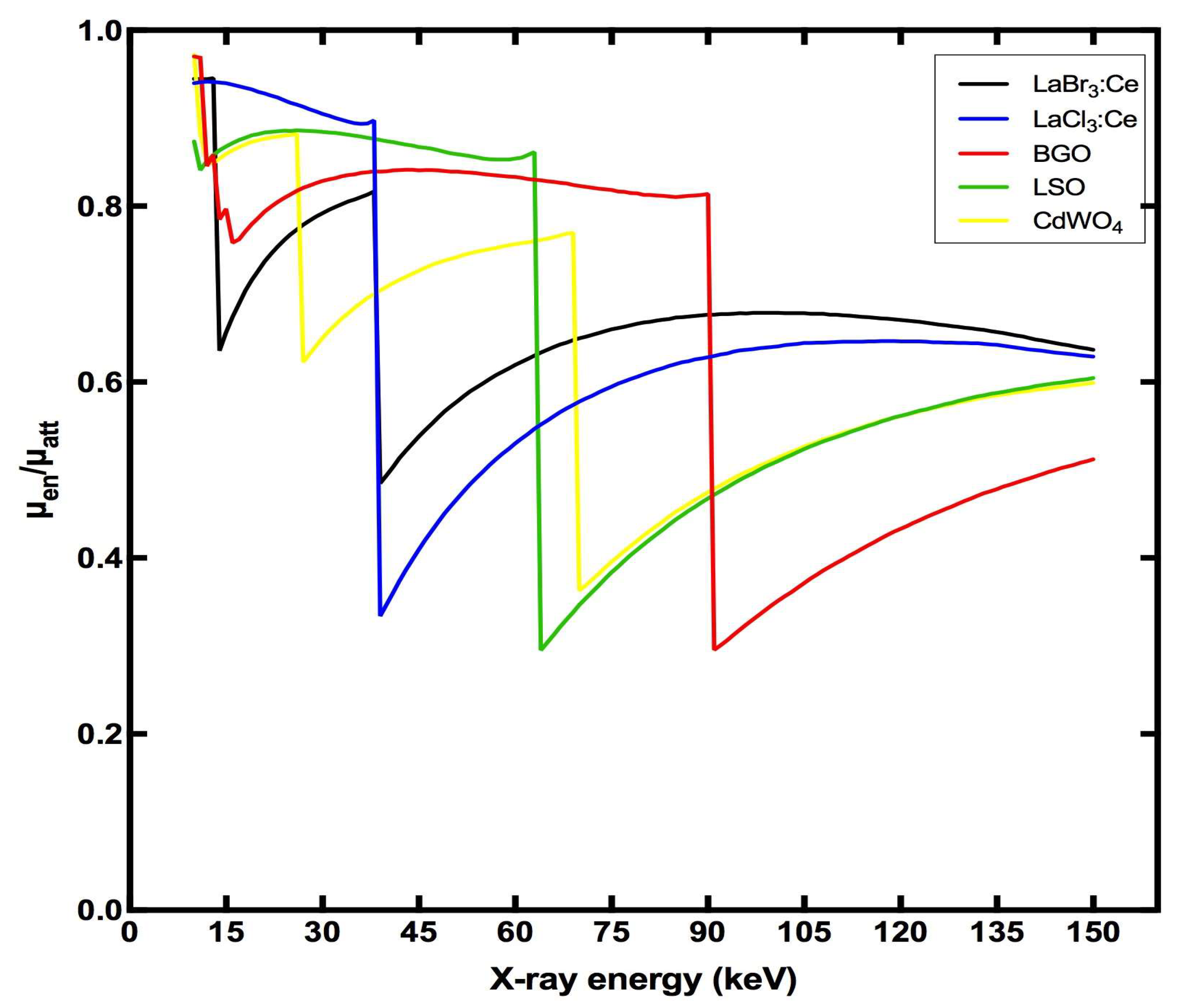
While μatt expresses the abovementioned probability, μεn expresses the probability of transfer and absorption of this energy within the crystal. Their calculated ratio with respect to energy is represented graphically in Figure 4. A sharp drop of this ratio at the absorption-edge positions of the crystals is observed, resulting in the corresponding reduction of EAE in the graph of Figure 2. The crystals of LaCl3:Ce and LaBr3:Ce show a common K edge at 39 keV, due to La, while LaBr3:Ce shows a second one, due to bromide at 14 keV, which, however, does not affect the EAE in the range of energies we are studying. The EAE of both crystals decreases after 40 kVp, and in fact the decrease in LaCl3:Ce is greater, due to the greater drop in the μenatt ratio, as shown in the graph. After 60 kVp, the EAE increases slightly, and stabilizes, due to the gradual increase in the μenatt ratio after 39 keV. Similarly, in BGO, LSO and CdWO4, with K-edges at 91 keV, 64 keV and 70 keV, respectively, the EAE decreases after 91 kVp, 64 kVp and 70 kVp, respectively. After 110 kVp for LSO and CdWO4 and 130 kVp for BGO, the EAE almost stabilizes, due to the partial recovery of the μenatt ratio values.
Figure 5 shows the effect of tube voltage on QDE. LaBr3:Ce demonstrates almost perfect efficiency in detecting the incident photons, as shown by the QDE values, which range from 0.998 to 1. The QDE is maximum at X-ray tube voltages up to 120 KVp. This is mainly due to the thickness of the crystal, since the attenuation coefficients drop after the K-edge of LaBr3:Ce, above 39 keV. For this energy range, the radiation absorption is maximum, and all the incident radiation may contribute to signal generation. For higher energies, however, the continuous drop of the attenuation coefficient leads to a smaller number of X photons absorbed in the crystal.
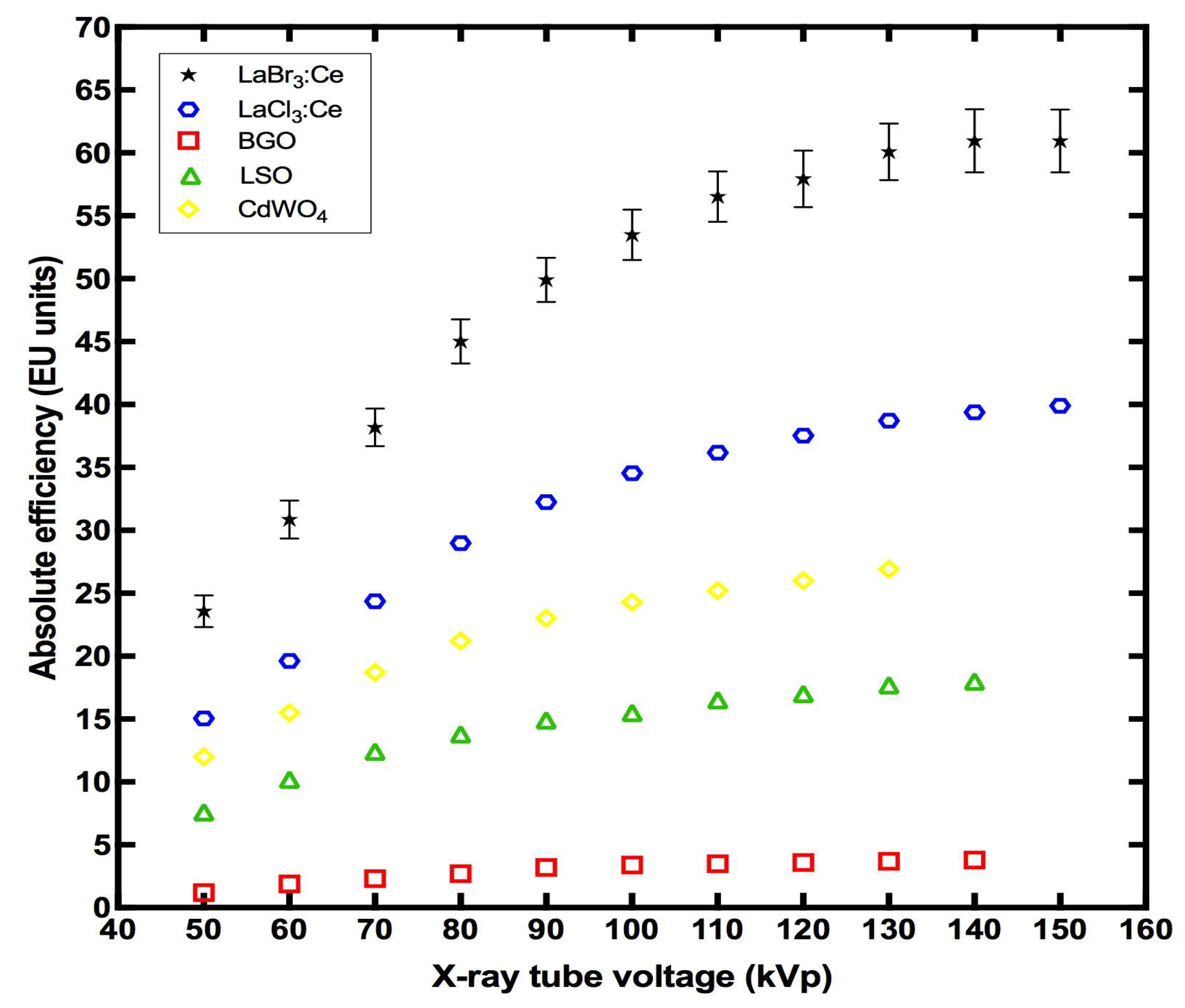
Figure 6 shows the variation of AE of the LaBr3:Ce crystal in the X-ray tube-voltage range from 50 to 150 kVp, compared with previously published data for the LaCl3:Ce, BGO, LSO, and CdWO4 crystals of similar dimensions [25,26,27]. The experimental error is 4.25%. As can be seen, the AE of all crystals increases continuously with increasing tube voltage; however, the LaBr3:Ce crystal’s values are the highest at all energies. Specifically, at 50 kVp, the values were 23.5 EU for LaBr3:Ce, 15.1 EU for LaCl3:Ce, 1.2 EU for BGO, 7.6 EU for LSO, and 12 EU for CdWO4. At 130 kVp, the difference was even greater, measuring 60.1 EU, 38.7 EU, 3.7 EU, 17.7 EU and 26.9 EU, respectively [25,26,27].
The very good absolute-efficiency values of LaBr3:Ce, compared to the other crystals reported for comparison in this paper (even though it has lower EAE values, as shown in Figure 2), can be explained by its higher LY [27].
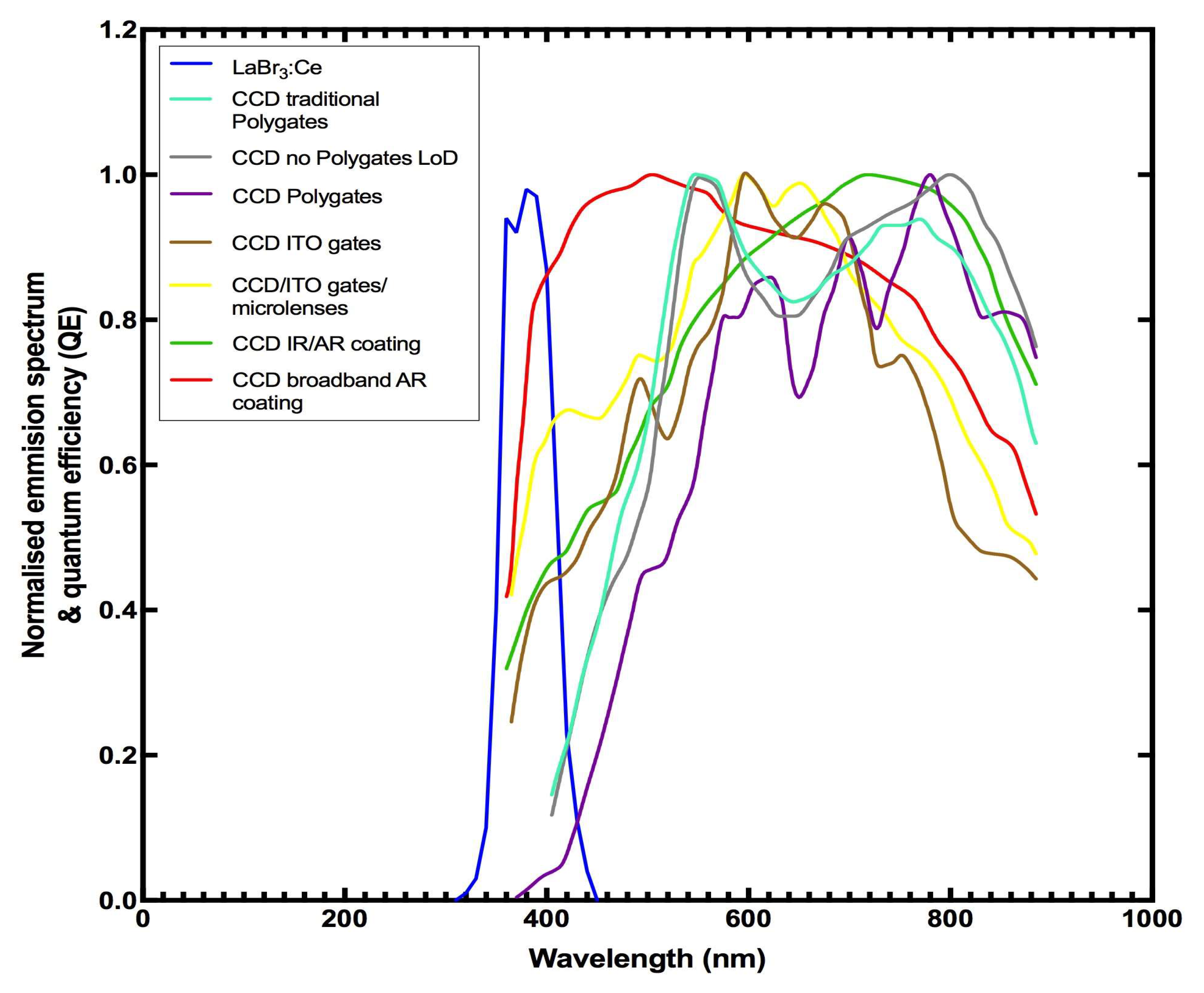
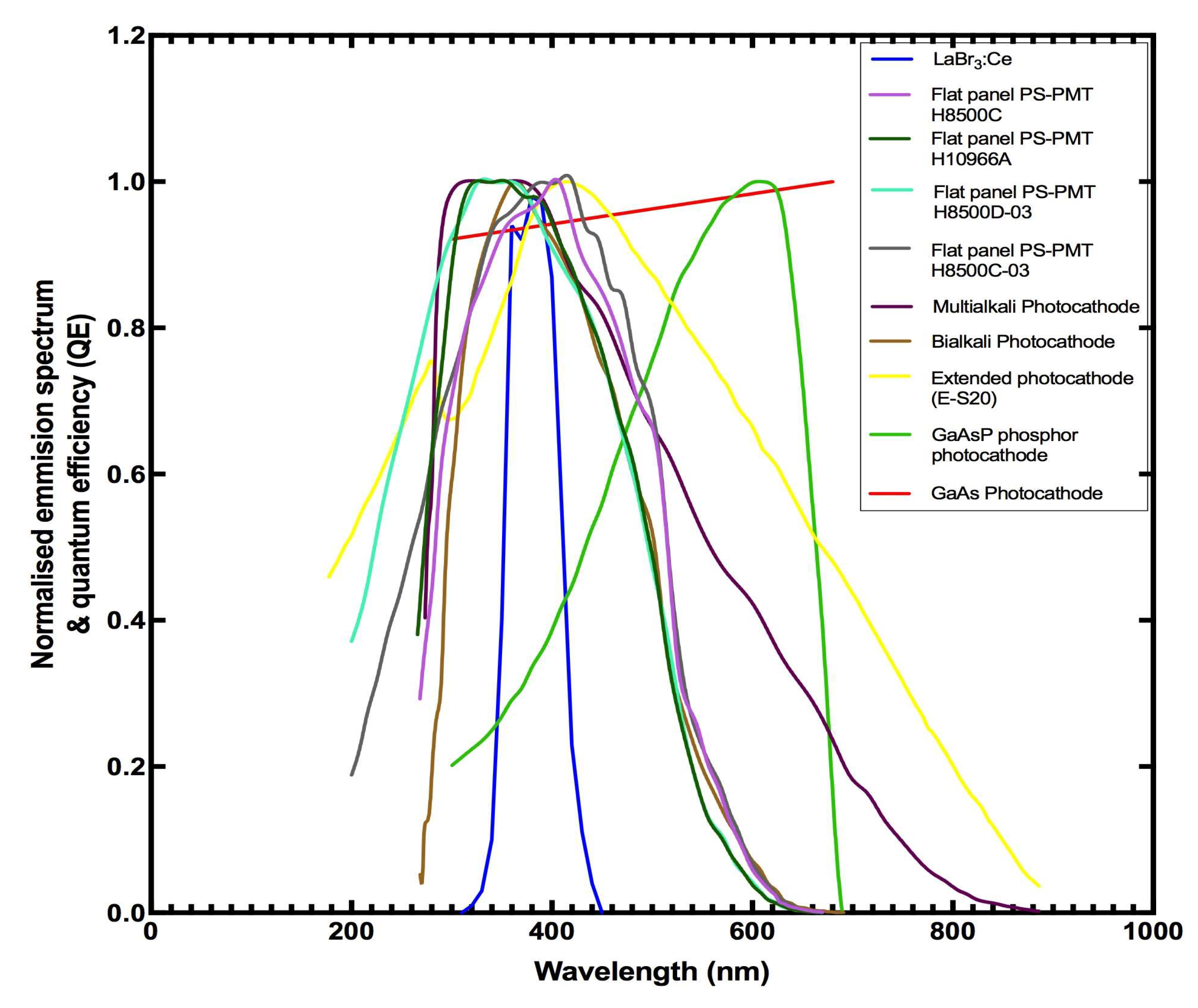
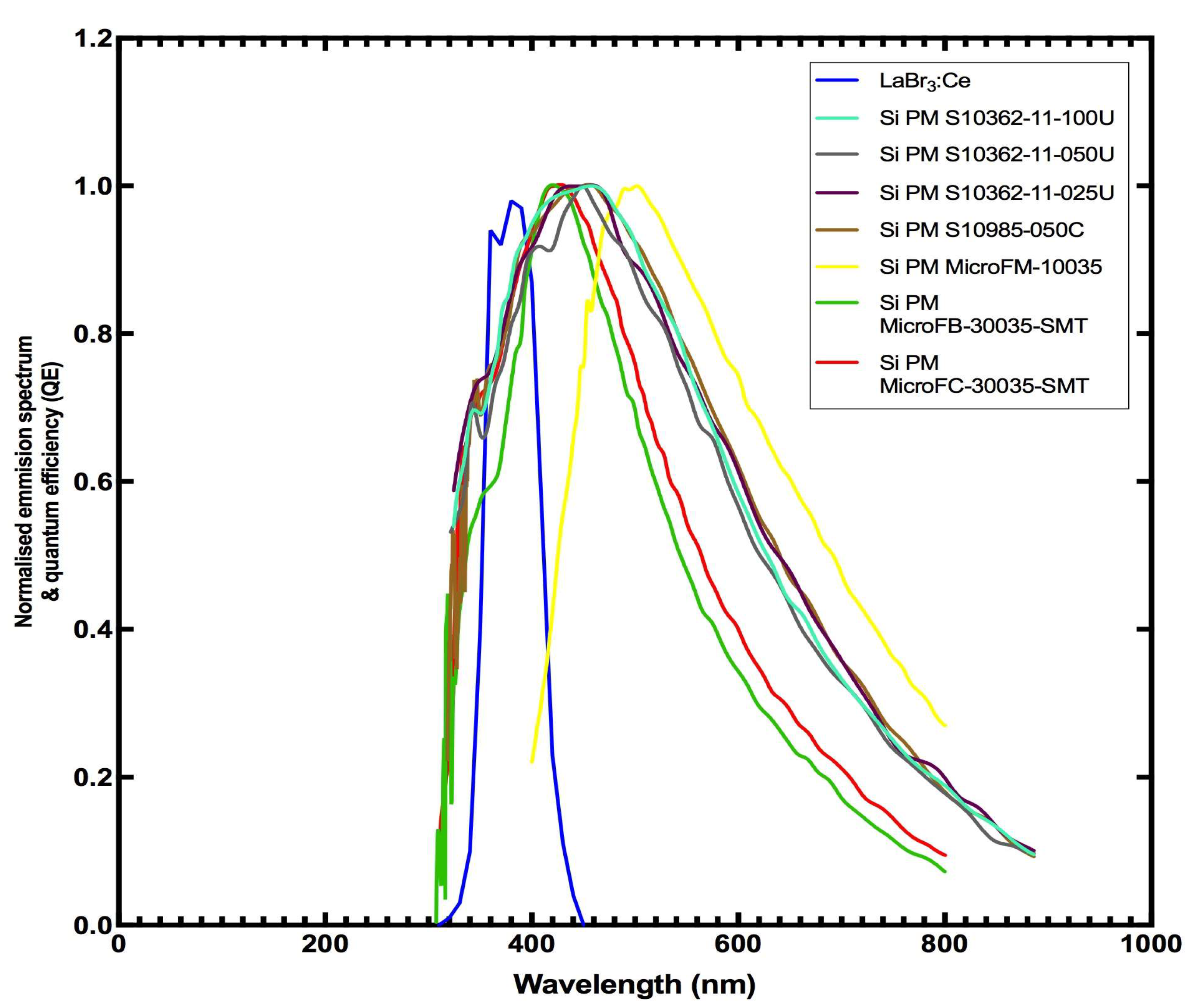
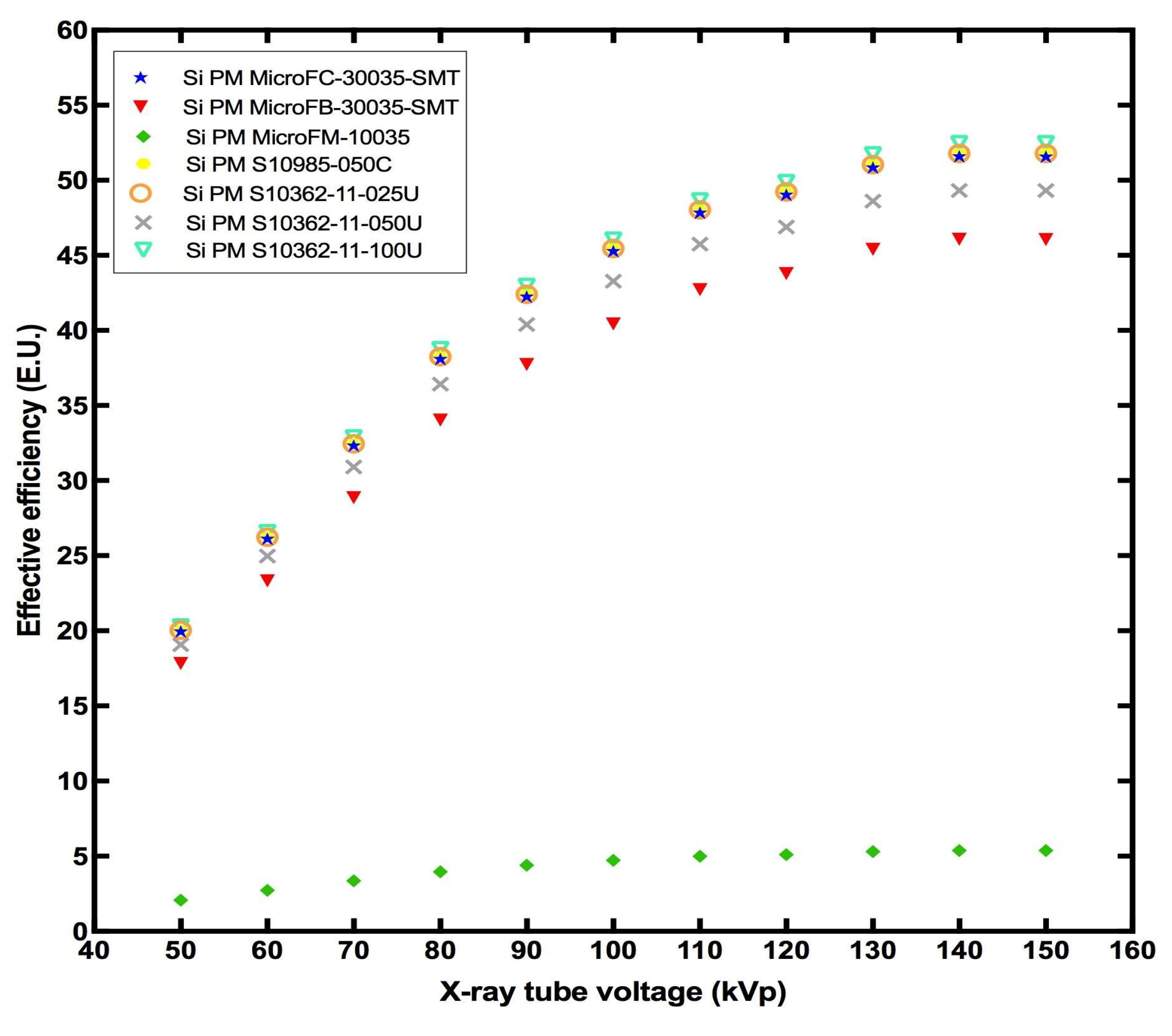
The emitted optical-spectrum of LaBr3:Ce, normalized to unity, compared to the spectral sensitivities of several commonly used optical photon-sensors, is illustrated in Figure 7, Figure 8, Figure 9 and Figure 10 [33,40,41,42,43]. The sensors listed are silicon photomultipliers (SiPMs), frequently employed in nuclear-medicine applications, charge-coupled devices (CCD) and complementary metal-oxide semiconductors (CMOS) utilized in imaging applications, and various photocathodes. The width of the LaBr3:Ce spectrum is approximately 120 nm, from 320 to 440 nm, with a maximum at 380 nm [18].
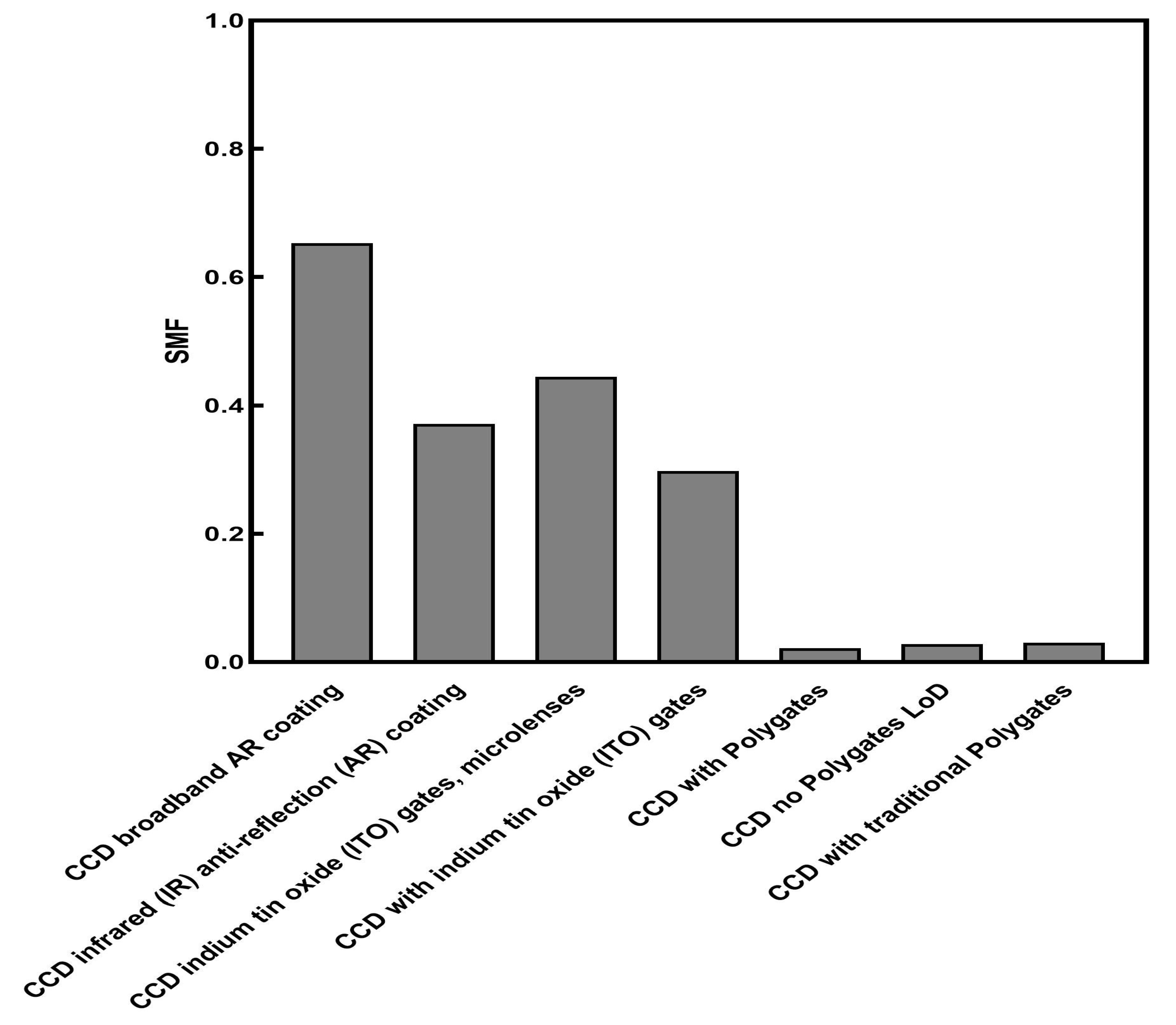
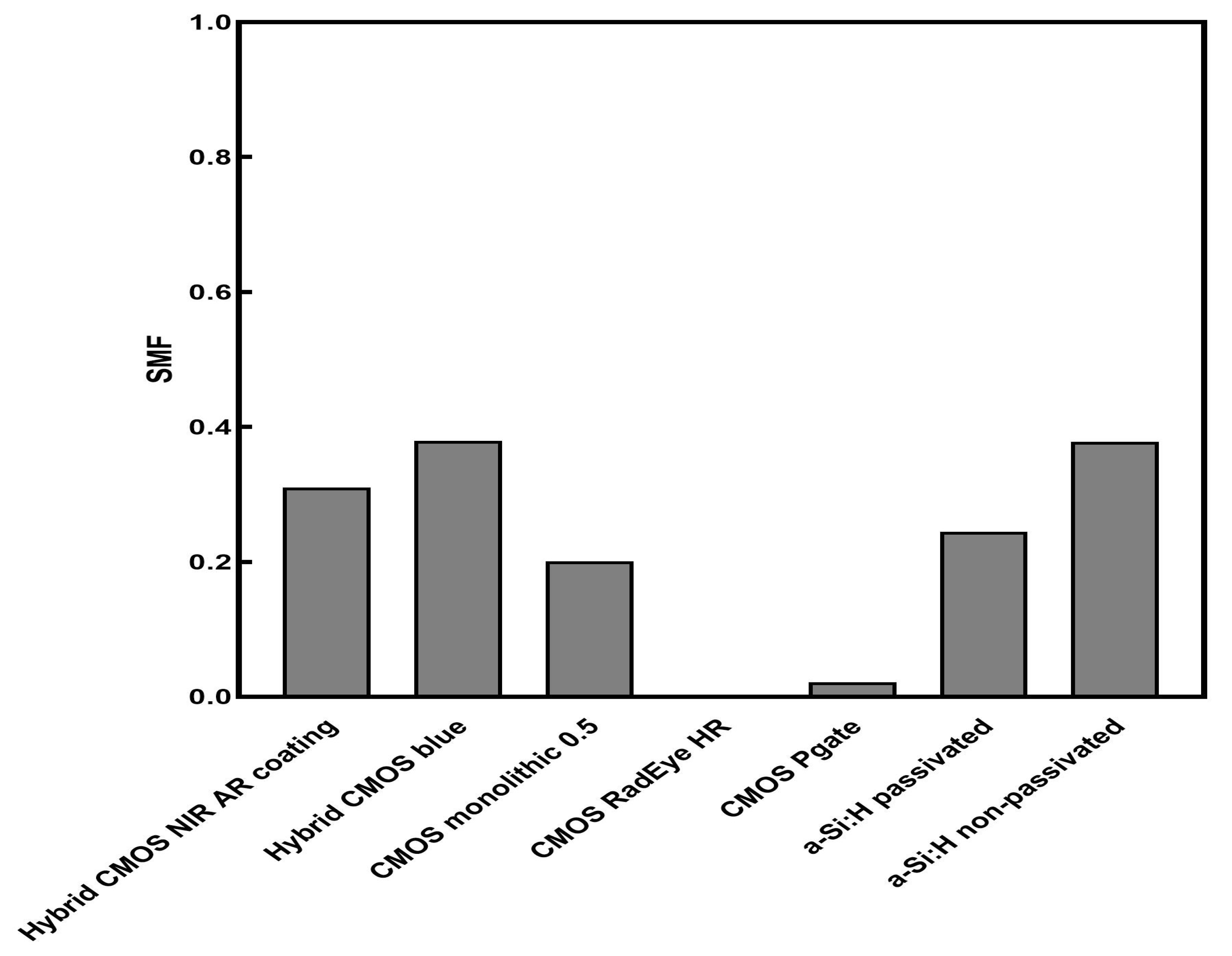
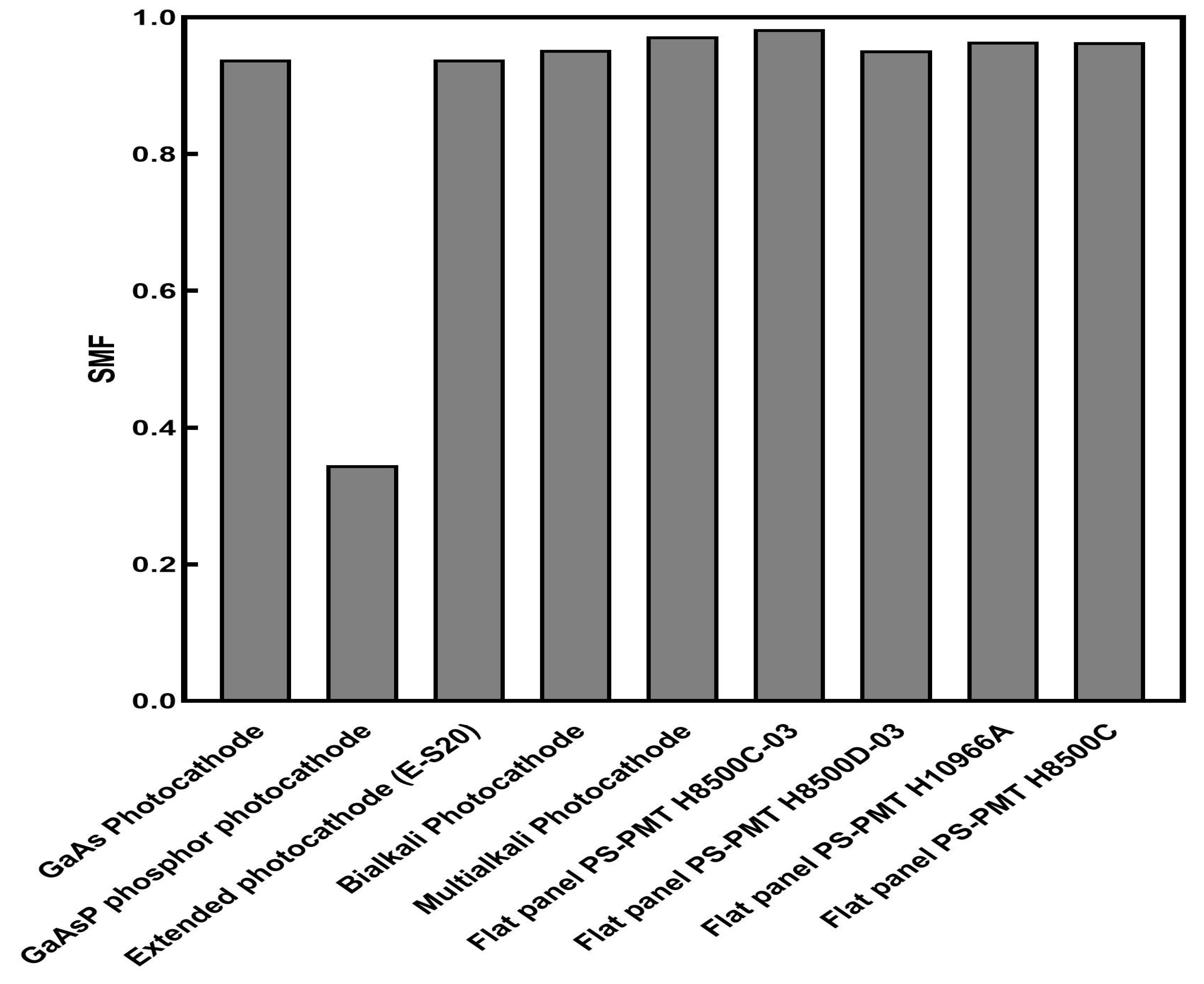
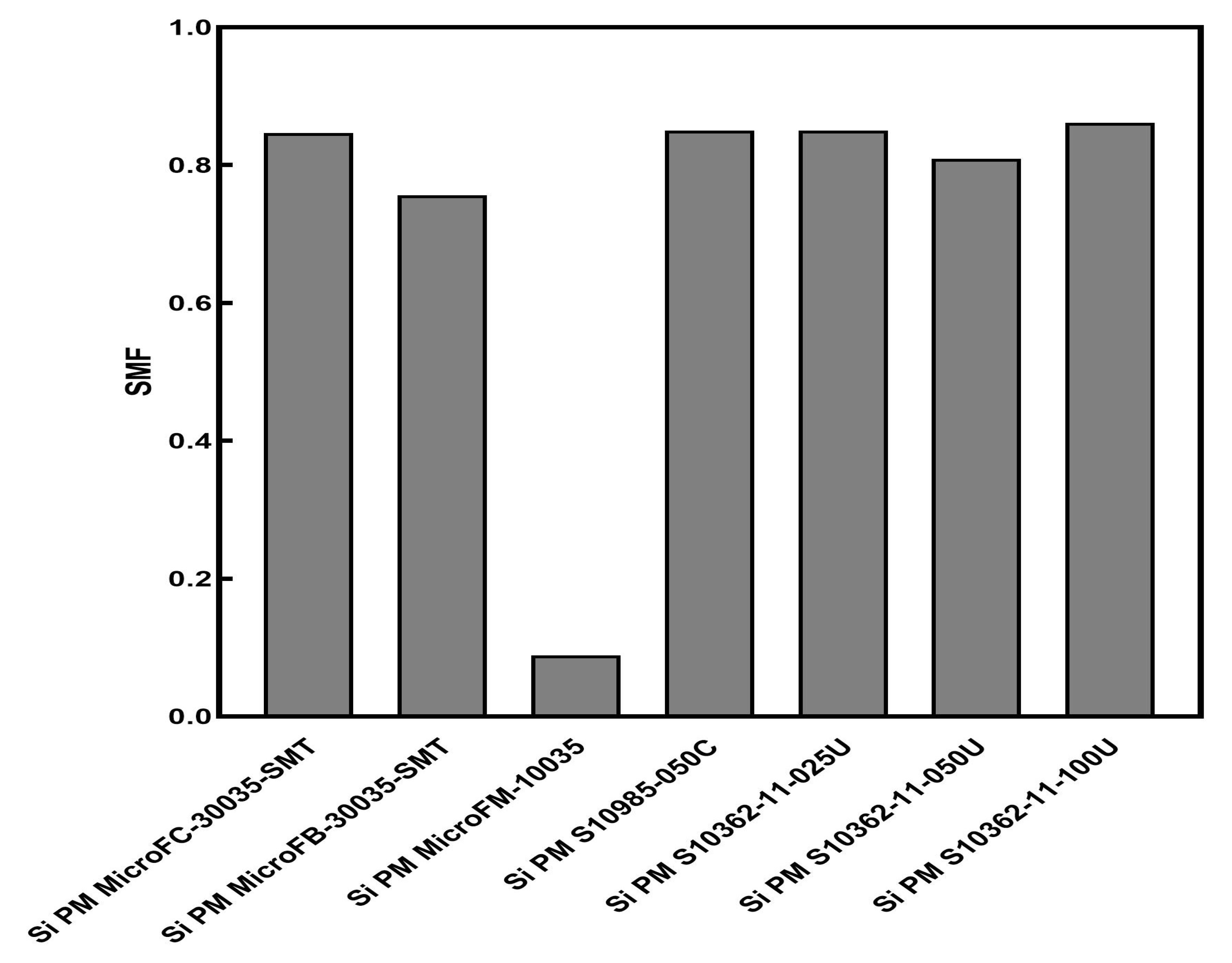
The compatibility of the crystal-detector combination, shown in Figure 7, Figure 8, Figure 9 and Figure 10, is directly related to the calculated SMFs demonstrated in Figure 11, Figure 12, Figure 13 and Figure 14. LaBr3:Ce provides excellent compatibility with various flat-panel (FP) photocathodes, with the SMF values ranging from 0.95 to 0.98, as well as with the Multialkali photocathode (0.97) and the Bialkali photocathode (0.95). LaBr3:Ce is a suitable choice for applications evolving silicon photomultipliers, since the SMF values ranged from 0.75 to 0.86 (0.86 for SiPM S10362-11-100U). On the other hand, LaBr3:Ce was found to be incompatible with CCDs and CMOS detectors, with the SMF values ranging from 0.02 to 0.65 for the CCDs detectors, and from 0.0 to 0.37 for the CMOS detectors. More specifically, the lowest values were registered when coupled with the CCD with Polygates and the CCD no-Polygates LoD (both at 0.02), the CCD with traditional Polygates (0.03), the CMOS Pgate (0.02) and the CMOS RadEye HR (0.0).
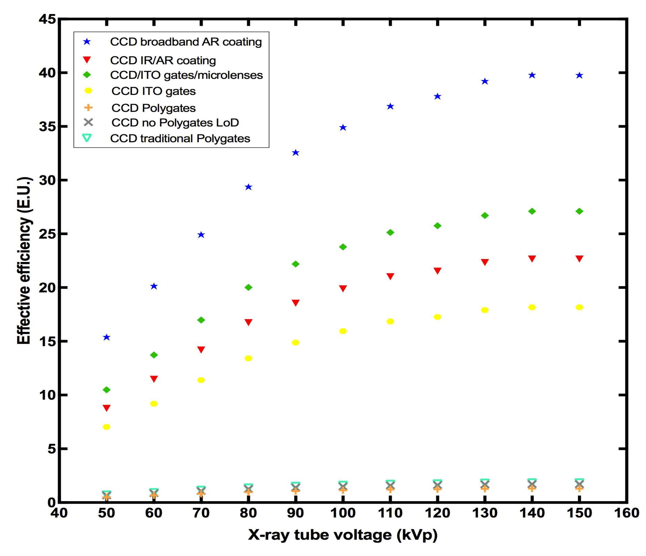
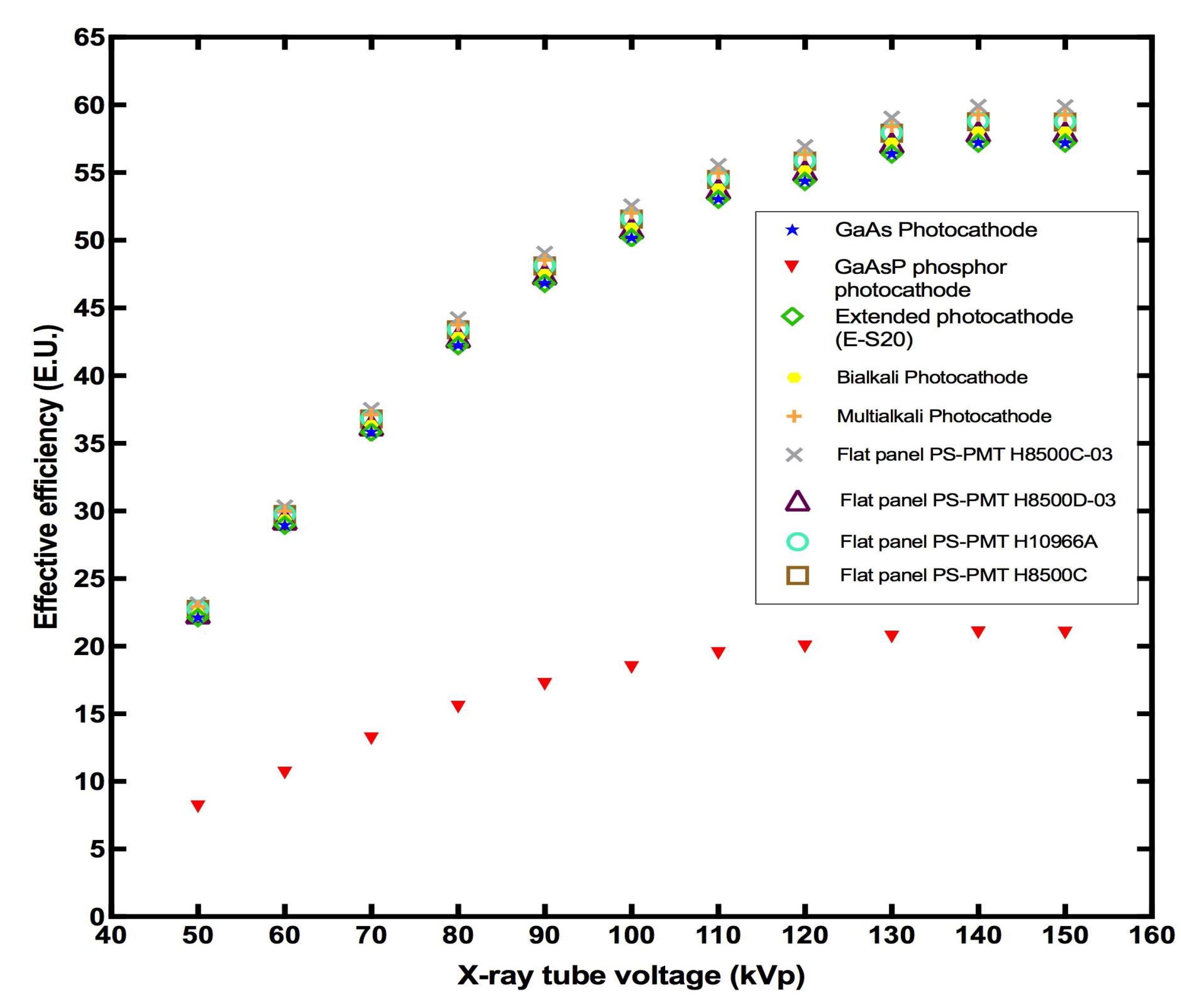
Figure 15, Figure 16, Figure 17 and Figure 18 show effective-efficiency data corresponding to every optical sensor mentioned before. Following the SMF results, EE values confirm the excellent compatibility of the LaBr3:Ce crystal with several photocathodes and silicon photomultipliers (SiPMs). Specifically, the highest EE values were attributed to the Multialkali photocathode, the Bialkali photocatode, and the flat-panel (FP) photocathodes. On the other hand, the lowest values were shown when coupled with various CCDs and CMOS detectors, with the decrease (with kVp) in the detected signal ranging from 99.3% to 100% when the crystal was matched with the CCD with Polygates, the CCD no-Polygates LoD, the CCD with traditional Polygates, the CMOS Pgate and the CMOS RadEye HR.
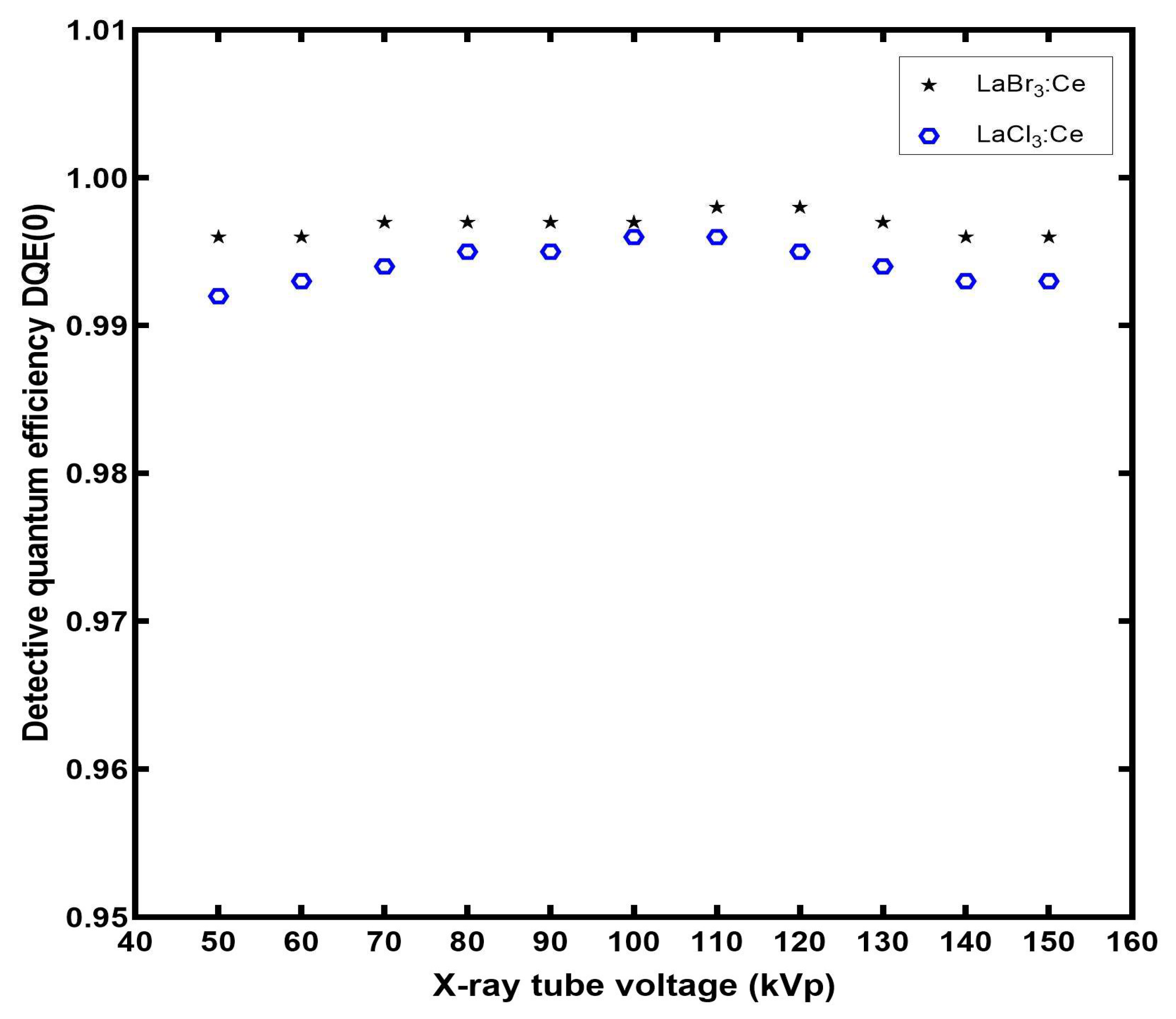
Figure 19 presents the calculated values of DQE(0) for the LaBr3:Ce crystal. For comparison purposes, the DQE(0) of a recently studied halide crystal (LaCl3:Ce) [27], which exhibits the second best AE value, is also shown. Both crystals show excellent performance, with LaBr3:Ce slightly outperforming LaCl3:Ce, possibly due to its higher light-yield [18,28] and AE values. All the DQE(0) values are above 0.99.

4. Discussion

The shape of the AE curve of LaBr3:Ce is a combination of the X-ray absorption and optical-photon production and escape from the crystal. At lower energies, the increased QDE enables a high optical-photon production. As the X-ray energy increases, and while QDE remains approximately 1, the number of optical photons generated increases, in proportion to the light yield; thus, the AE values are also measured as increasing. For X-ray tube voltages above 120 kVp, where the QDE drops below 1, the percentage of the X-rays absorbed is reduced, which in turn reduces the energy offered for optical-photons production. This concludes with a smaller slope, resembling a plateau, in the AE-value increase. If the QDE value was 1 for these energies, the rate of AE increase for X-ray tube voltages above 120 kVp might be similar to the increase rate between 50 kVp and 110 kVp.
It is worth commenting that the use of the LaBr3:Ce scintillator can give an equivalent result to the rest of the crystals regarding the signal, but with a significant reduction in radiation and therefore in the dose received by the examinee. In effect, compared to the BGO and LSO crystals used in hybrid SPECT/CT and PET/CT imaging systems, LaBr3:Ce will give the same output but, as calculated at 140 kVp, with 6.2% and 29.5% of the radiation, respectively. In addition, the reduction in emitted radiation means a reduction in the current-time mAs, and therefore a reduction in the operational cost of the X-ray tube, without affecting the quality of the examination. Although LaBr3:Ce is hygroscopic, other scintillators that were used in the past for radiation-imaging purposes, such as CsI:Na and NaI, were also hygroscopic. This suggests that the technology for managing this type of scintillators is applicable. The high light-yield of the LaBr3:Ce crystalline scintillator, compared to other scintillators, can lead to radiation-dose savings for the examinees, which is of importance in radiation imaging.
The LaBr3:Ce crystal’s higher efficiency when compared to the BGO and LSO crystals across the whole energy range, as well as its exceptional performance at high energies, demonstrate its potential for use in medical-imaging applications, especially in nuclear medicine.
Concerning the SMF values, when the LaBr3:Ce crystal was compared to the LaCl3:Ce crystal, a halide scintillator of similar characteristics, it presented almost the same values when coupled with various photocathodes, but considerably better compatibility with silicon photomultipliers, CCDs and CMOS, as the SMF values of LaCl3:Ce range from 0.62 to 0.67 for silicon photomultipliers, from 0.002 to 0.22 for CCDs, and from 0.0 to 0.28 for CMOS [27].
The DQE values are a function of the radiation-absorption performance of the scintillator crystal as described by QDE, and the escaping optical-photon signal fluctuation. Both these parameters are embedded in Equations (7) and (13). At lower X-ray energies, where QDE is approximately 1 and AE is low, DQE(0) presents its lowest values. As the X-ray energy increases up to approximately 120 kVp, QDE is still almost 1, but the optical-photon production is increased, leading to a corresponding increase in DQE(0) values. At higher energies however, the drop in QDE as shown in Figure 5, combined with a tendency to AE saturation, as shown in Figure 6, leads to somewhat lower DQE(0) values. In every case, LaBr3:Ce is superior to LaCl3:Ce, mainly due to its better radiation to optical conversion characteristics.
It may be deduced that the LaBr3:Ce scintillator under study performs better, both in terms of ionizing radiation to optical-photon-emission efficiency and in terms of SNR transfer at X-ray tube-voltages over 110 kVp. This range is the one utilized in computed tomography imaging and it is closer to the energy emitted by radioisotopes used in nuclear-medicine applications. This makes LaBr3:Ce a prominent candidate as a radiation sensor for hybrid medical-imaging systems such as SPECT/CT and PET/CT.

5. Conclusions

In the current study, the efficiency properties of a LaBr3:Ce crystal were investigated under X-ray excitation with X-ray tube-voltages (50–150 kVp) and compared with previously published data for BGO, LSO, CdWO4 and LaCl3:Ce crystals. The maximum AE of the examined crystal was obtained at 150 kVp (60.9 EU). The values of AE of the LaBr3:Ce were found to be considerably higher than those of the BGO, LSO, CdWO4 and LaCl3:Ce crystals in the examined energy range. These results are also considered important from the point of view of radiation protection, as the use of the crystal can reduce the dose received by the examinee. The emission spectrum of LaBr3:Ce showed an exceptional match with the spectral sensitivities of many commonly employed photocathodes and silicon photomultipliers (SiPMs). In addition, the LaBr3:Ce crystal exhibited excellent performance concerning its detective quantum efficiency for zero spatial-frequency. Taking into account the high AE values, the spectral compatibility with several optical-sensors, the DQE(0) values, the short decay-time, as well as the optimum X-ray tube-voltage range, which was found to be above 120 kVp, the LaBr3:Ce scintillation crystal might be considered as a radiation-detector option for hybrid medical-imaging modalities, such PET/CT and SPECT/CT scanners.

Author Contributions

Conceptualization, C.M. and S.T.; methodology, I.V.; software, S.T.; validation, N.K., A.B. and G.F.; formal analysis, N.K.; investigation, S.T. and K.N.; resources, G.F.; data curation, I.V.; writing—original draft preparation, S.T.; writing—review and editing, I.K., N.K. and C.M.; visualization, N.K.; supervision, N.K., C.M. and I.V; project administration, N.K. All authors have read and agreed to the published version of the manuscript.

Funding

This research received no external funding.

Institutional Review Board Statement

Not applicable.

Informed Consent Statement

Not applicable.

Data Availability Statement

Not applicable.

Conflicts of Interest

The authors declare no conflict of interest.

References

  1. Van Eijk, C.W.E. Inorganic scintillators in medical imaging. Phys. Med. Biol. 2002, 47, R85–R106. [Google Scholar] [CrossRef] [PubMed]
  2. Grammaticos, P.; Fountos, G. The physician should benefit, not harm the patient. Hell. J. Nucl. Med. 2006, 9, 82–84. [Google Scholar] [PubMed]
  3. Spanoudaki, V.C.; Levin, C.S. Photo-Detectors for Time of Flight Positron Emission Tomography (ToF-PET). Sensors 2010, 10, 10484–10505. [Google Scholar] [CrossRef] [PubMed]
  4. Moses, W.W. Scintillator Requirements for Medical Imaging. In Proceedings of the International Conference on Inorganic Scintillators and Their Applications, Moscow, Russia, 16–20 August 1999; p. 11. [Google Scholar]
  5. Van Loef, E.V.D.; Dorenbos, P.; van Eijk, C.W.E.; Krämer, K.; Güdel, H.U. High-Energy-Resolution Scintillator: Ce3+ Activated LaBr3. Appl. Phys. Lett. 2001, 79, 1573–1575. [Google Scholar] [CrossRef]
  6. Van Loef, E.V.D.; Dorenbos, P.; van Eijk, C.W.E.; Krämer, K.W.; Güdel, H.U. Scintillation Properties of LaBr3:Ce3+ Crystals: Fast, Efficient and High-Energy-Resolution Scintillators. Nucl. Instrum. Methods Phys. Res. Sect. A Accel. Spectrometers Detect. Assoc. Equip. 2002, 486, 254–258. [Google Scholar] [CrossRef]
  7. Shah, K.S.; Glodo, J.; Klugerman, M.; Moses, W.W.; Derenzo, S.E.; Weber, M.J. LaBr3:Ce Scintillators for Gamma Ray Spectroscopy. IEEE Trans. Nucl. Sci. 2004, 50, 2410–2413. [Google Scholar] [CrossRef]
  8. Dorenbos, P.; de Haas, J.T.M.; van Eijk, C.W.E. Gamma Ray Spectroscopy with a O19 × 19 mm3 LaBr3:0.5% Ce3+ Scintillator. IEEE Trans. Nucl. Sci. 2004, 51, 1289–1296. [Google Scholar] [CrossRef] [Green Version]
  9. Glodo, J.; Moses, W.W.; Higgins, W.M.; van Loef, E.V.D.; Wong, P.; Derenzo, S.E.; Weber, M.J.; Shah, K.S. Effects of Ce Concentration on Scintillation Properties of LaBr3:Ce. IEEE Trans. Nucl. Sci. 2005, 52, 1805–1808. [Google Scholar] [CrossRef]
  10. Iltis, A.; Mayhugh, M.R.; Menge, P.; Rozsa, C.M.; Selles, O.; Solovyev, V. Lanthanum Halide Scintillators: Properties and Applications. Nucl. Instrum. Methods Phys. Res. Sect. A Accel. Spectrometers Detect. Assoc. Equip. 2006, 563, 359–363. [Google Scholar] [CrossRef]
  11. Bizarri, G.; de Haas, J.T.M.; Dorenbos, P.; van Eijk, C.W.E. Scintillation Properties of O1 x 1 Inch3 LaBr3: 5%Ce3+ Crystal. IEEE Trans. Nucl. Sci. 2006, 53, 615–619. [Google Scholar] [CrossRef]
  12. Higgins, W.M.; Glodo, J.; Van Loef, E.; Klugerman, M.; Gupta, T.; Cirignano, L.; Wong, P.; Shah, K.S. Bridgman Growth of LaBr3:Ce and LaCl3:Ce Crystals for High-Resolution Gamma-Ray Spectrometers. J. Cryst. Growth 2006, 287, 239–242. [Google Scholar] [CrossRef]
  13. Chen, J.; Hu, Z.; Zhang, X.; Chen, Z.; Yuan, X.; Sun, Z.; Guo, Z.; Xu, H. Investigation of LaBr3: Ce Scintillator with Excellent Property. J. Phys. Conf. Ser. 2014, 488, 142013. [Google Scholar] [CrossRef] [Green Version]
  14. Dorenbos, P. Light Output and Energy Resolution of Ce3+-Doped Scintillators. Nucl. Instrum. Methods Phys. Res. Sect. A Accel. Spectrometers Detect. Assoc. Equip. 2002, 486, 208–213. [Google Scholar] [CrossRef]
  15. Alzimami, K.; Abuelhia, E.; Podolyak, Z.; Ioannou, A.; Spyrou, N.M. Characterization of LaBr3:Ce and LaCl3:Ce Scintillators for Gamma-Ray Spectroscopy. J. Radioanal. Nucl. Chem. 2008, 278, 755–759. [Google Scholar] [CrossRef]
  16. Chewpraditkul, W.; Moszynski, M. Scintillation Properties of Lu3Al5O12, Lu2SiO5 and LaBr3 Crystals Activated with Cerium. Phys. Procedia 2011, 22, 218–226. [Google Scholar] [CrossRef] [Green Version]
  17. Kuhn, A.; Surti, S.; Karp, J.S.; Raby, P.S.; Shah, K.S.; Perkins, A.E.; Muehllehner, G. Design of a Lanthanum Bromide Detector for Time-of-Flight PET. IEEE Trans. Nucl. Sci. 2004, 51, 2550–2557. [Google Scholar] [CrossRef]
  18. Advatech, U.K. LaBr3:Ce—Lanthanum Bromide (Ce). Available online: https://www.advatech-uk.co.uk/labr3_ce.html (accessed on 12 July 2022).
  19. Dey Chaudhuri, S.; Banerjee, D.; Bhattacharjee, T.; Wasim Raja, S.; Acharya, R.; Pujari, P.K. Performance Study of LaBr3:Ce Detectors Coupled to R2083 PM Tube for Energy and Timing Characteristics. J. Radioanal. Nucl. Chem. 2020, 324, 829–835. [Google Scholar] [CrossRef]
  20. Alzimami, K.S.; Spyrou, N.M.; Sassi, S.A. Investigation of LaBr3:Ce and LaCl3:Ce Scintillators for Spect Imaging. In 2008 5th IEEE International Symposium on Biomedical Imaging: From Nano to Macro; IEEE: Paris, France, 2008; pp. 1243–1246. [Google Scholar] [CrossRef]
  21. Surti, S.; Karp, J.S.; Muehllehner, G. Image Quality Assessment of LaBr3 -Based Whole-Body 3D PET Scanners: A Monte Carlo Evaluation. Phys. Med. Biol. 2004, 49, 4593–4610. [Google Scholar] [CrossRef] [Green Version]
  22. Kandarakis, I.; Cavouras, D.; Kalivas, N.; Nomicos, C.D.; Panayiotakis, G.S. Estimation of the Information Content of Medical Images Produced by Scintillators Interacting with Diagnostic X-Ray Beams. Nucl. Instrum. Methods Phys. Res. Sect. B Beam Interact. Mater. At. 1999, 155, 199–205. [Google Scholar] [CrossRef]
  23. Owens, A.; Bos, A.J.J.; Brandenburg, S.; Dorenbos, P.; Drozdowski, W.; Ostendorf, R.W.; Quarati, F.; Webb, A.; Welter, E. The Hard X-Ray Response of Ce-Doped Lanthanum Halide Scintillators. Nucl. Instrum. Methods Phys. Res. Sect. A Accel. Spectrometers Detect. Assoc. Equip. 2007, 574, 158–162. [Google Scholar] [CrossRef] [Green Version]
  24. Khodyuk, I.V.; Dorenbos, P. Nonproportional Response of LaBr3: Ce and LaCl3: Ce Scintillators to Synchrotron x-Ray Irradiation. J. Phys. Condens. Matter 2010, 22, 485402. [Google Scholar] [CrossRef] [PubMed]
  25. Michail, C.; Kalyvas, N.; Bakas, A.; Ninos, K.; Sianoudis, I.; Fountos, G.; Kandarakis, I.; Panayiotakis, G.; Valais, I. Absolute Luminescence Efficiency of Europium-Doped Calcium Fluoride (CaF2:Eu) Single Crystals under X-Ray Excitation. Crystals 2019, 9, 234. [Google Scholar] [CrossRef] [Green Version]
  26. Michail, C.; Koukou, V.; Martini, N.; Saatsakis, G.; Kalyvas, N.; Bakas, A.; Kandarakis, I.; Fountos, G.; Panayiotakis, G.; Valais, I. Luminescence Efficiency of Cadmium Tungstate (CdWO4) Single Crystal for Medical Imaging Applications. Crystals 2020, 10, 429. [Google Scholar] [CrossRef]
  27. Tseremoglou, S.; Michail, C.; Valais, I.; Ninos, K.; Bakas, A.; Kandarakis, I.; Fountos, G.; Kalyvas, N. Efficiency Properties of Cerium-Doped Lanthanum Chloride (LaCl3:Ce) Single Crystal Scintillator under Radiographic X-Ray Excitation. Crystals 2022, 12, 655. [Google Scholar] [CrossRef]
  28. Advatech, U.K. LaCl3:Ce—Lanthanum Chloride (Ce). Available online: https://www.advatech-uk.co.uk/lacl3_ce.html (accessed on 12 July 2022).
  29. Advatech, U.K. LSO:Ce—Lutetium Oxyorthosilicate (Ce). Available online: https://www.advatech-uk.co.uk/lso_ce.html (accessed on 12 July 2022).
  30. Advatech, U.K. BGO—Bismuth Germanate Scintillator Crystal. Available online: https://www.advatech-uk.co.uk/bgo.html (accessed on 12 July 2022).
  31. Linardatos, D.; Michail, C.; Kalyvas, N.; Ninos, K.; Bakas, A.; Valais, I.; Fountos, G.; Kandarakis, I. Luminescence Efficciency of Cerium bromide Single Crystal under X-ray Radiation. Crystals 2022, 12, 909. [Google Scholar] [CrossRef]
  32. Valais, I.G.; Kandarakis, I.S.; Nikolopoulos, D.N.; Michail, C.M.; David, S.L.; Loudos, G.K.; Cavouras, D.A.; Panayiotakis, G.S. Luminescence Properties of (Lu, Y)2SiO5:Ce and Gd2SiO5: Ce Single Crystal Scintillators Under X-Ray Excitation for Use in Medical Imaging Systems. IEEE Trans. Nucl. Sci. 2007, 54, 11–18. [Google Scholar] [CrossRef]
  33. Linardatos, D.; Konstantinidis, A.; Valais, I.; Ninos, K.; Kalyvas, N.; Bakas, A.; Kandarakis, I.; Fountos, G.; Michail, C. On the Optical Response of Tellurium Activated Zinc Selenide ZnSe:Te Single Crystal. Crystals 2020, 10, 961. [Google Scholar] [CrossRef]
  34. Boone, J. X-ray production, interaction, and detection in diagnostic imaging. In Handbook of Medical Imaging. Volume 1: Physics and Psychophysics; Beutel, J., Kundel, H.L., Van Metter, R.L., Eds.; SPIE Press: Bellingham, WA, USA, 2000; Volume 1, pp. 36–57. ISBN 978-0-8194-7772-9. [Google Scholar]
  35. Nowotny, R. XMuDat: Photon Attenuation Data on PC (IAEA-NDS-195); International Atomic Energy Agency: Vienna, Austria, 1998. [Google Scholar]
  36. TASMIP Spectra Calculator. Available online: https://www.solutioinsilico.com/medical-physics/applications/tasmip-app.php?ans=0 (accessed on 10 December 2022).
  37. Kandarakis, I.; Cavouras, D.; Panayiotakis, G.S.; Nomicos, C.D. Evaluating X-Ray Detectors for Radiographic Applications: A Comparison of ZnSCdS:Ag with and Screens. Phys. Med. Biol. 1997, 42, 1351–1373. [Google Scholar] [CrossRef]
  38. Michail, C.; Valais, I.; Seferis, I.; Kalyvas, N.; David, S.; Fountos, G.; Kandarakis, I. Measurement of the Luminescence Properties of Gd2O2S: Pr, Ce, F Powder Scintillators under X-Ray Radiation. Radiat. Meas. 2014, 70, 59–64. [Google Scholar] [CrossRef]
  39. Kandarakis, I.; Cavouras, D.; Nomicos, C.D.; Panayiotakis, G.S. X-Ray Luminescence of ZnSCdS:Au, Cu Phosphor Using X-Ray Beams for Medical Applications. Nucl. Instrum. Methods Phys. Res. Sect. B Beam Interact. Mater. At. 2001, 179, 215–224. [Google Scholar] [CrossRef]
  40. Hamamatsu Photonics, MPPC (Multi-Pixel Photon Counters). Available online: http://www.hamamatsu.com/us/en/product/category/3100/4004/4113/index.html# (accessed on 4 September 2022).
  41. SensL. Silicon Photomultipliers. Available online: http://sensl.com/products/silicon-photomultipliers/ (accessed on 4 September 2022).
  42. Magnan, P. Detection of Visible Photons in CCD and CMOS: A Comparative View. Nucl. Instrum. Methods Phys. Res. Sect. A Accel. Spectrometers Detect. Assoc. Equip. 2003, 504, 199–212. [Google Scholar] [CrossRef]
  43. Rowlands, J.A.; Yorkston, J. Flat Panel Detectors for Digital Radiography. In Handbook of Medical Imaging Physics and Psychophysics; Beutel, J., Kundel, H., Van Metter, R., Eds.; SPIE: Bellingham, WA, USA, 2000; Volume 1, pp. 223–328. ISBN 978-08-194-7772-9. [Google Scholar]
  44. Cavouras, D.; Kandarakis, I.; Bakas, A.; Triantis, D.; Nomicos, C.D.; Panayiotakis, G.S. An Experimental Method to Determine the Effective Luminescence Efficiency of Scintillator-Photodetector Combinations Used in X-Ray Medical Imaging Systems. Br. J. Radiol. 1998, 71, 766–772. [Google Scholar] [CrossRef] [PubMed]
  45. Valais, I. Systematic Study of the Light Emission Efficiency and the Corresponding Intrinsic Physical Characteristics of Single Crystal Scintillators, Doped with the Trivalent Cerium (Ce3+) Activator, in Wide Energy Range (from 20 kV–18 MV) for Medical Applications. Ph.D. Thesis, University of Patras, Patras, Greece, 2008. [Google Scholar]
  46. Attix, F.H. Introduction of Radiological Physics and Radiation Dosimetry; John Wiley & Sons: New York, NY, USA, 1987; p. 31. [Google Scholar]
  47. Abbene, L.; La Manna, A.; Fauci, F.; Gerardi, G.; Stumbo, S.; Raso, G. X-Ray Spectroscopy and Dosimetry with a Portable CdTe Device. Nucl. Instrum. Methods Phys. Res. Sect. A Accel. Spectrometers Detect. Assoc. Equip. 2007, 571, 373–377. [Google Scholar] [CrossRef]
  48. Hendee, W.R.; Ritenour, E.R. Medical Imaging Physics; John Wiley & Sons: New York, NY, USA, 2002. [Google Scholar] [CrossRef]
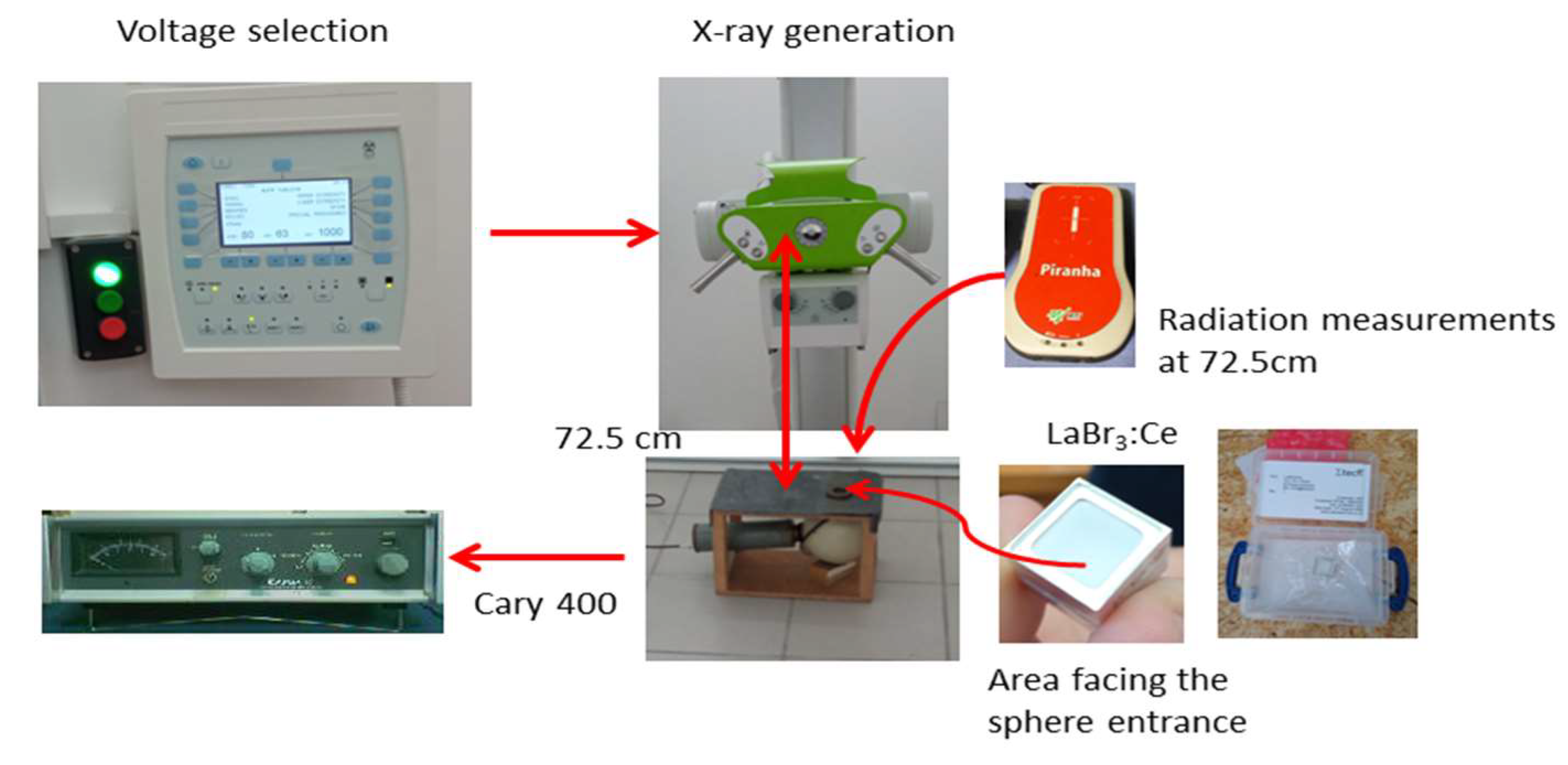
Figure 1. Experimental measurement setup.
Applsci 13 00419 g001
Figure 2. EAE of LaBr3:Ce, LaCl3:Ce, BGO, LSO and CdWO4 single crystals.
Applsci 13 00419 g002
Figure 3. Attenuation coefficients of the LaBr3:Ce, LaCl3:Ce, BGO, LSO and CdWO4 single crystals.
Applsci 13 00419 g003
Figure 4. The ratio μenatt of the LaBr3:Ce, LaCl3:Ce, BGO, LSO and CdWO4 single crystals.
Applsci 13 00419 g004
Figure 5. QDE of the LaBr3:Ce crystal in comparison with LaCl3:Ce, BGO, LSO and CdWO4 crystals.
Applsci 13 00419 g005
Figure 6. AE values of the LaBr3:Ce (4.25% error bar) crystal in comparison with previously published data for the LaCl3:Ce, BGO, LSO and CdWO4 crystals.
Applsci 13 00419 g006
Figure 7. Optical spectral distribution of LaBr3:Ce crystal, and spectral sensitivity of CCD photoreceptors.
Applsci 13 00419 g007
Figure 8. Optical spectral distribution of LaBr3:Ce crystal, and spectral sensitivity of CMOS photoreceptors.
Applsci 13 00419 g008
Figure 9. Optical spectra distribution of LaBr3:Ce, and spectral sensitivity of photocathodes.
Applsci 13 00419 g009
Figure 10. Optical spectra distribution of LaBr3:Ce, and spectral sensitivity of SiPMs.
Applsci 13 00419 g010
Figure 11. SMF values of LaBr3:Ce with various CCDs.
Applsci 13 00419 g011
Figure 12. SMF values of LaBr3:Ce with various CMOS.
Applsci 13 00419 g012
Figure 13. SMF values of LaBr3:Ce with various photocathodes.
Applsci 13 00419 g013
Figure 14. SMF values of LaBr3:Ce with various SiPMs.
Applsci 13 00419 g014
Figure 15. EE of LaBr3:Ce combined with various CCDs.
Applsci 13 00419 g015
Figure 16. EE of LaBr3:Ce combined with various CMOS.
Applsci 13 00419 g016
Figure 17. EE of the LaBr3:Ce combined with various photocathodes.
Applsci 13 00419 g017
Figure 18. EE of the LaBr3:Ce combined with various Si PMs.
Applsci 13 00419 g018
Figure 19. DQE(0) of the LaBr3:Ce and LaCl3:Ce single crystals.
Applsci 13 00419 g019
Table 1. BGO, LSO, CdW04, LaCl3:Ce and LaBr3:Ce crystal properties [18,26,28,29,30].
PropertiesBGOLSO:CeCdWO4LaCl3:CeLaBr3:Ce
Wavelength, max emission (nm)480420490350380
Decay Time (ns)3004050002825
Light Yield (photons/MeV)890030,00028,00049,00063,000
Radiation Length (cm)1.1181.141.062.8131.881
Density (g/cm3)7.137.47.93.865.2
Effective Atomic Number7475744746
Melting Point (°C)10442050132511351116
Hardness (Mho)55.84-4.533
HygroscopicityNoNoNoYesYes
Disclaimer/Publisher’s Note: The statements, opinions and data contained in all publications are solely those of the individual author(s) and contributor(s) and not of MDPI and/or the editor(s). MDPI and/or the editor(s) disclaim responsibility for any injury to people or property resulting from any ideas, methods, instructions or products referred to in the content.