
Upper Extremity Kinematics and Electromyographic Activity in Uninjured Tennis Players

 ,
,
Abstract
:Featured Application
Abstract
1. Introduction
2. Materials and Methods
2.1. Subjects
2.2. Data Collection
2.3. Data Analysis
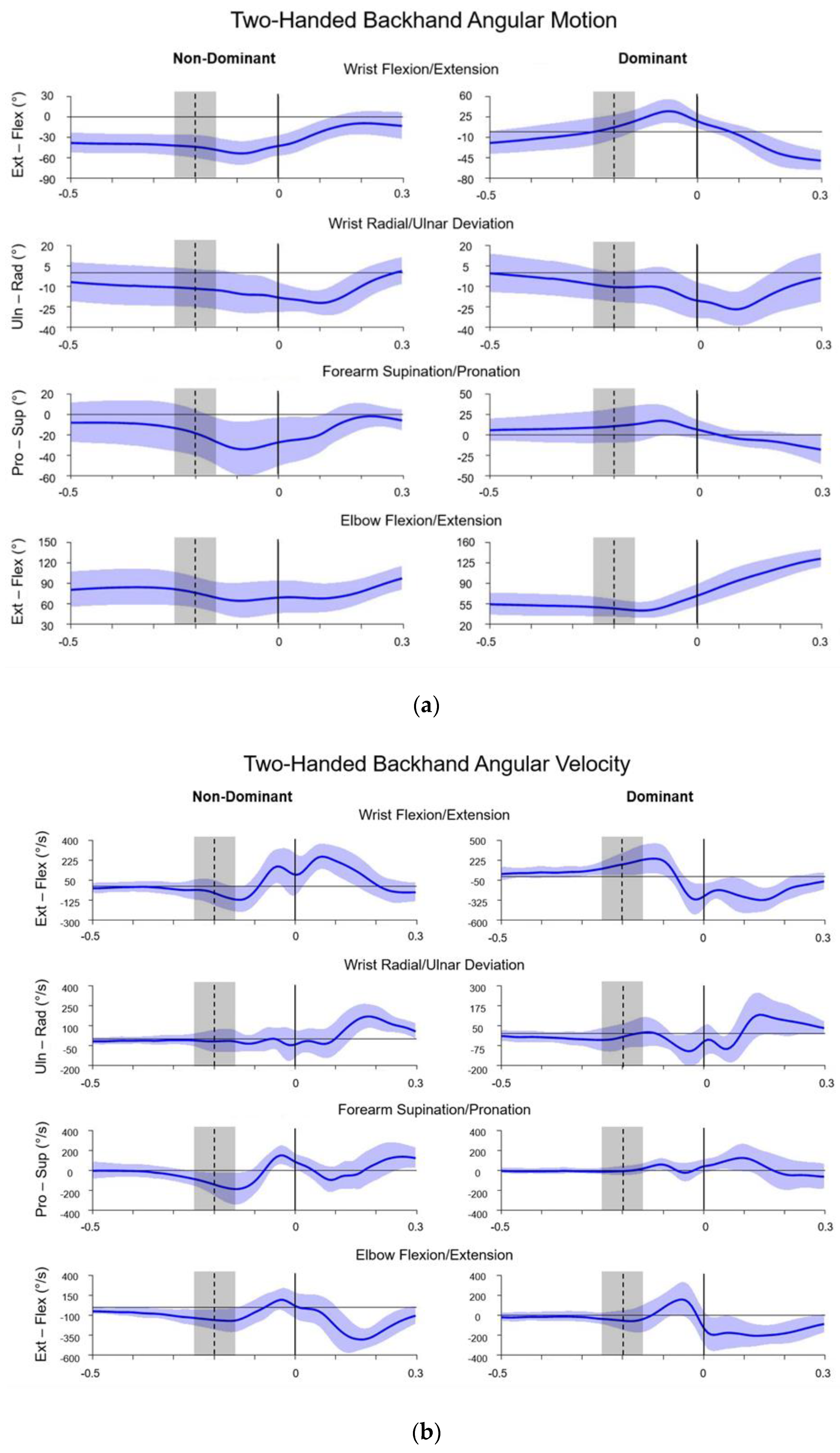
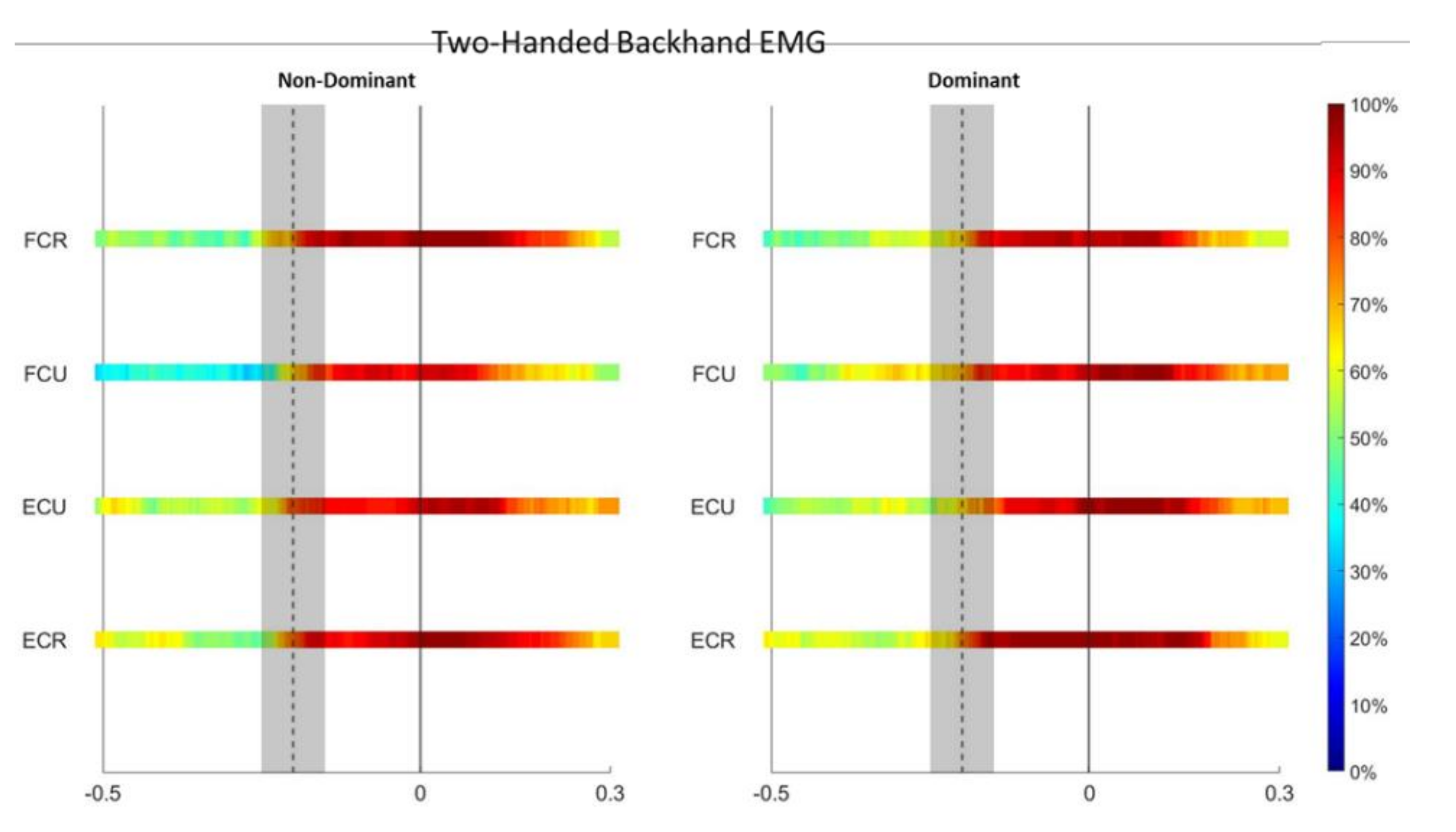
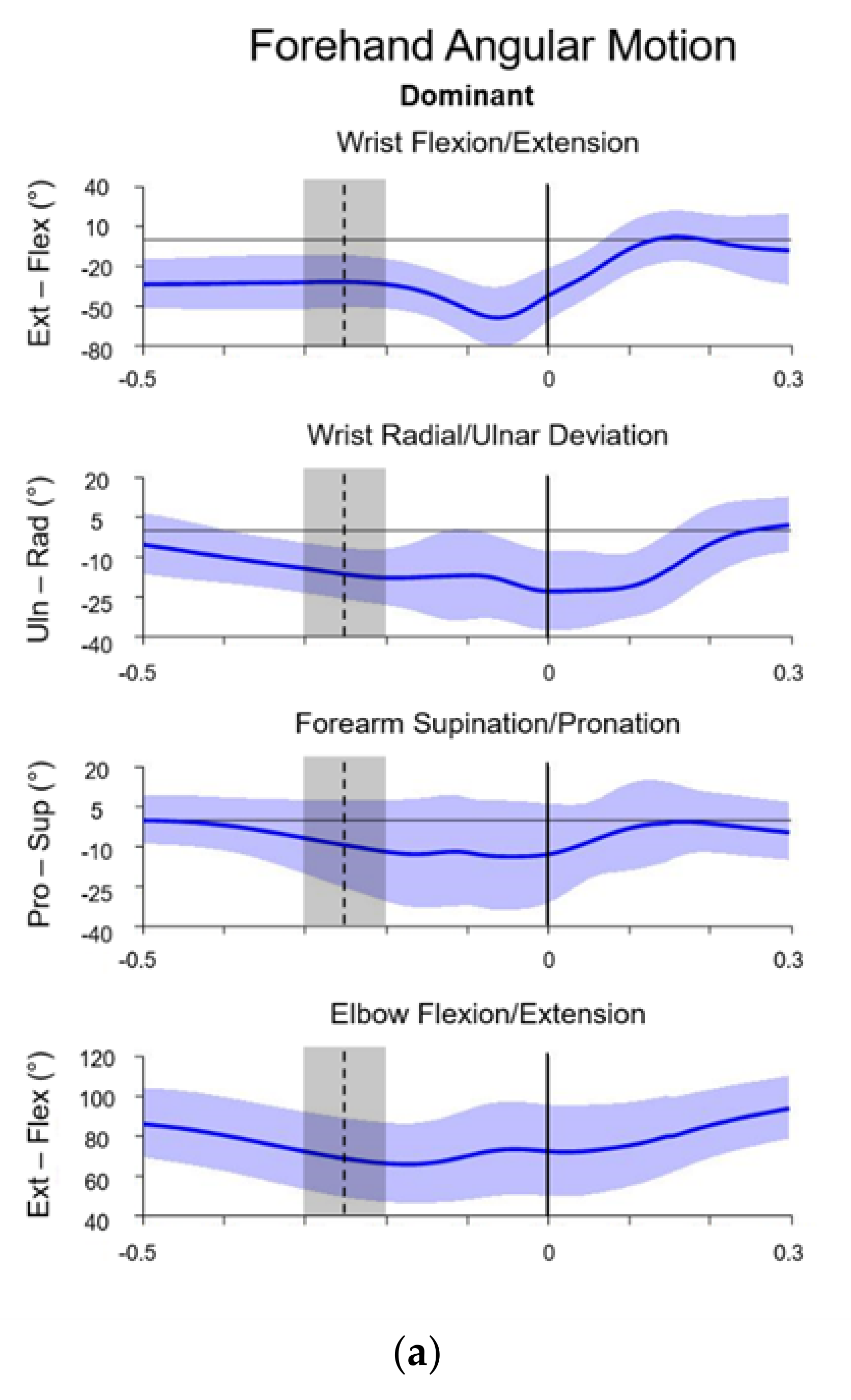
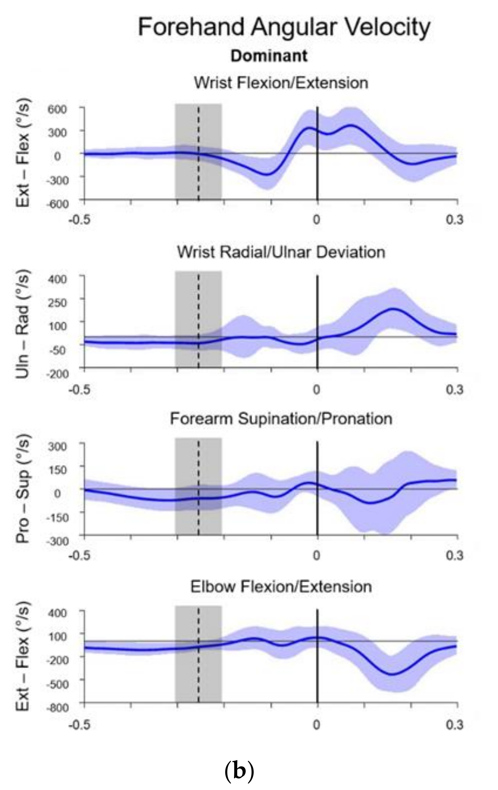
3. Results
4. Discussion
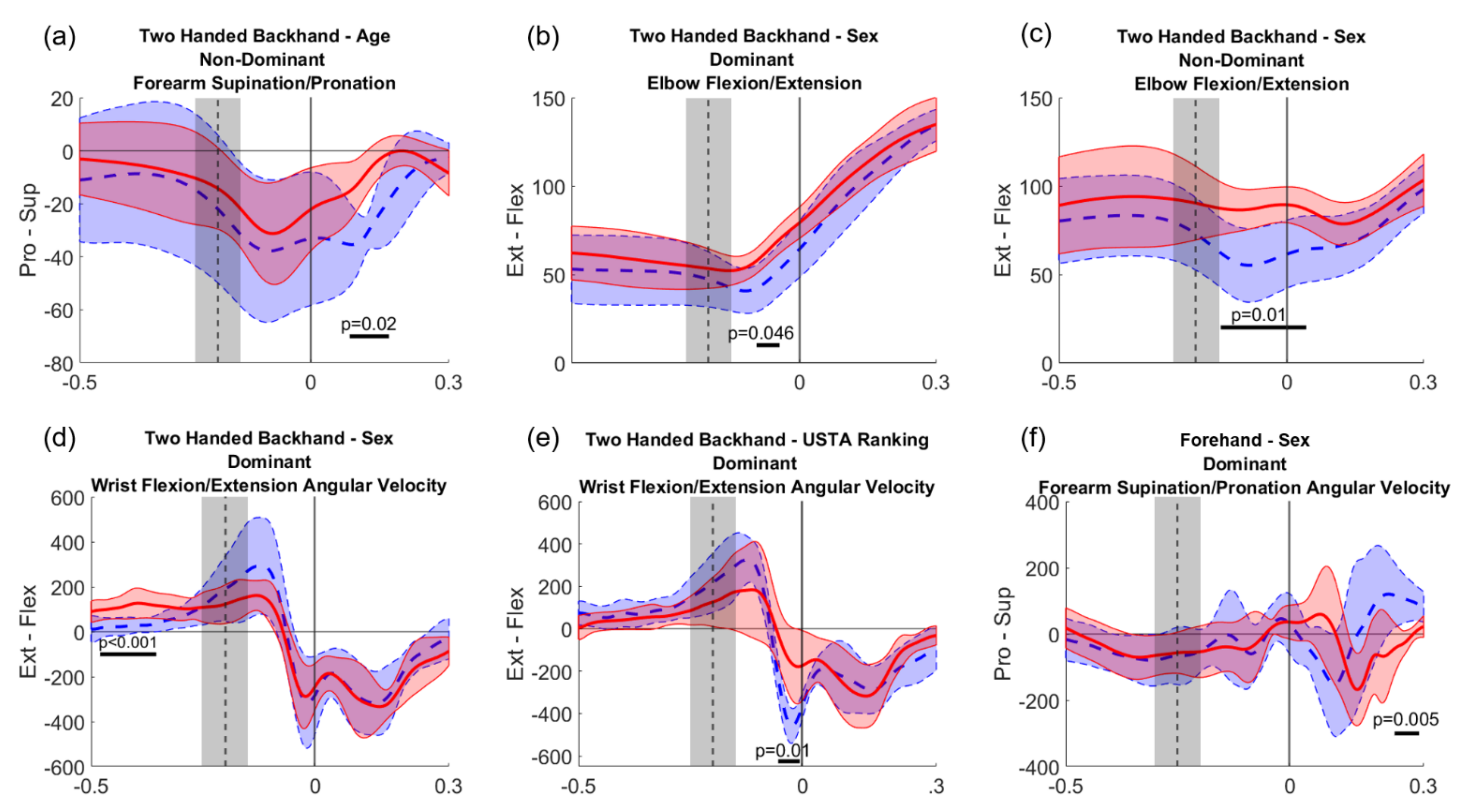
4.1. Two-Handed Backhand
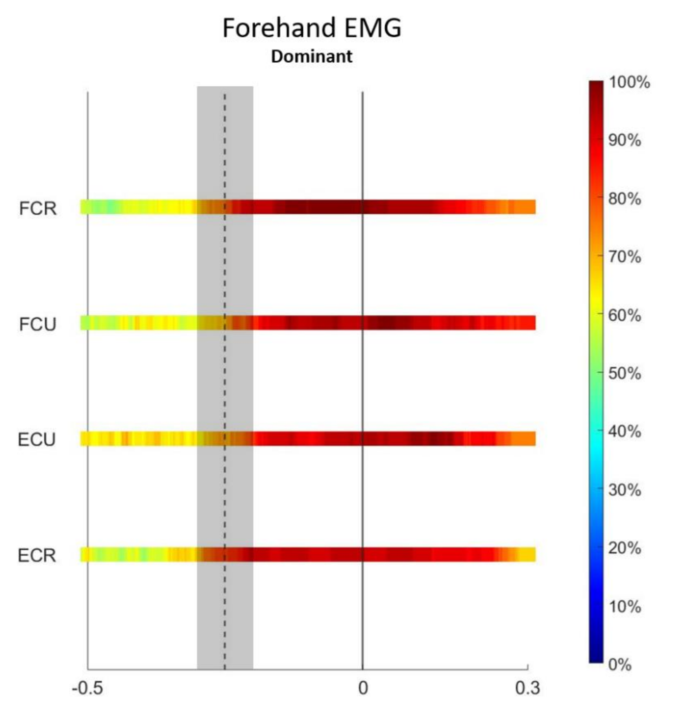
4.2. Forehand
5. Conclusions
Author Contributions
Funding
Institutional Review Board Statement
Informed Consent Statement
Data Availability Statement
Conflicts of Interest
References
- ITF Global Tennis Report 2021. Available online: http://itf.uberflip.com/i/1401406-itf-global-tennis-report-2021/0 (accessed on 19 October 2021).
- Abrams, G.D.; Renstrom, P.A.; Safran, M.R. Epidemiology of musculoskeletal injury in the tennis player. Br. J. Sports Med. 2012, 46, 492–498. [Google Scholar] [CrossRef] [PubMed]
- Pluim, B.M.; Staal, J.B.; Windler, G.E.; Jayanthi, N. Tennis injuries: Occurrence, aetiology, and prevention. Br. J. Sports Med. 2006, 40, 415–423. [Google Scholar] [CrossRef] [PubMed]
- Kibler, W.B.; Safran, M. Tennis injuries. Med. Sport Sci. 2005, 48, 120–137. [Google Scholar] [CrossRef] [PubMed] [Green Version]
- Gil, J.A.; Kakar, S. Hand and wrist injuries in tennis players. Curr. Rev. Musculoskelet. Med. 2019, 12, 87–97. [Google Scholar] [CrossRef] [PubMed]
- Stuelcken, M.; Mellifont, D.; Gorman, A.; Sayers, M. Wrist injuries in tennis players: A narrative review. Sports Med. 2017, 47, 857–868. [Google Scholar] [CrossRef] [PubMed]
- Montalvan, B.; Parier, J.; Brasseur, J.L.; Le Viet, D.; Drape, J.L. Extensor carpi ulnaris injuries in tennis players: A study of 28 cases. Br. J. Sports Med. 2006, 40, 424–429. [Google Scholar] [CrossRef] [PubMed]
- Bahamonde, R.E.; Knudson, D. Kinematic analysis of the open and square stance tennis forehand. Med. Sci. Sports 1998, 30, 29. [Google Scholar] [CrossRef]
- Hennig, E.M.; Rosenbaum, D.; Milani, T.L. Transfer of tennis racket vibrations onto the human forearm. Med. Sci. Sports Exerc. 1992, 24, 1134–1140. [Google Scholar] [CrossRef]
- Gruchow, H.W.; Pelletier, D. An epidemiologic study of tennis elbow. Incidence, recurrence, and effectiveness of prevention strategies. Am. J. Sports Med. 1979, 7, 234–238. [Google Scholar] [CrossRef]
- Kitai, E.; Itay, S.; Ruder, A.; Engel, J.; Modan, M. An epidemiological study of lateral epicondylitis (tennis elbow) in amateur male players. Ann. Chir. Main 1986, 5, 113–121. [Google Scholar] [CrossRef]
- Lambert, C.; Ritzmann, R.; Akoto, R.; Lambert, M.; Pfeiffer, T.; Wolfarth, B.; Lachmann, D.; Shafizadeh, S. Epidemiology of injuries in olympic sports. Int. J. Sports Med. 2021, 43, 473–481. [Google Scholar] [CrossRef] [PubMed]
- Elliott, B.C.; Marshall, R.N.; Noffal, G.J. Contributions of upper limb segment rotations during the power serve in tennis. J. Appl. Biomech. 1995, 11, 433–442. [Google Scholar] [CrossRef] [Green Version]
- Fleisig, G.; Nicholls, R.; Elliott, B.; Escamilla, R. Kinematics used by world class tennis players to produce high-velocity serves. Sports Biomech 2003, 2, 51–64. [Google Scholar] [CrossRef]
- Girard, O.; Micallef, J.P.; Millet, G.P. Lower-limb activity during the power serve in tennis: Effects of performance level. Med. Sci. Sports Exerc. 2005, 37, 1021–1029. [Google Scholar] [PubMed]
- Reid, M.; Whiteside, D.; Elliott, B. Effect of skill decomposition on racket and ball kinematics of the elite junior tennis serve. Sports Biomech. 2010, 9, 296–303. [Google Scholar] [CrossRef]
- Busuttil, N.A.; Reid, M.; Connolly, M.; Dascombe, B.J.; Middleton, K.J. A kinematic analysis of the upper limb during the topspin double-handed backhand stroke in tennis. Sports Biomech. 2020, 6, 1–19. [Google Scholar] [CrossRef]
- Dillman, C.J.; Fleisig, G.S.; Andrews, J.R. Biomechanics of pitching with emphasis upon shoulder kinematics. J. Orthop. Sports Phys. Ther. 1993, 18, 402–408. [Google Scholar] [CrossRef] [Green Version]
- Fisher, D.; Louw, Q.; Cockcroft, J.; Tawa, N. Three-dimensional kinematics of the thorax during over-ground running. J. Bodyw. Mov. Ther. 2018, 22, 300–307. [Google Scholar] [CrossRef]
- Rumpf, M.C.; Cronin, J.; Oliver, J.; Hughes, M. Sprint running kinematics and kinetics in pre-peak-height-velocity male children on a non-motorised treadmill: Reliability and normative data. Sports Biomech. 2019, 18, 256–263. [Google Scholar] [CrossRef]
- Werner, S.L.; Fleisig, G.S.; Dillman, C.J.; Andrews, J.R. Biomechanics of the elbow during baseball pitching. J. Orthop. Sports Phys. Ther. 1993, 17, 274–278. [Google Scholar] [CrossRef]
- Werner, S.L.; Jones, D.G.; Guido, J.A., Jr.; Brunet, M.E. Kinematics and kinetics of elite windmill softball pitching. Am. J. Sports Med. 2006, 34, 597–603. [Google Scholar] [CrossRef] [PubMed]
- Woltring, H.J. A Fortran package for generalized, cross-validatory spline smoothing and differentiation. Adv. Eng. Softw. 1986, 8, 104–113. [Google Scholar] [CrossRef]
- Wu, G.; van der Helm, F.C.; Veeger, H.E.; Makhsous, M.; Van Roy, P.; Anglin, C.; Nagels, J.; Karduna, A.R.; McQuade, K.; Wang, X.; et al. ISB recommendation on definitions of joint coordinate systems of various joints for the reporting of human joint motion--Part II: Shoulder, elbow, wrist and hand. J. Biomech. 2005, 38, 981–992. [Google Scholar] [CrossRef] [PubMed]
- Friston, K. Statistical Parametric Mapping: The Analysis of Functional Brain Images; Academic Press: London, UK, 2007; pp. 10–31. [Google Scholar] [CrossRef]
- Rogowski, I.; Creveaux, T.; Faucon, A.; Rota, S.; Champely, S.; Guillot, A.; Hautier, C. Relationship between muscle coordination and racket mass during forehand drive in tennis. Eur. J. Appl. Physiol. 2009, 107, 289–298. [Google Scholar] [CrossRef] [PubMed]
- Rota, S.; Morel, B.; Saboul, D.; Rogowski, I.; Hautier, C. Influence of fatigue on upper limb muscle activity and performance in tennis. J. Electromyogr. Kinesiol. 2014, 24, 90–97. [Google Scholar] [CrossRef]
- Elliott, B. Biomechanics and tennis. Br. J. Sports Med. 2006, 40, 392–396. [Google Scholar] [CrossRef]
- Blackwell, J.R.; Cole, K.J. Wrist kinematics differ in expert and novice tennis players performing the backhand stroke: Implications for tennis elbow. J. Biomech. 1994, 27, 509–516. [Google Scholar] [CrossRef]
- Zlatkin, M.B.; Rosner, J. MR imaging of ligaments and triangular fibrocartilage complex of the wrist. Radiol. Clin. North. Am. 2006, 44, 595–623, ix. [Google Scholar] [CrossRef]
- Tagliafico, A.S.; Ameri, P.; Michaud, J.; Derchi, L.E.; Sormani, M.P.; Martinoli, C. Wrist injuries in nonprofessional tennis players: Relationships with different grips. Am. J. Sports Med. 2009, 37, 760–767. [Google Scholar] [CrossRef]
- Tagliafico, A.; Rubino, M.; Autuori, A.; Bianchi, S.; Martinoli, C. Wrist and hand ultrasound. Semin. Musculoskelet. Radiol. 2007, 11, 95–104. [Google Scholar] [CrossRef]
- Rogowski, I.; Rouffet, D.; Lambalot, F.; Brosseau, O.; Hautier, C. Trunk and upper limb muscle activation during flat and topspin forehand drives in young tennis players. J. Appl. Biomech. 2011, 27, 15–21. [Google Scholar] [CrossRef] [PubMed] [Green Version]
- Rios-Russo, J.L.; Lozada-Bado, L.S.; de Mel, S.; Frontera, W.; Micheo, W. Ulnar-Sided Wrist Pain in the Athlete: Sport-Specific Demands, Clinical Presentation, and Management Options. Curr. Sports Med. Rep. 2021, 20, 312–318. [Google Scholar] [CrossRef] [PubMed]

| Swing Metrics | Two-Handed Backhand | Forehand | |
|---|---|---|---|
| TOTAL SWING TIME | 1.6 ± 0.4 s | 1.8 ± 0.2 s | |
| RACKET PREPARATION | 1.0 ± 0.3 s | 1.1 ± 0.2 s | |
| ACCELERATION | 0.2 ± 0.1 s | 0.3 ± 0.1 s | |
| FOLLOW-THROUGH | 0.4 ± 0.1 s | 0.4 ± 0.1 s | |
| KINEMATICS AT PEAK BACKSWING (°) | Non-Dominant | Dominant | Dominant |
| WRIST RADIAL–ULNAR DEVIATION | −13 ± 13° | −11 ± 12° | −16 ± 11° |
| WRIST FLEXION–EXTENSION | −43 ± 17° | 10 ± 20° | −35 ± 19° |
| FOREARM SUPINATION–PRONATION | −18 ± 25° | 11 ± 20° | −8 ± 17° |
| ELBOW FLEXION–EXTENSION | 77 ± 26° | 48 ± 16° | 69 ± 22° |
| KINEMATICS AT BALL IMPACT (°) | Non-Dominant | Dominant | Dominant |
| WRIST RADIAL–ULNAR DEVIATION | −18 ± 11° | −20 ± 13° | −23 ± 16° |
| WRIST FLEXION–EXTENSION | −42 ± 15° | 19 ± 14° | −41 ± 20° |
| FOREARM SUPINATION–PRONATION | −27 ± 24° | 7 ± 12° | −11 ± 15° |
| ELBOW FLEXION–EXTENSION | 69 ± 24° | 69 ± 19° | 73 ± 22° |
Publisher’s Note: MDPI stays neutral with regard to jurisdictional claims in published maps and institutional affiliations. |
© 2022 by the authors. Licensee MDPI, Basel, Switzerland. This article is an open access article distributed under the terms and conditions of the Creative Commons Attribution (CC BY) license (https://creativecommons.org/licenses/by/4.0/).
Share and Cite
Loushin, S.R.; Kakar, S.; Tetzloff, S.U.; Lubbers, P.; Ellenbecker, T.S.; Kaufman, K.R. Upper Extremity Kinematics and Electromyographic Activity in Uninjured Tennis Players. Appl. Sci. 2022, 12, 4638. https://doi.org/10.3390/app12094638
Loushin SR, Kakar S, Tetzloff SU, Lubbers P, Ellenbecker TS, Kaufman KR. Upper Extremity Kinematics and Electromyographic Activity in Uninjured Tennis Players. Applied Sciences. 2022; 12(9):4638. https://doi.org/10.3390/app12094638
Chicago/Turabian StyleLoushin, Stacy R., Sanjeev Kakar, Sabine U. Tetzloff, Paul Lubbers, Todd S. Ellenbecker, and Kenton R. Kaufman. 2022. "Upper Extremity Kinematics and Electromyographic Activity in Uninjured Tennis Players" Applied Sciences 12, no. 9: 4638. https://doi.org/10.3390/app12094638
APA StyleLoushin, S. R., Kakar, S., Tetzloff, S. U., Lubbers, P., Ellenbecker, T. S., & Kaufman, K. R. (2022). Upper Extremity Kinematics and Electromyographic Activity in Uninjured Tennis Players. Applied Sciences, 12(9), 4638. https://doi.org/10.3390/app12094638