Abstract
Staphylococcus bacteria are ubiquitous microorganisms. They occur in practically all environments. They also show the ability to colonize the skin and mucous membranes of humans and animals. The current trend is to look for new natural factors (e.g., plant extracts rich in polyphenols) limiting the growth of undesirable bacteria in food and cosmetics or use as an adjunct in antibiotic therapy. The aim of this study was to evaluate the effect of extracts from Rosa rugosa Thunb. on the antagonistic properties of selected lactic acid bacteria strains in relation to Staphylococcus spp. isolates. The biological material consisted of seven strains of lactic acid bacteria (LAB) and seven strains of bacteria of the Staphylococcus genus. The anti-staphylococcal properties of the Rosa rugosa Thunb. pomace extracts (the tested extracts were characterized by a high content of polyphenols, namely 8–34 g/100 g DM/dm) were tested using the well method. The conducted research showed that the pomace extracts of the pseudo-fruit (Rosa rugosa Thunb.) had the ability to inhibit the growth of Staphylococcus spp. bacteria. The minimum concentration of polyphenols inhibiting the growth of staphylococci was in the range of 0.156–0.625 mg/mL. The conducted research showed that combined lactic acid bacteria and extracts from the pomace from the pseudo-fruit Rosa rugosa Thunb. (LR systems) may be factors limiting the growth of Staphylococcus spp. bacteria. As a result of the research, two-component antagonist systems consisting of LAB cultures and extracts from Rosa rugosa Thunb. pomace were developed, which effectively limited the growth of the test strains of Staphylococcus spp. In 41% of all tested cases, the zone of inhibition of growth of bacteria of the genus Staphylococcus spp. after the use of two-component antagonist systems was higher than that as a result of the control culture (without the addition of extracts).
1. Introduction
Staphylococcus bacteria are ubiquitous microorganisms. They occur in practically all environments. They also show the ability to colonize the skin and mucous membranes of humans and animals. Often in the form of biofilm, they inhabit environmental, medical material and implant, and food production surfaces (polypropylene, polystyrene, and stainless steel) [1,2,3]. Staphylococcus spp. strains can be brought into food with raw materials and by humans participating in the production process. For example, enterotoxic strains of the genus Staphylococcus are often isolated from dairy products. Staphylococcus aureus is a widespread causative agent of mastitis in cattle [4]. In the process of obtaining milk from cows with staphylococcal mastitis, even with good filtration, the number of staphylococci in milk is 100–200 CFU/mL. In cases where the appropriate hygienic standards are not complied with during the milking of cows, this number may increase to 104–108 CFU/mL [5]. In the dairy industry, especially in the production of cheese (especially with low acidity products such as lump cheese), staphylococci may contaminate the final product. On the other hand, with the selection of inappropriate milk pasteurization temperatures, bacteria of the genus Staphylococcus are isolated directly from the raw material and the products for which it was used [6,7]. According to Asperger and Zangerl [8], staphylococci may also be present in cottage cheese, and their concentration depends on the number of bacteria in raw milk. Staphylococcus bacteria can also be a threat in many other industries. According to data presented by the European Union Rapid Information System for dangerous non-food products (Rapex), in 2005–2018, more than 100 cosmetics (including 20 for children) were withdrawn due to microbiological contamination. Although the majority of the infections were caused by gram-negative bacteria, it was found that in the group of gram-positive bacteria, S. aureus was most often responsible for contamination of the product (no data on whether the strain belongs to the methicillin-resistant species (MRSA) group) [9].
It should also be noted that a significant part of the world’s population (about 30%) are carriers of S. aureus in the nasopharyngeal cavity, which occasionally causes severe infections [10] but may be transferred to other environments, such as the food environment. It is not only S. aureus that can be transmitted by humans. This also applies to the skin microflora, where S. saprophyticus and S. epidermidis are a natural component of the human skin microbiota. Unfortunately, these strains may be opportunistic [11,12].
The constant exposure to antimicrobial drugs has led many microorganisms to develop antibiotic resistance mechanisms: changes in the permeability of cell membranes, pump efflux, enzymes that break down antibiotics, modification of the target site, or membrane vesicles. Multi-drug resistance (MDR) has become a global problem. Over the past 10 years, multi-drug resistance has increased significantly. Although MDR is a natural process, it makes disease-causing organisms resistant to many chemotherapeutic agents, resulting in increased mortality in patients. Thus, MDR is associated with fewer and fewer therapeutic chemicals. It is believed that traditional herbal medicines (rich in bioactive phytonutrients such as flavonoids and flavonols) may become an alternative to the use of chemical compounds. Thus, there is a great need for an alternative treatment strategy for staphylococcal infections. Resistance to antibiotics of bacteria of the genus Staphylococcus involves various mechanisms. Thus, strains resistant to methicillin, vancomycin, penicillin, linezolid, chloramphenicol, tetracycline, rifampicin, lincomycin, and drugs classified as cephalosporins, macrolides, and sulfonamides are isolated from the environment [13,14].
Given the current interest in finding alternative antimicrobial compounds to antibiotics, more and more research groups are exploring the antimicrobial potential of natural plant extracts. There are many reports in the literature on the antimicrobial properties of plant extracts (stems, roots, leaves, and flowers) [15]. The research group of Milala et al. [16] showed the antagonistic activity of pseudo-fruit extracts of three rose cultivars against the following strains: S. epidermidis A5, S. xylosus M5, S. haemolyticus M6, S. capitis KR6, and S. warneri KR2A, which were isolated from the food environment. The main advantage of plant extracts is the fact that they have a high content of phenolic compounds that can induce metabolic changes within the bacterial cell. First of all, polyphenols are capable of inducing cracks in the cell walls of bacteria (especially gram-positive species). The weakened cell wall is a pathway for intracellular components (such as proteins, enzymes, and ATP) to leak, resulting in abnormal replication processes. Moreover, the weakened bacterial cell is unable to efficiently synthesize the compounds necessary for the proper growth of biomass (impaired synthesis of enzymes, polysaccharides, and lipids). It has been observed that bacteria weakened by polyphenols are unable to grow and function properly. They often lose the ability to form biofilm and adhere to biotic surfaces [17].
Rosa rugosa belongs to the family Rosaceae Juss. and the genus Rosa L. The species is native to eastern Asia, China, Japan, Korea, and southeastern Siberia (Kamtschatka). Rugose rose has become a widely cultivated species all around the world, especially for production of flowers, fruit, and other parts of plants. Pseudo-fruits are valuable in processing for the preparation of jams, juices, sauces, syrups, wines, and jellies. Moreover, they are widely used to make infusion [18,19]. Chile is the main producer of rose hip material in the world market. Every year, nearly 8000 kg of Rosa mosqueta are exported from Chile. The United States is also one of the most important exporters of rose hips in the world. In the European market, the large producers of rose hips (200–1000 tons) are Sweden, Turkey, Hungary, and Germany. The commercial planting of wild rose in Poland consists of 1200 ha of Rosa rugosa with a yield of approximately 3000 tons per year [20].
Rose pseudo-fruits are known as a potential source of natural antioxidants and other bioactive substances, including polyphenols, carotenoids, polysaccharides, essential oils, polyunsaturated fatty acids (in seeds), vitamins A, B1, B2, B6, D, E, and K, and mineral nutrients [21]. The extracts obtained from Rosa spp. show not only high antioxidant activity but also antiallergic, antihypertensive, antidiabetic, antiinflammatory, antiviral, and antimicrobial properties [21,22]. Taking into account the valuable properties of rose extracts, pomace can be a good material for obtaining extracts rich in polyphenols. Rose extracts, which are characterized by high levels of polyphenols in their compositions, can also be a valuable source of substances with antimicrobial potential. Rosa spp. fruit extracts may have a different composition of polyphenols. Hvattum [23] identified in rose hip extracts inter alia the presence of anthocyanin, catechin, several glucosides of quercetin, glycosides of taxiofilin and eriodictyol phloridizin, and several conjugates of methyl gallate. However, in the extracts of Rosa canina, Ghendov-Mosanu et al. [24] identified compounds such as flavonoids, cinnamic acids, flavonols, procyanidin, chlorogenic acid, epicatechin, gallic acid, and catechin. The common feature of most phenolic compounds is the ability to inhibit the multiplication of pathogenic and nonpathogenic microorganisms and to slow down the oxidative processes in the food matrix. The bioactive compounds found in plants exhibit antioxidant properties, which lead to them being willingly used in the sector of natural preservation of food and cosmetics [25,26].
Due to the dynamic development of the functional food sector, lactic acid fermentation products combined with polyphenols began to appear on the market. Both metabolites of lactic acid bacteria (LAB) and secondary metabolites of plants also show high antimicrobial potential. Polyphenols largely affect the integrity of the cell membranes of pathogenic microorganisms. The rupture of cell membranes results in the leakage of many components necessary for the proper growth and development of microorganisms. By inducing changes in the metabolic processes of pathogenic microflora, polyphenols become a natural factor, limiting growth [27]. The antimicrobial mechanism of LAB action is mainly based on the synthesis of many specific metabolites. LAB are able to produce organic acids (mainly lactic acid, acetic acid, and propionic acid). They have the ability to produce compounds such as carbon monoxide (VI), hydrogen peroxide, diacetyl, and ethanol. In addition, many species show the ability to synthesize specific compounds with antimicrobial activity, such as bacteriocins, reuterin, and reutericycline [28,29]. Numerous in vitro studies have proven that LAB exhibit antioxidant activity, have the ability to chelate iron ions, and degrade nitrites and cholesterol [30,31]. LAB metabolites show antibacterial activity and are able to inhibit the multiplication of some fungi [32,33]. Lactic acid bacteria often show the ability to break down polyphenolic compounds (which has a positive effect on the consumer’s microflora). LAB contained in food, through the metabolism of plant compounds, provide the host organism with better absorbed (in the small intestine) compounds with antioxidant activity. As a result of this process, bacteria also gain access to substances with a prebiotic effect [34,35,36,37]. It is worth emphasizing that in the presence of polyphenols, LAB are able to synthesize lactic acid at a higher level, which at the same time positively influences the stabilization of phytochemicals. Due to the complex process of interaction between LAB and polyphenols, they can become a natural environmental system to fight staphylococci [38].
The aim of this study was to assess the anti-staphylococcal potential of lactic acid bacteria and extracts of Rosa rugosa Thunb. pseudo-fruit pomace both singly and in two-component antagonist systems. This goal was achieved by creating 28 2-component antagonist environmental systems (composed of LAB and Rosa rugosa Thunb. extracts).
2. Materials and Methods
2.1. Research Material
The research material consisted of four extracts (two water-ethanol extracts—crude (EC) and purified (EP)—and two water-acetone extracts: crude (AC) and purified (AP)) made of frozen Rosa rugosa Thunb. pseudo-fruit pomace. The exact preparation of the extracts was described by Piekarska-Radzik et al. [39]. For the frozen pomace of Rosa rugosa Thunb. pseudo-fruit, two portions of 2 kg (after the production of rose juice produced on the same day) (Polska Róża, Falenty New) were subjected to 0.5 h of water extraction at a ratio of 1:2 (v/v). Next, the pomace was extracted twice statically for 24 h with a 60% percent acetone solution or 60% ethanol at a ratio of 1:3 (v/v). The received ethanol and acetone extracts were concentrated in a vacuum evaporator (Hei-VAP Precision, Heidolph, Schwabach, Germany). The concentrated acetone or ethanol extracts were then divided into two parts. One of them was purified by column chromatography on Amberlite XAD 1600 N (divinylbenzene copolymer) (30.0 × 3.7 cm). The concentrated crude extracts were applied to the column (flow rate: 3–5 mL/min). Then, the column was washed with water, and the adsorbed polyphenols were eluted with 10% and 68% ethanol. The alcohol fractions were combined and concentrated on a Heidolph 24/7 automatic evaporator. The crude and purified extracts were freeze-dried using a freeze dryer (Christ, Alpha 1–2 LD plus, Osterode am Harz, Germany). In the obtained freeze-dried extracts, the content of the selected groups of polyphenols was determined by using HPLC chromatographic methods. The concentrations of ellagitannins, ellagic acid, and flavonols were determined in the extracts by dissolving 100 mg in 80% methanol in 10-mL volumetric flask, diluted, and analyzed as described by Karlińska et al. [40,41] and Sójka et al. [42]. Procyanidins and free catechin were determined as described by Milala et al. [16].
The tested extracts had the following composition. EC: total of measured polyphenolic compounds = 8.8 (g/100 g DM/dm), 4714.3 (mg/100 g DM/dm) including ellagitannins; free ellagic acid = 168.1 (mg/100 g DM/dm), flavonols = 357.5 (mg/100 g DM/dm), procyanidins = 3377.5 (mg/100 g DM/dm), free catechins = 196.2 (mg/100 g DM/dm). EP: total of measured polyphenolic compounds = 29.2 (g/100 g DM/dm), 15,379.7 (mg/100 g DM/dm) including ellagitannins; free ellagic acid = 496.6 (mg/100 g DM/dm); flavonols = 993.7 (mg/100 g DM/dm); procyanidins = 10,750 (mg/100 g DM/dm), free catechins = 1529.6 (mg/100 g DM/dm). AC: total of measured polyphenolic compounds = 14.9 (g/100 g DM/dm), of which ellagitannins = 8649.1 (mg/100 g DM/dm); free ellagic acid = 278.7 (mg/100 g DM/dm), flavonols = 603.2 (mg/100 g DM/dm); procyanidins = 5144.3 (mg/100 g DM/dm); free catechins = 205.5 (mg/100 g DM/dm). AP: total of measured polyphenolic compounds = 33.2 (g/100 g DM/dm), 17,680.9 (mg/100 g DM/dm) including ellagitannins; free ellagic acid = 645.8 (mg/100 g DM/dm); flavonols = 978.4 (mg/100 g DM/dm), procyanidins = 12,475.5 (mg/100 g DM/dm); free catechins = 1399.9 (mg/100 g DM/dm) [39].
2.2. Biological Material
The biological material consisted of six lactic acid bacteria strains deposited in the Pure Culture Collection of Industrial Microorganisms of the Institute of Fermentation Technology and Microbiology ŁOCK 105 (Łódź, Poland) and the Levilactobacillus brevis MG451814 strain, the nucleotide sequence which was deposited in the GenBank NCBI database. Due to the reclassification of the Lactobacillus genus carried out in 2020, the current and new taxonomic names of the strains used in the research are summarized in Table 1.

Table 1.
Change of the nomenclature and culture conditions of the lactic acid bacteria strains used in the research.
All strains of lactic acid bacteria (stored in MAST CRYOBANK; Mast Group Ltd., Merseyside, UK) were activated by transfer to a fresh Broth MRS medium (Merck, Darmstadt, Germany) and cultured at 30 °C or 37 °C (depending on the strain) for 24 h.
Seven strains of bacteria of the genus Staphylococcus were used as test strains. The five strains are environmental isolates. The strains were obtained by surface swabbing from the skin of healthy volunteers aged 24 to 30 years (2 female and 1 male). No invasive or inconvenient methods were used to collect the samples. In non-invasive sampling, our research institution (Institute of Fermentation Technology and Microbiology, Lodz University of Technology) did not require the approval of the ethics committee to conduct research using this microorganism in in vitro tests. All donors were volunteers who were informed about the planned analytical procedures and agreed to publication of the obtained data. The species affiliation of the Staphylococcus isolates was confirmed by molecular methods based on sequence analysis of the 16S ribosomal RNA gene. The nucleotide sequences of the strains obtained from the wild were deposited in the GenBank database (The National Center for Biotechnology Information). The species affiliation and accession numbers of the tested strains are presented in Table 2. In contrast, two strains, S. aureus ATCC 25923 and S. epidermidis DSMZ 3270, were used as reference strains in the tests of antagonist activity.
Table 2.
Species affiliations of environmental isolates of the genus Staphylococcus.
All strains of Staphylococcus spp. (stored in MAST CRYOBANK; Mast Group Ltd., Merseyside, UK) were activated by transfer to a fresh nutrient medium (Merck, Darmstadt, Germany) and culturing at 37 °C for 24 h.
2.3. The Minimum Concentration of Polyphenols (MIC) That Inhibits the Growth of Staphylococcus Bacteria
In order to determine the minimum concentration of polyphenols inhibiting the growth of bacteria of the genus Staphylococcus, a deep culture of the studied microorganisms was performed. For this purpose, 6 wells were cut (10 mm in diameter) into a plate from a 24-h culture of Staphylococcus bacteria (density 108 CFU/mL) with a sterile crank and removed with a sterile preparation needle. Added to the holes created in this way was 100 µL of appropriately diluted extracts from the pseudo-fruit pomace Rosa rugosa Thunb., with a content in the extracts in the range of 0.0006–2.5 mg/mL. The negative control was 5% DMSO. The plates were then placed in an incubator (37 °C) for 18–24 h. After this time, the zone of inhibition of growth of the bacteria of the Staphylococcus genus was measured. The results were given in mm ± standard deviation (SD). The lowest concentration of polyphenols at which growth inhibition of the test bacteria was still observed was assumed to be the MIC value (mg/mL).
2.4. Antagonistic Activity of Lactic Acid Bacteria
The antagonistic properties of lactic acid bacteria were investigated using the bar method [43,44]. The method is based on the observation of parallel growth of the strains (the indicator and the antagonistic ones). For this purpose, 1 mL was taken from a 24-h LAB culture with a density of 109 CFU/mL and plunged into a sterile Petri dish (by pouring over cooled MRS Agar, pouring it to the upper limit of the plate height). Next, from solidified MRS medium overgrown with LAB, 10-mm (diameter) bars were cut out and put on the prepared agar (nutrient agar, Merck) containing test microorganisms (Staphylococcus strain) (105–106 CFU/mL). The dishes were incubated at 37 °C for 24 h. Following incubation, the diameter of the test strain growth inhibition zone was measured. The results were given in mm as the mean value of three repetitions ± standard deviation. In this way, a control culture was obtained.
On the other hand, the two-component antagonist (LR) system was obtained by LAB culture with Rosa rugosa Thunb. pseudo-fruit pomace extracts. MRS Agar with the addition of Rosa rugosa Thunb. pseudo-fruit pomace extracts with a final polyphenol concentration of 0.156 mg/mL (the concentration did not inhibit LAB growth, as shown in the publication by Piekarska-Radzik et al. [39]). The plates prepared in this way were incubated for 24 h at the temperature optimal for the growth of each of the tested lactic acid bacteria strains. After incubation with a sterile cork, bars were cut from the plates and placed on the culture plates of the test Staphylococcus spp. isolates. Cultures of the test strains were prepared identically to those in Section 2.3. After the plates were incubated with the antagonist test, the zones of inhibition of growth of Staphylococcus spp. around the bars were measured. The results were given in mm as the mean value of three repetitions ± standard deviation.
2.5. The Acidity of Lactic Acid Bacteria
The effect of Rosa rugosa Thunb. pseudo-fruit pomace extracts on the acidic activity of the LAB was assessed by measuring the pH. The extracts were added to the stock culture in the Broth MRS medium bed so that the final concentration of polyphenols in each of the samples was 0.156 mg/mL. After inoculation by 1% (v/v) LAB inoculum (density of inoculum: 108–109 CFU/mL), the samples were cultivated for 48 h at the temperature optimal for LAB growth. After 24 and 48 h of culturing, the pH of the culture was tested (ELMETRON pH-meter, Poland, with a glass electrode (ELMETRON CP-411, Warsaw, Poland)).
2.6. Statistical Analysis
All experiments were performed in at least three independent replicates, and the result was reported as the mean value. For the obtained results, the value of the standard deviation was also calculated.
The obtained data were statistically analyzed using STATISTICA 12 software (StatStoft, San Francisco, CA, USA) using the one-way ANOVA and Tukey’s post hoc test with a confidence interval of p ≤ 0.05.
3. Results
3.1. The Minimum Concentration of Polyphenols That Inhibit the Growth of Staphylococcus Bacteria
The effect of Rosa rugosa Thunb. pseudo-fruit pomace extracts on the growth of bacteria of the genus Staphylococcus is presented in Table 3. Both the water-ethanol and water-acetone extracts showed a high antagonistic potential. The minimum concentration of polyphenols (MIC) inhibiting the growth of bacteria of the genus Staphylococcus was in the range of 0.156–0.625 mg/mL for most of the tested strains. In the case of the Staphylococcus epidermidis R1A strain, the MIC value of the water-ethanol extracts was above 2.5 mg/mL.
Table 3.
The minimum concentrations of polyphenols that inhibit the growth of Staphylococcus bacteria.
The sensitivity of the studied Staphylococcus spp. strains to the presence of polyphenols contained in the extracts of Rosa rugosa Thunb. pseudo-fruit pomace in the environment of their growth is an individual (strain) feature. Nevertheless, it should be noted that the type of extraction influences the antagonistic potential of the obtained extracts. The extracts obtained with the use of acetone showed a slightly higher antagonistic activity than the water-ethanol extracts. Purifying the extracts (regardless of the solvent used) also influenced the antagonistic activity of the obtained extracts.
3.2. Antagonistic Activity of Lactic Acid Bacteria
Table A1 (in Appendix A) and Figure 1 show the antagonistic activity of lactic acid bacteria against Staphylococcus spp. strains. The tested LAB showed a high antagonistic potential against the test bacteria of the Staphylococcus genus.
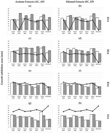
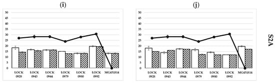
Figure 1.
Antagonistic activity of lactic acid bacteria and two-component LR systems against environmental isolates of the genus Staphylococcus.  : LR system with the addition of crude extracts;
  : LR system with the addition of purified extracts;
  : LAB basic culture. AC = crude water-acetone extract, AO = purified water-acetone extract, EC = crude water-ethanol extract, and EO = purified water-ethanol extract. (a,c,e,g,i) Antagonistic activity of two-component LR systems with the addition of acetone extracts and antagonistic activity of LAB stock culture. (b,d,f,h,j) Antagonistic activity of two-component LR systems with the addition of ethanol extracts and antagonistic activity of LAB stock culture. Lactic acid bacteria: Lactobacillus acidophilus ŁOCK 0928, Lacticaseibacillus rhamnosus ŁOCK 0943, Levilactobacillus brevis ŁOCK 0944, Lacticaseibacillus casei ŁOCK 0979, Levilactobacillus brevis ŁOCK 0980, Levilactobacillus brevis ŁOCK 0992, and Levilactobacillus brevis MG451814. Bacteria of the genus Staphylococcus spp. R1A: S. epidermidis R1A; R2A: S. haemolyticus R2A; R3A: S. saprophyticus R3A; S1A: S. saprophyticus S1A; S2A: S. aureus S2A).
: LR system with the addition of crude extracts; : LR system with the addition of purified extracts; : LAB basic culture. AC = crude water-acetone extract, AO = purified water-acetone extract, EC = crude water-ethanol extract, and EO = purified water-ethanol extract. (a,c,e,g,i) Antagonistic activity of two-component LR systems with the addition of acetone extracts and antagonistic activity of LAB stock culture. (b,d,f,h,j) Antagonistic activity of two-component LR systems with the addition of ethanol extracts and antagonistic activity of LAB stock culture. Lactic acid bacteria: Lactobacillus acidophilus ŁOCK 0928, Lacticaseibacillus rhamnosus ŁOCK 0943, Levilactobacillus brevis ŁOCK 0944, Lacticaseibacillus casei ŁOCK 0979, Levilactobacillus brevis ŁOCK 0980, Levilactobacillus brevis ŁOCK 0992, and Levilactobacillus brevis MG451814. Bacteria of the genus Staphylococcus spp. R1A: S. epidermidis R1A; R2A: S. haemolyticus R2A; R3A: S. saprophyticus R3A; S1A: S. saprophyticus S1A; S2A: S. aureus S2A).
Both the S. aureus ATCC 25923 and S. epidermidis DSMZ 3270 reference strains were sensitive to the metabolites of the LABs used. The growth inhibition zone for the ATCC 25923 strain was from 14.33 ± 0.58 mm to 21.00 ± 0.00 mm, while for the DSMZ 3270 strain, the values ranged from 17.22 ± 0.58 mm to 20.33 ± 0.58 mm. When analyzing the sensitivity of the environmental test strains of Staphylococcus spp. to the metabolites of the LAB tested, it was noted that the R1A strain showed resistance to the metabolites of two lactic acid bacteria strains: Lacticaseibacillus rhamnosus ŁOCK 0943 and Levilactobacillus brevis MG451814. In the case of the Levilactobacillus brevis MG451814 strain, its antagonistic potential was also limited in relation to the Staphylococcus spp. strains designated as S. haemolyticus R2A and S. aureus S2A. However, in the case of the test strain S. saprophyticus S1A, the strain MG451814 was characterized by the highest antagonistic activity (the zone of growth inhibition was 50.00 ± 0.00 mm). Moreover, it was found that the S1A strain was characterized by a higher sensitivity to the metabolites of the LAB bacteria studied (mean value of the growth inhibition, statistically significant) compared with the remaining Staphylococcus spp.
Table A2 (Appendix A) shows the antagonistic properties of the two-component LR systems. By combining 7 different LABs with four Rosa rugosa Thunb pseudo-fruit pomace extracts, 28 2-component LR systems were obtained, for which the anti-staphylococcal potential was tested (Figure 1). Each of the 28 LR systems was effective in inhibiting the growth of Staphylococcus bacteria. The anti-staphylococcal potential of the 28 LR systems (expressed in terms of growth inhibition zones) was in the range of 12–28 mm. Interestingly, each of the 28 systems inhibited the growth of the environmental isolates tested. This was especially important in relation to the control sample, in which in four cases saw the tested lactic acid bacteria not showing anti-staphylococcal properties. It should be noted that in these cases, the addition of extracts may positively affect the anti-staphylococcal properties of lactic acid bacteria.
In the case of the S. epidermidis R1A strain, the highest anti-staphylococcal activity was observed in the system consisting of the Lacticaseibacillus rhamnosus ŁOCK 0943 strain with the addition of purified acetone extract and Levilactobacillus brevis ŁOCK 0992 with the addition of purified ethanol extract (Figure 1a,b). Both zones of growth inhibition were close to 40 mm. On the other hand, the weakest effect limiting the growth of staphylococci was observed for systems based on the culture of Levilactobacillus brevis ŁOCK 0980 (regardless of the addition of the extract). The greatest antimicrobial potential was observed in LR systems based on the culture of Levilactobacillus brevis ŁOCK 0992 with crude extracts and Lacticaseibacillus rhamnosus ŁOCK 0943 with purified extracts. Due to the addition of acetone and ethanol extracts, the systems constructed on the basis of Lacticaseibacilluss brevis ŁOCK 0943 and Levilactobacillus brevis MG451814 cultures showed anti-staphylococcal potential (zone of growth inhibition in the range of 20–40 mm), despite the fact that the basic culture did not show anti-staphylococcal properties in this strain. Moreover, in most cases (20/28), the two-component LR systems showed a statistically significant higher antimicrobial potential than the stock culture (Figure 2 and Table A2, Appendix A). It should also be noted that neither of the systems exhibited a statistically significantly lower antimicrobial potential than the stock culture.
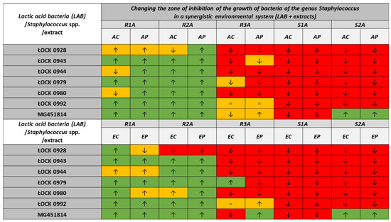
Figure 2.
The changes in the zones of growth inhibition of Staphylococcus bacteria under the influence of polyphenols contained in the Rosa rugosa Thunb. pseudo-fruit pomace extracts. The control sample was the anti-staphylococcal properties of lactic acid bacteria without the addition of Rosa spp. extracts (AC = crude water-acetone extract, AP = purified water-acetone extract, EC = crude water-ethanol extract, and EP = purified water-ethanol extract). Strains: Lactobacillus acidophilus ŁOCK 0928, Lacticaseibacillus rhamnosus ŁOCK 0943, Levilactobacillus brevis ŁOCK 0944, Lacticaseibacillus casei ŁOCK 0979, Levilactobacillus brevis ŁOCK 0980, Levilactobacillus brevis ŁOCK 0992, Levilactobacillus brevis MG451814, S. epidermidis R1A, S. haemolyticus R2A, S. saprophyticus R3A, and S. saprophyticus S1A, S. aureus S2A. The selected symbols show the difference in the size of the zone: “↓” = growth inhibition zone after using the LR system, lower than in the case of control culture; “↑” = growth inhibition zone after the application of the environmental LR system, higher than in the case of the control culture; and “=” = the zone of inhibition of growth after the application of the LR system, the same as in the case of control culture. Statistically significant changes are marked by different colors:  = statistically significant reduction in the size of the growth inhibition zone in the LR systems;
  = statistically significant increase in the size of the zones of growth inhibition in the LR systems;
  = no statistically significant differences in the size of the growth inhibition zones.
= statistically significant reduction in the size of the growth inhibition zone in the LR systems; = statistically significant increase in the size of the zones of growth inhibition in the LR systems; = no statistically significant differences in the size of the growth inhibition zones.
In the case of the S. haemolyticus R2A strain, it was not possible to clearly define which two-component LR system had the highest and lowest anti-staphylococcal potentials (Figure 1c,d). The highest anti-staphylococcal potential (33–38-mm growth inhibition zone) was demonstrated by the systems consisting of all tested extracts (both crude and purified) with the culture of Levilactobacillus brevis ŁOCK 0992, a combination of crude extracts (both ethanol and acetone) with the culture of Levilactobacillus brevis ŁOCK0944. The same anti-staphyloccal potential was observed in the culture of Levilactobacillus brevis ŁOCK0980 bacteria with the addition of purified acetone extract and the culture of Lacticaseibacillus rhamnosus ŁOCK 0943 bacteria with crude ethanol extract. On the other hand, the lowest antimicrobial potential in relation to the S. haemolyticus R2A strain (growth inhibition zone: 21–28 mm) was observed in the systems consisting of Levilactobacillus brevis MG451814 bacterial cultures with the addition of acetone extracts (crude and purified) and purified ethanol extract, as well as in combining the culture of Levilactobacillus brevis ŁOCK0980 with crude extracts (acetone and ethanol) and the culture of Lactobacillus acidophilus ŁOCK 0928 with purified acetone extract and crude ethanol extract. All tested extracts used to create the LR systems based on the Levilactobacillus brevis MG451814 bacterial culture showed antagonistic activity (21–28-mm growth inhibition zone), although the stock culture did not show anti-staphylococcal properties. Moreover, in the vast majority of cases (24/28), the addition of extracts resulted in a statistically significant increase in the anti-staphylococcal properties compared with the anti-staphylococcal properties of the stock culture (Figure 2 and Table A2, Appendix A). On the other hand, a statistically significant reduction in the antagonistic properties was observed only in the case of combining ethanol extracts with the Lactobacillus acidophilus ŁOCK0928 culture.
The studies of the antagonistic properties of the LR systems in the case of the S. saprophyticus R3A strain showed that the systems consisting of the Lactobacillus acidophilus ŁOCK 0928 strain (26–29-mm growth inhibition zone) and the systems constructed on the basis of the Levilactobacillus brevis ŁOCK 0992 strain (inhibition zone: 27–29 mm) were characterized by the highest antagonistic activity (Figure 1e,f). The lowest antagonistic activity (zone of growth inhibition in the range of 16–20 mm) was recorded for the system consisting of the Levilactobacillus brevis MG451814 culture and polyphenols contained in extracts from the Rosa rugosa Thunb. pseudo-fruit pomace. Low antimicrobial potential (growth inhibition zone: 17–19 mm) was also observed in the systems consisting of Levilactobacillus brevis ŁOCK 0980 cultures and cleaned crude ethanolic and acetone extracts, as well as for the system based on the culture of the Lacticaseibacillus casei ŁOCK 0979 strain and purified acetone extract (growth inhibition zone equal to 19.67 mm). It is worth mentioning that the addition of extracts to the culture of lactic acid bacteria would significantly affect their anti-staphylococcal properties in relation to the S. saprophyticus R3A strain (in 18/28 cases, the 2-component LR system showed statistically significantly lower anti-staphylococcal properties than the basic culture) (Figure 2 and Table A2, Appendix A).
In the case of the S. saprophyticus S1A strain, the highest antagonistic activity was recorded in the LR system consisting of Levilactobacillus brevis ŁOCK 0992 and polyphenols contained in the crude acetone extract (zone of growth inhibition equal to 36 mm) (Figure 1g). On the other hand, the system consisting of purified ethanol extract and the Levilactobacillus brevis MG4511814 strain was characterized by the lowest anti-staphylococcal activity (Figure 1h). In the case of the purified acetone extracts, it was not possible to clearly indicate which environmental system showed the greatest antimicrobial potential in relation to the S. saprophyticus S1A strain (combination of purified acetone extract with the Levilactobacillus brevis ŁOCK 0992 strain, with a growth inhibition zone equal to 27.67 mm, and the combination of purified acetone extract and the Lactobacillus acidophilus ŁOCK 0928 strain, with a growth inhibition zone equal to 28.33 mm). In the case of ethanol extracts, the highest anti-staphylococcal activity was obtained by combining them with the Levilactobacillus brevis ŁOCK 0992 strain (zone of growth inhibition equal to 30.33 mm). The system based on the culture of Lactobacillus acidophilus ŁOCK 0928 bacteria and crude ethanol extract (zone of growth inhibition equal to 27.33 mm) also showed a high antimicrobial potential. The weakest anti-staphylococcal properties in relation to S. saprophyticus S1A were demonstrated by the combination of acetone extracts with the Levilactobacillus brevis ŁOCK 0980 strain and ethanol extracts with the Levilactobacillus brevis MG451814 strain (zones of growth inhibition equal to 15–17 mm). It should be noted that the antimicrobial activity of the 28 LR systems against the S. saprophyticus S1A strain was largely due to the antagonistic activity of the LAB strains used to create the system. In all cases (28/28), the addition of the extract resulted in a statistically significant reduction in anti-staphylococcal properties compared with the stock culture (Figure 2).
The S. aureus S2A strain, compared with the other environmental isolates, was the most resistant to the effects of the two-component LR systems (Figure 1i,j). This was evidenced by the fact that the highest zone of inhibition of growth (not exceeding 20 mm) was comparable to the value of the minimum zones of inhibition of growth of other staphylococci. In the case of the S. aureus S2A strain, an anti-staphylococcal potential was observed in the systems consisting of the culture of Levilactobacillius brevis ŁOCK 0992 with acetone extracts (zone of growth inhibition of about 19 mm) and the culture of Levilactobacillus brevis MG451814 with the addition of ethanol extracts (zone of growth inhibition in the interval 17–20 mm). A moderate anti-staphylococcal potential in relation to the S. aureus S2A strain (growth inhibition zone in the range of 17–19 mm) was also noted in the case of the Lactobacillus acidophilus ŁOCK 0928 bacteria cultures with crude extracts (ethanol and acetone) and in the system consisting of Levilactobacillus brevis ŁOCK0944 cultures with the addition of purified ethanol extract. The lowest antimicrobial potential in relation to the S. aureus S2A strain was demonstrated by the two-component LR systems consisting of Levilactobacillus brevis ŁOCK 0992 cultures in combination with ethanol extracts (crude and purified) and the combination of purified ethanol extract with the Levilactobacillus brevis ŁOCK 0980 culture. A low anti-staphylococcal potential was also characterized by systems consisting of the Levilactobacillus brevis MG451814 and Levilactobacillus brevis ŁOCK 0980 cultures in combination with acetone extracts (growth inhibition zone equal to 13.33 mm), as well as a combination of purified acetone extract with Lacticaseibacillus casei ŁOCK 0979 and Lactobacillus acid ŁOCK 0928 (growth inhibition zone in the range of 13–15 mm). It is worth noting that although 28 2-component LR systems were obtained to inhibit the growth of S. aureus S2A bacteria, the overall antagonistic activity of the LR systems toward this strain should be assessed as low. Four systems based on the Levilactobacillus brevis MG451814 strain showed anti-staphylococcal activity, although it was not observed in the control sample.
Out of all 28 LR systems tested, the strongest antagonistic activity was observed in relation to the S. epidermidis R1A strain using a system consisting of purified acetone extract and the Lacticaseibacillus rhamnosus ŁOCK 0943 strain. On the other hand, the weakest antagonistic properties of the LR systems were observed in the case of S. aureus S2A (a combination of both ethanol extracts with the Levilactobacillus brevis ŁOCK 0992 strain and a combination of purified ethanol extract with the Levilactobacillus brevis ŁOCK 0980 strain). Of all the environmental isolates tested, S. epidermidis R1A and S. haemolyticus R2A turned out to be the most sensitive strains to the effects of environmental systems consisting of polyphenols and LAB cultures. It is worth emphasizing that in the case of the antagonistic properties of the two-component LR systems, the antimicrobial activity against the S. epidermidis R1A and S. haemolyticus R2A strains was largely due to the addition of extracts. On the other hand, the S. aureus S2A strain was most resistant to the antagonistic effects of co-cultures of LABs and polyphenols.
Figure 2 shows the differences in the size of the zones of inhibition of the growth of bacteria of the genus Staphylococcus caused by the action of LR systems in relation to the antagonistic properties of basic lactic acid bacteria cultures. The combination of lactic acid bacteria and polyphenols contained in the Rosa rugosa Thunb. pseudo-fruit pomace in a common culture system had a positive or negative effect, depending on the tested strain of the genus Staphylococcus. It is interesting that the lack of statistically significant differences in the operation of the LR system accounted for only 15% of all examined cases. Among the results with a statistically significant difference, nearly 41% showed a favorable effect from polyphenols on the LAB antagonistic properties (increase in the zone of growth inhibition). LR systems consisting of LAB and rose extracts significantly influenced the growth of the isolates S. epidermidis R1A and S. haemolyticus R2A. Both strains were more sensitive to the effects of LAB systems in combination with ethanol and acetone extracts than in the case of stock cultures. In the case of the remaining isolates (S. saprophyticus R3A, S. saprophyticus S1A, and S. aureus S2A), the use of LR systems resulted in a statistically significant reduction in the growth inhibition zones compared with the control sample. It is worth noting, however, that in the case of the LR systems, in each of the studied cases, the anti-staphylococcal properties of the systems were observed (while not all LAB strains showed anti-staphylococcal activity). Only in 2% of the cases did the addition of polyphenols to LAB cultures not change their anti-staphylococcal potentials. It should be noted that in the LR systems, a concentration of polyphenols equal to 0.156 mg/mL was used, which for 14 out of 20 cases permitted the use of a concentration lower than the MIC value for the Staphylococcus spp. strains tested.
3.3. Change in the pH of the LAB Culture in the Presence of Rosa rugosa Thunb. Pseudo-Fruit Pomace Extracts
The changes in the pH value during the culture of seven LAB strains with the addition of polyphenols contained in Rosa rugosa Thunb. pseudo-fruit pomace extracts are presented in Table 4. The extracts in the concentration of 0.156 mg/mL (converted to the concentration of polyphenols) added to the initial culture (t = 0 h) did not significantly affect the pH value in any tested cases (no statistically significant differences). At the time of inoculation, the lowest pH was observed in the culture of Lactobacillus acidophilus ŁOCK 0928 (5.2–5.3). On the other hand, the starter culture of the Levilactobacillus brevis MG451814 strain was characterized by the highest pH value (5.7). The pH value at the time of inoculation in the case of the remaining cultures fluctuated in the range of 5.4–5.6. During the 48-h incubation (under optimal conditions for the growth of each of the microorganisms), the pH value of the culture gradually decreased. For all the tested strains, the highest decrease in the pH value was observed after the first day of culturing Lacticaseibacillus rhamnosus ŁOCK 0943 with the addition of crude ethanol extract (∆pH = 1.5 units). In turn, the lowest decrease in the pH value was recorded after a 24-h culture of Levilactobacillus brevis ŁOCK 0980 bacteria with the addition of crude ethanol extract (∆pH = 0.7 units). On the second day, the decrease in pH was much lower in all cases and did not exceed ∆ = 0.3 units. The highest decrease in pH value was observed in the culture of the Levilactobacillus brevis ŁOCK 0980 strain with the addition of crude ethanol extract (∆pH = 0.25 units). On the other hand, in the case of Levilactobacillus brevis ŁOCK 0944 with the addition of crude ethanol extract and the Lacticaseibacillus casei ŁOCK 0979 and Lactobacillus acidophilus ŁOCK 0928 cultures with the addition of purified acetone extract, the decrease in pH was practically imperceptible (∆pH = 0.02 units).
Table 4.
Changes in pH of 48-h LAB cultures in the presence of Rosa rugosa Thunb. pseudo-fruit pomace extracts.
It should be noted, however, that extending the culture from 24 to 48 h for all 35 cultures resulted in a significant decrease in the pH value, and in 63% of cases, this value was statistically significant (in all cultures with the addition of purified ethanol extract and almost all control cultures, where the exception was Levilactobacillus brevis ŁOCK 0992).
After 48 h of incubation, the pH of the tested cultures was the highest for the Levilactobacillus brevis MG451814 strain in the culture with the addition of purified ethanol extract. It should be noted that the observed pH value (4.67) was statistically different from the pH value in the stock culture without the addition of extracts. However, there were no statistically significant differences with the pH value in the culture with crude acetone extract. On the other hand, the lowest pH value after 48 h of incubation was observed for the Lactobacillus acidophilus ŁOCK 0928 strain grown with the addition of crude extracts (3.97). However, this difference was not statistically significant compared with the cultures with purified extracts.
4. Discussion
In recent years, a systematic increase in the number of microorganisms resistant to routinely used drugs has been observed. The spread of antibiotic resistance among staphylococci (including coagulase-negative strains) may pose a threat to human health. Many scientists believe that food may be the habitat of multi-drug resistant strains of the genus Staphylococcus and may be the transfer of antibiotic resistance between the environment and people [45]. The substrate for the development of antibiotic-resistant bacteria can be, among others, ready-to-eat, fermented milk products (for the preparation of which unpasteurized milk is used) [46]. Currently, it is methicillin resistance that is considered to be one of the most important problems in the control of staphylococci. Methicillin-resistant species are often phenotypically resistant to other β-lactam antibiotics used so far (including penicillin, oxacillin, nafcillin, or cephalosporins) [47]. According to the research conducted by Chajęcka-Wierzchowska et al. [47], more than 83% of methicillin-resistant Staphylococcus isolates (from ready-to-eat food) also showed resistance to tetracycline, rifampicin, and clindamycin. Moreover, the studies by Chajęcka-Wierzchowska et al. [47] showed that over 30% of food isolates are strains that have experienced multi-drug resistance (antibiotic resistance belonging to three or more classes). Hence, the aim of this study was to understand the anti-staphylococcal properties of four polyphenol-rich extracts from Rosa rugosa Thumb. pseudo-fruit pomace and seven strains of lactic acid bacteria.
The tested extracts from the pseudo-fruit pomace of Rosa rugosa Thunb. were characterized by a total content of polyphenols at the level of 8–33 mg per 100 g DM. The content of polyphenols in the extracts of rose pseudo-fruit depended primarily on the species of rose but also on the method of extraction. The extracts obtained from the pomace through the use of various solvents differed in the content of polyphenols. The content of polyphenols in the acetone extracts was higher than in the ethanolic extracts by 1.69 times (crude) and 1.14 times (purified). Ellagitannins and procyanidins dominated in all obtained extracts, and their shares were similar. According to research by Yi et al. [48], the methanol extracts of Rosa nutkana, Rosa pisocarpa, and Rosa woodsii contained 6–13 mg of polyphenols (GAE equivalent) per liter of extract. On the other hand, Nowak and Gawlik-Dziki [49] determined the content of polyphenols in 17 different species of roses (including Rosa canina, Rosa rugosa, Rosa villosa, and Rosa gallica). The total content of polyphenols in the extracts was in the range of 6–16% dry matter (which was 5–19 mg of flavonols per g of dry matter and 9–20 mg of ellagic acid per g of dry matter). The number of polyphenols (and their types) contained in rose extracts is primarily influenced by the part of the plant that was used to produce them. For example, the concentration of polyphenols in the extracts of the Rosa nutkana and Rosa woodsii rose hips obtained by Yi et al. [48] was higher than in the seed extracts.
Four extracts of pomace from Rosa rugosa Thunb. pseudo-fruit were used in the study, for which the antagonistic activity against Staphylococcus spp. was tested. The tested extracts, with a concentration of polyphenols ranging from 0.156 to 0.625 mg/mL, effectively inhibited the growth of the tested Staphylococcus bacteria. The research group of Yi et al. [48] investigated the antimicrobial potential of rose extracts (sourced from British Columbia (Canada)). The extracts inhibited the growth of gram-positive bacteria Enterococcus faecalis, Bacillus subtilis, S. aureus ATCC 25923, and methicillin-resistant clinical staphylococcal isolates. The studies of Yi et al. [48] showed that rose hip extracts demonstrate selective antimicrobial activity. The mean zone of inhibition of growth (including the diameter of the disc (6 mm)) of S. aureus ATCC 25923 bacteria was in the range of 9–15 mm in the case of whole extracts and in the range of 11–18 mm in the case of pericarp extracts (with the addition of 20 μL of extract per filter paper disc). Cendrowski et al. [21] studied the antibacterial properties of extracts made from the Rosa rugosa pseudo-fruit. For this purpose, they prepared various types of extracts (aqueous, ethanolic, supercritical, and enzymatic). The aqueous extracts showed antimicrobial activity against gram-positive bacteria such as Bacillus cereus or S. epidermidis and against gram-negative species (Escherichia coli). Interestingly, the minimum inhibitory concentration (MIC) of the tested aqueous extracts for S. aureus was over 128 mg/mL. Ethanol extracts strongly inhibited the growth of Bacillus subtilis bacteria, but only at a concentration of 32 mg/mL did they limit the growth of S. epidermidis (the MIC for the aqueous extracts was 16 mg/mL). Cendrowski et al. [21] did not determine the concentration of the Rosa rugosa ethanol extracts, which would have a bacteriostatic effect on staphylococci (the MBC exceeded the range of tested concentrations at 128 mg/mL). In contrast, the MIC of the enzymatic and supercritical extracts for S. aureus ATCC 25923 was 64 mg/mL. In the research conducted by the authors of the present work, two different solvents were used to extract polyphenols from rose pomace: ethanol and acetone. The highest concentration of polyphenols used was 2.5 mg/mL. Thus, it was the concentration in the case of ethanol extracts that turned out to be insufficient for limiting the growth of only one of the tested strains (S. epidermidis R1A). It should be noted, however, that the concentration of polyphenols of 2.5 mg/mL corresponded to the concentration of the ethanol extracts at the level of 8–30 mg/mL (depending on the degree of purification). Thus, the result obtained by the authors of the study is comparable to the anti-staphylococcal properties of rose extracts determined by Cendrowski et al. [21]. In the analyses by Adwan et al. [50], the effect of plant extracts on the growth of microorganisms was also investigated. One of the extracts was made from damask rose. The influence of the extract on the growth of the gram-positive bacteria S. aureus was analyzed. The ethanol extract of damask rose showed stronger bactericidal activity against staphylococci than the other extracts tested: lemon balm, mint, and marshmallow (MIC: 0.395–0.780 mg/mL). It should also be noted that rose extracts sometimes show stronger bactericidal properties compared with extracts prepared from other plants. The results obtained by Mishra et al. [51] showed that extracts (prepared with methanol and ethyl acetate) obtained from various parts of Rosa indica (leaves, stem, and flowers) also showed bactericidal activity against microorganisms considered pathogenic. All methanol extracts showed anti-staphylococcal properties against S. aureus. The greatest zone of inhibition of growth was recorded after the application of methanolic extract from flowers and stems (16–17 mm). The leaf extracts showed a lower antagonistic property: a growth inhibition diameter of 13 mm. Thus, the MIC of each of the Rosa indica extracts was 4.5 mg/mL. It is worth mentioning that the method of extracting polyphenols has a decisive influence on the antagonistic properties of Rosa spp. fruit extracts. Two solvents were used in this research: ethanol and acetone. The extracts were further purified by column chromatography on Amberlite XAD 1600 N. Stronger anti-staphylococcal activity was observed in the extracts obtained with the use of acetone and the purified extracts. Interestingly, according to the literature data, the ethanol extracts showed a higher antimicrobial potential. Halawani [52] not only investigated the antimicrobial properties of Rosa damascena extracts but also tried to determine the effect of the extraction solvent on the antimicrobial properties of the extracts (water, methanol, ethanol, and hexane). Their research showed that the highest anti-staphylococcal properties were obtained in the case of ethanol extracts (growth inhibition zone in the range of 27–30 mm; MIC: 62.5 µg/mL). A similar antimicrobial activity was observed in water (MIC: 62.5–125 µg/mL) and methanol (MIC: 62.5–250 µg/mL) extracts. The weakest antimicrobial property was observed in extracts prepared with the use of hexane. Shohayeb et al. [53] used many popular solvents to prepare Rosa damascena flower extracts (water, hexane, and ethanol and alcoholic extracts were suspended in chloroform, butanol, and ethyl acetate). Then, the bactericidal properties of the extracts against Streptococcus pyogenes, B. subtilis, and S. aureus were examined. The MIC value for the S. aureus strain ranged from 0.125 to 2.0 mg/mL. Nevertheless, the methanol and acetone extracts of Rosa damascena prepared by El-Shouny et al. [54] also demonstrated anti-staphylococcal properties. The antagonistic activity of the extracts at a concentration of 100 mg/mL resulted in the formation of zones of growth inhibition of S. aureus isolates at the level of 11–18 mm for the methanol extract and 14–18 mm for the acetone extract.
There are also reports in the literature examining the antimicrobial potential of roses as a by-product in the food industry. An example of a product that is waste from the production of fruit juices is pomace. The work investigated extracts made of frozen Rosa rugosa Thunb. pseudo-fruit pomace resulting from the production of natural juices. Ren et al. [55] also used an extract from Rosa rugosa Thunb. var. plena Regal (by-product of production of rose tea or dried rose petals; Fragrant Rose Biological Technology Co., LTD in Pingyin) for testing antimicrobial properties. The total content of the compounds in the extract was divided into phenolics (0.31 mg/mL) and flavonoids (0.43 mg/mL). The three main components of the extract are hyperoside, kaempferol-3-O-rutoside, and rutin. Rosa rugosa Thunb. var. plena Regal extract showed antimicrobial properties against Cutibacterium acnes (called Propionibacterium acnes by Ren, et al. [55]) and S. aureus (MIC: 125 µg/mL). For the remaining bacteria (e.g., Listeria ivanovii, Salmonella Enteritidis, or E. coli) the MIC value of the tested extract exceeded 250 µg/mL.
Lactic acid produced as a result of fermentation makes LAB bacteria antagonistic to pathogenic microorganisms that pollute not only food but also other ecological niches [56].
The selection of an appropriate carbohydrate in the medium may increase the acidifying activity. Therefore, the addition of polyphenols from Rosa rugosa Thunb. pseudo-fruit pomace extracts in a concentration below 0.156 mg/mL does not affect the metabolic processes leading to acidification of the environment by the tested LAB bacteria. Yoon et al. [57] also proved that lowering the pH of LAB cultures is an obvious consequence of metabolic processes. The pH of Lactobacillus casei (Lacticaseibacillus casei) cultures decreased from 5 to 3.4, while in the case of Lactobacillus plantarum (Lactiplantibacillus plantarum) and Lactobacillus delbruecki subsp. delbruecki, the pH of the environment decreased from 5.8 to 3.6.
The tolerance of LAB to low pH levels is therefore a strain feature. It is worth noting that the intracellular pH of the LAB drops simultaneously with the decrease in the extracellular pH, which is an individual feature compared to other bacterial cells (e.g., pathogenic microorganisms) and may constitute the basis for their antimicrobial activity [58]. Measurement of the pH of LAB cultures is an indirect method for monitoring the acidity of the strains used for fermentation. Ren et al. [55] showed that the amount of acid (pH < 3.7) synthesized by two strains, T30 and S6, was higher than in the case of other LAB strains, for which the pH value was higher than 3.7 (the tested LAB strains belonged to the following species: Lactobacillus plantarum, Lactobacillus pentosus, and Lactobacillus paracasei (Lactiplantibacillus plantarum, Lactiplantibacillus pentosus, and Lacticaseibacillus paracaei)). The strains of lactic acid bacteria studied by the authors of the present study were characterized by a much higher antimicrobial potential compared with environmental isolates of the Staphylococcus genus and reference strains. The observed diameters of growth inhibition were in the range of 14–50 mm. It should be emphasized that not all lactic acid bacteria show anti-staphylococcal properties. The tested Levilactobacillus brevis MG 451,814 strain was not able to inhibit the growth of the following isolates: R1A, R2A, and S2A. However, in the case of the Lacticaseibacillus rhamnosus ŁOCK 0943 strain and the R1A isolate, the zones of growth inhibition were not determined either. Karska-Wysocki et al. [59], during their study of the antimicrobial potential of Lactobacillus acidophilus, Lactobacillus casei (Lactocaseibacillus casei), and Lactococcus cremoris in relation to 10 isolates of the genus Staphylococcus (methicillin-resistant strains) and the reference strain of S.aureus ATCC 43300, did not observe the anti-staphylococcal potential of Lactcoccus cremoris, whereas Mohamed et al. [60] did not demonstrate the anti-staphylococcal properties of Lactobacillus reuteri ATCC 55730 (Limosilactobacillus reuteri) (regardless of aerobic conditions).
During the present study, it was decided to create two-component antagonist environmental systems. The studies of Fang et al. [61] showed no effect of the base medium on the antagonistic activity of lactic acid bacteria. Therefore, a commercial MRS Agar (Merck, Germany) was used for the preparation of systems, which is optimal for the growth of lactic acid bacteria. LR systems were created by combining polyphenols contained in extracts of pseudo-fruit pomace Rosa rugosa Thunb. and selected strains of lactic acid bacteria. Earlier studies have shown that polyphenols at a concentration of 0.156 mg/mL have a prebiotic effect on the microorganisms used in the study [39]. It is worth noting that other scientists also looked for plant extracts that would inhibit the growth of pathogenic microflora while not limiting the growth of lactic acid bacteria. Chan et al. [62] investigated the potential of spice and herb extracts (e.g., Japanese knotweed, pomegranate peel, and cloves). The obtained extracts showed the ability to combat five species of pathogenic bacteria (B. cereus, E. coli, Salmonella enterica subsp. enterica, Shigella flexneri, and S. aureus). Thus, the extracts showed a prebiotic effect for four LAB species (the prebiotic effect was tested for the following strains of lactic acid bacteria: Lactobacillus acidophilus, Lactobacillus casei (Lacticaseibacillus casei), Lactobacillus plantarum (Lactiplantibacillus plantarum), and Lactobacillus rhamnosibus (Lacticaseibacillus rhamnosus)). In our work, we managed to compose antagonistic two-component antagonist systems based on the culture of selected LAB and pseudo-fruit pomace extracts from Rosa rugosa Thunb., which effectively limited the growth of both collection strains and environmental isolates belonging to the genus Staphylococcus spp.
5. Conclusions
Test extracts of pseudo-fruit pomace Rosa rugosa Thunb. constitute a valuable reservoir of compounds with anti-staphylococcal activity. The LR systems developed in the study showed a high antagonistic potential toward Staphylococcus spp. strains. Nevertheless, it should be noted that the effectiveness of the LR system depends on the LAB bacterial strain but above all on the sensitivity of the Staphylococcus spp. The extraction environment and the level of preparation purification are also important factors.
The creation of environmental two-component antagonist systems, in this case consisting of LAB and polyphenols from Rosa spp. (LR system), may therefore represent the beginning of the development of a new trend in the production of functional food or cosmetics or in the design of dietary supplements.
Author Contributions
Conceptualization, E.K., R.K. and J.M.; methodology, L.P.-R., J.M., M.S. and R.K.; formal analysis, L.P.-R., N.R., J.M., A.O. and M.S.; preparing plant material for research, B.M. and N.R.; writing—original draft preparation, L.P.-R., J.M. and E.K.; writing—review and editing, L.P.-R., E.K., R.K., M.S., B.M. and A.O.; visualization, L.P.-R.; supervision, E.K.; project administration, E.K. All authors have read and agreed to the published version of the manuscript.
Funding
This work was financed through statutory funds from the Institute of Food Technology and Analysis and the Institute of Fermentation Technology and Microbiology of the Faculty of Biotechnology and Food Sciences at Lodz University of Technology in Poland.
Institutional Review Board Statement
Not applicable.
Informed Consent Statement
Not applicable.
Data Availability Statement
The data presented in this study are available on request from the corresponding author.
Conflicts of Interest
The authors declare no conflict of interest.
Appendix A
Table A1 shows the antagonistic activity of lactic acid bacteria against Staphylococcus spp. strains. The tested LABs showed high antagonistic potential against the test bacteria of the Staphylococcus genus. The mean values of the growth inhibition of Staphylococcus spp. showed no statistically significant differences. Nevertheless, it should be remembered that the antagonistic interactions between antagonistic LABs and the Staphylococcus spp. test strains were highly individual. Table A2 shows the antagonistic properties of the LR systems.
Table A1.
Antagonistic activity of lactic acid bacteria.
Table A1.
Antagonistic activity of lactic acid bacteria.
| Growth Inhibition Zone (mm) | |||||||
|---|---|---|---|---|---|---|---|
| ATCC 25923 | DSMZ 3270 | R1A | R2A | R3A | S1A | S2A | |
| ŁOCK 0928 | 17.00 ± 0.00 Aa | 20.33 ± 0.58 Ab | 24.00 ± 1.00 Ac | 25.67 ± 0.58 Ad | 30.00 ± 0.00 Ee | 36.67 ± 0.58 Af | 27.00 ± 0.00 ACd |
| ŁOCK 0943 | 19.67 ± 0.58 Ba | 18.33 ± 0.58 Ba | 0.00 ± 0.00 Bb | 19.33 ± 0.58 BCa | 25.67 ± 0.58 Ac | 42.33 ± 1.53 Bd | 28.33 ± 1.53 ACc |
| ŁOCK 0944 | 19.67 ± 0.58 Babd | 17.67 ± 0.58 Bb | 27.33 ± 1.53 Cc | 21.00 ± 0.00 Bd | 28.33 ± 1.53 Ec | 37.33 ± 1.15 Ae | 28.33 ± 1.15 ACc |
| ŁOCK 0979 | 14.33 ± 0.58 Ca | 15.33 ± 0.58 Ca | 22.33 ± 0.58 ADEbc | 20.00 ± 1.00 Bb | 23.67 ± 0.58 BCc | 38.33 ± 1.53 ADd | 24.00 ± 0.00 Bc |
| ŁOCK 0980 | 21.00 ± 0.00 Da | 18.33 ± 0.58 Ba | 20.33 ± 0.58 Da | 17.67 ± 1.15 Ca | 25.67 ± 0.58 Cbd | 41.46 ± 1.53 BDc | 28.00 ± 0.00 Cd |
| ŁOCK 0992 | 20.33 ± 0.58 BDda | 18.33 ± 0.58 Bb | 23.67 ± 0.58 AEc | 21.00 ± 1.00 Ba | 27.67 ± 0.58 Ed | 37.67 ± 0.58 Ae | 30.67 ± 0.58 Df |
| MG451814 | 17.00 ± 0.00 Aa | 17.33 ± 0.58 Ba | 0.00 ± 0.00 BFb | 0.00 ± 0.00 Db | 18.00 ± 1.00 Da | 50.00 ± 0.00 Cc | 0.00 ± 0.00 Eb |
| Mean | 18.43 ± 2.21 | 17.93 ± 1.39 | 16.81 ± 10.81 | 17.81 ± 7.62 | 25.57 ± 3.64 | 40.54 ± 4.34 # | 23.76 ± 9.87 |
A, B, C, D, E, F: statistical differences (ANOVA, Tukey’s post-hoc test (p ≤ 0.05)) between zones of growth inhibition within the same strain of Staphylococcus spp.; a, b, c, d, e, f: statistically significant differences (ANOVA, Tukey’s post-hoc test (p ≤ 0.05)) between the zone of growth inhibition within the same LAB strain; #: statistical difference between mean values for Staphylococcus spp. strains (ANOVA, Tukey’s post-hoc test (p ≤ 0.05). Strains: Lactobacillus acidophilus ŁOCK 0928, Lacticaseibacillus rhamnosus ŁOCK 0943, Levilactobacillus brevis ŁOCK 0944, Lacticaseibacillus casei ŁOCK 0979, Levilactobacillus brevis ŁOCK 0980, Levilactobacillus brevis ŁOCK 0992, Levilactobacillus brevis MG451814, S. aureus ATCC 25923, S. epidermidis DSMZ 3270, S. epidermidis R1A, S. haemolyticus R2A, S. saprophyticus R3A, S. saprophyticus S1A, and S. aureus S2A.
Table A2.
Antagonistic activity of LR systems: LAB with addition of Rosa rugosa Thunb. pseudo-fruit pomace extracts.
Table A2.
Antagonistic activity of LR systems: LAB with addition of Rosa rugosa Thunb. pseudo-fruit pomace extracts.
| Extract or LAB | AC | AP | EC | EP |
|---|---|---|---|---|
| Growth inhibition zone (mm): S. epidermidis R1A | ||||
| ŁOCK 0928 | 24.67 ± 0.58 Aa* | 25.33 ± 1.53 Aa* | 28.00 ± 1.00 Ab* | 23.33 ± 0.58 Ba* |
| ŁOCK 0943 | 24.00 ± 1.00 Aa* | 39.67 ± 0.58 Ba* | 32.33 ± 1.53 Ab* | 34.67 ± 0.58 Ab# |
| ŁOCK 0944 | 26.67 ± 0.58 Aa* | 34.33 ± 1.53 Ba* | 29.00 ± 1.00 Aa# | 30.33 ± 0.58 Ab# |
| ŁOCK 0979 | 27.33 ± 1.53 Aa* | 37.33 ± 0.58 Ba* | 31.00 ± 1.00 Ab# | 27.33 ± 0.58 Bb* |
| ŁOCK 0980 | 18.33 ± 1.53 Aa* | 23.33 ± 0.58 Ba* | 26.00 ± 1.00 Ab# | 21.67 ± 0.58 Ba# |
| ŁOCK 0992 | 31.67 ± 0.58 Aa* | 36.33 ± 1.53 Ba* | 33.00 ± 1.00 Aa# | 39.00 ± 1.00 Ba# |
| MG451814 | 23.67 ± 1.53 Aa* | 30.00 ± 1.00 Ba* | 29.67 ± 0.58 Ab* | 23.00 ± 1.00 Bb* |
| Growth inhibition zone (mm): S. haemolyticus R2A | ||||
| ŁOCK 0928 | 25.33 ± 0.58 Aa* | 27.33 ± 0.58 Ba* | 23.33 ± 0.58 Ab# | 23.67 ± 0.58 Ab# |
| ŁOCK 0943 | 28.33 ± 1.53 Aa* | 34.33 ± 1.53 Ba* | 33.00 ± 1.00 Ab* | 29.33 ± 0.58 Bb* |
| ŁOCK 0944 | 34.33 ± 0.58 Aa* | 34.67 ± 0.58 Aa* | 33.33 ± 1.53 Aa* | 30.33 ± 0.58 Bb# |
| ŁOCK 0979 | 33.00 ± 1.00 Aa* | 33.67 ± 0.58 Aa* | 28.33 ± 1.53 Ab# | 28.00 ± 1.00 Ab# |
| ŁOCK 0980 | 23.67 ± 0.58 Aa* | 37.33 ± 0.58 Ba* | 21.33 ± 1.53 Aa# | 26.00 ± 1.00 Bb* |
| ŁOCK 0992 | 34.00 ± 1.00 Aa* | 37.67 ± 0.58 Ba* | 32.67 ± 0.58 Aa# | 33.00 ± 1.00 Ab* |
| MG451814 | 23.00 ± 1.00 Aa* | 28.00 ± 1.00 Ba* | 26.33 ± 0.58 Ab* | 21.00 ± 1.00 Bb* |
| Growth inhibition zone (mm): S. saprophyticus R3A | ||||
| ŁOCK 0928 | 28.00 ± 0.00 Aa* | 29.00 ± 1.00 Ba* | 27.00 ± 0.00 Ab# | 26.67 ± 0.58 Ab# |
| ŁOCK 0943 | 24.00 ± 1.00 Aa* | 25.33 ± 0.58 Ba* | 22.33 ± 0.58 Ab# | 22.33 ± 0.58 Ab# |
| ŁOCK 0944 | 22.67 ± 0.58 Aa* | 24.00 ± 1.00 Aa* | 25.67 ± 1.53 Ab* | 25.33 ± 0.58 Aa# |
| ŁOCK 0979 | 23.11 ± 0.58 Aa* | 19.67 ± 0.58 Ba* | 25.67 ± 0.58 Ab# | 22.33 ± 0.58 Bb* |
| ŁOCK 0980 | 17.33 ± 0.58 Aa* | 22.22 ± 0.58 Ba* | 19.67 ± 0.58 Aa# | 18.67 ± 0.58 Ab* |
| ŁOCK 0992 | 27.67 ± 0.58 Aa* | 27.67 ± 0.58 Aa* | 27.67 ± 0.58 Aa* | 28.00 ± 1.00 Aa* |
| MG451814 | 16.67 ± 0.58 Aa* | 19.33 ± 0.58 Ba* | 16.00 ± 1.00 Aa# | 19.67 ± 0.58 Ba# |
| Growth inhibition zone (mm): S. saprophyticus S1A | ||||
| ŁOCK 0928 | 29.33 ± 1.15 Aa* | 28.33 ± 1.53 Aa* | 27.33 ± 1.15 Ab* | 27.00 ± 0.00 Aa# |
| ŁOCK 0943 | 24.00 ± 1.00 Aa* | 26.00 ± 1.00 Aa* | 21.67 ± 0.58 Ab# | 22.67 ± 0.58 Ab* |
| ŁOCK 0944 | 24.33 ± 0.58 Aa* | 25.00 ± 0.00 Aa* | 27.33 ± 0.58 Ab# | 26.67 ± 0.58 Ab# |
| ŁOCK 0979 | 25.67 ± 0.58 Aa* | 22.67 ± 1.15 Ba* | 23.67 ± 0.58 Aa* | 25.00 ± 1.00 Ab* |
| ŁOCK 0980 | 17.33 ± 0.58 Aa* | 15.67 ± 0.58 Aa* | 17.00 ± 0.00 Aa* | 17.00 ± 1.00 Aa* |
| ŁOCK 0992 | 36.00 ± 0.00 Aa* | 27.67 ± 0.58 Ba* | 30.33 ± 0.58 Ab# | 30.33 ± 0.58 Ab# |
| MG451814 | 18.00 ± 1.73 Aa* | 19.33 ± 0.58 Aa* | 16.00 ± 0.00 Ab* | 15.00 ± 0.00 Bb# |
| Growth inhibition zone (mm): S. aureus S2A | ||||
| ŁOCK 0928 | 18.33 ± 1.53 Aa* | 14.33 ± 0.58 Ba* | 18.00 ± 1.73 Aa# | 15.00 ± 0.00 Ba# |
| ŁOCK 0943 | 16.67 ± 0.58 Aa* | 15.67 ± 0.58 Aa* | 14.00 ± 1.00 Aa* | 16.00 ± 0.00 Aa* |
| ŁOCK 0944 | 16.33 ± 0.58 Aa* | 16.33 ± 0.58 Aa* | 17.33 ± 0.58 Aa* | 17.00 ± 0.00 Aa* |
| ŁOCK 0979 | 15.00 ± 0.00 Aa* | 13.00 ± 0.00 Ba# | 16.67 ± 1.15 Ab* | 12.33 ± 0.58 Ba# |
| ŁOCK 0980 | 13.33 ± 0.58 Aa* | 13.33 ± 0.58 Aa* | 14.33 ± 0.58 Ab# | 12.00 ± 0.00 Bb# |
| ŁOCK 0992 | 19.67 ± 0.58 Aa* | 19.33 ± 0.58 Aa* | 12.00 ± 0.00 Ab# | 12.00 ± 0.00 Ab# |
| MG451814 | 13.33 ± 0.58 Aa* | 13.33 ± 0.58 Aa* | 19.67 ± 0.58 Ab# | 17.00 ± 0.00 Bb# |
A, B: statistical differences (ANOVA, Tukey’s post-hoc test (p ≤ 0.05)) between acetone extracts and between ethanol extracts (AC-AP, EC-EP) within the same LAB species; a, b: statistical differences (ANOVA, Tukey’s post-hoc test (p ≤ 0.05)) between crude and purified extracts (AC-EC, AP-EP) within the same LAB species; *, #: other statistical differences (ANOVA, Tukey’s post-hoc test (p ≤ 0.05)) between extracts (AC-EP, AP-EC) within one LAB species. Strains: Lactobacillus acidophilus ŁOCK 0928, Lacticaseibacillus rhamnosus ŁOCK 0943, Levilactobacillus brevis ŁOCK 0944, Lacticaseibacillus casei ŁOCK 0979, Levilactobacillus brevis ŁOCK 0980, Levilactobacillus brevis ŁOCK 0992, and Levilactobacillus brevis MG451814.
References
- Johler, S.; Stephan, R. Staphylococcal Food Poisoning: A current review. Arch. Für Lebensm. 2010, 61, 1053–1061. [Google Scholar] [CrossRef]
- De Souza, E.L.; Meira, Q.G.; de Medeiros Barbosa, I.; Athayde, A.J.; da Conceição, M.L.; de Siqueira Júnior, J.P. Biofilm formation by Staphylococcus aureus from food contact surfaces in a meat-based broth and sensitivity to sanitizers. Braz. J. Microbiol. 2014, 45, 67–75. [Google Scholar] [CrossRef]
- Di Ciccio, P.; Vergara, A.; Festino, A.R.; Paludi, D.; Zanardi, E.; Ghidini, S.; Ianieri, A. Biofilm formation by Staphylococcus aureus on food contact surfaces: Relationship with temperature and cell surface hydrophobicity. Food Control 2015, 50, 930–936. [Google Scholar] [CrossRef]
- Hoque, M.N.; Das, Z.C.; Rahman, A.; Haider, M.G.; Islam, M.A. Molecular characterization of Staphylococcus aureus strains in bovine mastitis milk in Bangladesh. Int. J. Vet. Sci. Med. 2018, 6, 53–60. [Google Scholar] [CrossRef] [PubMed]
- Medveďová, A.; Valík, Ľ.; Studenicova, A. The Effect of Temperature and Water Activity on the Growth of Staphylococcus aureus. Czech J. Food Sci. 2009, 27, S228–S235. [Google Scholar] [CrossRef]
- García, P.; Madera, C.; Martínez, B.; Rodríguez, A. Biocontrol of Staphylococcus aureus in curd manufacturing processes using bacteriophages. Int. Dairy J. 2007, 17, 1232–1239. [Google Scholar] [CrossRef]
- Lindqvist, R.; Sylven, S.; Vagsholm, I. Quantitative microbial risk assessment exemplified by Staphylococcus aureus in unripened cheese made from raw milk. Int. J. Food Microbiol. 2002, 78, 155–170. [Google Scholar] [CrossRef]
- Asperger, H.; Zangerl, P. Pathogens in Milk | Staphylococcus aureus—Dairy. In Encyclopedia of Dairy Sciences, 2nd ed.; Fuquay, J.W., Ed.; Academic Press: San Diego, CA, USA, 2011; pp. 111–116. [Google Scholar] [CrossRef]
- Michalek, I.M.; John, S.M.; Caetano Dos Santos, F.L. Microbiological contamination of cosmetic products—Observations from Europe 2005–2018. J. Eur. Acad Dermatol. Venereol. 2019, 33, 2151–2157. [Google Scholar] [CrossRef]
- Lu, Y.J.; Sasaki, T.; Kuwahara-Arai, K.; Uehara, Y.; Hiramatsu, K. Development of a New Application for Comprehensive Viability Analysis Based on Microbiome Analysis by Next-Generation Sequencing: Insights into Staphylococcal Carriage in Human Nasal Cavities. Appl. Environ. Microbiol. 2018, 84, e00517-18. [Google Scholar] [CrossRef]
- Silva, V.; Capelo, J.L.; Igrejas, G.; Poeta, P. Molecular Epidemiology of Staphylococcus aureus Lineages in Wild Animals in Europe: A Review. Antibiotics 2020, 9, 122. [Google Scholar] [CrossRef]
- Di Lodovico, S.; Menghini, L.; Ferrante, C.; Recchia, E.; Castro-Amorim, J.; Gameiro, P.; Cellini, L.; Bessa, L.J. Hop Extract: An Efficacious Antimicrobial and Anti-biofilm Agent Against Multidrug-Resistant Staphylococci Strains and Cutibacterium acnes. Front Microbiol. 2020, 11, 1852. [Google Scholar] [CrossRef] [PubMed]
- Mukherjee, R.; Priyadarshini, A.; Pandey, R.P.; Raj, V.S. Antimicrobial Resistance in Staphylococcus aureus. In Insights Into Drug Resistance in Staphylococcus aureus; Intech Open: London, UK, 2021. [Google Scholar]
- Guo, Y.; Song, G.; Sun, M.; Wang, J.; Wang, Y. Prevalence and Therapies of Antibiotic-Resistance in Staphylococcus aureus. Front. Cell Infect. Microbiol. 2020, 10, 2235–2988. [Google Scholar] [CrossRef] [PubMed]
- Gonelimali, F.D.; Lin, J.; Miao, W.; Xuan, J.; Charles, F.; Chen, M.; Hatab, S.R. Antimicrobial Properties and Mechanism of Action of Some Plant Extracts Against Food Pathogens and Spoilage Microorganisms. Front. Microbiol. 2018, 9, 1639. [Google Scholar] [CrossRef] [PubMed]
- Milala, J.; Piekarska-Radzik, L.; Sojka, M.; Klewicki, R.; Matysiak, B.; Klewicka, E. Rosa spp. Extracts as a Factor That Limits the Growth of Staphylococcus spp. Bacteria, a Food Contaminant. Molecules 2021, 26, 4590. [Google Scholar] [CrossRef]
- Piekarska-Radzik, L.; Klewicka, E. Mutual Influence of Polyphenols and Lactobacillus Spp. Bacteria in Food: A Review. Eur. Food Res. Technol. 2021, 247, 9–24. [Google Scholar] [CrossRef]
- Fatrcová-Šramková, K.; Brindza, J.; Ivanišová, E.; Juríková, T.; Schwarzová, M.; Horčinová Sedláčková, V.; Grygorieva, O. Morphological and antiradical characteristics of rugosarose (Rosa rugosa Thunb.) Fruits canned in different kind ofhoneys and in beverages prepared from honey Potravinarstvo. Slovak J. Food Sci. 2019, 13, 497–506. [Google Scholar] [CrossRef]
- Cendrowski, A.; Królak, M.; Kalisz, S. Polyphenols, L-Ascorbic Acid, and Antioxidant Activity in Wines from Rose Fruits (Rosa rugosa). Molecules 2021, 26, 2561. [Google Scholar] [CrossRef]
- Werlemark, G. Dogrose: Wild plant, bright future. Chron. Hortic. 2009, 40, 8–13. [Google Scholar]
- Cendrowski, A.; Kraśniewska, K.; Przybył, J.L.; Zielińska, A.; Kalisz, S. Antibacterial and Antioxidant Activity of Extracts from Rose Fruits (Rosa rugosa). Molecules 2020, 25, 1365. [Google Scholar] [CrossRef]
- Turan, I.; Demir, S.; Kilinc, K.; Yaman, S.O.; Misir, S.; Kara, H.; Genc, B.; Mentese, A.; Aliyazicioglu, Y.; Deger, O. Cytotoxic effect of Rosa canina extract on human colon cancer cells through repression of telomerase expression. J. Pharm. Anal. 2018, 8, 394–399. [Google Scholar] [CrossRef]
- Hvattum, E. Determination of phenolic compounds in rose hip (Rosa canina) using liquid chromatography coupled to electrospray ionisation tandem mass spectrometry and diode-array detection. Rapid Commun. Mass Spectrom. 2002, 16, 655–662. [Google Scholar] [CrossRef] [PubMed]
- Ghendov-Mosanu, A.; Cristea, E.; Patras, A.; Sturza, R.; Niculaua, M. Rose Hips, a Valuable Source of Antioxidants to Improve Gingerbread Characteristics. Molecules 2020, 25, 5659. [Google Scholar] [CrossRef] [PubMed]
- Beya, M.M.; Netzel, M.E.; Sultanbawa, Y.; Smyth, H.; Hoffman, L.C. Plant-Based Phenolic Molecules as Natural Preservatives in Comminuted Meats: A Review. Antioxidants 2021, 10, 263. [Google Scholar] [CrossRef] [PubMed]
- Martinengo, P.; Arunachalam, K.; Shi, C. Polyphenolic Antibacterials for Food Preservation: Review, Challenges, and Current Applications. Foods 2021, 10, 2469. [Google Scholar] [CrossRef] [PubMed]
- Silhavy, T.J.; Kahne, D.; Walker, S. The bacterial cell envelope. Cold Spring Harb. Perspect. Biol. 2010, 2, a000414. [Google Scholar] [CrossRef] [PubMed]
- Ren, D.; Zhu, J.; Gong, S.; Liu, H.; Yu, H. Antimicrobial Characteristics of Lactic Acid Bacteria Isolated from Homemade Fermented Foods. Biomed Res. Int. 2018, 2018, 5416725. [Google Scholar] [CrossRef]
- Vieco-Saiz, N.; Belguesmia, Y.; Raspoet, R.; Auclair, E.; Gancel, F.; Kempf, I.; Drider, D. Benefits and Inputs From Lactic Acid Bacteria and Their Bacteriocins as Alternatives to Antibiotic Growth Promoters During Food-Animal Production. Front. Microbiol. 2019, 10, 57. [Google Scholar] [CrossRef]
- Kuda, T.; Takahashi, H.; Kimura, B. Alcohol-brewing properties of acid- and bile-tolerant yeasts co-cultured with lactic acid bacteria isolated from traditional handmade domestic dairy products from Inner Mongolia. LWT Food Sci. Technol. 2016, 65, 62–69. [Google Scholar] [CrossRef]
- Kim, S.-H.; Kang, K.; Kim, S.; Lee, S.; Lee, S.-H.; Ha, E.-S.; Sung, N.-J.; Kim, J.; Chung, M. Lactic acid bacteria directly degrade N-nitrosodimethylamine and increase the nitrite-scavenging ability in kimchi. Food Control 2016, 71, 101–109. [Google Scholar] [CrossRef]
- Shim, Y.H.; Lee, S.J.; Lee, J.W. Antimicrobial activity of lactobacillus strains against uropathogens. Pediatr. Int. 2016, 58, 1009–1013. [Google Scholar] [CrossRef]
- Klewicka, E.; Lipinska-Zubrycka, L. Aktywność przeciwgrzybowa bakterii fermentacji mlekowej z rodzaju Lactobacillus [Antifungal activity of lactic acid bacteria of Lactobacillus genus]. ZNTJ 2016, 1, 17–31. [Google Scholar] [CrossRef]
- Cueva, C.; Moreno-Arribas, M.V.; Martin-Alvarez, P.J.; Bills, G.; Vicente, M.F.; Basilio, A.; Rivas, C.L.; Requena, T.; Rodriguez, J.M.; Bartolome, B. Antimicrobial activity of phenolic acids against commensal, probiotic and pathogenic bacteria. Res. Microbiol. 2010, 161, 372–382. [Google Scholar] [CrossRef] [PubMed]
- Tabasco, R.; Sanchez-Patan, F.; Monagas, M.; Bartolome, B.; Victoria Moreno-Arribas, M.; Pelaez, C.; Requena, T. Effect of grape polyphenols on lactic acid bacteria and bifidobacteria growth: Resistance and metabolism. Food Microbiol. 2011, 28, 1345–1352. [Google Scholar] [CrossRef] [PubMed]
- Coman, M.M.; Oancea, A.M.; Verdenelli, M.C.; Cecchini, C.; Bahrim, G.E.; Orpianesi, C.; Cresci, A.; Silvi, S. Polyphenol content and in vitro evaluation of antioxidant, antimicrobial and prebiotic properties of red fruit extracts. Eur. Food Res. Technol. 2018, 244, 735–745. [Google Scholar] [CrossRef]
- Santamaria, L.; Reveron, I.; de Felipe, F.L.; de Las Rivas, B.; Munoz, R. Ethylphenol Formation by Lactobacillus plantarum: Identification of the Enzyme Involved in the Reduction of Vinylphenols. Appl. Environ. Microbiol. 2018, 84, e01064-18. [Google Scholar] [CrossRef]
- Zhang, X.; Zhu, X.; Sun, Y.; Hu, B.; Sun, Y.; Jabbar, S.; Zeng, X. Fermentation in vitro of EGCG, GCG and EGCG3”Me isolated from Oolong tea by human intestinal microbiota. Int. Food Res. J. 2013, 54, 1589–1595. [Google Scholar] [CrossRef]
- Piekarska-Radzik, L.; Klewicka, E.; Milala, J.; Klewicki, R.; Rosół, N.; Matysiak, B.; Sójka, M.; Markowski, J. Wpływ polifenoli z wytłoków z pseudoowoców Rosa rugosa Thunb. na wzrost bakterii z rodzaju Lactobacillus [Impact of polyphenols from Rosa rugosa Thunb. pseudofruits pomace on growth of Lactobacillus bacteria]. ZNTJ 2019, 26, 73–87. [Google Scholar] [CrossRef]
- Karlińska, E.; Masny, A.; Cieślak, M.; Macierzyński, J.; Pecio, Ł.; Stochmal, A.; Kosmala, M. Ellagitannins in roots, leaves, and fruits of strawberry (Fragaria × ananassa Duch.) vary with developmental stage and cultivar. Sci. Hortic. 2021, 275, e109665. [Google Scholar] [CrossRef]
- Karlińska, E.; Romanowska, B.; Kosmala, M. The Aerial Parts of Agrimonia procera Wallr. and Agrimonia eupatoria L. as a Source of Polyphenols, and Especially Agrimoniin and Flavonoids. Molecules 2021, 26, 7706. [Google Scholar] [CrossRef]
- Sójka, A.; Karlińska, E.; Klewicki, R. Ellagitannin and Anthocyanin Retention in Osmotically Dehydrated Blackberries, Food Sci. Technol. Res. 2017, 23, 801–810. [Google Scholar] [CrossRef][Green Version]
- Strus, M. Nowa metoda oceny antagonistycznego dzialania bakterii kwasu mlekowego [LAB] na wybrane, chorobotworcze bakterie wskaznikowe. Med. Dosw. Mikrobiol. 1998, 50, 123–130. [Google Scholar]
- Klewicki, R.; Klewicka, E. Antagonistic activity of lactic acid bacteria as probiotics against selectedbacteria of the Enterobaceriacae family in the presence of polyols and their galactosyl derivatives. Biotechnol. Lett. 2004, 26, 317–320. [Google Scholar] [CrossRef] [PubMed]
- Chajęcka-Wierzchowska, W.; Zadernowska, A.; Nalepa, B.; Laniewska-Trokenheim, L. Occurrence and antibiotic resistance of enterococci in ready-to-eat food of animal origin. Afr. J. Microbiol. Res. 2012, 6, 6773–6780. [Google Scholar] [CrossRef]
- Chajęcka-Wierzchowska, W.; Zadernowska, A.; Gajewska, J. S. epidermidis strains from artisanal cheese made from unpasteurized milk in Poland-Genetic characterization of antimicrobial resistance and virulence determinants. Int. J. Food Microbiol. 2019, 2, 55–59. [Google Scholar] [CrossRef] [PubMed]
- Chajęcka-Wierzchowska, W.; Zadernowska, A.; Nalepa, B.; Sierpińska, M.; Łaniewska-Trokenheim, Ł. Coagulase-negative staphylococci (CoNS) isolated from ready-to-eat food of animal origin-phenotypic and genotypic antibiotic resistance. Food Microbiol. 2015, 46, 222–226. [Google Scholar] [CrossRef]
- Yi, O.; Jovel, E.M.; Towers, G.H.; Wahbe, T.R.; Cho, D. Antioxidant and antimicrobial activities of native Rosa sp. from British Columbia, Canada. Int. J. Food Sci. Nutr. 2007, 58, 178–189. [Google Scholar] [CrossRef]
- Nowak, R.; Gawlik-Dziki, U. Polyphenols of Rosa L. leaves extracts and their radical scavenging activity. Z. Für Nat. C 2007, 62, 32–38. [Google Scholar] [CrossRef]
- Adwan, G.; Abu -Shanab, B.; Jarrar, N.; Abu-Hijleh, A.; Adwan, K. Antibacterial Activity of Four Plant Extracts Used in Palestine in Folkloric Medicine against Methicillin-Resistant Staphylococcus Aureus. Turk. J. Biol. 2005, 30, 195–198. [Google Scholar]
- Mishra, R.P.; Arshad, M.; Sami, A. Antibacterial Properties of Rosa indica (L.) Stem, Leaves and Flowers. J. Pharm. Biomed. Sci. 2011, 12, 1–3. [Google Scholar]
- Halawani, E.M. Antimicrobial activity of Rosa damascena petals extracts and chemical composition by gas chromatography-mass spectrometry (GC/MS) analysis. Afr. J. Microbiol. Res. 2014, 8, 2359–2367. [Google Scholar]
- Shohayeb, M.; Saleh, E.-S.; Bazaid, S.A.; Maghrabi, I. Antibacterial and antifungal activity of Rosa damascena MILL. essential oil, different extracts of rose petals. Glob. J. Pharmacol. 2014, 8, 1–7. [Google Scholar] [CrossRef]
- El-Shouny, W.; Ali, S.; Alnabarawy, A. In vitro Antibacterial Potential of Rosa damascena and Terminalia chebula against Bacterial Peritonitis. Glob. J. Biol. Agric. Health Sci. 2016, 5, 40–49. [Google Scholar]
- Ren, G.; Xue, P.; Sun, X.; Zhao, G. Determination of the volatile and polyphenol constituents and the antimicrobial, antioxidant, and tyrosinase inhibitory activities of the bioactive compounds from the by-product of Rosa rugosa Thunb. var. plena Regal tea. BMC Complement. Altern. Med. 2018, 18, 307. [Google Scholar] [CrossRef] [PubMed]
- Perez, R.H.; Zendo, T.; Sonomoto, K. Novel bacteriocins from lactic acid bacteria (LAB): Various structures and applications. Microb. Cell Fact. 2014, 13 (Suppl. 1), S3. [Google Scholar] [CrossRef]
- Yoon, K.Y.; Woodams, E.E.; Hang, Y.D. Production of probiotic cabbage juice by lactic acid bacteria. Bioresour. Technol. 2006, 97, 1427–1430. [Google Scholar] [CrossRef]
- O’Sullivan, E.; Condon, S. Intracellular pH is a major factor in the induction of tolerance to acid and other stresses in Lactococcus lactis. Appl. Environ. Microbiol. 1997, 63, 4210–4215. [Google Scholar] [CrossRef]
- Karska-Wysocki, B.; Bazo, M.; Smoragiewicz, W. Antibacterial activity of Lactobacillus acidophilus and Lactobacillus casei against methicillin-resistant Staphylococcus aureus (MRSA). Microbiol. Res. 2010, 165, 674–686. [Google Scholar] [CrossRef]
- Mohamed, S.; Elmohamady, M.; Abdelrahman, S.; Amer, M.; Abdelhamid, A. Antibacterial effects of antibiotics and cell-free preparations of probiotics against Staphylococcus aureus and Staphylococcus epidermidis associated with conjunctivitis. Saudi. Pharm. J. 2020, 28, 1558–1565. [Google Scholar] [CrossRef]
- Fang, W.; Shi, M.; Huang, L.; Chen, J.; Wang, Y. Antagonism of lactic acid bacteria towards Staphylococcus aureus and Escherichia coli on agar plates and in milk. Vet. Res. 1996, 27, 3–12. [Google Scholar]
- Chan, C.L.; Gan, R.-Y.; Shah, N.; Corke, H. Polyphenols from selected dietary spices and medicinal herbs differentially affect common food-borne pathogenic bacteria and lactic acid bacteria. Food Control 2018, 92, 437–443. [Google Scholar] [CrossRef]
Publisher’s Note: MDPI stays neutral with regard to jurisdictional claims in published maps and institutional affiliations. |
© 2022 by the authors. Licensee MDPI, Basel, Switzerland. This article is an open access article distributed under the terms and conditions of the Creative Commons Attribution (CC BY) license (https://creativecommons.org/licenses/by/4.0/).