Article

Antioxidant, Antimicrobial and Antibiofilm Properties of Glechoma hederacea Extracts Obtained by Supercritical Fluid Extraction, Using Different Extraction Conditions

by
Daniela Gwiazdowska
1,*,
Pascaline Aimee Uwineza
2,
Szymon Frąk
1,
Krzysztof Juś
1,
Katarzyna Marchwińska
1,
Romuald Gwiazdowski
3 and
Agnieszka Waśkiewicz
2
1
Department of Natural Science and Quality Assurance, Institute of Quality Science, Poznań University of Economics and Business, Niepodległości 10, 61-875 Poznań, Poland
2
Department of Chemistry, Poznań University of Life Sciences, Wojska Polskiego 75, 60-625 Poznań, Poland
3
Research Centre for Registration of Agrochemicals, Institute of Plant Protection-National Research Institute, Władysława Węgorka 20, 60-318 Poznań, Poland
*
Author to whom correspondence should be addressed.
Appl. Sci. 2022, 12(7), 3572; https://doi.org/10.3390/app12073572
Submission received: 8 February 2022 / Revised: 29 March 2022 / Accepted: 29 March 2022 / Published: 31 March 2022

Abstract

:

Featured Application

Ensuring safe food and care for the health of consumers and animal welfare are among the objectives of the EU’s policy and food production sectors. Moreover, there is a great need for the introduction of new, environmentally friendly technologies, including the extraction of antimicrobial substances. Supercritical fluid extraction is becoming an increasingly popular method for the recovery of bioactive compounds, representing a non-toxic, cheap, and generally recognized as safe (GRAS) technique, compared to conventional extraction methods, which often require higher temperatures and large amounts of organic solvents. The research presented here is the first to describe the biological activity of Glechoma hederacea extracts obtained by means of supercritical fluid extraction. Therefore, it provides new information and broadens the existing knowledge in the study of the properties of SC-CO2 plant extracts and their potential application.

Abstract

Glechoma hederacea var. longituba is a herbaceous plant from the Lamiaceae family, used in herbal medicine. In this work, we aimed to assess the total phenolic content, antioxidant, antimicrobial and antibiofilm activity of extracts obtained from G. hederacea via supercritical dioxide extraction with methanol as a co-solvent under different extraction conditions. The results showed that the activity of the obtained SC-CO2 extracts is strongly dependent on the extraction temperature. Significantly higher total polyphenol content, as well as antioxidant and antimicrobial activity towards bacteria and yeasts, was observed in the extract obtained at 40 °C, compared to extracts obtained at 50 °C and 60 °C; however, antifungal activity against filamentous fungi was not dependent on the extraction conditions. Antimicrobial activity also depended on the microorganism type. Higher sensitivity was exhibited by Gram-positive bacteria than by Gram-negative bacteria, with S. aureus and P. aeruginosa being the most sensitive species among each group. The most susceptible fungi were Candida albicans and Sclerotinia sclerotiorum. The antibiofilm activity was differentiated and depended on the extraction conditions, the microorganism and the method of biofilm treatment. All tested extracts inhibited biofilm formation, with the extract obtained at 40 °C showing the highest value, whereas only extract obtained at 60 °C efficiently removed mature biofilm.

1. Introduction

Food safety is concerned with protecting the food supply chain against the introduction, development or survival of hazardous microorganisms and their metabolites, as well as chemical agents [1]. Therefore, maintaining high quality and safety of food is a public health priority. According to WHO reports [2] 18 million disability-adjusted life years (DALYs) have been lost due to foodborne pathogens worldwide, with nontyphoidal Salmonella enterica and enteropathogenic Escherichia coli being the dominant microorganisms. Research shows that non-optimal food hygiene practices may contribute to microbial contamination of food [3] and thus to bacterial food poisoning, which is one of the most common causes of illness and death all over the world [4]. The majority of food poisoning cases are linked to bacterial contamination, particularly Salmonella species, Bacillus cereus, Staphylococcus aureus, Escherichia coli species, Clostridium species, Campylobacter jejuni and Listeria monocytogenes [5,6,7]. Fungi also pose a serious risk to food safety at every stage of the food chain. Filamentous fungi such as Fusarium, Alternaria, Botrytis and Sclerotinia are responsible for plant diseases and can be dangerous to humans and animals among others due to mycotoxins production. These metabolites decrease nutritional properties and pose a threat to human and animal health by causing acute or chronic problems such as carcinogenicity, mutagenicity, teratogenicity and hepatotoxicity [8,9,10,11].
In general, chemical compounds are used at different stages of food production to prevent and control microbial contamination and spoilage. Agrotechnical procedures include the application of fungicides, whereas in the food industry disinfectants or preservatives are used. The disadvantage of chemicals is the possibility of the accumulation of their residues in the food and feed chain, the development of microbial resistance to the applied compounds and other side effects on human and animal health [4,12]. Therefore, efforts have been made to develop potentially effective, healthy, safer and natural substances as alternatives to the commonly used compounds. One solution was found in the use of plant extracts, which have been extensively studied for their antimicrobial properties [13]. For example, roselle (Hibiscus sabdariffa), rosemary (Rosmarinus officinalis), clove (Syzygium aromaticum), thyme (Thymus vulgaris) and other herbs demonstrate both antibacterial and antifungal activity against various microorganisms [14,15].
Plant extracts obtained from aromatic, medicinal or herbal plants consist of compounds that are increasingly being used as preservatives in the food industry, in pharmaceuticals and cosmetics and as natural fungicides in agriculture. Glechoma hederacea var. longituba, commonly known as ground ivy, is a herbaceous plant from the Lamiaceae family, that is widely available throughout Asia, Europe and North America [16]. In some regions, G. hederacea is known as a weed plant that grows in shaded areas, fallow lands, dry ditches, around fences and hedges and along the edges of wet meadows [17]. According to the literature, G. hederacea leaves or flowering herbs have long been used as a traditional medicine in the treatment of various diseases, such as abscess, arthritis, asthma, cold, cough, diabetes, influenza, gastric disorders, headaches, hypochondria, inflammation, jaundice and scurvy [17,18].
Different chemical compositions of G. hederacea have been described, and a variety of active compounds, such as phenolic acids, flavonoids, terpenoids, alkaloids, steroids and fatty acids, have been identified [17,19,20,21,22]. Phenolic compounds, including rosmarinic acid, chlorogenic acid, caffeic acid, rutin, genistin and ferulic acid, have been confirmed as major constituents of G. hederacea extracts [21,23]. Moreover, norlignans, tropane alkaloids (hederacins), sesquiterpenes, sesquiterpene lactones, triterpenoids (such as ursolic and oleanolic acids), essential oils and lectins have also been identified in Glechoma [22,24,25,26].
Literature data revealed that the biological effects of this plant include anti-melanogenic, anti-inflammatory, antibacterial, antimutagenic, genoprotective, antigenotoxic and anti-tumor effects [17,18,27,28,29]. Moreover, its antioxidant properties have been reported with respect to its use in the food industry [26,30]. According to the literature data, Glechoma is a promising source of bioactive constituents that can be beneficial in a sustainable manner, acting as natural antioxidants and antibacterial agents, but it has not been associated with phytopathogenic fungal efficacy.
Extraction methods of bioactive compounds from aromatic or medicinal plants have a significant impact on the quality of the extracts and their chemical composition [12]. The literature usually describe G. hederacea extracts prepared using traditional procedures such as distillation, with different parameters and solvents applied [21,26,31,32]. However, these extraction methods have a number of limitations, including the fact that they are time-consuming, labor-intensive procedures that require a lot of solvents and, in some cases, produce low yields. Therefore, new potential extraction methods have emerged in recent years that provide some type of additional energy to enable the faster transfer of solutes from the sample to the solvent. Supercritical fluid extraction (SFE) is one of the alternative methods to conventional systems that has gained acceptance in the extraction of bioactive compounds from a variety of materials [33]. It is considered a clean method due to the exceptional purity of the extracts obtained. Carbon dioxide (CO2) is usually the preferred solvent in supercritical fluid extraction. It offers several advantages over other solvents, including its low cost, nonflammability, chemical inertness and lack of toxicity [33,34]. However, polar chemicals such as phenolics cannot be extracted directly using supercritical-CO2 (SC-CO2) due to the non-polarity of CO2. Therefore, methanol, water or ethanol are added as co-solvents or modifiers to improve the solvation power, affinity for weakly soluble solutes (alkaloids, phenolics and glycosidic chemicals), solubility and extraction yield, depending on the operating pressure and temperature [35]. When compared to conventional separation techniques, SFE with carbon dioxide (SC-CO2) has several advantages, including physicochemical properties that are halfway between a liquid and a gas, with low viscosity, high density and diffusivity; the fact that thermally sensitive compounds can be separated at low temperatures; the solvent can be easily removed from the extracts through pressure reduction or temperature elevation; and that some studies have indicated that SC-CO2 extracts have the highest antioxidant and antifungal activities [34,35,36,37].
In the relation to the data mentioned above, the aim of the present work was to investigate the total phenolic content and antioxidant, antimicrobial and antibiofilm activity of G. hederacea extracts obtained via supercritical dioxide extraction with methanol as the co-solvent under differential extraction conditions.

2. Materials and Methods

2.1. Material and Sample Extraction

Dried, ground G. hederacea var. longituba herbs were purchased from a Polish manufacturer of high-quality natural herbal products, FLOS Elżbieta and Jan Głąb Spółka Jawna, Poland. The experiment was performed using a supercritical fluid extraction system, specifically, SC-CO2, following the procedure described by Uwineza et al. (2021) [38]. The grounded and dried G. hederacea herbs of 5 g were placed in an extraction vessel of 25 mL and kept in an oven set at different temperatures (40, 50, and 60 °C) and constant pressure (250 bar). The CO2 flow rate was set to 4 mL/min and 1 mL/min of pure methanol (99.5% purity) was used as a co-solvent. The extraction process was started automatically after the system reached the established conditions and was carried out for 180 min in each experimental run which was composed of 1st dynamic time—45 min, static time—15 min and 2nd dynamic time—120 min. G. hederacea extracts were collected in flasks placed in a fraction collection module, and stored at −20 °C for further analysis.

2.2. Chemicals

Methanol for the HPLC-super gradient was purchased from POCh (Gliwice, Poland) and Folin–Ciocalteu’s reagent and hydrochloric acid of 35–38% purity were purchased from Chempur (Piekary Śląskie, Poland). 3,4,5-trihydroxybenzoic acid (gallic acid), 2,2-dyphenyl-1-picrylhydrazyl (DPPH), iron (III) chloride, sodium acetate, sodium carbonate anhydrous, potassium acetate, acetic acid glacial, phosphate buffered saline tablet, potassium persulfate, 2,4,6-tris(2-pyridyl)-s-triazine (TPTZ), 2,2′-azino-bis(3-ethylbenzothiazoline-6-sulfonic acid (ABTS) and (±)-6-hydroxy-2,5,7,8-tetramethylchromane-2-carboxylic acid (Trolox) were purchased from Sigma-Aldrich (Poland). Carbon dioxide (CO2, SFE grade), was purchased from Air Products Sp, Poland. Microbiological media were purchased from BioMaxima (Poland) and A&A Biotechnology (Poland). All chemicals were of analytical grade.

2.3. Total Phenolic Content of G. hederacea Extracts

The total phenolic content (TPC) of G. hederacea extracts was measured using the Folin–Ciocalteu assay [39]. In a test tube, 1.60 mL of distilled water was mixed with 20 µL of the sample extract, blank or standard to be analyzed. After that, 100 µL of the Folin–Ciocalteu reagent was added and vortexed. After 3 min, 300 µL of 75 g/L Na2CO3 was added and stirred vigorously. After incubating the solution at room temperature for 45 min in the absence of light, absorbance values at 760 nm were acquired using a Varian Cary 300bio UV-VIS spectrophotometer (Agilent, Santa Clara, CA, USA). Gallic acid was used as a standard and a calibration curve was plotted in the range 50–500 mg/mL [40] and the blank sample was distilled water. All measurements were performed at least in triplicate and the total phenolic content estimation was calculated using the following formula according to Mabrouki et al. (2018) [41]:
TPC = c × v/m
where c is the concentration of gallic acid established from the calibration curve (mg/mL), v is the volume of extract solution (mL) and m is the weight of the sample extract (g). The results were expressed as mg gallic acid equivalent per gram of the extract (mg GAE/g of extract).

2.4. Evaluation of Antioxidant Activity

2.4.1. Ferric Reducing Antioxidant Power (FRAP) Assay

A modified version of the FRAP assay was used to assess the ferric reducing capacity of G. hederacea extracts [42]. The reduction of a pale-yellow-colored ferric complex (Fe3+-tripyridyl triazine) to a blue-colored ferrous complex (Fe2+-tripyridyl triazine) by electron-donating antioxidants at low pH is the basis for this approach. The working FRAP reagent was prepared daily by mixing 10 mL of 300 mM acetate buffer of 3.6 pH, with 1 mL of 10 mM TPTZ (2,4,6-tri(2-pyridyl)-s-triazine) in 40 mM hydrochloric acid and with 1 mL of 20 mM ferric chloride. The reaction mixture was incubated for 15 min at 37 °C in a water bath before use. An aliquot of 3 mL of the freshly prepared FRAP reagent was added to 100 µL of plant extracts or standard (Trolox) and incubated for 5 min at 37 °C in a water bath before analysis. Then, the absorbance of the samples was measured at 593 nm using a Varian Cary 300bio UV-VIS spectrophotometer (Agilent, Santa Clara, CA, USA). All measurements were carried out in triplicate. Trolox was used as the standard and the calibration curve was plotted in the range of 150–3000 µmol/L [42]. The FRAP value was determined as previously reported [38]. Based on the obtained FRAP value, the final antioxidant activity (AA) in each sample was expressed as a Trolox equivalent (TE) in terms of µmol Trolox equivalent TE/g of extract, according to the following equation [43]:
AA (µmol TE/g extract) = FRAP value (µmol/L)/sample (g/L)

2.4.2. Free Radical Scavenging by ABTS Assay

The free radical scavenging capacity of G. hederacea extracts was also studied using the ABTS radical cation decolorization assay [44], which is based on the reduction of ABTS+ radicals by the antioxidants of the tested plant extracts. The ABTS+ radical was prepared by mixing equal amounts of two stock solutions (7 mM ABTS solution and 2.45 mM potassium persulfate solution) and allowing them to react for 16 h at room temperature in the dark. The working solution was then prepared by mixing 3.9 mL of ABTS+ with 140 mL of 5 mM phosphate buffered saline (pH 7.4) to obtain an absorbance of (0.70 ± 0.02) at 734 nm using a spectrophotometer. Fresh ABTS working solution was prepared daily. The amount of 2850 µL of ABTS working solution was allowed to react with 150 µL of plant extracts in a test tube for 8 min in water bath incubation at 30 °C. The absorbance was measured at 734 nm using a Varian Cary 300bio UV-VIS spectrophotometer (Agilent, Santa Clara, CA, USA). Trolox was used as the standard and PBS as the blank. The calibration curve was plotted in the range of 0.9–10 µg/mL. The results are expressed in µg Trolox equivalents/g of extract mass (µg(TE)/g) based on the calibration curve according to the following formula:
ABTS value (µg TE/g of extract) = (c × v)/m
where c is the Trolox concentration (µg/mL) of the corresponding standard curve of the plant extract, v is the sample volume (mL) and m is the weight of the plant extract (g) [45].

2.4.3. Free Radical Scavenging Ability by DPPH Assay

The antioxidant activity of G. hederacea extracts was determined based on the free radical scavenging activity of the DPPH assay (2,2-diphenyl-1-picrylhydrazyl radical), which was modified slightly from the method described by Moradi et al. (2016) [46]. The working solution was prepared by preparing a methanolic solution of DPPH (0.1 mM). An aliquot of 2850 μL of this solution was mixed with 150 μL of the sample, the standard (Trolox) under different concentrations or the blank (methanol). The reaction mixture was thoroughly mixed before incubation in the dark for 30 min at room temperature. After that, the absorbance at 517 nm was measured using a Varian Cary 300bio UV-VIS spectrophotometer (Agilent, Santa Clara, CA, USA). Low absorbance of the reaction mixture indicated high free radical scavenging activity. The experiment was repeated three times, and the findings presented are averages of the three values. The scavenging activity was estimated based on the percentage of DPPH radical scavenged according to the following equation:
DPPH inhibition % = (B − S/B) × 100
where B is the absorbance of the blank and S is the absorbance of the sample.

2.5. Antimicrobial Activity of G. hederacea Extracts

2.5.1. Indicator Microorganisms

In the experiment, four Gram-positive bacteria—Micrococcus luteus ATCC 10240, Staphylococus aureus ATCC 33862, Bacillus subtilis ATCC 11774 and Enterococcus faecalis ATCC 19433—as well as three Gram-negative bacteria—Escherichia coli ATCC 8739, Pseudomonas aeruginosa ATCC 9027 and Salmonella enterica ser. Enteritidis ATCC 13076—were used. All bacterial strains were purchased from the American Type Culture Collection (ATCC) and cultivated on liquid media: trypticasein soy broth (TSB) for M. luteus, nutrient broth (NB) for S. aureus, B. subtilis, E. coli, P. aeruginosa and brain heart infusion (BHI) for E. faecalis and S. enteritidis under optimal temperature conditions (30 °C for M. luteus and 37 °C for the remaining bacteria). The fungistatic activity of the tested extracts was determined against one strain of yeast and five filamentous fungi. The yeast strain (C. albicans ATCC 10231) was purchased from the American Type Culture Collection and cultured on Sabouraud Dextrose Broth (SAB) at 37 °C under aerobic conditions. Two species of the genus Fusarium (F. graminearum KZF 1 and F. culmorum KZF 5), Alternaria alternata KZF 13 and Sclerotinia sclerotiorum KZF 23 were obtained from the collection of the Research Centre for Registration of Agrochemicals, whereas Botrytis cinerea BPR 187 was from the Bank of Plant Pathogens and Research on their Biodiversity, Institute of Plant Protection, National Research Institute in Poznań, Poland. The tested filamentous fungi were cultivated in Petri dishes (55 mm diameter) on a Potato Dextrose Agar (PDA) at 25 °C for 5–10 days.

2.5.2. Inoculum Preparation and Standardization

Bacteria and yeasts were cultured for 24 h on agar media (according to Section 2.5.1). The bacteria and yeast inocula were prepared in Mueller–Hinton broth (MHB) for bacteria and SAB for yeasts, with optical density adjusted to 0.5 McFarland standard. In the case of filamentous fungi, hyphae and conidia suspensions were prepared in sterile PDB by mixing harvested mycelium from mature cultures with medium to achieve a final cell concentration of 106 cells/mL, determined with a hemocytometer.

2.5.3. Minimal Inhibitory Concentration (MIC), Minimal Bactericidal Concentration (MBC), Minimal Fungicidal Concentration (MFC) Determination

The MIC, as well as the MBC/MFC, of the tested G. hederacea extracts were determined using the microdilution method according to Gwiazdowski et al. (2018) [47] and Rzemieniecki et al. (2019) [48] with some modifications. Twofold dilutions of the extracts were prepared in 96-well microtiter plates in MHB for bacteria, SAB for yeast and PDB for filamentous fungi. The final concentration of tested extracts was established in the range of 0.04–5 mg/mL; in the case of Fusarium species the range was 0.08–10 mg/mL. The final concentration of methanol in control samples was established in the range of 0.2–25% in proportion to its content in the samples. Next, 100 µL of the microorganism solutions were added to each well. The plates inoculated with bacteria and yeasts were covered and incubated for 24 h at 30 °C or 37 °C, depending on the microorganism. In the case of filamentous fungi, microtiter plates were sealed with parafilm (to minimize the risk of extracts evaporation) and incubated at 25 °C ± 2 °C for 5–10 days under aerobic conditions. Culture media containing G. hederacea extracts without microbial inoculum were used as negative controls, whereas bacterial or fungal cultures without extracts were used as positive controls. After incubation, the optical density of the bacterial and yeasts samples was determined at a 600 nm wavelength using the BioTek Epoch 2 microplate reader. The MIC value was defined as the lowest concentration of extract that exhibited at least 90% growth inhibition. The MBC/MFC value was determined via spot inoculation of 10 μL of microbial culture with the addition of an extract at a concentration equal to or higher than the MIC value (100% inhibition based on spectrophotometric measurements using BioTek Epoch 2). MIC/MFC values for filamentous fungi were determined through a visual assessment of the fungal growth on the plate. All tests were performed in triplicate.

2.6. Antibiofilm Activity of G. hederacea Extracts

2.6.1. Biofilm Formation

Biofilm formation experiments for selected bacteria: P. aeruginosa, B. subtilis, E. coli and E. faecalis were carried out according to the modified Somrani (2020) [49] method. Standardized bacteria cultures of 106 CFU/mL were prepared in the appropriate broth medium (TSB, BHI or NB) and in amounts of 60 µL added into each well. Samples were prepared in triplicate and incubated at 37 °C for 24 h. After incubation, the suspension was carefully removed from the plate and the wells were rinsed three times with water to remove non-adherent cells and the residual medium. The plates were air-dried for 2 h.

2.6.2. Assessment of Antibiofilm Activity of G. hederacea Extracts

The antibiofilm activity of G. hederacea extracts was examined in two ways: as a factor preventing biofilm formation and as a biofilm removal factor. In the first case, the extracts were added to the wells before microbial incubation, whereas in second case the extracts were used after biofilm formation to remove them. To determine the effect of the tested extracts on the ability to form bacterial biofilms, solutions of plant extracts were prepared. Into each well of flat-bottom 96-well microtiter plates, 60 µL of each extract and 60 µL of the bacterial suspension, prepared as described above, were added. The plates were incubated for 24 h at 37 °C. The final concentrations of the tested extracts were equal to the MIC values. Methanol was added as a negative control. After incubation, the suspension was removed from the plates and the wells were rinsed three times. The plates were air-dried for 2 h.
To determine the effect of the tested plant substances on the removal of the mature biofilm, the biofilm was first formed by adding 60 µL of water and 60 µL of the bacterial suspension into each well and incubating for 24 h at 37 °C. After washing with water and air-drying, the biofilm was washed three times with 125 µL of the extract at a corresponding MIC value concentration at room temperature (25 °C ± 1 °C). After 15 min, the tested substances were removed and the plates were washed with water and air-dried for 2 h.

2.6.3. Biofilm Staining and Quantifying

Biofilm biomass was determined using the modified crystal violet method developed by O’Toole (2011) [50]. The dried plates with formed biofilms were flooded with 125 µL of a 0.1% crystal violet solution for 15 min. The plates were then washed again with water and dried overnight. For the biofilm quantification, 125 µL of 30% acetic acid was added into each well and left at room temperature for 10–15 min. The contents of the wells were transferred to a new microtiter plate and the optical density of each well was analyzed spectrophotometrically at a 550 nm wavelength using a BioTek Epoch 2 microplate reader. As a blank, 30% acetic acid in water was used. Samples were conducted in triplicate parallel repetitions. Results were expressed as a percentage of inhibition of biofilm formation.

2.7. Statistical Analysis

The experimental data concerning polyphenol content and antioxidant activity were statistically evaluated using the Statgraphics 4.1 software package (Graphics Software System, STCC, Inc., Rockville, MD, USA). A one-way ANOVA was used to assess the significance of differences in antioxidant activity and polyphenol concentration in the tested extracts. Fisher’s least significant difference (LSD) test at α = 0.05 was used for the paired tests. The effect of the tested extracts on the formation and removal of bacterial biofilms was estimated via a one-way analysis of variance (ANOVA) using the IBM SPSS Statistics program. Homogeneity of variance was tested using Levene’s test. Furthermore, for homogeneous samples Tukey’s test was applied and for nonhomogeneous samples the Games–Howell test with a p-value < 0.05 was applied.

3. Results

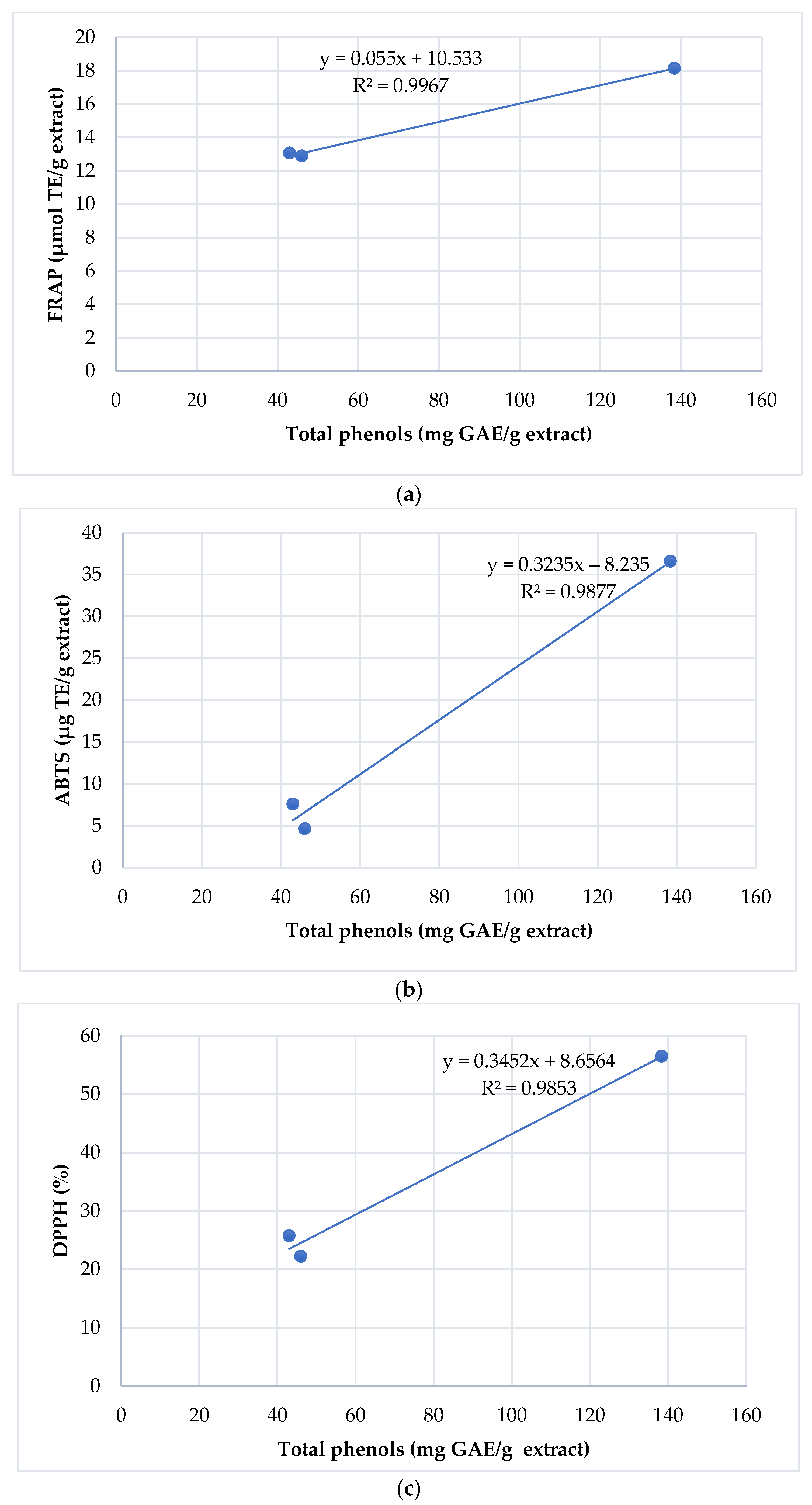
3.1. Phenolic Content of G. hederacea Extracts Obtained at Different Extraction Variants

G. hederacea CO2 extracts obtained with different extraction conditions were tested for their biological activity. The yield of SC-CO2 extraction with methanol as a co-solvent was established in the presented work at 9.58%. In SC-CO2 extraction, density is extremely important and highly dependent on temperature and pressure variation [51,52]. An increase in pressure favors an increase in density at a constant temperature, whereas an increase in temperature decreases the density at a constant pressure. In our study, we tested three temperatures (40, 50 and 60 °C) during the extraction process at constant pressure. At the lowest temperature we obtained the highest level of total phenolic content. As reported Bezerra et al. (2020) [52] the increase in the temperature causes a greater intermolecular distance, and consequently the reduction of the solubility power of CO2, by decreasing the density.
In this study, the TPC in the obtained G. hederacea extracts was analyzed spectrophotometrically using the Folin–Ciocalteu method. The results showed that the extract obtained in conditions of 40 °C/250 bar had the highest TPC value (138.33 ± 5.00 mg GAE/g) compared with the samples obtained at temperatures of 50 °C and 60 °C (Table 1). The TPC values of the other two tested G. hederacea extracts were lower than ⅓ of the value obtained at 40 °C (43.00 ± 3.04 mg GAE/g for 50 °C and 46.00 ± 9.26 mg GAE/g for 60 °C).
The most reported method for obtaining G. hederacea extracts is aqueous extraction and the total phenolic content obtained in this study cannot be directly compared to those extracts, as different parameters (extraction method, time, pressure, solvent) had an influence on the final results. However, it is worth mentioning that Varga et al. (2016) [32] reported total phenols from the aqueous Glechoma extract in the range of 43.9 ± 3.2–109.8 ± 5.8 mg GAE/g, whereas Hahm et al. (2021) [53] reported an average phenolic content of 14.81 ± 4.53 mg/g, and Chou et al. (2012) [18] found a total phenol content of 79.70 ± 0.193 mg GAE/g.
The literature data indicate that SC-CO2 can be used to obtain extracts rich in phenolic compounds [54]. However, due to the nonpolar nature of CO2, some polar cosolvents such as ethanol, methanol or ethyl acetate need to be added to increase the extraction yield of phenolic compounds via the increase in solvation power [52,55]. In the presented work, methanol was used as an example of a polar solvent due to its properties and costs. Moreover, the effect of temperature, pressure, flow rate and density of the supercritical CO2 on the bioactive compound extraction process using different plant materials was analyzed previously [52]. According to some authors [51,56], solvent density or pressure may influence the mass yield of the extract. According to a study by García-Abarrio (2014), overall SFE yield increases with CO2 density and co-solvent ratio [56]. Silva et al. (2021) [51] reported that at 40 °C the solvent density affected the mass yields, whereas after increasing temperature to 50 °C, the solvent density did not influence yields, but rather affected the vapor pressure of the solute. Based on the obtained results, we can conclude that a temperature increase has a negative effect on phenolic solubility during extraction due to the decrease in the solvent’s density. Akowuah et al. (2009) [57] observed a decrease in the total phenolic compounds determined in an extract from Gynura procumbens leaves when the temperature was increased in a conventional solvent extraction system. When compared to other authors’ studies, the high TPC value achieved for the obtained extracts (particularly for the fraction at 40 °C) shows that the extraction method employing the supercritical fluid extraction technique was very successful.

3.2. Antioxidant Effect of G. hederacea Extracts

Plants are a rich source of natural antioxidants, mainly phenolic compounds, that may delay, inhibit, or prevent oxidative processes that contribute to the deterioration of food quality or to the onset and development of degenerative diseases in the body [58]. Generally, antioxidant activity is primarily based on two chemical mechanisms: single-electron transfer and hydrogen atom transfer. However, due to the various mechanisms, reaction characteristics and variable phase localizations that are typically involved in the process, there is currently no single standardized method for determining antioxidant activity. It is worth noting that the DPPH radical is a stable free radical that is commonly used to assess antioxidants’ free radical scavenging capacities [23]. In this study, the antioxidant activity of G. hederacea extracts obtained using SC-CO2 with methanol as a co-solvent was analyzed spectrophotometrically using three different assays (DPPH, ABTS and FRAP).
The findings showed that the antioxidant activity of the G. hederacea extracts was significantly different at p < 0.05 between the extraction temperatures. However, any of these assays could be used for the analysis of the antioxidant activity of Glechoma because all tested assays confirmed 40 °C/250 bar to be the best conditions for the extraction of antioxidants in this study. The results are summarized in Table 2. Compared with other authors, Chou et al. (2012) [18] reported that the antioxidant activities of the hot water extract of G. hederacea (HWG) were significantly higher than those of vitamin C and Trolox in terms of superoxide anion radical-scavenging activity and Fe2+-chelating ability (p < 0.05). Similarly, Oalđe et al. (2021) [29] investigated the antioxidant activity of methanolic, ethanolic and aqueous extracts of Glechoma hederacea, Hyssopus officinalis, Lavandula angustifolia, Leonurus cardiaca, Marrubium vulgare and Sideritis scardica (Lamiaceae) using several experimental models. Their findings revealed that the ethanolic extract of G. hederacea had the highest DPPH scavenging activity among the investigated extracts, which was comparable to that of the positive control, 2-tert-butyl-4-hydroxyanisole (BHA) [29]. Furthermore, Matkowski (2008) [59] examined the antioxidant capacity of extracts and various solvent fractions of Glechoma hederacea L. and Orthosiphon stamineus (Benth.) Kudo. The results demonstrated that the methanolic extracts of O. stamineus exhibited much higher activity than those of G. hederacea [59].
The correlation of total phenolic content with FRAP, ABTS and DPPH scavenging activities is shown in Figure 1a–c, respectively. The analysis revealed that the results of all three assays best correlated with the gallic acid equivalent values, estimated using the Folin−Ciocalteu method. The Pearson’s correlation coefficient (r) and coefficient of determination (R2) were the highest (r  =  0.9983, R2  =  0.9967) between total phenolic content and FRAP activity than those of total phenolic content and ABTS activity (r  =  0.9938, R2  =  0.9877), followed by total phenolic content and DPPH activity (r  =  0.9926, R2  =  0.9853). These results suggest that the total phenols in the G. hederacea extracts were the primary contributor to the antioxidant activities of Glechoma extracts obtained using the SC-CO2 set at different temperatures.

3.3. Antibacterial Activity

The antibacterial properties of the tested G. hederacea extracts, expressed as MIC and MBC values, against Gram-positive and Gram-negative bacteria, are presented in Table 3. All tested extracts exhibited the inhibition of indicator bacteria; however, the results depended on both the bacterial strain and the sample extraction temperature. The strongest antagonistic effect was observed for the herb extract obtained at 40 °C/250 bar for most of the Gram-positive bacteria and one of the tested Gram-negative species. Stronger antibacterial properties were detected mainly in relation to Gram-positive bacteria, whereas the effect was weaker in relation to Gram-negative bacteria. MIC ranged from 0.3 to >5.0 mg/mL for Gram-positive bacteria, and from 1.25 to 2.5 mg/mL for Gram-negative bacteria, whereas MBC ranged from 0.6 to >5 mg/mL and from 2.5 to 5.0 mg/mL, respectively. The MIC values for all extracts against Gram-positive M. luteus (1.25 mg/mL), as well as Gram-negative P. aeruginosa (2.5 mg/mL) and E. coli (2.5 mg/mL), were the same for all the different extraction temperatures. The strongest antibacterial activity was observed towards S. aureus with an MIC of 0.3 mg/mL and an MBC of 0.6 mg/mL for the G. hederacea extract obtained at 40 °C/250 bar. Among Gram-negative bacteria, the strongest inhibition of bacterial growth was observed for P. aeruginosa for all tested extracts.
The antibacterial potential of G. hederacea extracts obtained by SC-CO2, depending on the process conditions, has not been established in the literature; however, the influence of different extraction conditions of SC-CO2 on the antibacterial activity of plant extracts is well described. Cadena-Carrera et al. (2019) [60] studied the biological activity of SC-CO2 guayusa leaf (Ilex guayusa Loes.) extracts obtained with differentiated process conditions. The authors tested, among others, antibacterial properties against B. subtilis, S. aureus, E. coli and P. aeruginosa. On the contrary to the results obtained in the studies with guayusa leaves, the lower extraction temperature of G. hederacea extracts resulted in higher antibacterial properties for most of the bacterial strains. The obtained results indicate the influence of the different extraction temperatures of SC-CO2 on the total polyphenol composition of G. hederacea extracts and consequently on the antibacterial activity. Mendiola et al. (2008) [61] determined antimicrobial activity of SC-CO2 extracts of green alga (Dunaliella salina) obtained using different extraction parameters against E. coli, S. aureus, C. albicans and Aspergillus niger. The results showed that all tested alga extracts presented antimicrobial activity against selected bacteria and yeasts. The authors confirmed the findings that when comparing the activities of the extracts obtained under the different experimental conditions, the sample obtained at the lowest temperature (9.8 °C) was the most active. Therefore, it can be clearly stated that the lower the SC-CO2 extraction temperature, the higher antibacterial properties the extract demonstrated. It has been reported that G. hederacea extracts show antimicrobial activity against some microorganisms; however, their antibacterial activity is related to the extraction techniques used, to the different parameters of the process and therefore the concentration of active substances, and finally with the tested microorganism strains [22,62,63].

3.4. Fungistatic Activity—MIC and MFC Values

The fungistatic activity of G. hederacea extracts obtained via SC-CO2 against five filamentous fungi, expressed as MIC and MFC values, are presented in Table 4 and Figure S1a–e in the Supplementary Materials. Based on the results, it can be stated that the tested extracts exhibited fungistatic activity towards the tested fungi; however, this activity was dependent mainly on the fungal species, whereas the extraction conditions generally did not affect the activity of the extracts. An exception was the extract obtained at 40 °C, which demonstrated activity against B. cinerea (MIC and MFC values were 5 mg/mL), whereas extracts obtained at 50 °C and 60 °C showed no inhibition of this species in the tested concentration range. Similar results can be observed in the case of C. albicans, where the extract obtained at 40 °C demonstrated the highest antagonistic activity compared to the other tested extracts. The strongest activity of all extracts was observed towards S. sclerotiorum with MIC and MFC values of 1.25 mg/mL, as well as towards C. albicans (MIC and MFC at the level of 1.25 mg/mL), but only for the extract obtained at 40 °C. Among the Fusarium strains, higher sensitivity to the tested G. hederacea extracts was observed for F. graminearum (MIC and MFC at 2.5 mg/mL) compared to F. culmorum (MIC and MFC at 5.0 mg/mL level). No fungistatic activity of the tested extracts was observed against A. alternata.
Interesting data about the diversified fungistatic activity of different plant extracts obtained via SC-CO2 can be found in the literature. Many authors reported that the antagonistic activity of the extracts strongly depends on the fungal genus, which was also underlined in this study. Confortin et al. (2019) [64] showed no differences in the fungistatic activity of extracts obtained via SC-CO2 from Lupinus albescens against F. oxysporum and F. verticillioides, which is similar to the result obtained in the present work (the same MIC and MFC values were obtained for F. graminearum and F. culmorum). The high dependence of the antifungal activity of extracts of guayusa leaves (Ilex guayusa Loes.) obtained by SC-CO2 on the type of indicator microorganism was shown by Cadena-Carrera et al. (2019) [60]. The studies presented in the mentioned paper showed little impact (in the case of E. floccosum, M. canis, M. gypseum and T. mentagrophytes) or no influence (in the case of A. fumigatus, Rhizopus and C. albicans) of the extraction conditions tested on the fungistatic activity of the extracts obtained via SC-CO2 from guayusa leaves [60]. In the presented study, a minor influence of extraction conditions on the fungistatic activity of G. hederacea extracts was also noted. However, in the case of C. albicans, a significantly stronger effect of the extract obtained at 40 °C was observed compared to the other temperature variants.

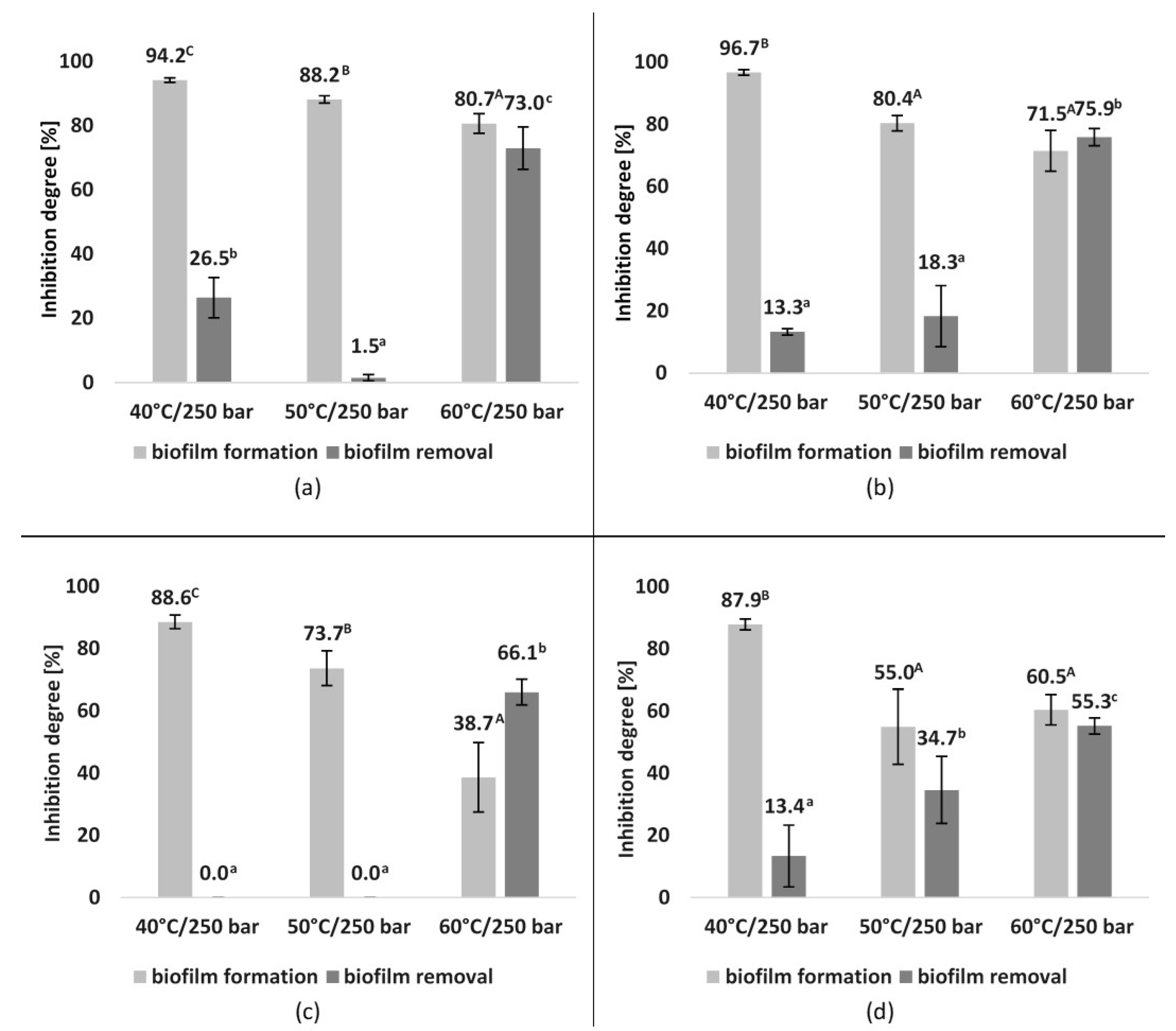
3.5. Antibiofilm Activity

The antibiofilm activity of the tested G. hederacea extracts, expressed as a percentage of inhibition of biofilm formation or biofilm removal, is presented in Figure 2. Four strains of bacteria were used for this experiment. All tested extracts reduced the biofilm formation, with the highest ratio demonstrated by extract obtained at the 40 °C compared to the other temperature variants. The percentage inhibition of the biofilm formation by E. coli and E. faecalis exceeded 90%, whereas in the case of P. aeruginosa and B. subtilis it was almost 90% (88.6% and 87.9%, respectively). The extract obtained at 50 °C also had a strong inhibitory effect on biofilm formation, displaying the strongest inhibition of biofilm formed by E. coli (88.2%). The percentage reduction of biofilm formation by other bacteria was lower, with the lowest percentage of biofilm inhibition by B. subtilis (55.0%). In the case of the extract obtained at 60 °C, the best results in the reduction of biofilm formation were observed for E. coli (80.7%) and E. faecalis (71.5%), whereas the inhibitory efficiency was lower for B. subtilis (60.5%) and P. aeruginosa (38.7%).
The tested extracts showed variable effects on the removal of mature biofilm. In contrast to the inhibition of biofilm formation, the best results in biofilm removal were observed after the application of extracts obtained at 60 °C, within the range of 55.3–75.9%, depending on the microorganism. Only a small percentage of biofilm was removed by extracts obtained at 40 °C and 50 °C. Depending on the extract and bacterial strain, the efficiency of biofilm destruction ranged from 1.5% to 34.7%, with no effect on the biofilm formed by P. aeruginosa.
The antibiofilm activity of G. hederacea has not been described in the literature; however, several studies have described the effect of plant extracts obtained via supercritical fluid extraction. Al-Maqtari et al. (2020) [65] reported that extracts of Artemisia arborescens, Artemisia abyssinica, Pulicaria jaubertii and Pulicaria petiolaris were effective as anti-biofilm formation agents for all tested bacteria at 1/2 MIC. The highest inhibition rate of biofilm formation by the extracts was observed against B. subtilis, whereas the lowest inhibition ratio was noted on K. pneumoniae, S. typhimurium, P. aeruginosa and E. faecalis. Abdullah et al. (2021) [66] found that green cardamom essential oil obtained via supercritical fluid extraction prevented the biofilm formation of E. coli O157:H7 and S. typhimurium JSG 1748. In the presented work, the antibiofilm activity also depended on the bacterial strain, with the highest inhibition rate observed against E. coli and E. faecalis, whereas the biofilm prevention performance was slightly lower for P. aeruginosa and B. subtilis. Moreover, the extraction conditions had an impact on the degree of reduction of biofilm formation. In a study by Santos et al. (2021) [67], propolis extracts obtained via the supercritical and ethanolic extraction methods were effective in interfering with bacterial biofilm formation, whereas only little activity was observed on the consolidated film, which is consistent with the results obtained in the presented work. As the literature data suggest, one of the reasons for the extracts’ weak efficacy in disrupting the biofilm maybe due to the structure of the biofilm, as the exopolymeric matrix may prevent the penetration of antibacterial agents [68].

4. Conclusions

The results of antioxidant, antimicrobial and antibiofilm activity analysis of G. hederacea extracts obtained via SC-CO2 under different conditions of the process using methanol as a co-solvent show that the temperature of extraction affects the biological activity of the tested product. Overall, the results indicate that the SC-CO2 extracts are characterized by high TPC values that differ depending on the extraction conditions. The high TPC values correlate with high antioxidant properties, as well as antimicrobial (excluding filamentous fungi) and antibiofilm activity (the prevention of biofilm formation). Among the tested extracts, the most promising results were obtained for the extract obtained at 40 °C, including the highest TPC value, as well as the best-performing antimicrobial and antioxidant properties. All tested extracts were effective in controlling biofilm formation of the studied bacteria at MIC concentration, but only the extract obtained at 60 °C efficiently removed formed biofilm. G. hederacea extracts obtained using SC-CO2 could have promising applications at different stages of food production, as well as in the industry as a safe alternative to chemical preservatives or disinfectants due to their demonstrated properties.

Supplementary Materials

The following supporting information can be downloaded at: https://www.mdpi.com/article/10.3390/app12073572/s1, Figure S1: The fungistatic activity of obtained G. hederacea CO2 extracts against (a) F. graminearum, (b) F. culmorum, (c) A. alternata, (d) B. cinerea and (e) S. sclerotiorum, expressed as MIC and MFC values. C—control; M—methanol; GH 40—G. hederacea CO2 extract (extraction conditions: 40 °C/250 bar); GH 50—G. hederacea CO2 extract (extraction conditions: 50 °C/250 bar); GH 60—G. hederacea CO2 extract (extraction conditions: 60 °C/250 bar); Cp—percentage concentration of CO2 extracts.

Author Contributions

Conceptualization, D.G., K.M., K.J. and A.W.; methodology, D.G., K.J., K.M., P.A.U., S.F. and A.W.; validation, K.J., S.F. and A.W.; formal analysis, K.M., D.G., R.G. and A.W.; investigation, D.G., K.J., P.A.U., S.F., K.M. and A.W.; resources, D.G., K.M., K.J., R.G. and A.W.; data curation, K.J. and A.W.; writing—original draft preparation, D.G., K.M., K.J., P.A.U., S.F., R.G. and A.W.; writing—review and editing, D.G., K.M. and K.J.; visualization, D.G., K.J., P.A.U., S.F. and K.M.; supervision, D.G. and A.W.; project administration, R.G. All authors have read and agreed to the published version of the manuscript.

Funding

This research was funded by Polish National Science Centre, grant number 2018/31/B/NZ9/03485.

Institutional Review Board Statement

Not applicable.

Informed Consent Statement

Not applicable.

Data Availability Statement

Publicly archived datasets are not available.

Conflicts of Interest

The authors declare no conflict of interest.

References

  1. Uyttendaele, M.; Franz, E.; Schlüter, O. Food Safety, a Global Challenge. Int. J. Environ. Res. Public Health 2016, 13, 67. [Google Scholar] [CrossRef]
  2. World Health Organization. Estimates of the Global Burden of Foodborne Diseases: Food Borne Disease Burden Epidemiology Reference Group 2007–2015; WHO: Geneva, Switzerland, 2015. [Google Scholar]
  3. Soares, L.S.; Almeida, R.C.; Cerqueira, E.S.; Carvalho, J.S.; Nunes, I.L. Knowledge, attitudes and practices in food safety and the presence of coagulase-positive staphylococci on hands of food handlers in the schools of Camaçari, Brazil. Food Control 2012, 27, 206–213. [Google Scholar] [CrossRef] [Green Version]
  4. Mostafa, A.A.; Al-Askar, A.A.; Almaary, K.S.; Dawoud, T.M.; Sholkamy, E.; Bakri, M.M. Antimicrobial activity of some plant extracts against bacterial strains causing food poisoning diseases. Saudi J. Biol. Sci. 2017, 25, 361–366. [Google Scholar] [CrossRef] [PubMed]
  5. Pandey, A.; Singh, P. Antibacterial activity of Syzygium aromaticum (clove) with metal ion effect against food borne pathogens. Asian J. Plant Sci. 2011, 1, 69–80. [Google Scholar]
  6. European Food Safety Authority (EFSA); European Centre for Disease Prevention and Control (ECDC). The European Union summary report on trends and sources of zoonoses, zoonotic agents and food-borne outbreaks in 2017. EFSA J. 2018, 16, e05500. [Google Scholar] [CrossRef]
  7. Galiè, S.; García-Gutiérrez, C.; Miguélez, E.M.; Villar, C.J.; Lombó, F. Biofilms in the Food Industry: Health Aspects and Control Methods. Front. Microbiol. 2018, 9, 898. [Google Scholar] [CrossRef]
  8. Chilaka, C.A.; De Boevre, M.; Atanda, O.O.; De Saeger, S. The Status of Fusarium Mycotoxins in Sub-Saharan Africa: A Review of Emerging Trends and Post-Harvest Mitigation Strategies towards Food Control. Toxins 2017, 9, 19. [Google Scholar] [CrossRef] [Green Version]
  9. Wild, C.P.; Gong, Y.Y. Mycotoxins and human disease: A largely ignored global health issue. Carcinogenesis 2009, 31, 71–82. [Google Scholar] [CrossRef]
  10. Zain, M.E. Impact of mycotoxins on humans and animals. J. Saudi Chem. Soc. 2011, 15, 129–144. [Google Scholar] [CrossRef] [Green Version]
  11. Khaneghah, A.M.; Farhadi, A.; Nematollahi, A.; Vasseghian, Y.; Fakhri, Y. A systematic review and meta-analysis to investigate the concentration and prevalence of trichothecenes in the cereal-based food. Trends Food Sci. Technol. 2020, 102, 193–202. [Google Scholar] [CrossRef]
  12. Sales, M.D.C.; Costa, H.B.; Fernandes, P.M.B.; Ventura, J.A.; Meira, D.D. Antifungal activity of plant extracts with potential to control plant pathogens in pineapple. Asian Pac. J. Trop. Biomed. 2016, 6, 26–31. [Google Scholar] [CrossRef]
  13. Gnat, S.; Majer-Dziedzic, B.; Nowakiewicz, A.; Trościańczyk, A.; Ziółkowska, G.; Jesionek, W.; Choma, I.; Dziedzic, R.; Zięba, P. Antimicrobial activity of some plant extracts against bacterial pathogens isolated from faeces of red deer (Cervus elaphus). Pol. J. Vet. Sci. 2017, 20, 697–706. [Google Scholar] [CrossRef] [PubMed]
  14. Gonelimali, F.D.; Lin, J.; Miao, W.; Xuan, J.; Charles, F.; Chen, M.; Hatab, S.R. Antimicrobial Properties and Mechanism of Action of Some Plant Extracts Against Food Pathogens and Spoilage Microorganisms. Front. Microbiol. 2018, 9, 1639. [Google Scholar] [CrossRef] [PubMed]
  15. Manandhar, S.; Luitel, S.; Dahal, R.K. In Vitro Antimicrobial Activity of Some Medicinal Plants against Human Pathogenic Bacteria. J. Trop. Med. 2019, 2019, 1–5. [Google Scholar] [CrossRef] [PubMed] [Green Version]
  16. Kim, J.; Song, S.; Lee, I.; Kim, Y.; Yoo, I.; Ryoo, I.; Bae, K. Anti-inflammatory activity of constituents from Glechoma hederacea var. longituba. Bioorgan. Med. Chem. Lett. 2011, 21, 3483–3487. [Google Scholar] [CrossRef]
  17. An, H.-J.; Jeong, H.-J.; Um, J.-Y.; Kim, H.-M.; Hong, S.-H. Glechoma hederacea inhibits inflammatory mediator release in IFN-γ and LPS-stimulated mouse peritoneal macrophages. J. Ethnopharmacol. 2006, 106, 418–424. [Google Scholar] [CrossRef]
  18. Chou, S.T.; Chan, Y.R.; Chung, Y.C. Studies on the Antimutagenicity and Antioxidant Activity of the Hot Water Extract of Glechoma hederacea. J. Food Drug Anal. 2012, 20, 637–645. [Google Scholar] [CrossRef]
  19. Belščak-Cvitanović, A.; Durgo, K.; Bušić, A.; Franekić, J.; Komes, D. Phytochemical Attributes of Four Conventionally Extracted Medicinal Plants and Cytotoxic Evaluation of Their Extracts on Human Laryngeal Carcinoma (HEp2) Cells. J. Med. Food 2014, 17, 206–217. [Google Scholar] [CrossRef]
  20. Chao, W.; Chan, W.; Ma, H.; Chou, S. Phenolic acids and flavonoids-rich Glechoma hederacea L. (Lamiaceae) water extract against H2O2—Induced apoptosis in PC12 cells. J. Food Biochem. 2021, 46, e14032. [Google Scholar] [CrossRef]
  21. Cho, K.; Choi, Y.-J.; Ahn, Y.H. Identification of Polyphenol Glucuronide Conjugates in Glechoma hederacea var. longituba Hot Water Extracts by High-Performance Liquid Chromatography-Tandem Mass Spectrometry (HPLC-MS/MS). Molecules 2020, 25, 4713. [Google Scholar] [CrossRef]
  22. Zhou, Y.; He, Y.-J.; Wang, Z.-J.; Hu, B.-Y.; Xie, T.-Z.; Xiao, X.; Zhou, Z.-S.; Sang, X.-Y.; Luo, X.-D. A review of plant characteristics, phytochemistry and bioactivities of the genus Glechoma. J. Ethnopharmacol. 2021, 271, 113830. [Google Scholar] [CrossRef] [PubMed]
  23. Chou, S.-T.; Lin, T.-H.; Peng, H.-Y.; Chao, W.-W. Phytochemical profile of hot water extract of Glechoma hederacea and its antioxidant, and anti-inflammatory activities. Life Sci. 2019, 231, 116519. [Google Scholar] [CrossRef] [PubMed]
  24. Kumarasamy, Y.; Cox, P.J.; Jaspars, M.; Nahar, L.; Sarker, S.D. Isolation, structure elucidation and biological activity of hederacine A and B, two unique alkaloids from Glechoma hederaceae. Tetrahedron 2003, 59, 6403–6407. [Google Scholar] [CrossRef]
  25. Zhu, Q.-F.; Wang, Y.-Y.; Jiang, W.; Qu, H.-B. Three new norlignans from Glechoma longituba. J. Asian Nat. Prod. Res. 2013, 15, 258–264. [Google Scholar] [CrossRef]
  26. Grabowska, K.; Żmudzki, P.; Wróbel-Biedrawa, D.; Podolak, I. Simultaneous Quantification of Ursolic and Oleanolic Acids in Glechoma hederacea and Glechoma hirsuta by UPLC/MS/MS. Planta Medica 2021, 87, 305–313. [Google Scholar] [CrossRef]
  27. Chou, S.-T.; Lai, C.-C.; Lai, C.-P.; Chao, W.-W. Chemical composition, antioxidant, anti-melanogenic and anti-inflammatory activities of Glechoma hederacea (Lamiaceae) essential oil. Ind. Crop. Prod. 2018, 122, 675–685. [Google Scholar] [CrossRef]
  28. Hwang, J.; Erkhembaatar, M.; Gu, D.; Lee, S.; Lee, C.; Shin, D.M.; Lee, Y.; Kim, M. Glechoma hederacea Suppresses RANKL-mediated Osteoclastogenesis. J. Dent. Res. 2014, 93, 685–690. [Google Scholar] [CrossRef] [Green Version]
  29. Oalđe, M.; Kolarević, S.; Živković, J.; Aradski, A.A.; Marić, J.J.; Kolarević, M.K.; Đorđević, J.; Marin, P.D.; Šavikin, K.; Vuković-Gačić, B.; et al. A comprehensive assessment of the chemical composition, antioxidant, genoprotective and antigenotoxic activities of Lamiaceae species using different experimental models in vitro. Food Funct. 2021, 12, 3233–3245. [Google Scholar] [CrossRef]
  30. Milovanovic, M.; Zivkovic, D.; Vucelic-Radovic, B. Antioxidant Effects of Glechoma hederacea as a Food Additive. Nat. Prod. Commun. 2010, 5. [Google Scholar] [CrossRef] [Green Version]
  31. Chao, W.W.; Liou, Y.J.; Ma, H.T.; Chen, Y.H.; Chou, S. Phytochemical composition and bioactive effects of ethyl acetate fraction extract (EAFE) of Glechoma hederacea L. J. Food Biochem. 2021, 45, e13815. [Google Scholar] [CrossRef]
  32. Varga, L.; Engel, R.; Szabo, K.; Abrankó, L.; Gosztola, B.; Németh, Z.; Sárosi, S. Seasonal Variation in Phenolic Content and Antioxidant Activity of Glechoma hederacea L. Harvested from Six Hungarian Populations. Acta Aliment. 2016, 45, 268–276. [Google Scholar] [CrossRef] [Green Version]
  33. Benito-Román, O.; Rodríguez-Perrino, M.; Sanz, M.T.; Melgosa, R.; Beltrán, S. Supercritical carbon dioxide extraction of quinoa oil: Study of the influence of process parameters on the extraction yield and oil quality. J. Supercrit. Fluids 2018, 139, 62–71. [Google Scholar] [CrossRef] [Green Version]
  34. Fornari, T.; Vicente, G.; Vázquez, E.; Garcia-Risco, M.R.; Reglero, G. Isolation of essential oil from different plants and herbs by supercritical fluid extraction. J. Chromatogr. A 2012, 1250, 34–48. [Google Scholar] [CrossRef] [PubMed] [Green Version]
  35. Uwineza, P.A.; Waśkiewicz, A. Recent Advances in Supercritical Fluid Extraction of Natural Bioactive Compounds from Natural Plant Materials. Molecules 2020, 25, 3847. [Google Scholar] [CrossRef]
  36. Bai, X.; Aimila, A.; Aidarhan, N.; Duan, X.; Maiwulanjiang, M. Chemical constituents and biological activities of essential oil from Mentha longifolia: Effects of different extraction methods. Int. J. Food Prop. 2020, 23, 1951–1960. [Google Scholar] [CrossRef]
  37. Gonçalves, R.M.; Lemos, C.O.T.; Leal, I.C.R.; Nakamura, C.V.; Cortez, D.A.G.; Da Silva, E.A.; Cabral, V.F.; Cardozo-Filho, L. Comparing Conventional and Supercritical Extraction of (−)-Mammea A/BB and the Antioxidant Activity of Calophyllum brasiliense Extracts. Molecules 2013, 18, 6215–6229. [Google Scholar] [CrossRef]
  38. Uwineza, P.A.; Gramza-Michałowska, A.; Bryła, M.; Waśkiewicz, A. Antioxidant Activity and Bioactive Compounds of Lamium album Flower Extracts Obtained by Supercritical Fluid Extraction. Appl. Sci. 2021, 11, 7419. [Google Scholar] [CrossRef]
  39. Arabshahi-Delouee, S.; Urooj, A. Antioxidant properties of various solvent extracts of mulberry (Morus indica L.) leaves. Food Chem. 2007, 102, 1233–1240. [Google Scholar] [CrossRef]
  40. Li, H.-B.; Cheng, K.-W.; Wong, C.-C.; Fan, K.-W.; Chen, F.; Jiang, Y. Evaluation of antioxidant capacity and total phenolic content of different fractions of selected microalgae. Food Chem. 2007, 102, 771–776. [Google Scholar] [CrossRef]
  41. Mabrouki, H.; Duarte, C.; Akretche, D.E. Estimation of Total Phenolic Contents and In Vitro Antioxidant and Antimicrobial Activities of Various Solvent Extracts of Melissa officinalis L. Arab. J. Sci. Eng. 2017, 43, 3349–3357. [Google Scholar] [CrossRef]
  42. Benzie, I.F.; Devaki, M. The ferric reducing/antioxidant power (FRAP) assay for non-enzymatic antioxidant capacity: Concepts, procedures, limitations and applications. Meas. Antioxid. Act. Capacit. 2018, 77–106. [Google Scholar] [CrossRef]
  43. Tomasina, F.; Carabio, C.; Celano, L.; Thomson, L. Analysis of two methods to evaluate antioxidants. Biochem. Mol. Biol. Educ. 2012, 40, 266–270. [Google Scholar] [CrossRef] [PubMed]
  44. Re, R.; Pellegrini, N.; Proteggente, A.; Pannala, A.; Yang, M.; Rice-Evans, C. Antioxidant activity applying an improved ABTS radical cation decolorization assay. Free Radic. Biol. Med. 1999, 26, 1231–1237. [Google Scholar] [CrossRef]
  45. Xiao, F.; Xu, T.; Lu, B.; Liu, R. Guidelines for antioxidant assays for food components. Food Front. 2020, 1, 60–69. [Google Scholar] [CrossRef] [Green Version]
  46. Moradi, M.-T.; Karimi, A.; Alidadi, S.; Hashemi, L. In Vitro Anti-adenovirus Activity, Antioxidant Potential and total Phenolic Compounds of Melissa officinalis L. (Lemon Balm) Extract. Int. J. Pharmacogn. Phytochem. Res. 2016, 8, 1471–1477. [Google Scholar]
  47. Gwiazdowski, R.; Gwiazdowska, D.; Marchwińska, K.; Juś, K.; Szutowska, J.; Bednarek-Bartsch, A.; Danielewicz, B. Fungistatic activity of essential oils towards selected oilseed rape pathogens. Prog. Plant Prot. 2018, 58, 300–305. [Google Scholar] [CrossRef]
  48. Rzemieniecki, T.; Gwiazdowska, D.; Rybak, K.; Materna, K.; Juś, K.; Pernak, J. Synthesis, Properties, and Antimicrobial Activity of 1-Alkyl-4-hydroxy-1-methylpiperidinium Ionic Liquids with Mandelate Anion. ACS Sustain. Chem. Eng. 2019, 7, 15053–15063. [Google Scholar] [CrossRef]
  49. Somrani, M.; Inglés, M.-C.; Debbabi, H.; Abidi, F.; Palop, A. Garlic, Onion, and Cinnamon Essential Oil Anti-Biofilms’ Effect against Listeria monocytogenes. Foods 2020, 9, 567. [Google Scholar] [CrossRef]
  50. O’Toole, G.A. Microtiter Dish Biofilm Formation Assay. J. Vis. Exp. 2011, 47, e2437. [Google Scholar] [CrossRef]
  51. Silva, S.G.; de Oliveira, M.S.; Cruz, J.N.; da Costa, W.A.; da Silva, S.H.M.; Maia, A.A.B.; de Sousa, R.L.; Junior, R.N.C.; Andrade, E.H.D.A. Supercritical CO2 extraction to obtain Lippia thymoides Mart. & Schauer (Verbenaceae) essential oil rich in thymol and evaluation of its antimicrobial activity. J. Supercrit. Fluids 2020, 168, 105064. [Google Scholar] [CrossRef]
  52. Bezerra, F.; de Oliveira, M.; Bezerra, P.; Cunha, V.; Silva, M.; da Costa, W.; Pinto, R.; Cordeiro, R.; da Cruz, J.; Neto, A.C.; et al. Extraction of bioactive compounds. In Green Sustainable Process for Chemical and Environmental Engineering and Science; Elsevier: Amsterdam, The Netherlands, 2020; pp. 149–167. [Google Scholar]
  53. Hahm, Y.H.; Cho, K.; Ahn, Y.H. Compositional Characteristics of Glucuronide Conjugates in Regional Glechoma hederacea var. longituba Herbal Extracts Using a Set of Polyphenolic Marker Compounds. Plants 2021, 10, 2353. [Google Scholar] [CrossRef] [PubMed]
  54. De Melo, M.M.R.; Silvestre, A.J.D.; Silva, C.M. Supercritical fluid extraction of vegetable matrices: Applications, trends and future perspectives of a convincing green technology. J. Supercrit. Fluids 2014, 92, 115–176. [Google Scholar] [CrossRef]
  55. Feliciano, R.P.; Meudt, J.J.; Shanmuganayagam, D.; Metzger, B.T.; Krueger, C.G.; Reed, J.D. Supercritical Fluid Extraction (SFE) of Cranberries Does Not Extract Oligomeric Proanthocyanidins (PAC) but Does Alter the Chromatography and Bioactivity of PAC Fractions Extracted from SFE Residues. J. Agric. Food Chem. 2014, 62, 7730–7737. [Google Scholar] [CrossRef]
  56. García-Abarrio, S.; Martin, L.; Burillo, J.; Della Porta, G.; Mainar, A. Supercritical fluid extraction of volatile oil from Lippia alba (Mill.) cultivated in Aragón (Spain). J. Supercrit. Fluids 2014, 94, 206–211. [Google Scholar] [CrossRef]
  57. Akowuah, G.; Mariam, A.; Chin, J. The effect of extraction temperature on total phenols and antioxidant activity of Gynura procumbens leaf. Pharmacogn. Mag. 2009, 5, 81. [Google Scholar]
  58. Shahidi, F.; Zhong, Y. Measurement of antioxidant activity. J. Funct. Foods 2015, 18, 757–781. [Google Scholar] [CrossRef]
  59. Matkowski, A. Antioxidant activity of extracts and different solvent fractions of Glechoma hederacea L. and Orthosiphon stamineus (Benth.) Kudo. Adv. Clin. Exp. Med. 2008, 17, 615–624. [Google Scholar]
  60. Cadena-Carrera, S.; Tramontin, D.P.; Cruz, A.B.; Cruz, R.C.B.; Müller, J.M.; Hense, H. Biological activity of extracts from guayusa leaves (Ilex guayusa Loes.) obtained by supercritical CO2 and ethanol as cosolvent. J. Supercrit. Fluids 2019, 152, 104543. [Google Scholar] [CrossRef]
  61. Mendiola, J.A.; Santoyo, S.; Cifuentes, A.; Reglero, G.; Ibáñez, E.; Señoráns, F.J. Antimicrobial Activity of Sub- and Supercritical CO2 Extracts of the Green Alga Dunaliella salina. J. Food Prot. 2008, 71, 2138–2143. [Google Scholar] [CrossRef]
  62. Tian, F.M.; Huang, Z.H.; Wang, H.; Chen, Q.; Zhuo, P.Q.; Chen, W.D. The study of antibacterial activity of Glechoma Alcohol extraction in vitro. J. Gansu Norm. Coll. 2016, 21, 42–45. [Google Scholar]
  63. Coss, E.; Kealey, C.; Brady, D.; Walsh, P. A laboratory investigation of the antimicrobial activity of a selection of western phytomedicinal tinctures. Eur. J. Integr. Med. 2018, 19, 80–83. [Google Scholar] [CrossRef]
  64. Confortin, T.C.; Todero, I.; Soares, J.F.; Luft, L.; Brun, T.; Rabuske, J.E.; Nogueira, C.U.; Mazutti, M.A.; Zabot, G.L.; Tres, M.V. Extracts from Lupinus albescens: Antioxidant power and antifungal activity in vitro against phytopathogenic fungi. Environ. Technol. 2018, 40, 1668–1675. [Google Scholar] [CrossRef] [PubMed]
  65. Al-Maqtari, Q.A.; Al-Ansi, W.; Mahdi, A.A.; Al-Gheethi, A.A.S.; Mushtaq, B.S.; Al-Adeeb, A.; Wei, M.; Yao, W. Supercritical fluid extraction of four aromatic herbs and assessment of the volatile compositions, bioactive compounds, antibacterial, and anti-biofilm activity. Environ. Sci. Pollut. Res. 2021, 28, 25479–25492. [Google Scholar] [CrossRef] [PubMed]
  66. Abdullah; Asghar, A.; Algburi, A.; Huang, Q.; Ahmad, T.; Zhong, H.; Javed, H.U.; Ermakov, A.M.; Chikindas, M.L. Anti-biofilm Potential of Elletaria cardamomum Essential Oil against Escherichia coli O157:H7 and Salmonella Typhimurium JSG 1748. Front. Microbiol. 2021, 12, 620227. [Google Scholar] [CrossRef] [PubMed]
  67. Santos, L.M.; Rodrigues, D.M.; Kalil, M.A.; Azevedo, V.; Meyer, R.; Umsza-Guez, M.A.; Machado, B.A.; Seyffert, N.; Portela, R.W. Activity of Ethanolic and Supercritical Propolis Extracts in Corynebacterium pseudotuberculosis and Its Associated Biofilm. Front. Vet. Sci. 2021, 8, 965. [Google Scholar] [CrossRef]
  68. Davenport, E.K.; Call, D.R.; Beyenal, H. Differential Protection from Tobramycin by Extracellular Polymeric Substances from Acinetobacter baumannii and Staphylococcus aureus Biofilms. Antimicrob. Agents Chemother. 2014, 58, 4755–4761. [Google Scholar] [CrossRef] [PubMed] [Green Version]
Figure 1. Correlation between TPC and (a) FRAP, (b) ABTS and (c) DPPH results of G. hederacea extracts.
Applsci 12 03572 g001
Figure 2. Effect of G. hederacea SC-CO2 extracts on biofilm formation and removal: (a) E coli, (b) E. faecalis, (c) P. aeruginosa, (d) B. subtilis. Averages with different letters (A–C) for biofilm formation and (a–c) for biofilm removal are significantly different at p < 0.05.
Applsci 12 03572 g002
Table 1. Total phenolic content of G. hederacea extracts obtained from different extraction variants.
Extraction ConditionsTPC (mg GAE/g Extract)
40 °C138.33 a ± 5.00
50 °C43.00 b ± 3.04
60 °C46.00 b ± 9.26
Values are mean ± standard deviation, n = 3, values with the same lowercase letters in the same column indicating no significant difference at the level of 5% (p < 0.05).
Table 2. Antioxidant activities estimated via DPPH, ABTS and FRAP assays of G. hederacea extracts.
Extraction ConditionsDPPH
(%)
ABTS
(µg TE/g)
FRAP
(µmol TE/g)
40 °C56.48 a ± 3.9836.58 a ± 1.2018.15 a ± 0.21
50 °C25.74 b ± 0.437.60 b ± 0.6913.06 b ± 0.04
60 °C22.21 c ± 0.394.66 c ± 0.1212.88 b ± 0.07
Values are mean ± standard deviation, n = 3, values with the same lowercase letters in the same column indicate no significant difference at the level of 5% (p < 0.05).
Table 3. Antibacterial activity of G. hederacea, displayed by SC-CO2 extracts, against tested indicator microorganisms.
MicroorganismMIC/
MBC/
(mg/mL)
Glechoma hederacea Extracts,
Extraction Conditions:
40 °C50 °C60 °C
Gram-positive bacteria
S. aureus ATCC 33862MIC0.32.52.5
MBC0.6>5.02.5
B. subtilis ATCC 11774MIC0.65.0>5.0
MBC1.25>5.0>5.0
E. faecalis ATCC 19433MIC0.62.52.5
MBC1.255.02.5
Micrococcus luteus ATCC 4698MIC1.251.251.25
MBC5.0>5.0>5.0
Gram-negative bacteria
P. aeruginosa ATCC 9027MIC1.251.251.25
MBC2.55.02.5
S. Enteritidis ATCC 13076MIC1.252.52.5
MBC2.55.02.5
E. coli ATCC 8739MIC2.52.52.5
MBC5.05.02.5
Table 4. Fungistatic activity of G. hederacea extracts obtained via SC-CO2 against tested indicator fungi.
MicroorganismMIC/
MFC
(mg/mL)
Glechoma hederacea Extracts,
Extraction Conditions:
40 °C50 °C60 °C
F. graminearum KZF 1MIC2.52.52.5
MFC2.52.52.5
F. culmorum KZF 5MIC5.05.05.0
MFC5.05.05.0
A. alternata KZF 13MIC5.05.05.0
MFC5.05.05.0
S. sclerotiorum KZF 23MIC1.251.251.25
MFC1.251.251.25
B. cinerea BPR 187MIC5.0>5.0>5.0
MFC5.0>5.0>5.0
C. albicans ATCC 10231MIC1.252.5>5.0
MFC1.25>5.0>5.0
Publisher’s Note: MDPI stays neutral with regard to jurisdictional claims in published maps and institutional affiliations.

