Abstract
Poly(lactic acid)(PLA) is an aliphatic polyester that can be derived from natural and renewable resources. Owing to favorable features, such as biocompatibility, biodegradability, good thermal and mechanical performance, and processability, PLA has been considered as one of the most promising biopolymers for biomedical applications. Particularly, electrospun PLA nanofibers with distinguishing characteristics, such as similarity to the extracellular matrix, large specific surface area and high porosity with small pore size and tunable mechanical properties for diverse applications, have recently given rise to advanced spillovers in the medical area. A variety of PLA-based nanofibrous structures have been explored for biomedical purposes, such as wound dressing, drug delivery systems, and tissue engineering scaffolds. This review highlights the recent advances in electrospinning of PLA-based structures for biomedical applications. It also gives a comprehensive discussion about the promising approaches suggested for optimizing the electrospun PLA nanofibrous structures towards the design of specific medical devices with appropriate physical, mechanical and biological functions.
1. Introduction
Poly(lactic acid) or polylactide (PLA) is a synthetic biopolymer that is widely used in the biomedical field. PLA can be derived from natural raw materials, such as sugar cane, rice, and corn starch. It is produced via condensation polymerization or ring-opening polymerization of the lactic acid. PLA has been approved by the Food and Drug Administration (FDA) for human usage in diverse applications, such as implantable medical devices, drug delivery carriers, and tissue regeneration scaffolds. PLA involves three isomeric forms of Poly(d-lactide) (PDLA), Poly(l-lactide) (PLLA), and racemic poly(DL-lactide) (PDLLA) [1,2,3,4,5].
PLA is a biodegradable, bioabsorbable, and biocompatible thermoplastic aliphatic polyester with good thermal and mechanical performance. These characteristics, including non-toxicity for humans, make it an ideal material for bioengineering applications [3,6,7,8,9]. In some applications, such as medical implants, targeted drug delivery, and tissue engineering scaffolds, stereocomplex PLA (Sc-PLA) has gained great attention. Sc-PLA can be formed by the strong interaction created by the side-by-side order of the molecular chains of enantiomeric PLA polymers, such as PDLA and PLLA. The creation of stereocomplex improves the mechanical performance, thermal, and hydrolysis resistance of the PLA-based materials [10,11,12].
Recently, producing PLA-based nanofibrous structures through the electrospinning technique has received growing attention [13]. Electrospinning is an efficient procedure to fabricate ultrafine fibers with unique features. This method is established based on the electrostatic drawing of the polymeric jet. A droplet of a polymer solution or melt is stretched under a strong electrical field to fine fibers, which deposit on a collector to generate a desired structure. Due to flexibility and the possibility of selecting the materials and altering the ultimate features, the electrospinning method has been widely applied to develop biomedical devices [9,14,15].
PLA nanofibers with distinguished features, such as similarity to the extracellular matrix (ECM), large specific surface area and high porosity with small pore size and appropriate mechanical properties, have recently found advanced applications in the medical area. The morphological, physical, mechanical, and biological characteristics of the PLA-based nanofibrous structures for specified applications can be tuned by altering the solution features, process variables, and controlling the methods of electrospinning. Electrospun nanofibrous structures based on PLA have been extensively investigated as sutures [8,16,17], artificial blood vessels [18,19], wound dressings [7,20,21,22], tissue engineering scaffolds [23,24,25], and drug delivery carriers [2,8,13,26,27].
Highlighting and explaining the recent progress in electrospinning of PLA-based structures, with a focus on their applications in the biomedical field, are the intention of this review (Figure 1). In the biomedical field, the optimization of polymer-based systems for diverse applications is an interesting issue. This review emphasizes the promising strategies used to optimize the electrospun PLA nanofibrous structures for designing the specified medical devices with appropriate physical, mechanical, and biological functions. Firstly, the fundamental concepts about PLA, with emphasis on specific needs and unique properties for biomedical applications, are presented; further, the electrospinning procedure, advantages of electrospun PLA nanofibers for medical use, and the relevance of the electrospinning parameters and efficiency of the designed structure are considered. Engineering of the PLA-based nanofibrous structures for biomedical applications by considering the different methods of electrospinning, controlling the morphology, architecture, and structure of the nanofiber deposits is another issue that is intended in this review. For this purpose, the particular studies conducted with the aim of achieving improvement in functional features are discussed; moreover, an overview of recently developed multifunctional PLA-based electrospun structures for biomedical applications are discussed. The last section of the review involves the future prospects of PLA electrospinning for advanced medical applications.
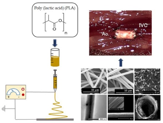
Figure 1.
Schematic to explain the recent progress in electrospinning diverse PLA-based structures, with a focus on their biomedical applications (IVC: inferior vena cava; Ao: aorta).
2. PLA Synthesis
Poly(lactic acid) (PLA) is a synthetic biopolymer derived from natural renewable resources (such as rice, sugar cane, and starch). Lactic acid (LA) is the constituent monomer for the synthesis of PLA. Two common synthesis ways of direct polycondensation or ring-opening polymerization of lactic acid are applicable for producing PLA. For industrial production, ring-opening polymerization is mostly employed [4,28,29].
The chiral molecule of LA exists in both forms of L and D isomers. The isotactic homopolymers of PLLA, PDLA, or racemic poly(dl-lactide) (PDLLA) can be obtained from the polymerization of pure L- or D-lactides. This offers possibilities for tuning the ultimate properties of the polymer depending on its final application [4,28,30,31,32]. Figure 2 represents the stereoisomers of the polymer from D-lactide and L-lactide [28].
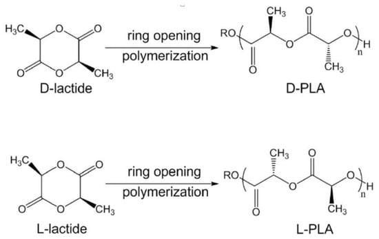
Figure 2.
Stereoisomers of the polymer start from different lactides. Reproduced with permission from [28] (License number: 5235811357024).
For the L or D stereoisomer as starting compounds, the synthesized polymer is semicrystalline with a melting temperature (Tm) around 180 °C. The racemic form of PDLLA is quite amorphous. The synthesis of stereoregular PLA is an effective approach to alter its properties. Using this strategy, by changing the chain length and/or the block succession, it is possible to tailor the mechanical and physical behavior of the PLA-based material [28].
Low molecular weight PLA was firstly polymerized by Carothers in 1932. Through subsequent attempts by DuPont, higher molecular weight PLA was synthesized and patented in 1954. For biomedical applications, for the first time, LA-based polymers were commercially produced as fiber materials for resorbable sutures; further, the mass production was followed by the development of different prosthetic devices [30].
3. PLA Unique Features for Biomedical Applications
Over the years, PLA has attracted growing attention as a base material for various biomedical applications [28]. Its medical application was firstly reported for the repair of mandibular fractures in dogs [5]. PLA has been applied in surgery as suture materials and bone fixation devices for about three decades. For the first time in 1973, LA and glycolic acid were offered as degradable materials for the sustained delivery of bioactive agents. PLA has been proved to be a proper bioabsorbable material for medical implantable devices, such as resorbable plates and screws. Likewise, synthetic bioabsorbable polymers, such as PLA can arouse cells to regenerate tissues and release drugs, which has recently been attended as tissue engineering scaffolds [30].
There are several advantages offered by PLA-based materials for biomedical applications. PLA is a biopolymer obtained from renewable resources at relatively low costs. PLA-based materials have been approved by the FDA for direct contact with biological fluids. PLA is biocompatible, biodegradable, and bioabsorbable; it is not toxic both in solid form and when degraded. Compared to other similar polymers, PLA is easily thermal processable and needs low energy consumption during the procedure, typically in processes, such as film casting, foaming, extrusion and fiber spinning. The use of PLA in biomedical applications is also based on its thermomechanical features, likely its shape memory effect. Considering these characteristics, PLA can efficiently fulfill the requirements for medical implantable devices; hence, different medical matrices have been designed from different PLA-based structures, such as degradable sutures, drug delivery devices, nanoparticles, and scaffolds for tissue regeneration [4,11,28,30,31,33,34].
4. Electrospinning of PLA Fibers
Recently, the design of PLA-based nanofibrous structures through the electrospinning process has been widely proposed for biomedical applications, such as wound dressings, drug carriers, tissue engineering scaffolds, and implants [4]. The electrospinning method is an efficient and tunable approach for fabricating ultrafine fibers with distinguished characteristics. As presented in Figure 3, the typical set-up for electrospinning consists of a high voltage power supply, a syringe pump, a nozzle, and a conductive collector. During this procedure, a liquid droplet of solution or melt polymer is electrically charged to form a jet; further, by uniaxial stretching of the viscoelastic jet under the electrostatic forces, nanofibers are generated and deposited onto a grounded collector [35].
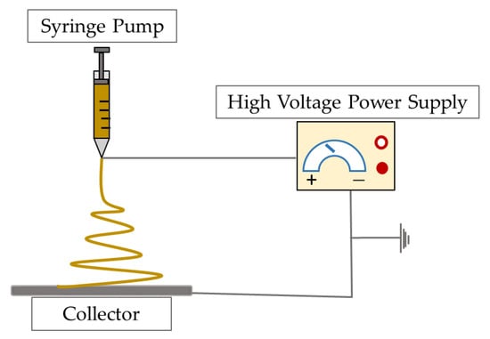
Figure 3.
Schematic illustration of the electrospinning set-up.
4.1. PLA Electrospinnability
As one of the most important advantages of the electrospinning process, it offers different options via tuning the fiber characteristics, such as morphology, diameter, and porosity by controlling the process variables and type of materials. This possibility practically allows for adjustment of the ultimate features of the fibrous structures and thus, modify their performance and functional properties in the specified application [35,36]. Several researchers investigated the electrospinnability of PLA. Several parameters and variables could influence the ultimate features of fibers electrospun from PLA-based solutions, and accordingly, their end-use functions in biomedical applications [37]. Depending on the specifications of the solution (e.g., solvent system, concentration, viscosity, and surface tension), procedure variables (e.g., flow rate, distance, and applied voltage), and ambient state (e.g., temperature and humidity), the characteristics of the electrospun fibers, comprising morphology, diameter, molecular orientation, and crystallinity will control [35,36,37].
4.1.1. Solution Parameters
Generally, the electrospinnability of the PLA-based solutions, and the morphological features of the fabricated nanofibers, are specified by a set of factors relevant to the solution, such as viscosity, conductivity, polymer concentration, surface tension, and solvent system [35,37].
Solvent Systems
Different research works performed on basic concepts of electrospinning have been approved so that the solvent system has a great influence on the ultimate properties of the electrospun fibers and structures, including morphology and mechanical properties. The vapor pressure of the solvent characterizes its evaporation velocity and so the solidification speed of the polymer jet. Generally, a very high evaporation rate is not proper for the electrospinning, because the jet may solidify immediately after exiting from the nozzle. Since the volatility is too low, the fibers may still be wet when deposited on the collector. In addition, the dielectric constant of the solvent is also important; this feature controls the value of the electrostatic repulsion. Depending on the dielectric constant of the solvent, the required applied voltage during the electrospinning process will differ [35]. Dichloromethane (DCM), chloroform (CHCl3), dimethylformamide (DMF), hexafluoroisopropanol (HFIP), and 2,2,2-trifluoroethanol (TFE), are the commonly used solvents for electrospinning of PLA-based solutions [35]. Several attempts have been made to consider the effect of the solvent system on the electrospinnability of the PLA and the ultimate properties of the fibers. In research works performed by Maleki et al., the effect of solvent type on the electrospinning of PLA has been studied [36,37]. For this purpose, different solvents of CHCl3, DCM, TFE, or HFIP were considered for electrospinning of the PLA-based structures applicable for medical devices. The scanning electron microscopy (SEM) images of electrospun PLA structures from different solvents are illustrated in Figure 4 and Figure 5.
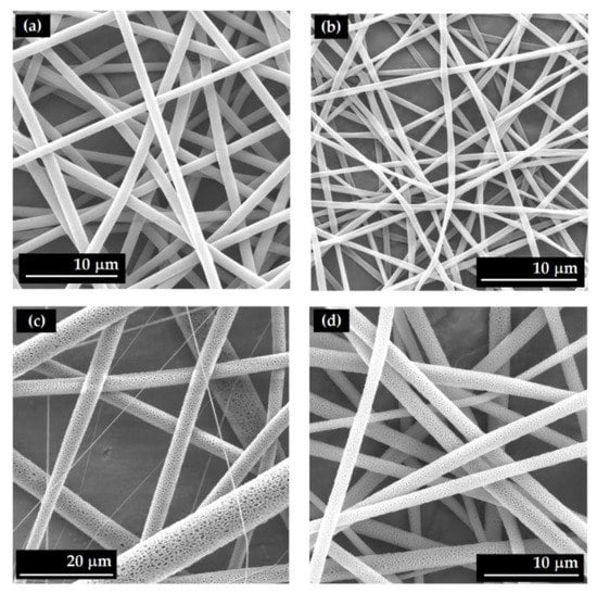
Figure 4.
SEM images of fibers electrospun from PLA solutions using different solvents of (a) HFIP (unpublished original picture by the authors), (b) TFE (unpublished original picture by the authors), (c) CHCl3 (unpublished original picture by the authors), (d) DCM (unpublished original picture by the authors).
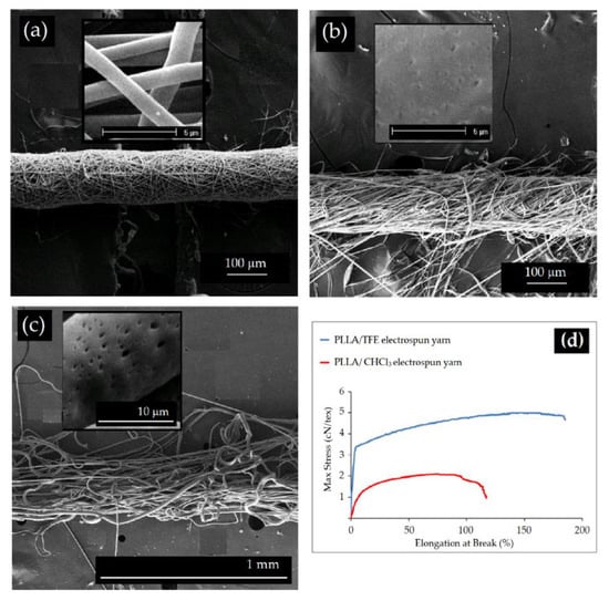
Figure 5.
SEM images of electrospun yarn structures from PLA solutions using different solvents; (a) TFE (unpublished original picture by the authors), (b) DCM (unpublished original picture by the authors), (c) CHCl3 (unpublished original picture by the authors), (d) Stress–strain curves of electrospun structures (unpublished original picture by the authors).
The use of solvents with different features led to notable variations in the shape, surface morphology, and diameter of the electrospun fibers. HFIP (Figure 4a) and TFE (Figure 4b and Figure 5a) based electrospun fibers had smaller diameters with a smooth surface morphology and were more uniform compared to those obtained with other solvents. In contrast, using CHCl3 and DCM as solvents, broad fiber diameter distributions were obtained with higher diameters (Figure 3 and Figure 4) [36,37]; moreover, the electrospinning PLA solutions using solvents with high vapor pressure and lower boiling points, such as DCM (Figure 4d and Figure 5b) and CHCl3 (Figure 4c and Figure 5c), may produce nanofibers with porous surface morphology. These pores form due to the solvent evaporation and successive phase separation during the procedure [36,37]. In addition to the morphology and diameter, the studies have revealed that the characteristics of the solvent affect the crystallinity and consequently the mechanical properties of the electrospun structure. In a study performed by Maleki et al. [36,37], it was found that in the case of the solvents with high vapor pressure, such as DCM, it is not enough time for crystal growth, thus the electrospun fibers had lower crystallinity. When using solvents with lower volatility, such as TFE, however, the crystals have more time to grow and this caused to development of a structure with relatively high crystallinity; in addition, the dielectric constant of the solvent is also effective. The solution conductivity influences the electrostatic forces which are required to apply during the electrospinning. For example, the lower conductivity of PLA/DCM solution caused a decrease in repulsive coulombic forces on the charged jet and resulted in less stretching on the way between the spinneret and the collector, whereas the higher conductivity of the TFE solution boosted the molecular orientation in the electrospun PLA fibers [36,37]. The mechanical properties of the electrospun fibers are also affected by the solvent system (Figure 5).
The difference in surface morphologies as well as in crystallinity influenced by solvent vapor pressure and dielectric constant can influence the mechanical properties of the electrospun structures. Maleki et al. [37] found that the electrospun PLLA yarns produced from the solvent with a low evaporation rate like TFE showed a higher degree of molecular orientation and crystallinity, and smaller diameter, and consequently higher modulus and tensile strength (Figure 5).
Concentration
In addition to the type of solvent, the electrospinnability of PLA-based solutions mainly depends on their concentration. The concentration can also affect the viscosity and surface tension of the solution, and both factors may change the morphology and diameter of the electrospun fibers. Aiming to form fibers, a minimum concentration is required to attain the chain entanglement, which is critical to shift from electrospray (i.e., droplet forming) to electrospinning (i.e., continuous jet forming). At a concentration lower than this limit, the jet will break into droplets and thus polymeric beads will form, instead of continuous fibers. By gradually increasing the concentration, the jet may not break up and the shape of the beads will change from spherical to spindle-like (Figure 6a). At high enough concentrations, the adequate viscosity and surface tension allow fabricating uniform and beadles fibers, while at very high concentrations, it is extremely hard to overcome the viscoelastic forces and no jet will be formed [35,36,38].
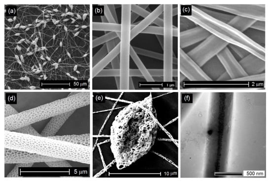
Figure 6.
Different morphologies of PLA fibers are obtained by tuning the electrospinning parameters: (a) bead-on-string morphology (unpublished original picture by the authors), (b) cylindrical shape with a smooth surface (reproduced from an open access paper [4] distributed under the terms of the Creative Commons CC BY license, (c) flat ribbon-like morphology, reproduced from an open access paper [4] distributed under the terms of the Creative Commons CC BY license, (d) porous reproduced from an open access paper [4] distributed under the terms of the Creative Commons CC BY license, (e) bead with the porous surface (unpublished original picture by the authors), (f) core–shell structure reproduced from an open access paper [9] distributed under the terms of the Creative Commons CC BY license.
In an appropriate range, decreasing the concentration leads to thin fibers. The research revealed that at high solution concentration, the average diameter of fibers increased [36]. The low concentrations with reduced viscosity facilitate the elongation of the jet under the electrical field. At high concentrations, under the same electrical forces, the viscous nature of the solution hinders the jet to be completely stretched and results in fibers with increased diameters [36,38].
In addition, the solution concentration may affect the crystallinity of the PLA nanofibers. In a research work performed by Maleki et al., it was reported that the increase of the solution concentration led to a significant reduction in crystallinity of electrospun PLA fibers. With increasing solution concentrations from 5 wt% to 9 wt%, PLA fibers showed a decrease of around 47%. The lower viscosity of solutions with low concentrations boosts the mobility of molecular chains to arrange to the fiber axis during electrospinning; this allows a higher degree of molecular orientation. In addition, a lower solution concentration has a slower solidification procedure, so affords more time for jet stretching which also leads to molecular orientation in the fiber structure [36].
The solution concentration also appeared to have a considerable influence on the mechanical performance of the electrospun PLA structures. The experimental results obtained by Maleki et al. [38] showed an increase in tensile strength and modulus of electrospun PLA yarns by increasing the solution concentration from 5 wt% up to around 7.5 wt%. Beaded fibers were formed at a lower concentration of 5 wt %, which almost influenced the mechanical features of the electrospun PLA structures. By further increasing the concentration from ~8 wt % up to 9 wt %, the fiber diameter enhanced and both tensile strength and modulus were reduced [38].
4.1.2. Processing Parameters
The electrospinning and ultimate properties of the PLA fibers are significantly determined by the processing parameters, such as solution feed rate, applied voltage, and the distance between the nozzle and the collector [36,38].
The applied voltage actually assesses the number of electrical charges carried by the jet [35]. A high voltage generally favors the formation of fibers with small diameters, while it can also cause more solution ejection from the nozzle, resulting in thicker fibers than those obtained at a low voltage under the same processing conditions [35,38]. The experimental works on electrospinning of PLA fibers have revealed an interdependence of concentration and applied voltage on fiber diameter; however, the evaluation of diameter data shows that the solution concentration is the most important parameter, the applied voltage can change some factors, such as volume of solution ejected from the nozzle, stretching of the jet by the electrostatic force, and also the morphology of the electrospun fibers. To regulate the morphology and diameter of the fibers, an equivalency between applied voltage and concentration should be made [38].
The distance between the tip of the nozzle and the grounded collector gives the amount of instability to the jet before deposition on the collector. Enough distance is required to ensure complete elongation and solidification of the jet. In a certain range, generally, by increasing the distance, fibers with a smaller diameter will be formed. Regarding the flow rate of the solution, any increase will usually lead to the formation of fibers with higher diameters [35,38].
In general, the interdependence of all the processing parameters tunes the morphology and diameter of the electrospun fibers. For example, with the increase of flow rate, it is necessary to enhance the applied voltage, and also the working distance between the nozzle and the collector to ensure complete extension and solidification of the jet and fabricate the uniform fibers. Therefore, it is required to optimize all the processing parameters to control an electrospinning process [35,36,38].
5. Engineering of PLA-Based Electrospun Structures for Biomedical Applications
Considering the wide range of potential applications of PLA in the biomedical field, one of the main challenges in the electrospinning approach is the design of structures with controllable features. By tuning the substances and methods of electrospinning, the composition, architecture, and characteristics of the PLA-based nanofibrous structures can be engineered for specific applications [35]. In this context, several process and post-treatment modifications are proposed, such as the employ of multiple jets [39], manipulating the alignment and/or patterning using different collection devices or manipulating the electric field [37,38,40,41], coaxial electrospinning [9,20,42,43], yarn electrospinning [37,44,45], blends with other biopolymers [8,9,15], the use of micro or nanoparticles, and biomolecules in the polymer matrices [22,26,46], and surface modifications [4,38,40,41]. In addition, by controlling electrospinning process parameters, environmental conditions (e.g., temperature, humidity), and precise choice of solvent system, the electrospun PLA structures may exhibit distinguished features, such as wrinkles, porous, beaded, or hollow morphologies [35,36,37,38,47,48].
5.1. Control of Morphology
Commonly, research works are focused on determining the optimized electrospinning conditions to fabricate PLA structures composed of uniform bead-less fibers with a cylindrical shape, and smooth surfaces (Figure 6b), while to fulfill the requirements of some specified applications, it is also necessary to obtain electrospun nanofibers with other morphologies [35]. By precise control of the electrospinning parameters and applying spinnerets and collectors with specific shapes, nanofibers with diverse morphologies have been explored; for example, it is found that the drug release from electrospun fibers with flat ribbon morphology (Figure 6c) with higher surface area to volume ratio, may happen via diffusion from a shorter distance. In addition, the flat ribbon-like morphology limits the volume of entrapped air at the fiber-water interface, and thus influences the surface hydrophobicity which is important for their biological function in some applications like drug release, and cell adhesion and proliferation [4].
Generally, nanofibers fabricated by electrospinning have a solid structure with a smooth surface. Since the fiber morphology changes to a porous state (Figure 6d,e), some fiber characteristics likely specific surface area, network porosity, and thus functional flexibility will enhance [4,35]. For many biomedical applications, such as scaffolds for tissue engineering, wound dressing, and carriers for drug delivery, PLA-based fibers are electrospun purposefully to have porous morphology. The porous fibers can mimic the native ECM and the increased network porosity assists cell attachment and creates adequate space for cell penetration and thus improving their efficiency as tissue engineering scaffold. The network porosity of the electrospun matrices with tiny pores can increase hemostasis, and successfully preserve the wound site from bacterial permeation. The porosity of electrospun fibers may increase drug loading efficacy. Small pores and enhanced surface-to-volume ratio can also affect the release behavior from porous fibrous structures [4,8,36,47].
Several types of research have been focused on electrospinning of PLA fibers with porous morphology using a volatile solvent or solvent/non-solvent system [47,49,50,51]. As mentioned in Section Solvent Systems by proper choice of solvent system, and within appropriate environmental conditions whereby porous fibers can be electrospun from PLA-based solutions. Figure 4c,d show SEM images of the nanofibers with porous surface morphology were fabricated by electrospinning of PLA solutions in highly volatile solvents of CHCl3 and DCM, respectively [35,36,37]. Solvent and non-solvent combination is another approach that has also been proposed to induce porosity in PLA fibers; for example, Natarajan et al. [50] found that the addition of DMF to the PLA/DCM system led to forming fibers with porous morphology, and the same system for producing porous PLA fibers has been applied by Li et al. [52]. Qi et al. [49] used 1-butanol (BuOH) as a non-solvent to create porosity on PLA fibers. In another research work, Rezabeigi et al. [53] fabricated PLA porous fibers, by electrospinning of PLA-DCM-hexane solution.
However, the uniform and beadles’ fibers are usually preferred to form, but bead-on-string nanofibers (Figure 6a,e), with versatility in the bead diameter, shape, and surface morphology, have been proposed as a potential for some specific applications. Electrospun fibers with beaded morphology can provide efficient encapsulation of biomolecules and a controlled release profile as a drug carrier for tissue engineering and wound dressing applications. The diameter of the formed beads affects the release behavior of the bead-on-string nanofibers [54]. As discussed in Section Concentration, PLA-based nanofibers with beaded morphology can be obtained by changing the concentration, and thus surface tension and viscosity of the electrospinning solution, and also by tuning the density of electrical charges on the jet. Generally, lower viscosity and lower density of surface charge both facilitate the formation of beaded nanofibers, whiles a decrease in the surface tension induces the beads to vanish gradually [35].
Core-Shell Structure
Electrospinning of PLA nanofibers with core–shell structure is an attractive strategy for biomedical applications. The coaxial electrospinning makes it possible to produce multicomponent fibers with a core–shell structure. Core−shell nanofibers compose of diverse materials for the core and the sheath (or shell). This method is also a versatile approach for producing hollow nanofibers by optionally eliminating the core part from the core–shell fibers. Coaxial electrospinning has boosted several challenges in the medical area, such as sustained drug release [9,35,42]. The ordinary use of this strategy is to encapsulate biological agents in the core site. Therefore, the shell part preserves the biomolecules and the delivery function occurs in a controlled manner by minimizing the burst release. Enhanced mechanical properties and the feasibility to functionalize the surface without affecting the core component are the other advantages of this technology; moreover, this approach is suggested to alleviate some limitations of PLA, such as its brittleness and hydrophobic nature [9,42]. In a research work performed by Maleki et al. [9], coaxial electrospinning was applied to fabricate core–shell (PVA)-PLA nanofibers (Figure 6f). This allowed the hydrophilic feature of PVA and the biocompatibility of PLA to be combined in one structure.
5.2. Control of the Architecture of PLA-Based Nanofibrous Structures
In a typical electrospinning process, nanofibers are deposited randomly to form a nonwoven web with no orientation [8,36]. Besides the morphological features, the architecture and arrangement of the fibers in the structure also greatly affect the ultimate function of the nanofibrous matrices. For example, it influences adhesion, proliferation, and penetration of the cells, and also releases the behavior of the biomolecules from the matrix; furthermore, the architecture and structure of scaffolds also considerably affect the wound healing procedure. In addition, the mechanical performance of the matrices is important and can notably be regulated by the arrangement of the fibers in the structure [8,35]; hence, many attempts have been performed to control the alignment and patterning of PLA-based- electrospun structures.
By employing various collection devices or by manipulating the electrical field during the electrospinning procedure, fiber deposition and structure architecture can be tuned, in the form of random (Figure 7a), up to uniaxial orientation (Figure 7b). Compared to randomly oriented nonwovens, in aligned structures, cells adhere and grow in the direction of the fiber arrangement. Aligned fiber orientation in the structure can also control the release of incorporated biomolecules by tuning the network porosity [4,8].
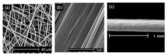
Figure 7.
Different arrangements of fiber deposits produced via electrospinning of PLA: (a) random. Reproduced with permission from [40] (License number: 5235950005044), (b) oriented, reproduced from an open access paper [4] distributed under the terms of the Creative Commons CC BY license, and (c) yarn, Reproduced with permission from [45] (License number: 5235871036609).
In a research work performed by Lopresti et al. [40], the effect of the fiber arrangement on the morphology and mechanical performance of PLA-based electrospun scaffolds has been investigated.
In this work, the electrospun nanofibers were collected on a grounded rotary drum with the speed of 10 rpm and 200 rpm to form the randomly and uniaxially oriented fibrous depositions, respectively. The stress-strain curves of the PLA-based scaffolds were strongly affected by the fiber orientation. The tensile strength and elastic modulus of the randomly oriented structures were significantly lower than those of aligned matrices. In vitro biological tests confirmed that the aligned arrangement induced homogeneous colonization of the pre-osteoblastic cells. Electrospinning of the PLA-based structures with the oriented arrangement of fibers, such as aligned bundles or twisted yarns (Figure 7c), is a promising approach to develop a new generation of materials for various medical devices.
5.3. PLA-Based Electrospun Yarns
Initial research on the electrospinning of PLA for biomedical engineering has been centralized on the fabrication of randomly oriented fibrous webs which is suitable for wound dressings, tissue regeneration scaffolds, and drug carriers. Nevertheless, the low mechanical performance of such random fibrous webs has restricted their efficiency; hence, in addition to the optimized morphology and uniformity, it is a substantial challenge to develop PLA nanofibrous structures with improved mechanical functions [11]. In order to meet the requirements for particular loadbearing applications in biomedical engineering, in recent years, electrospinning of aligned structures and yarns with improved mechanical features has received attention. Aiming to increase the adhesion and frictional forces between the fibers, and thus enhance their strength, twist was inserted into the fibrous bundle during the electrospinning procedure [37]. The superior lateral interaction and cohesion between fibers in twisted yarns significantly improve their mechanical properties.
Electrospun PLA yarns have recently attracted great attention and offered advanced applications as medical devices in the form of sutures and implants, artificial blood vessels, high-performance and functional fabrics, drug delivery carriers, and tissue scaffolding materials [8,37,38]. In recent years, a variety of electrospinning technologies have been proposed by researchers to produce uniaxially aligned bundles and yarns. In several research works performed by Maleki and coworkers, a double-nozzle electrospinning set-up has been applied to produce PLA-based twisted yarns for biomedical application.
This set-up includes a high voltage power supply, two syringe nozzles, a neutral surface, and a take-up twister unit (Figure 8a). Using this device, a PLA fibrous structure in the form of continuous twisted yarn was electrospun (Figure 8b). Through a comprehensive study, they have investigated the effects of processing parameters (i.e., voltage, take-up rate, and distances) [38], twist level [45], and post-draw treatment [55] on the morphology, diameter, crystallinity, and mechanical features of electrospun PLA yarns. Finally, they concluded that the mechanical behavior of yarns was affected by the yarn geometry and characteristics of individual fibers in the yarn structure [8,38,45,55]. In a recently published work, Sharifisamani et al. [17], have suggested the same electrospinning set-up to produce PLA-based twisted yarns with a core–shell structure for suture application (Figure 8c). The investigations revealed that the electrospun core–shell drug-loaded yarn with desirable physical and mechanical properties can use as a suitable drug carrier for wound healing. Additionally, the loading efficiency of drug and release profile could versatility be controlled by tuning the structural characteristics of electrospun yarns as an effective tool for developing suture yarns with appropriate functions.
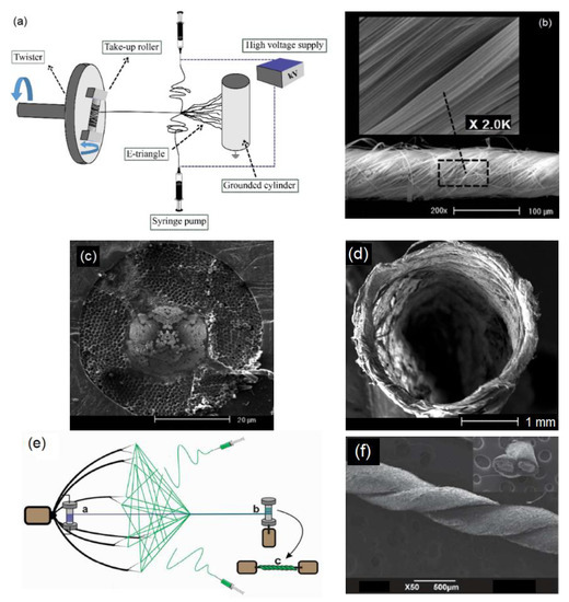
Figure 8.
(a) Double-nozzle electrospinning set-up to produce continuous twisted yarns, Reproduced with permission from [45] (License number: 5235871036609), (b) SEM images of the PLA twisted yarn prepared by double-nozzle electrospinning set-up Reproduced with permission from [45] (License number: 5235871036609), (c) cross-section image of electrospun PLA yarn with core–shell structure, reproduced from an open access paper [17] distributed under the terms of the Creative Commons CC BY license, (d) SEM image of the cross-section of hollow PLA nanofiber yarn reproduced from [56], distributed under the terms of the Creative Commons CC BY license, (e) electrospinning set-up composed of a rotating collector having a plurality of point electrodes to produce, adapted from [16], reprinted with permission from [16] Copyright @2022 American Chemical Society. (f) Dual plied PLA suture yarn, reprinted (adapted) with permission from [16] Copyright @2022 American Chemical Society.
Padamkumar et al. [16] produced electrospun core–shell yarns as surgical sutures, with PLLA core, and drug-loaded Poly-Lactic-co-Glycolic acid (PLGA) as a shell. Firstly, the core part in the form of continuous yarn was separately electrospun using an apparatus made from a rotating collector, and an infusion pump equipped with a syringe (Figure 8e). The fibrous bundle was twisted to form a yarn and drawn using a guidewire and further wound on a rotating bobbin. To produce yarn with core–shell structure, PLGA/drug solution electrospun upon the pre-spun PLLA core yarn (Figure 8f).
The double-nozzle electrospinning set-up also has been applied by Banitaba et al. [48,56] to collect PLA-based nanofibrous yarns with hollow structures as vascular scaffolds (Figure 8d). For this purpose, firstly, a three-layered nanofibrous structure composed of PVA multifilament, PVA nanofibers, and electrospun PLA fibers, as the core, middle, and shell layers respectively, were fabricated. Afterward, in order to form a hollow PLA nanofibrous structure, the middle layer was eliminated in water, and the core part was also extracted. In this study, besides the mechanical properties, cell attachment, biocompatibility, and blood compatibility of PLA hollow yarn were considered to evaluate its potential as a vascular graft.
6. Melt Electrospinning
Recent attempts have also offered the use of the melt electrospinning technique (Figure 9a) for the production of PLA-based nanofibers with desired features (Figure 9b) [35].
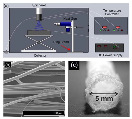
Figure 9.
(a) A schematic illustration of the melt electrospinning apparatus, reproduced with permission from [57] (License number: 5236501189506), (b) Morphology of PLA melt-electrospun nanofibers Reproduced with permission from [57] (License number: 5236501189506), (c) Macroscopic view of PLA tubular model produce via melt electrospinning procedure, reproduced from an open access paper [58] distributed under the terms of the Creative Commons CC BY license.
This method has been presented as an eco-friendly approach that eliminates the cytotoxic effects of solvents in the solution electrospinning for utilization of medical-grade polymers. Electrospinning of a polymer melt has recently attracted scientific attention to fabricate highly porous nano- or micro-fibrous scaffolds for biomedical applications [59,60].
Morphology control of PLA fibers via melt electrospinning was performed by Yu et al. [60]; they reported that the average diameter of melt-electrospun PLA fibers decreased by increasing the process temperature (from 200 °C to 250 °C); moreover, it was found that the average diameter of the electrospun PLA fiber reduced with enhancing the applied voltage, while the fiber diameter increased at higher spinning distances. In another study by Nazari et al. [59], the influences of processing parameters (applied voltage, spinning temperature, and nozzle to collector distance) on the morphology of melt-electrospun PLA-based fibers have been evaluated. Effects of process parameters on macromolecular orientation and crystallinity of melt-electrospun PLA fibers for future biomedical studies have also been investigated by Li et al. [57].
In a research work by Lee et al., to examine the solvent-free effect on cellular activities, melt-electrospun poly(lactic acid) (PLA) fibrous structures were successfully produced with a gas-assisted melt electrospinning set-up for bone tissue regeneration; they concluded that the possible residual solvent in solvent-based electrospun scaffolds decreased cell viability and osteogenic expression; hence, melt electrospinning can be employed as a suitable process for spinning PLA nanofibers [61]. Mazalevska et al. have also applied the melt electrospinning process to prepare PLA webs with tubular structures for potential application as cardiovascular implants (Figure 9c) [58,62].
7. Electrospinning of PLA Stereocomplex Nanofibers
The biocompatible, biodegradable, and non-toxic electrospun PLA fibers with desired thermomechanical properties are suitable materials to develop various medical devices; however, PLA-based nanofibrous structures have several limitations that considerably narrow their applications [11,12]. PLA is brittle with a low impact toughness, which is a hindrance in mechanically intensive applications, such as medical implants. It has a low thermal resistance and is hydrolytically sensitive which may restrict its function on prolonged operations in physiological media. Accordingly, the improvement of the physical and mechanical properties of PLA nanofibers is an essential matter for their ultimate functions [11,12,32,63,64].
Several research attempts have been performed to modify the PLA’s physical limitation. Stereocomplexation, which was firstly reported by Ikada et al. in 1987 [65], has presented a suitable approach to control these restrictions. This strategy has attained increasing attention and offered a feasible procedure to design and develop the PLA-based biomaterials with specified and tunable features, such as degradation behavior, thermal stability, and mechanical properties [11,12].
Sc-PLA can be formed via crystallization of the mixture of enantiomers of PLLA and PDLA in solution or under melting conditions. In addition to the denser structure and higher melting temperature of the stereocomplex crystals than PLA homopolymers, the formation of stereocomplex also developed PLA-based materials with enhanced mechanical performance, thermal stability, and hydrolysis resistance [11,12,63,64]. Different research studies have focused on the fabrication of Sc-PLA fibers through the solution or melt spinning. Since, in the fibers spun via these methods, in addition to the stereocomplex crystallites, the homocrystallites also exist in the structure [11,12]. Tsuji et al. [66] realized that the electrostatic forces during the electrospinning may facilitate the molecular chain orientation in the polymer and thus enhance the possibility for Sc crystallites growth and hinder the homocrystallites formation (Figure 10a) [67]. For the first time, Tsuji and colleagues employed the electrospinning strategy to form the Sc-PLA crystallites in the nanofibers; in line with this, several research efforts have been performed to modify the PLA limitations by creating Sc crystallites in the PLA electrospun fibers. In comparison to homo-crystallized, Sc-PLA nanofibers exhibited distinguished characteristics likely lower degradation rate, improved mechanical performance, and enhanced thermal stabilities [68]. Electrospun Sc-PLA structures have been positively considered in the biomedical field as tissue engineering scaffolds, drug delivery matrices, among others [68].
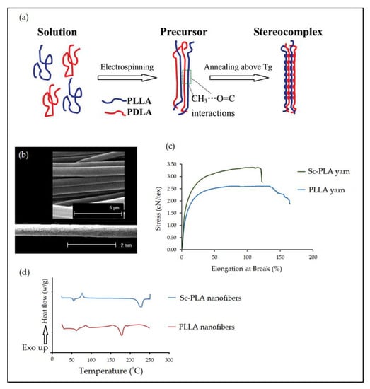
Figure 10.
(a) Intermolecular ordering for stereocomplex formation in the electrospun PLA fibers Reproduced with permission from [67] (License number: 5237071296979), (b) SEM micrographs of electrospun Sc-PLA structures, unpublished original picture by the authors, (c) Stress–Elongation curves of PLA-based electrospun yarns, unpublished original picture by the authors (d) DSC curves of electrospun PLLA and Sc-PLA fibers, unpublished original picture by the authors.
In research works performed by Maleki et al., in order to provide the requirements for some loadbearing applications in biomedical engineering, the mechanical function of the nanofibrous structures has been improved. Their approach was to electrospun Sc-PLA- aligned structures in the form of twisted yarns (Figure 10b). Sc-PLA nanofibrous yarns prepared by electrospinning of a blend solution of high molecular weight PLLA and PDLA and their characteristics compared with pure PLLA fibers [11,12]. The results indicated that, upon heat treatment, stereocomplex crystals were completely formed in the Sc-PLA electrospun fibers, without any homocrystallites formation [11,12]. The formation of Sc led to form fibers and yarns with smaller diameters and also narrower diameter distributions [11,12,64]; moreover, the thermal analysis revealed that the strong interaction between PLLA and PDLA chains enhanced the melting temperature of the Sc-PLA nanofibrous structures by around 56 °C compared to homopolymers (Figure 10d) [11,12,64]. The stereocomplexation phenomenon also affects the mechanical performance of the electrospun PLA structures. The results of this study confirmed that the formation of stereocomplex crystals led to the higher stress-at-break and Young’s modulus for electrospun Sc-PLA yarns (Figure 10c) [11].
In the other work, Ishii et al. [69] investigated the in vivo biocompatibility and inflammation response of Sc-PLA and PLLA fibrous materials by subcutaneous implantation in rats, and the in vitro degradation of the fibrous materials was also evaluated; they found that the Sc-PLA fibrous matrices were degraded slower than PLLA, due to the differences in the crystal structure of the fibrous materials.
8. Biomedical Applications of PLA-Based Nanofibrous Structures
Biodegradability, biocompatibility, non-toxicity combined with good thermomechanical properties, make PLA a suitable candidate for the use in bioengineering applications especially, in medical implants, Sc-PLA has attracted growing attention [4,6,70,71,72]. Electrospun PLA nanofibers have been widely investigated for biomedical applications as wound dressings, drug carriers, and tissue engineering scaffolds [71,72,73]. The related researches are briefly reviewed in this section, with a focus on the application of electrospun PLA-based structures, its blends with other polymers, and electrospun PLA composite structures in wound healing, drug delivery, and tissue engineering applications.
8.1. Wound Healing
In view of research, the lack of antibacterial properties of traditional dressings, poor cell respiration at the wound infection, and slow wound healing rate during use would be possible easily. Wound dressing can replace damaged skin to act as a temporal barrier in the wound healing process. The ideal wound dressing should accelerate healing of the wound, prevent infection, and restore skin structure in a short period of time. Wound dressings based on functional PLA nanofiber have been studied by a wide range of scholars. A large specific surface area, a controllable porosity and pore size, good ductility, and good biological properties of PLA fibers are not only useful to cell respiration, but also can prevent bacterial infection of wounds, and can improve cell proliferation and speed up wound healing. Furthermore, due to the good air permeability, they can efficiently absorb wound exudate and keep the wound in an ideal moist state. At the same time, it is conducive to the full contact of the wound with external oxygen and promotes the timely gas exchange of cells, thereby accelerating wound healing; moreover, the biodegradability of PLA provides better meets the current new requirements for the development of environmentally friendly materials, which is promising in the field of wound dressing research and development in the future [4]. Since the composition of electrospun nanofibers is controllable, loading antibacterial agents in PLA electrospun nanofibers can effectively inhibit wound infection. In addition, PLA nanofiber wound dressings with biologically active substances can be prepared according to the needs of medical treatment, which can better promote the adhesion and proliferation of cells at the wound and accelerate wound recovery. Table 1 shows different studies in which electrospun PLA-based structures have been used as a wound dressing for wound healing applications.

Table 1.
Different studies in which electrospun PLA-based structures have been used as a wound dressing.
In most of these studies, bioactive PLA-based electrospun membranes have been developed as wound healing products with multifunctional properties, in which electrospun fibers are carrying different drugs; for example, Alves et al., demonstrated that PLA electrospun membranes are promising wound dressings with the ability of sustainable released drug delivery [2]. Physical adsorption and blend electrospinning techniques have been used to load anti-inflammatory agents, Betamethasone (BEX), and Dexamethasone acetate (DEX) into PLA fibers and their release efficiency has been compared (Figure 11a–c). Blend electrospinning resulted in a better-sustained release profile with a burst release during the first five hours, while physical adsorption of the drug on the PLA electrospun membranes resulted in a significant burst release. Pankongadisak et al. [74] incorporated curcumin as an anti-inflammatory and an antioxidant agent inside electrospun PLLA fibers for wound dressing application (Figure 11d,e). The fabricated membrane protected cell attachment and proliferation and was non-toxic to human adult dermal fibroblast (HDF) cells. Since curcumin is hydrophobic, in another attempt, different contents and molecular weights of poly(ethylene glycol) (PEG) were used to modify the hydrophilicity of curcumin-loaded electrospun PLA fiber for wound dressing applications (Figure 11f) [70]. Results showed that the increase in concentration and a decrease in the molecular weight of PEG led to an increase in the weight loss values. The curcumin-loaded PLA/PEG electrospun membrane improved the conditions for cell growth and speeded up the drug release through balancing the hydrophilicity–hydrophobicity of the medium. Zou et al. used the blend electrospinning of PLA/Poly(1,8-octanediol-co-citric acid) (POC) in order to improve the elasticity and hydrophilicity of the Aspirin-loaded PLA nanofibrous as wound dressings. The presence of POC significantly improved the elasticity, hydrophilicity, biodegradability, and namely tensile deformation of the nanofibrous membrane which are important characteristics in wound dressing applications [88]. Cui et al. [78] used doxycycline (DCH), a broad-spectrum antibiotic, as a model drug, inside PLA nanofibers (Figure 11g,h). The resulted fibers revealed favorable cytocompatibility to L929 mouse fibroblasts and showed reasonable antibacterial activity, which can be considered as a promising wound dressing for chronic wound healing. To avoid the disadvantages of wound dressings loaded with conventional antibiotics, Zhang et al. [79] developed an antibiotic-free wound dressing based on silver (I) metal-organic frameworks-PLA nanofibers with the effective antibacterial capability to promote tissue regeneration (Figure 11i).
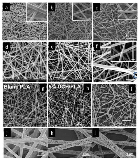
Figure 11.
SEM images of the PLA e(PLA-D) fibrous mats (a) pure, (b) with 14% of BET and (c) DEX, reproduced with permission from [2] (License number: 5232480112869), SEM images of neat (d) and curcumin-loaded PLLA fiber mats (e) reproduced with permission from [74] (License number: 5232490308135). (f) SEM micrographs of electrospun PLA/CUR/PEG nanofibers containing 10 wt% of PEG with a molecular weight of 400, reproduced with permission from [70] (License number: 5232490801255), Morphology of (g) the pure PLA nanofibers and (h) PLA nanofibers with 5% DCH contents, reproduced with permission from [78] (License number: 5232491264115). (i) SEM images of Ag2[HBTC][im]-PLA composite fibrous mat Reproduced with permission from [79] (License number: 5232491480582). SEM images of the electrospun fibers of (j) neat PLLA, (k) PLLA/POSS and (l) PLLA/POSS/pAng, reproduced with permission from [87] (License number: 5272940375894).
In vivo results showed that the electrospun fibrous composite could significantly speed up the healing rate of infected wounds. Ilomuanya et al. [77] used Aspalathus linearis (AL) fermented extract and silver sulphadiazine (Ag + S) in PLA and collagen-PLA electrospun fibrous scaffolds to improve the antibacterial activity for wound healing applications. Antibacterial properties and cellular biocompatibility were improved. The obtained composite fibers were non-toxic to the cells and presented favorable substrates for cell attachment and proliferation. Ghorbani et al. [80] used a wet electrospinning technique including a liquid coagulation bath collector to produce 3D porous PLA nanofibrous scaffolds for wound healing application. Wet electrospun nanofibers had higher porosity and surface area in comparison to the dry electrospinning technique. Rat bone marrow stem cells (BMSCs) were seeded on the fibers. In vitro and in vivo results demonstrated the potential of the 3D electrospun fibrous PLA as a suitable wound dressing. In another study, a novel wound dressing was designed based on PLA/PVA/SA (sodium alginate) electrospun fibers. The mouse fibroblasts (L929 cell line) were grown on both PLA and PLA/PVA/SA fibers with better adhesion and proliferation on PLA/PVA/SA than on the PLA membranes. The in vivo assay also confirmed the significant efficacy of PLA/PVA/SA electrospun membranes for wound healing compared to commercially available gauzes [7]. Li et al. [87] used the electrospinning technique to produce bioactive PLLA/polyhedral oligomeric silsesquioxane (POSS) nanofibers as a delivery vehicle of plasmid DNA encoding angiopoietin-1 (pAng) (Figure 11j–l). The nanofibrous scaffold could sustainably release pAng, considerably increase the gene transfection efficiency, and effectively promote wound healing.
In a series of studies, the potential of core–shell electrospun fibers has been investigated in wound healing applications. The coaxial electrospinning method has been used by Fang et al. [6] to produce core–shell nanofibers based on PLA, and PGA for wound healing application. Different core–shell structures with compact core structures and with porous core structures have been obtained (Figure 12a,b). The in vitro cell culture study and in vivo assay on PLA/-PGA core–shell nanofiber substrate represented desirable biocompatibility to the wound. In another study, poly(glycerol sebacate) (PGS) (core)/PLLA (shell) fibrous scaffolds were developed via coaxial electrospinning as a novel wound dressing. The shell fibers with surface pores showed excellent ability to repair tissues of the skin wound. The core–shell structure, compared to pure PLLA scaffold, showed superior cell proliferation, with a lower inflammatory response [1]. The coaxial electrospinning method was also applied by Yang et al. [90] to fabricate the core–shell PLLA/chitosan nanofibrous scaffolds for wound dressing applications. In order to present a synergistic microenvironment for wound healing, graphene oxide (GO) nanosheets have been coated on the surface of nanofibers which significantly increased the hydrophilicity of the electrospun scaffold and improved its antibacterial activity; they also promoted the growth of pig iliac endothelial cells (PIECs). In vivo results demonstrated the potential of GO-coated chitosan/PLLA nanofibrous scaffolds in wound healing applications. In a research work performed by Augustine et al. [91], a connective tissue growth factor (CTGF) was encapsulated inside the PVA fibers as core which was covered by a thin layer of PLA fibers as the shell (Figure 12c). The core–shell structure of the scaffold enhanced sustained release of CTGF which is crucial for diabetic wound healing procedures.

Figure 12.
TEM images of core–shell nanofibers: (a) with compact core structure; (b) with porous core structure Reproduced with permission from [6] (License number: 5232520656228), (c) SEM image of coaxial PLA-PVA-CTGF membranes, reproduced from an open access paper [91] distributed under the terms of the Creative Commons Attribution—Non-Commercial (v3.0) License.
8.2. Drug Delivery
In recent years, a lot of research has been done for discovering nanotechnology procedures (especially nanofibers) as a drug delivery system for transdermal uses by different scientists. Nanofibers are used to deliver drugs and are able to release the drugs under controlled conditions for a long period of time. In these cases, PLA is one of the well-known synthetic polymers in biomedical usage since it has good biodegradability and biocompatibility. PLA, which is a liner aliphatic polyester can be synthesized from 100% renewable materials, such as rice and wheat through fermentation and polymerization. The FDA has accepted PLA as a biomaterial application, such as, for instance, sutures, bone plates, abdominal mesh, and drug delivery systems [71].
There are some mechanisms, such as coating, embedding, or encapsulating for drug loading and drug releasing from nanofibers to achieve control over drug release kinetics [92,93,94]. It is possible to dissolve the drug and the polymer directly in the polymer solution if they both are soluble in the same solution [85]. On the other hand, when drugs and polymers are not soluble in the same solvent, the drug can be dissolved in another solvent and then added to the polymer solution [95]. There is another method for dissolving drugs and polymers which are not soluble in the same solvent: in this method, the drug is dissolved in a solvent that is immiscible with the solvent in which the polymer is dissolved, and both solutions are loaded in different capillaries to be electrospun coaxially, or both solutions can be mixed and the emulsion electrospun [96]; this method is an encapsulation tactic of the drug in the polymer matrix [97]. Another method has been suggested for loading the drug in nanofibers after the nanofiber was made; in this method, the nanofiber is immersed in a drug solution and the drug is absorbed by nanofibers through capillary property [84,98,99].
Several mechanisms are involved in the release of drugs from nanofibers, including desorption from the nanofiber surface layer, diffusion through the canals and pores of nanofibers, or matrix degradation [100,101]. The drug release kinetics can be changed with the type of polymer, geometry, control over the diameter of nanofibers, porosity, morphology, and adjustment of multiple processing variables during the production of nanofibers [92,102]. Figure 13 shows a scheme image of the different methods for loading the drug into nanofibers. PLA-based nanofiber structures have been investigated as drug carriers in different types of systems, such as nanofibers containing chemical drugs, nanofibers coated with drugs, porous nanofibers combined with growth factors, and core–shell fibrous structure containing drugs in the core [84,103,104]. Table 2 shows different studies in which electrospinning has been used for the production of drug-loaded PLA fibers for drug delivery application.
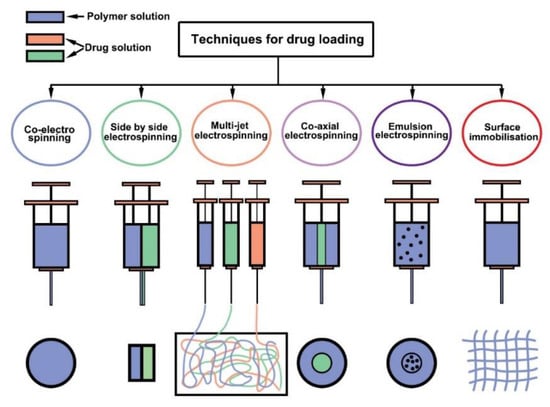
Figure 13.
The scheme image of the different methods for loading the drug in nanofibers, reproduced from an open access paper [71], under the terms of Creative Commons Attribution 4.0 International (CC BY 4.0) License.

Table 2.
Different electrospinning studies reporting the production of drug-loaded PLA fibers for drug delivery application.
Yuan et al. [107] investigated the possibility of loading the hydrophilic doxorubicin hydrochloride (Dox-HCl) and hydrophobic free doxorubicin (Dox-base) in a PLA carrier matrix. Drug–polymer miscibility, fiber wettability, and fiber degradability were investigated as a function of the drug release profile of the fibers. In the hydrophilic Dox-HCl/PLA mixture, there was a larger agglomerate on the fiber surface or inside the fiber due to poor drug–polymer compatibility (Figure 14a).
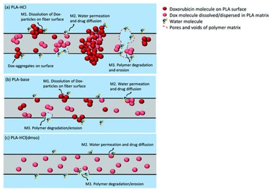
Figure 14.
Illustration of doxorubicin drug release mechanisms from a PLA fiber matrix. (a) PLA–HCl; (b) PLA–base; (c) PLA–HCl (DMSO). M1, dissolution of drug molecules; M2, water permeation followed by drug diffusion; M3, polymer degradation followed by drug dissolution/diffusion. Reproduced from an open access paper [107], distributed under the terms of the Creative Commons Attribution-NonCommercial 3.0 Unported Licence.
In this way, the drug is released so fast because of the accumulation of the drug at the surface of the fibers. The hydrophobic free doxorubicin was perfectly dispersed in the PLA matrix (Figure 14b) but the drug release was very slow. Dimethyl sulfoxide was used to solve the Dox-HCl, so then the drug mixing in the polymer matrix improved, and the electrospinning fibers containing drugs with uniform distribution in the polymer matrix were obtained (Figure 14c). The release of the drug from such fibers was very low and this low release caused low toxicity to hepatocellular carcinoma.
Doustgani et al. [108] developed doxorubicin-loaded PLA (PLA/DOX) nanofibers with a defined release mechanism. Some electrospinning parameters, such as distance, concentration, temperature, flow rate, and applied voltage, on the average diameter of PLA/DOX nanofibers, were investigated. The results of DSC showed that DOX was effectively loaded in the nanofibers. In vitro results showed that diffusion is the prevailing drug release mechanism for drug-loaded fibers.
Scaffaro et al. [103] investigated the possibility of mixing carvacrol (CAR) in PLA nanofibers; they effectively formulated PLA nanofibers that are capable to hold the dispersed CAR. The results demonstrated that PLA and CAR have good compatibility. CAR was released regularly from nanofibers and caused an inti-microbial activity up to 144 h. More than 60% of total collective CAR was released after 6 h when the samples were immersed in KBS at 37 °C. At the end of the examination, the total released CAR was about 90% of the loaded amount.
The novel Dexamethasone releasing PLLA/Pluronic P123 multilayer nanofibers constructed by Birhanu et al. [117] are ideal for drug delivery usage. The drug was loaded in the central layer. The scaffolds constructed have appropriate surface-layer properties, but with various mechanical strengths and osteogenic proliferation and differential. The drug release profiles of the scaffolds were entirely various: single layer scaffolds displayed burst release on the first day, whereas multilayer scaffolds a controlled delivery of Dexamethasone with better osteogenesis. Therefore, the existence of a layer covering the drug-loaded layer is necessary for the controlled release of drugs and or bioactive molecules at the position of scaffold implantation.
PEG is a hydrophilic polyether that is easily accessible in the market in various molecular weights. PEG with different molecular weights of 2000, 6000, 10,000, and 20,000 g/mol were selected as model molecules for investigating the effect of molecular weight on the release rate and the total released quantity. PEG has a significant effect on drug release; in one study, PEG was mixed into the polymer solutions and was included in PLA nanofibers through electrospinning. The release behavior of these molecules was investigated in water. The release experimental investigations showed two different styles: the release rate is depended on the molecular weight of model PEGs and the type of nanofibrous polymer. Large molecules were released very fast with the compression of small molecules. The release rate and total released quantity are affected by the molecular weight of incorporated molecules [109].
The combination of electrospinning and electrospray techniques for the fabrication of 3D polymeric and composite nanofibrous scaffolds creates added value for final product applications [124]. Accordingly, Bae et al. [110] fabricated PLA electrospun fibers coated with electrosprayed core–shell PVP-PLGA particles enables the scaffold to be degradable independently and at the same time deliver active ingredients (Figure 15a). The core–shell particles were uniformly located on the surface of fibers (Figure 15b). Their results showed that the composition of the binary solvent mixture ethyl acetate (EA) and benzaldehyde (BA) has a critical role in the degree of combination between the PLGA particles and the PLA fibers. This assembled particle-fiber system can improve the functional characteristics of the final scaffold for tissue engineering.
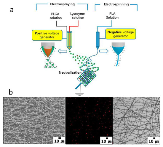
Figure 15.
(a) In situ combination of encapsulated particles and fibers via electrospinning and electrospray techniques. (b) SEM (left) and confocal microscopy (middle and right) images of particle-fiber composites produced with different solvent compositions: EA:BA = 1:9, Reproduced with permission from [110] (License number: 5232980582910).
8.3. Tissue Engineering
PLA nanofibrous scaffolds have a good potential for tissue engineering, as a substrate for bio-functionalization, suitable topographical surface for cell adhesion, and as a reservoir for drug delivery. The small diameter of PLA nanofiber closely matches that of ECM fibers, and the relatively large surface area is beneficial for cell attachment and bioactive factor loading. Electrospinning is a versatile technique for the production of nanofibrous scaffolds, and recent advancements in this process aided the creation of particular scaffolds addressing the needs of specific tissues, such as loading different bioactive molecules and having fiber alignment. Many studies have been investigated PLA nanofibrous scaffolds in vitro, but the application of these scaffolds into the human body will require more testing in suitable animal models. The perspective of PLA would likely be its combination with other biomaterials in order to improve its strengths and reduce its weaknesses, which would be a promising strategy for the engineering of complex tissues [72].
8.3.1. PLA Electrospun Structures for Musculoskeletal Tissue Engineering
Musculoskeletal Disorders are injuries that affect the human body’s movement or musculoskeletal system (i.e., bones, tendons, muscles, ligaments, nerves, etc.). Ignoring defects in bone, muscle, or tendon can lead to reduction and even loss of their function and subsequently lower quality of life for those affected. Due to the considerable role of PLA in the orthopedic field, the continued use of PLA in many musculoskeletal tissue engineering strategies is not surprising [125]. Worldwide, over two million bone grafting procedures are annually performed but in the cases of large defects and complex healing optimal bone regeneration and repair is still challenging [126]. Innovative approaches are based on the application of new materials or growth factors in order to mimic the physiology of the tissue. Scaffolds for bone tissue engineering should provide mechanical stability and an environment conducive to bone regeneration [127,128]. On the other hand, a lot of tissues in our body have been demonstrated piezoelectric effects [129]. Bone is a tissue with piezoelectric constants similar to those of quartz, the main component of the bone organic ECM. For this reason, the application of piezoelectric materials, such as polyvinylidene fluoride (PVDF) and PLA in bone tissue engineering has been invoked to support tissue function [126]. Scaffolds with (3D) nanofibrous structures have received a lot of attention in the field of bone regeneration since nanofiber mimic the bone ECM [130]. Electrospinning is one of the most important techniques for the fabrication of 3D scaffolds with nanoscale features and interconnected pores. Electrospun PLA nanofibrous scaffolds have a greater surface area than raw PLA material, which consequently results in higher protein adsorption but also faster degradation. The degradation kinetics and mechanical properties of PLA-based scaffolds can be adjusted by changing the racemic mixture of lactic acids composing the polymer chains [131]. Table 3 shows different studies in which electrospun PLA fibers have been used for bone tissue engineering.

Table 3.
Different studies in which electrospun PLA fibers have been used for tissue engineering applications.
Regarding the importance of topographic cues of electrospun membranes, such as alignment and diameter on cellular behaviors, Xie et al. [130] investigated the influence of PLLA fiber diameter and alignment on cellular responses of bone marrow mesenchymal stem cells (BMSCs), including cell attachment, migration, proliferation, and osteogenesis. The results verified that aligned nanofibers (AN) with smaller diameters had obvious advantages in promoting cell migration and proliferation and facilitating the osteogenic differentiation of BMSCs in comparison with random fibers. Scaffolds with randomly oriented fibers hinder effective cell infiltration (Figure 16a1–a8).
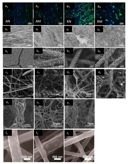
Figure 16.
Effect of fiber alignment on the morphology of BMSCs on electrospun membranes (a1–a4) Immunofluorescence images of AN, AM, RN and RM. (a5–a8) SEM images of AN, AM, RN and RM, Reproduced with permission from [130] (License number: 5232540107019). SEM images of (b1) SiNPs, (b2) porous PLLA membrane after DOP surface modification and porous PLLA/DOP/SiNP membranes with different SiNP concentrations (b3 = 0.05, b4 = 0.10) Reproduced with permission from [132] (License number: 5232550661164), SEM image of (c1) nanoHA-coated nanofibers, (c2) liposome-loaded scaffolds (liposcaffolds), Reproduced with permission from [133] (License number: 5232550143142). SEM images of the internal morphological structures of (d1) PLA/GEL, (d2) nHA/PLA/GEL, and (d3) nHA/PLA/GEL-PEP 3D nanofibrous scaffolds, respectively, Reproduced with permission from [134] (License number: 5232541047315). SEM images of surface morphology of the P3C1 scaffolds after biomineralization for (e1) 7 days, (e2) SEM image of mineral clusters after 7 days mineralization in SBF with high magnification, reproduced with permission from [140] (License number: 5232551100684). SEM images of electrospun fibers prepared at different temperatures (f1 = 35 °C), (f2 = 50 °C), (f3 = 60 °C) using a PLA/CS 70:30 weight ratio; Reprinted (adapted) with permission from [141]. Copyright (2022) American Chemical Society.
In order to improve the biocompatibility of PLA scaffolds, some additives have been applied on the surface of electrospun fibers [132,133]; for example, Lu et al., coated with silica nanoparticles (SiNPs), with high surface area and good biocompatibility, the surface of porous PLLA fibrous membrane in order to improve better bone cell attachment (Figure 16b). The membrane was designed to serve as substrates of SiNPs for bone tissue engineering. To improve the coating strength of SiNPs on PLLA fibers, Dopamine (DOP) was used to modify the surface of PLLA fibers. Hydrophilicity and mechanical properties of PLLA/DOP/SiNP composite membranes were significantly improved due to the presence of SiNPs; they concluded that PLLA/DOP/SiNP membrane with improved cellular biocompatibility, and more cell adhesion and proliferation are promising materials for bone regeneration applications [132].
In another study, Mohammadi et al. [133] used surface grafting to modify the surface of PLA fibrous scaffold to better immobilize BMP-2 peptides liposomes on the surface of fibers (Figure 16c). The treated scaffolds showed higher attachment efficiency of liposomes. In another study, a polydopamine (pDA)-assisted coating strategy was used in order to immobilize bone morphogenetic protein-2 (BMP-2)-derived peptides on the surface of nano-hydroxyapatite/PLLA/gelatin (nHA/PLA/GEL) scaffolds (Figure 16d). In vitro and in vivo results showed that nanofibrous scaffolds containing nHA and BMP-2 peptides have suitable biocompatibility and osteoinductivity; they also showed sustained release ability, so they are excellent candidates in bone regenerative medicine [134]. Using blend electrospinning of PLA with other polymers, such as chitosan also has received attention in order to improve different properties of fibrous scaffold for bone tissue engineering application [72]; for example, Xu et al. [137] mixed PLLA and bioactive lecithin to fabricate fibrous scaffolds, with improved cytocompatibility and hydrophilicity, where great interaction between PLLA/lecithin scaffolds and cells took place, resulting in cells being induced on scaffolds for keeping morphological shape and integrating with the fibers in order to develop a 3D network. Wang et al. [138] developed a PLLA electrospun scaffold incorporated with Poly(3-hydroxybutyrate-co-3-hydroxyvalerate) (PHBV) to improve the shape memory functionality and mechanical properties. PLLA-PHBV scaffold considerably promoted the osteogenic commitment in BMSCs with osteoinductive factors in a synergistic manner, which has a great potential to be used as a multifunctional 3D scaffold for bone regeneration. One of the important challenges in bone tissue engineering is the manufacturing of biomimetic scaffolds, which are able to fill the gap between porous structure requirements and mechanical strength. Although electrospinning technology develops nanofibrous structures similar to ECM, the restricted shapes and pore size of the electrospun mesh limits its application in bone tissue engineering; regarding this, Chen et al. [140] combined electrospinning, freeze-drying, and crosslinking to fabricate a novel (3D) PLLA/regenerated cellulose (PLA/RC) scaffold (Figure 16e). Due to the presence of RC nanofibers, hydrophilicity and biological activity of the final scaffolds have been improved. PLA/RC scaffolds also exhibit excellent biomineralization ability in the SBF solution that can be considered as a promising scaffold bone tissue engineering application.
A core−shell and island-like structure scaffold composed of a bicomponent was developed by Xu et al. [141] to improve cell biocompatibility of the PLA membrane through designing nanosized topography with highly bioactive chitosan onto PLA electrospun fiber surface (Figure 16f). This method in which bioactive modification and topographic effects at the interface between cells and materials happens is based on automatic phase separation of two incompatible polymers during the electrospinning; indeed, by rapid solvent evaporation under a high temperature in jets, the chitosan molecular chains did not have enough time to migrate to the surface of electrospun jets to cover the fibrous shell. The structure of the intermittent island was therefore formed, which results in a controllable balance of the hydrophilicity and hydrophobicity of the surface, desirable surface roughness for cell attachment, and cell recognition sites.
8.3.2. PLA Scaffolds for Neural Tissue Engineering
Nerve tissue repair and regeneration strategies have become crucial challenges, because they directly affect the patient’s quality of life. Recent advances in nerve regeneration have included the use of tissue engineering principles, and this has opened up a new perspective on neurotherapy [152]. Synthetic scaffolds for nerve tissue engineering are usually either hollow or filled tubes developed to be biocompatible, immunologically inert, biodegradable, infection-resistant biomaterial, and mechanically matched to nerves to support neurite outgrowth [153]. Due to such design criteria, PLA and its composites have been used for the development of biodegradable nerve scaffolds. In recent years, a lot of research has been focused on changes in the topography, alignment, and mechanical properties of PLA fibrous scaffolds to aid in neural regeneration [72]. Table 3 shows different studies in which electrospun PLA fibers have been used for nerve tissue engineering. A bio-coaxial electrospinning technique has been used by Shih et al. [144] for the development of oriented tubular PLA scaffold containing PC-12 cells to be used as a nerve guide conduit (NGC). Randomly oriented and aligned hollow fibers have been developed with diameters in tens of micrometers and wall thicknesses of around a few micrometers. PC12 cells were successfully located inside the hollow fibers, and after the addition of the nerve growth factor, have been differentiated with neurite extended along the hollow fibers in the desired direction. Co-electrospinning was used also by Tian et al. [145] to develop PLA/Silk Fibroin/Nerve Growth Factor (called PS/N) through encapsulating nerve growth factor (NGF) along with silk fibroin (SF) as the core. The effect of air plasma treatment of PS/N scaffold on cell behavior was investigated and the results showed that plasma-treated PS/N scaffolds supported the attachment and differentiation of PC12 cells, and have a great potential to be used in nerve tissue engineering.
8.3.3. PLA Scaffolds for Cardiovascular Tissue Engineering
The development of novel tissue engineering strategies to replace cardiovascular tissues is very important since cardiovascular disease is the leading cause of death worldwide [154]. Like in nerve conduit scaffolds, a biocompatible and biodegradable tube with hollow lumen is required for designing vascular scaffolds that mechanically match with the graft site. The application of PLA nanofibrous scaffolds would aid the large-scale requirement of heart and blood vessels regeneration. Conduits for vascular tissue engineering are often limited by the tendency for the lumen to clot, especially in small diameter vessels, preventing blood flow from occurring; however, Kurobe et al. [147]. Constructed tissue-engineered vascular graft (TEVG) composed of PLA tubular nanofiber scaffolds and successfully implanted these grafts in mice as aortic interposition conduits without any graft stenosis or aneurysmal dilatation (Figure 17). Wang et al. [148] developed a bilayered electrospun scaffold containing PLA nanofibers mesh outer layer and silk fibroin–gelatin nanofibrous inner layer to improve scaffold biomimicry for blood vessel application. In another study, the potential of PLA/gelatin scaffold with aligned fibers was investigated for vascular tissue engineering application (Figure 18).
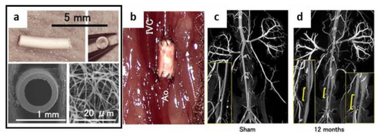
Figure 17.
(a) Structure of TEVG. Implanted TEVG was composed of biodegradable electrospun PLA nanofibers with a length of approximately 3 mm and an inner luminal diameter of 500–600 μm. Scanning electron microscopy (SEM) demonstrates macro- and micro-arrangement of graft. (b) TEVG after surgical implantation (IVC: inferior vena cava; Ao: aorta). (c,d) High-resolution post-mortem microCT angiography at 12 months post-implantation. The results indicated a smooth endoluminal surface and absence of aneurysmal dilatation or stenosis. Yellow bar indicates the approximate location of the vascular graft. TEVG resembles native aorta on microCT throughout the 12 month-experiment; (n = 5 in each group), reproduced from [147], distributed under the terms of Creative Commons Attribution License.
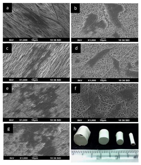
Figure 18.
SEM image of SMCs on the surface of the scaffolds after 120 h (a) PLLA alone, (b) nonwoven PLLA with 5% gelatin, (c) aligned PLLA with 5% gelatin, (d) nonwoven PLLA with 10% gelatin, (e) aligned PLLA with 10% gelatin (f) nonwoven PLLA with 20% gelatin and (g) aligned PLLA with 20% gelatin. (h)The electrospun PLLA/gelatin tubular constructs. Reproduced with permission from [149] (License number: 5232561220454).
Results showed that fiber alignment had a significant effect on endothelial cells orientation so that cells have been elongated with the direction of the fibers, and the presence of gelatin led to an enhancement of human umbilical vein endothelial cells and smooth muscle cell adhesion (Figure 18) [149]. Although PLA nanofibrous scaffolds were most commonly applied for vascular tissue engineering, there are cases in which PLA nanofibrous patches are developed for the treatment of cardiac muscle [155].
9. Conclusions and Future Trends
Electrospun PLA-based fibrous structures have shown considerable potential in biomedical applications (wound healing, controlled drug delivery, scaffolds for tissue engineering of different tissue, such as bone, nerve, and cardiac tissues) as a result of their specific properties, such as the high surface area to volume ratio, high porosity, biocompatibility, biodegradability, and sustainability. The electrospinning technique has advantages since some physical properties of nanofibers, such as fiber diameter, surface morphology, and porosity can be optimized, simply by controlling electrospinning process parameters to mimic the specific tissue in the body. Nevertheless, this technique has some limitations for biomedical applications including difficulties in the fabrication of 3D scaffolds with macropores and scaling up into industrial-scale. New strategies are used to overcome such limitations including the development of novel electrospinning systems.
For example, conventional electrospinning uses a needle spinneret for the generation of nanofibers and is associated with many limitations and drawbacks (i.e., needle clogging, limited production capacity, and low yield); needleless electrospinning (NLES) has been proposed to overcome these problems. Ultrasound-enhanced electrospinning (USES) is a novel NLES approach as a continuous orifice-less technique for the fabrication of nanofibers in which high-intensity focused ultrasound is used for the fabrication of nanofibers.
Edge electrospinning is another needleless technique that provides a straightforward approach for scaled-up production of high-quality nanofibers through the formation of many parallel jets. Near-field electrospinning (NFES) is a micro-additive manufacturing technique that uses DC electric fields to continuously print micro- and nanofibers onto moving collectors. Several notable examples of electrospinning in combination with other techniques, such as microfluidics and additive manufacturing have been performed but these combinations and their innovative products are still in their infancy.
Author Contributions
Conceptualization, H.M., B.A., S.I. and S.D.; methodology, H.M.; investigation, H.M., B.A., S.I.; writing—original draft preparation, H.M., B.A., S.I; writing—review and editing, S.D.; visualization, H.M., B.A.; supervision, S.D. All authors have read and agreed to the published version of the manuscript.
Funding
This research was funded by the Italian Ministry of University and Research (MIUR) under the framework of the project “BIONUTRA” (CUP B14I20001320005).
Institutional Review Board Statement
Not applicable.
Informed Consent Statement
Not applicable.
Data Availability Statement
Not applicable.
Conflicts of Interest
The authors declare no conflict of interest.
References
- Yang, X.; Li, L.; Yang, D.; Nie, J.; Ma, G. Electrospun Core–Shell Fibrous 2D Scaffold with Biocompatible Poly(Glycerol Sebacate) and Poly-L-Lactic Acid for Wound Healing. Adv. Fiber Mater. 2020, 2, 105–117. [Google Scholar] [CrossRef]
- Alves, P.E.; Soares, B.G.; Lins, L.C.; Livi, S.; Santos, E.P. Controlled delivery of dexamethasone and betamethasone from PLA electrospun fibers: A comparative study. Eur. Polym. J. 2019, 117, 1–9. [Google Scholar] [CrossRef]
- Fattahi, F.-S.; Khoddami, A.; Avinc, O. Poly(lactic acid) (PLA) Nanofibers for Bone Tissue Engineering. J. Text. Polym. 2019, 7, 47. [Google Scholar]
- Azimi, B.; Maleki, H.; Zavagna, L.; De la Ossa, J.G.; Linari, S.; Lazzeri, A.; Danti, S. Bio-Based Electrospun Fibers for Wound Healing. J. Funct. Biomater. 2020, 11, 67. [Google Scholar] [CrossRef]
- Tyler, B.; Gullotti, D.; Mangraviti, A.; Utsuki, T.; Brem, H. Polylactic acid (PLA) controlled delivery carriers for biomedical applications. Adv. Drug Deliv. Rev. 2016, 107, 163–175. [Google Scholar] [CrossRef]
- Fang, Y.; Zhu, X.; Wang, N.; Zhang, X.; Yang, D.; Nie, J.; Ma, G. Biodegradable core-shell electrospun nanofibers based on PLA and γ-PGA for wound healing. Eur. Polym. J. 2019, 116, 30–37. [Google Scholar] [CrossRef]
- Bi, H.; Feng, T.; Li, B.; Han, Y. In Vitro and In Vivo Comparison Study of Electrospun PLA and PLA/PVA/SA Fiber Membranes for Wound Healing. Polymers 2020, 12, 839. [Google Scholar] [CrossRef]
- Maleki, H.; Gharehaghaji, A.A.; Toliyat, T.; Dijkstra, P.J. Drug release behavior of electrospun twisted yarns as implantable medical devices. Biofabrication 2016, 8, 35019. [Google Scholar] [CrossRef]
- Maleki, H.; Mathur, S.; Klein, A. Antibacterial Ag containing core-shell polyvinyl alcohol-poly(lactic acid) nanofibers for biomedical applications. Polym. Eng. Sci. 2020, 60, 1221–1230. [Google Scholar] [CrossRef]
- Li, W.; Fan, X.; Wang, X.; Shang, X.; Wang, Q.; Lin, J.; Hu, Z.; Li, Z. Stereocomplexed micelle formation through enantiomeric PLA-based Y-shaped copolymer for targeted drug delivery. Mater. Sci. Eng. C 2018, 91, 688–695. [Google Scholar] [CrossRef]
- Maleki, H.; Rahbar, R.S.; Nazir, A. Improvement of physical and mechanical properties of electrospun poly(lactic acid) nanofibrous structures. Iran. Polym. J. 2020, 29, 841–851. [Google Scholar] [CrossRef]
- Maleki, H.; Barani, H. Stereocomplex electrospun fibers from high molecular weight of poly(L-lactic acid) and poly(D-lactic acid). J. Polym. Eng. 2019, 40, 136–142. [Google Scholar] [CrossRef]
- Jiang, F.; Yan, D.; Lin, J.; Kong, H.; Yao, Q. Implantation of multiscale silk fibers on poly(lactic acid) fibrous membrane for biomedical applications. Mater. Today Chem. 2021, 21, 100494. [Google Scholar] [CrossRef]
- Deeraj, B.D.S.; Jayan, J.S.; Saritha, A.; Joseph, K. Electrospun biopolymer-based hybrid composites. In Hybrid Natural Fiber Composites; Elsevier: Amsterdam, The Netherlands, 2021; pp. 225–252. [Google Scholar]
- Pisani, S.; Genta, I.; Dorati, R.; Modena, T.; Chiesa, E.; Bruni, G.; Benazzo, M.; Conti, B. A Design of Experiment (DOE) approach to correlate PLA-PCL electrospun fibers diameter and mechanical properties for soft tissue regeneration purposes. J. Drug Deliv. Sci. Technol. 2022, 68, 103060. [Google Scholar] [CrossRef]
- Padmakumar, S.; Joseph, J.; Neppalli, M.H.; Mathew, S.E.; Nair, S.V.; Shankarappa, S.A.; Menon, D. Electrospun Polymeric Core–sheath Yarns as Drug Eluting Surgical Sutures. ACS Appl. Mater. Interfaces 2016, 8, 6925–6934. [Google Scholar] [CrossRef] [PubMed]
- Sharifisamani, E.; Mousazadegan, F.; Bagherzadeh, R.; Latifi, M. PEG-PLA-PCL based electrospun yarns with curcumin control release property as suture. Polym. Eng. Sci. 2020, 60, 1520–1529. [Google Scholar] [CrossRef]
- Awad, N.K.; Niu, H.; Ali, U.; Morsi, Y.S.; Lin, T. Electrospun fibrous scaffolds for small-diameter blood vessels: A review. Membranes 2018, 8, 15. [Google Scholar] [CrossRef]
- Sukchanta, A.; Kummanee, P.; Nuansing, W. Development and study on mechanical properties of small diameter artificial blood vessel by using electrospinning and 3d printing. Proc. J. Phys. Conf. Ser. 2021, 2145, 12037. [Google Scholar] [CrossRef]
- Hajikhani, M.; Emam-Djomeh, Z.; Askari, G. Fabrication and characterization of mucoadhesive bioplastic patch via coaxial polylactic acid (PLA) based electrospun nanofibers with antimicrobial and wound healing application. Int. J. Biol. Macromol. 2021, 172, 143–153. [Google Scholar] [CrossRef]
- Croitoru, A.-M.; Karaçelebi, Y.; Saatcioglu, E.; Altan, E.; Ulag, S.; Aydoğan, H.K.; Sahin, A.; Motelica, L.; Oprea, O.; Tihauan, B.-M. Electrically Triggered Drug Delivery from Novel Electrospun Poly(Lactic Acid)/Graphene Oxide/Quercetin Fibrous Scaffolds for Wound Dressing Applications. Pharmaceutics 2021, 13, 957. [Google Scholar] [CrossRef]
- Echeverría, C.; Muñoz-Bonilla, A.; Cuervo-Rodríguez, R.; López, D.; Fernández-García, M. Antibacterial PLA fibers containing Thiazolium groups as wound dressing materials. ACS Appl. Bio Mater. 2019, 2, 4714–4719. [Google Scholar] [CrossRef] [PubMed]
- Peranidze, K.; Safronova, T.V.; Kildeeva, N.R. Fibrous Polymer-Based Composites Obtained by Electrospinning for Bone Tissue Engineering. Polymers 2022, 14, 96. [Google Scholar] [CrossRef] [PubMed]
- Ciarfaglia, N.; Laezza, A.; Lods, L.; Lonjon, A.; Dandurand, J.; Pepe, A.; Bochicchio, B. Thermal and dynamic mechanical behavior of poly(lactic acid)(PLA)-based electrospun scaffolds for tissue engineering. J. Appl. Polym. Sci. 2021, 138, 51313. [Google Scholar] [CrossRef]
- Lopresti, F.; Pavia, F.C.; Ceraulo, M.; Capuana, E.; Brucato, V.; Ghersi, G.; Botta, L.; La Carrubba, V. Physical and biological properties of electrospun poly(d, l-lactide)/nanoclay and poly(d, l-lactide)/nanosilica nanofibrous scaffold for bone tissue engineering. J. Biomed. Mater. Res. Part A 2021, 109, 2120–2136. [Google Scholar] [CrossRef] [PubMed]
- Herrero-Herrero, M.; Gómez-Tejedor, J.-A.; Vallés-Lluch, A. PLA/PCL electrospun membranes of tailored fibres diameter as drug delivery systems. Eur. Polym. J. 2018, 99, 445–455. [Google Scholar] [CrossRef]
- Viscusi, G.; Lamberti, E.; Vittoria, V.; Gorrasi, G. Coaxial electrospun membranes of poly(ε-caprolactone)/poly(lactic acid) with reverse core-shell structures loaded with curcumin as tunable drug delivery systems. Polym. Adv. Technol. 2021, 32, 4005–4013. [Google Scholar] [CrossRef]
- Gritsch, L.; Conoscenti, G.; La Carrubba, V.; Nooeaid, P.; Boccaccini, A.R. Polylactide-based materials science strategies to improve tissue-material interface without the use of growth factors or other biological molecules. Mater. Sci. Eng. C 2019, 94, 1083–1101. [Google Scholar] [CrossRef]
- Ghafari, R.; Scaffaro, R.; Maio, A.; Gulino, E.F.; Lo Re, G.; Jonoobi, M. Processing-structure-property relationships of electrospun PLA-PEO membranes reinforced with enzymatic cellulose nanofibers. Polym. Test. 2020, 81, 106182. [Google Scholar] [CrossRef]
- Singhvi, M.S.; Zinjarde, S.S.; Gokhale, D.V. Polylactic acid: Synthesis and biomedical applications. J. Appl. Microbiol. 2019, 127, 1612–1626. [Google Scholar] [CrossRef]
- Singhvi, M.; Gokhale, D. Biomass to biodegradable polymer (PLA). RSC Adv. 2013, 3, 13558–13568. [Google Scholar] [CrossRef]
- Jin, F.L.; Hu, R.R.; Park, S.J. Improvement of thermal behaviors of biodegradable poly(lactic acid) polymer: A review. Compos. Part B Eng. 2019, 164, 287–296. [Google Scholar] [CrossRef]
- Bayer, I.S. Thermomechanical properties of polylactic acid-graphene composites: A state-of-the-art review for biomedical applications. Materials 2017, 10, 748. [Google Scholar] [CrossRef] [PubMed]
- Polonio-Alcalá, E.; Rabionet, M.; Gallardo, X.; Angelats, D.; Ciurana, J.; Ruiz-Martínez, S.; Puig, T. PLA electrospun scaffolds for three-dimensional triple-negative breast cancer cell culture. Polymers 2019, 11, 916. [Google Scholar] [CrossRef] [PubMed]
- Xue, J.; Wu, T.; Dai, Y.; Xia, Y. Electrospinning and Electrospun Nanofibers: Methods, Materials, and Applications. Chem. Rev. 2019, 119, 5298–5415. [Google Scholar] [CrossRef] [PubMed]
- Maleki, H.; Semnani Rahbar, R.; Saadatmand, M.M.; Barani, H. Physical and morphological characterisation of poly(L-lactide) acid-based electrospun fibrous structures: Tunning solution properties. Plast. Rubber Compos. 2018, 47, 438–446. [Google Scholar] [CrossRef]
- Maleki, H.; Gharehaghaji, A.; Moroni, L.; Dijkstra, P.J. Influence of the solvent type on the morphology and mechanical properties of electrospun PLLA yarns. Biofabrication 2013, 5, 035014. [Google Scholar] [CrossRef]
- Maleki, H.; Gharehaghaji, A.A.; Criscenti, G.; Moroni, L.; Dijkstra, P.J. The influence of process parameters on the properties of electrospun PLLA yarns studied by the response surface methodology. J. Appl. Polym. Sci. 2015, 132, 41388. [Google Scholar] [CrossRef]
- Scaffaro, R.; Lopresti, F.; Botta, L. Preparation, characterization and hydrolytic degradation of PLA/PCL co-mingled nanofibrous mats prepared via dual-jet electrospinning. Eur. Polym. J. 2017, 96, 266–277. [Google Scholar] [CrossRef]
- Lopresti, F.; Carfì Pavia, F.; Vitrano, I.; Kersaudy-Kerhoas, M.; Brucato, V.; La Carrubba, V. Effect of hydroxyapatite concentration and size on morpho-mechanical properties of PLA-based randomly oriented and aligned electrospun nanofibrous mats. J. Mech. Behav. Biomed. Mater. 2020, 101, 103449. [Google Scholar] [CrossRef]
- Scaffaro, R.; Lopresti, F. Properties-morphology relationships in electrospun mats based on polylactic acid and graphene nanoplatelets. Compos. Part A Appl. Sci. Manuf. 2018, 108, 23–29. [Google Scholar] [CrossRef]
- Alharbi, H.F.; Luqman, M.; Khalil, K.A.; Elnakady, Y.A.; Abd-Elkader, O.H.; Rady, A.M.; Alharthi, N.H.; Karim, M.R. Fabrication of core-shell structured nanofibers of poly(lactic acid) and poly(vinyl alcohol) by coaxial electrospinning for tissue engineering. Eur. Polym. J. 2018, 98, 483–491. [Google Scholar] [CrossRef]
- Da Silva, T.N.; Gonçalves, R.P.; Rocha, C.L.; Archanjo, B.S.; Barboza, C.A.G.; Pierre, M.B.R.; Reynaud, F.; de Souza Picciani, P.H. Controlling burst effect with PLA/PVA coaxial electrospun scaffolds loaded with BMP-2 for bone guided regeneration. Mater. Sci. Eng. C 2019, 97, 602–612. [Google Scholar] [CrossRef] [PubMed]
- Wu, S.; Zhou, R.; Zhou, F.; Streubel, P.N.; Chen, S.; Duan, B. Electrospun thymosin Beta-4 loaded PLGA/PLA nanofiber/ microfiber hybrid yarns for tendon tissue engineering application. Mater. Sci. Eng. C 2020, 106, 110268. [Google Scholar] [CrossRef] [PubMed]
- Maleki, H.; Gharehaghaji, A.A.; Dijkstra, P.J. Electrospinning of continuous poly(L-lactide) yarns: Effect of twist on the morphology, thermal properties and mechanical behavior. J. Mech. Behav. Biomed. Mater. 2017, 71, 231–237. [Google Scholar] [CrossRef] [PubMed]
- Habibi Jouybari, M.; Hosseini, S.; Mahboobnia, K.; Boloursaz, L.A.; Moradi, M.; Irani, M. Simultaneous controlled release of 5-FU, DOX and PTX from chitosan/PLA/5-FU/g-C3N4-DOX/g-C3N4-PTX triaxial nanofibers for breast cancer treatment in vitro. Colloids Surf. B Biointerfaces 2019, 179, 495–504. [Google Scholar] [CrossRef]
- Huang, C.; Thomas, N.L. Fabricating porous poly(lactic acid) fibres via electrospinning. Eur. Polym. J. 2018, 99, 464–476. [Google Scholar] [CrossRef]
- Asghar, A.L.I.; Jeddi, A. Fabrication and characterization of hollow electrospun PLA structure through a modified electrospinning method applicable as vascular graft. Bull. Mater. Sci. 2021, 44, 158. [Google Scholar] [CrossRef]
- Qi, Z.; Yu, H.; Chen, Y.; Zhu, M. Highly porous fibers prepared by electrospinning a ternary system of nonsolvent/solvent/poly(l-lactic acid). Mater. Lett. 2009, 63, 415–418. [Google Scholar] [CrossRef]
- Natarajan, L.; New, J.; Dasari, A.; Yu, S.; Manan, M.A. Surface morphology of electrospun PLA fibers: Mechanisms of pore formation. RSC Adv. 2014, 4, 44082–44088. [Google Scholar] [CrossRef]
- Li, L.; Hashaikeh, R.; Arafat, H.A. Development of eco-efficient micro-porous membranes via electrospinning and annealing of poly(lactic acid). J. Membr. Sci. 2013, 436, 57–67. [Google Scholar] [CrossRef]
- Li, Y.; Lim, C.T.; Kotaki, M. Study on structural and mechanical properties of porous PLA nanofibers electrospun by channel-based electrospinning system. Polymer 2015, 56, 572–580. [Google Scholar] [CrossRef]
- Rezabeigi, E.; Sta, M.; Swain, M.; McDonald, J.; Demarquette, N.R.; Drew, R.A.L.; Wood-Adams, P.M. Electrospinning of porous polylactic acid fibers during nonsolvent induced phase separation. J. Appl. Polym. Sci. 2017, 134, 44862. [Google Scholar] [CrossRef]
- Li, T.; Ding, X.; Tian, L.; Hu, J.; Yang, X.; Ramakrishna, S. The control of beads diameter of bead-on-string electrospun nanofibers and the corresponding release behaviors of embedded drugs. Mater. Sci. Eng. C 2017, 74, 471–477. [Google Scholar] [CrossRef]
- Maleki, H.; Barani, H. Morphological and mechanical properties of drawn poly(l-lactide) electrospun twisted yarns. Polym. Eng. Sci. 2017, 58, 1091–1096. [Google Scholar] [CrossRef]
- Banitaba, S.N.; Amini, G.; Gharehaghaji, A.A.; Jeddi, A.A.A. Fabrication of hollow nanofibrous structures using a triple layering method for vascular scaffold applications. Fibers Polym. 2017, 18, 2342–2348. [Google Scholar] [CrossRef]
- Li, X.; Liu, Y.; Peng, H.; Ma, X.; Fong, H. Effects of hot airflow on macromolecular orientation and crystallinity of melt electrospun poly(L-lactic acid) fibers. Mater. Lett. 2016, 176, 194–198. [Google Scholar] [CrossRef]
- Mazalevska, O.; Struszczyk, M.H.; Krucinska, I. Design of vascular prostheses by melt electrospinning—Structural characterizations. J. Appl. Polym. Sci. 2013, 129, 779–792. [Google Scholar] [CrossRef]
- Nazari, T.; Garmabi, H. The effects of processing parameters on the morphology of PLA/PEG melt-electrospun fibers. Polym. Int. 2017, 67, 178–188. [Google Scholar] [CrossRef]
- Yu, S.-X.; Zheng, J.; Yan, X.; Wang, X.-X.; Nie, G.-D.; Tan, Y.-Q.; Zhang, J.; Sui, K.-Y.; Long, Y.-Z. Morphology control of PLA microfibers and spheres via melt electrospinning. Mater. Res. Express 2018, 5, 45019. [Google Scholar] [CrossRef]
- Lee, H.; Ahn, S.; Choi, H.; Cho, D.; Kim, G. Fabrication, characterization, and in vitro biological activities of melt-electrospun PLA micro/nanofibers for bone tissue regeneration. J. Mater. Chem. B 2013, 1, 3670–3677. [Google Scholar] [CrossRef]
- Chrzanowska, O.; Struszczyk, M.H.; Krucinska, I. Small diameter tubular structure design using solvent-free textile techniques. J. Appl. Polym. Sci. 2014, 131, 40147. [Google Scholar] [CrossRef]
- Bai, H.; Deng, S.; Bai, D.; Zhang, Q.; Fu, Q. Recent Advances in Processing of Stereocomplex-Type Polylactide. Macromol. Rapid Commun. 2017, 38, 1700454. [Google Scholar] [CrossRef] [PubMed]
- Kurokawa, N.; Hotta, A. Thermomechanical properties of highly transparent self-reinforced polylactide composites with electrospun stereocomplex polylactide nanofibers. Polymer 2018, 153, 214–222. [Google Scholar] [CrossRef]
- Ikada, Y.; Jamshidi, K.; Tsuji, H.; Hyon, S.H. Stereocomplex formation between enantiomeric poly(lactides). Macromolecules 1987, 20, 904–906. [Google Scholar] [CrossRef]
- Tsuji, H.; Nakano, M.; Hashimoto, M.; Takashima, K.; Katsura, S.; Mizuno, A. Electrospinning of Poly(lactic acid) Stereocomplex Nanofibers. Biomacromolecules 2006, 7, 3316–3320. [Google Scholar] [CrossRef]
- Paneva, D.; Spasova, M.; Stoyanova, N.; Manolova, N.; Rashkov, I. Electrospun fibers from polylactide-based stereocomplex: Why? Int. J. Polym. Mater. Polym. Biomater. 2019, 70, 270–286. [Google Scholar] [CrossRef]
- Jing, Y.; Quan, C.; Liu, B.; Jiang, Q.; Zhang, C. A Mini Review on the Functional Biomaterials Based on Poly(lactic acid) Stereocomplex. Polym. Rev. 2016, 56, 262–286. [Google Scholar] [CrossRef]
- Ishii, D.; Ying, T.H.; Mahara, A.; Yamaoka, T.; Lee, W.; Iwata, T.; Murakami, S. In Vivo Tissue Response and Degradation Behavior of PLLA and Stereocomplexed PLA Nanofibers In Vivo Tissue Response and Degradation Behavior of PLLA and Stereocomplexed PLA Nanofibers. Biomacromolecules 2009, 10, 237–242. [Google Scholar] [CrossRef]
- Moradkhannejhad, L.; Abdouss, M.; Nikfarjam, N.; Shahriari, M.H.; Heidary, V. The effect of molecular weight and content of PEG on in vitro drug release of electrospun curcumin loaded PLA/PEG nanofibers. J. Drug Deliv. Sci. Technol. 2020, 56, 101554. [Google Scholar] [CrossRef]
- Fattahi, F.S.; Khoddami, A.; Avinc, O. Poly(Lactic Acid) Nano-fibers as Drug-delivery Systems: Opportunities and Challenges. Nanomed. Res. J. 2019, 4, 130–140. [Google Scholar] [CrossRef]
- Santoro, M.; Shah, S.R.; Walker, J.L.; Mikos, A.G. Poly(lactic acid) nanofibrous scaffolds for tissue engineering. Adv. Drug Deliv. Rev. 2016, 107, 206–212. [Google Scholar] [CrossRef] [PubMed]
- Liu, X.; Xu, H.; Zhang, M.; Yu, D.G. Electrospun Medicated Nanofibers for Wound Healing: Review. Membranes 2021, 11, 770. [Google Scholar] [CrossRef] [PubMed]
- Pankongadisak, P.; Sangklin, S.; Chuysinuan, P.; Suwantong, O.; Supaphol, P. The use of electrospun curcumin-loaded poly(L-lactic acid) fiber mats as wound dressing materials. J. Drug Deliv. Sci. Technol. 2019, 53, 101121. [Google Scholar] [CrossRef]
- Nguyen, T.T.T.; Ghosh, C.; Hwang, S.G.; Tran, L.D.; Park, J.S. Characteristics of curcumin-loaded poly(lactic acid) nanofibers for wound healing. J. Mater. Sci. 2013, 48, 7125–7133. [Google Scholar] [CrossRef]
- Li, J.; Hu, Y.; He, T.; Huang, M.; Zhang, X.; Yuan, J.; Wei, Y.; Dong, X.; Liu, W.; Ko, F.; et al. Electrospun Sandwich-Structure Composite Membranes for Wound Dressing Scaffolds with High Antioxidant and Antibacterial Activity. Macromol. Mater. Eng. 2017, 303, 1700270. [Google Scholar] [CrossRef]
- Ilomuanya, M.O.; Adebona, A.C.; Wang, W.; Sowemimo, A.; Eziegbo, C.L.; Silva, B.O.; Adeosun, S.O.; Joubert, E.; De Beer, D. Development and characterization of collagen-based electrospun scaffolds containing silver sulphadiazine and Aspalathus linearis extract for potential wound healing applications. Appl. Sci. 2020, 2, 881. [Google Scholar] [CrossRef]
- Cui, S.; Sun, X.; Li, K.; Gou, D.; Zhou, Y.; Hu, J.; Liu, Y. Polylactide nanofibers delivering doxycycline for chronic wound treatment. Mater. Sci. Eng. C 2019, 104, 109745. [Google Scholar] [CrossRef]
- Zhang, S.; Ye, J.; Sun, Y.; Kang, J.; Liu, J.; Wang, Y.; Li, Y.; Zhang, L.; Ning, G. Electrospun fibrous mat based on silver (I) metal-organic frameworks-polylactic acid for bacterial killing and antibiotic-free wound dressing. Chem. Eng. J. 2020, 390, 124523. [Google Scholar] [CrossRef]
- Ghorbani, S.; Eyni, H.; Tiraihi, T.; Asl, L.S.; Soleimani, M.; Atashi, A.; Beiranvand, S.P.; Warkiani, M.E. Combined effects of 3D bone marrow stem cell-seeded wet-electrospun polylactic acid scaffolds on full-thickness skin wound healing. Int. J. Polym. Mater. Polym. Biomater. 2018, 67, 905–912. [Google Scholar] [CrossRef]
- Han, Y.; Jiang, Y.; Li, Y.; Wang, M.; Fan, T.; Liu, M.; Ke, Q.; Xu, H.; Yi, Z. An aligned porous electrospun fibrous scaffold with embedded asiatic acid for accelerating diabetic wound healing. J. Mater. Chem. B 2019, 7, 6125–6138. [Google Scholar] [CrossRef]
- Mouro, C.; Gomes, A.P.; Gouveia, I.C. Double-layer PLLA/PEO_Chitosan nanofibrous mats containing Hypericum perforatum L. as an effective approach for wound treatment. Polym. Adv. Technol. 2021, 32, 1493–1506. [Google Scholar] [CrossRef]
- Fan, T.; Daniels, R. Preparation and Characterization of Electrospun Polylactic Acid (PLA) Fiber Loaded with Birch Bark Triterpene Extract for Wound Dressing. AAPS PharmSciTech 2021, 22, 205. [Google Scholar] [CrossRef] [PubMed]
- Mohiti-Asli, M.; Saha, S.; Murphy, S.V.; Gracz, H.; Pourdeyhimi, B.; Atala, A.; Loboa, E.G. Ibuprofen loaded PLA nanofibrous scaffolds increase proliferation of human skin cells in vitro and promote healing of full thickness incision wounds in vivo. J. Biomed. Mater. Res. Part B Appl. Biomater. 2015, 105, 327–339. [Google Scholar] [CrossRef]
- Li, H.; Williams, G.R.; Wu, J.; Lv, Y.; Sun, X.; Wu, H.; Zhu, L.M. Thermosensitive nanofibers loaded with ciprofloxacin as antibacterial wound dressing materials. Int. J. Pharm. 2017, 517, 135–147. [Google Scholar] [CrossRef]
- Li, W.; Tan, X.; Luo, T.; Shi, Y.; Yang, Y.; Liu, L. Preparation and characterization of electrospun PLA/PU bilayer nanofibrous membranes for controlled drug release applications. Integr. Ferroelectr. 2017, 179, 104–119. [Google Scholar] [CrossRef]
- Li, W.; Wu, D.; Zhu, S.; Liu, Z.; Luo, B.; Lu, L.; Zhou, C. Sustained release of plasmid DNA from PLLA/POSS nanofibers for angiogenic therapy. Chem. Eng. J. 2019, 365, 270–281. [Google Scholar] [CrossRef]
- Zou, F.; Sun, X.; Wang, X. Elastic, hydrophilic and biodegradable poly(1, 8-octanediol-co-citric acid)/polylactic acid nanofibrous membranes for potential wound dressing applications. Polym. Degrad. Stab. 2019, 166, 163–173. [Google Scholar] [CrossRef]
- Pedersbæk, D.; Frantzen, M.T.; Fojan, P. Electrospinning of Core-Shell Fibers for Drug Release Systems. J. Self-Assem. Mol. Electron. 2017, 5, 17–30. [Google Scholar] [CrossRef][Green Version]
- Yang, C.; Yan, Z.; Lian, Y.; Wang, J.; Zhang, K. Graphene oxide coated shell-core structured chitosan/PLLA nanofibrous scaffolds for wound dressing. J. Biomater. Sci. Polym. Ed. 2020, 31, 622–641. [Google Scholar] [CrossRef]
- Augustine, R.; Zahid, A.A.; Hasan, A.; Wang, M.; Webster, T.J. CTGF loaded electrospun dual porous core-shell membrane for diabetic wound healing. Int. J. Nanomed. 2019, 14, 8573. [Google Scholar] [CrossRef]
- Pilehvar-Soltanahmadi, Y.; Dadashpour, M.; Mohajeri, A.; Fattahi, A.; Sheervalilou, R.; Zarghami, N. An Overview on Application of Natural Substances Incorporated with Electrospun Nanofibrous Scaffolds to Development of Innovative Wound Dressings. Mini-Rev. Med. Chem. 2017, 18, 414–427. [Google Scholar] [CrossRef] [PubMed]
- Kontogiannopoulos, K.N.; Assimopoulou, A.N.; Tsivintzelis, I.; Panayiotou, C.; Papageorgiou, V.P. Electrospun fiber mats containing shikonin and derivatives with potential biomedical applications. Int. J. Pharm. 2011, 409, 216–228. [Google Scholar] [CrossRef] [PubMed]
- Pillay, V.; Dott, C.; Choonara, Y.E.; Tyagi, C.; Tomar, L.; Kumar, P.; du Toit, L.C.; Ndesendo, V.M.K. A Review of the Effect of Processing Variables on the Fabrication of Electrospun Nanofibers for Drug Delivery Applications. J. Nanomater. 2013, 2013, 22. [Google Scholar] [CrossRef]
- Fayemi, O.E.; Ekennia, A.C.; Katata-Seru, L.; Ebokaiwe, A.P.; Ijomone, O.M.; Onwudiwe, D.C.; Ebenso, E.E. Antimicrobial and Wound Healing Properties of Polyacrylonitrile-Moringa Extract Nanofibers. ACS Omega 2018, 3, 4791–4797. [Google Scholar] [CrossRef] [PubMed]
- Felgueiras, H.P.; Amorim, M.T.P. Functionalization of electrospun polymeric wound dressings with antimicrobial peptides. Colloids Surf. B Biointerfaces 2017, 156, 133–148. [Google Scholar] [CrossRef] [PubMed]
- Chereddy, K.K.; Lopes, A.; Koussoroplis, S.; Payen, V.; Moia, C.; Zhu, H.; Sonveaux, P.; Carmeliet, P.; des Rieux, A.; Vandermeulen, G.; et al. Combined effects of PLGA and vascular endothelial growth factor promote the healing of non-diabetic and diabetic wounds. Nanomed. Nanotechnol. Biol. Med. 2015, 11, 1975–1984. [Google Scholar] [CrossRef] [PubMed]
- Liu, X.; Nielsen, L.H.; Kłodzińska, S.N.; Nielsen, H.M.; Qu, H.; Christensen, L.P.; Rantanen, J.; Yang, M. Ciprofloxacin-loaded sodium alginate/poly(lactic-co-glycolic acid) electrospun fibrous mats for wound healing. Eur. J. Pharm. Biopharm. 2018, 123, 42–49. [Google Scholar] [CrossRef]
- Zhao, Y.; Qiu, Y.; Wang, H.; Chen, Y.; Jin, S.; Chen, S. Preparation of Nanofibers with Renewable Polymers and Their Application in Wound Dressing. Int. J. Polym. Sci. 2016, 2016, 4672839. [Google Scholar] [CrossRef]
- Thenmozhi, S.; Dharmaraj, N.; Kadirvelu, K.; Kim, H.Y. Electrospun nanofibers: New generation materials for advanced applications. Mater. Sci. Eng. B 2017, 217, 36–48. [Google Scholar] [CrossRef]
- Tocco, I.; Zavan, B.; Bassetto, F.; Vindigni, V. Nanotechnology-based therapies for skin wound regeneration. J. Nanomater. 2012, 2012, 714134. [Google Scholar] [CrossRef]
- Pásztor, N.; Rédai, E.; Szabó, Z.-I.; Sipos, E. Preparation and Characterization of Levofloxacin-Loaded Nanofibers as Potential Wound Dressings. Acta Med. Marisiensis 2017, 63, 66–69. [Google Scholar] [CrossRef]
- Scaffaro, R.; Lopresti, F.; D’Arrigo, M.; Marino, A.; Nostro, A. Efficacy of poly(lactic acid)/carvacrol electrospun membranes against Staphylococcus aureus and Candida albicans in single and mixed cultures. Appl. Microbiol. Biotechnol. 2018, 102, 4171–4181. [Google Scholar] [CrossRef] [PubMed]
- Scaffaro, R.; Lopresti, F.; Marino, A.; Nostro, A. Antimicrobial additives for poly(lactic acid) materials and their applications: Current state and perspectives. Appl. Microbiol. Biotechnol. 2018, 102, 7739–7756. [Google Scholar] [CrossRef]
- Kenawy, E.-R.; Bowlin, G.L.; Mansfield, K.; Layman, J.; Simpson, D.G.; Sanders, E.H.; Wnek, G.E. Release of tetracycline hydrochloride from electrospun poly(ethylene-co-vinylacetate), poly(lactic acid), and a blend. J. Control. Release 2002, 81, 57–64. [Google Scholar] [CrossRef]
- Mao, Z.; Li, J.; Huang, W.; Jiang, H.; Zimba, B.L.; Chen, L.; Wan, J.; Wu, Q. Preparation of poly(lactic acid)/graphene oxide nanofiber membranes with different structures by electrospinning for drug delivery. RSC Adv. 2018, 8, 16619–16625. [Google Scholar] [CrossRef]
- Yuan, Y.; Choi, K.; Choi, S.O.; Kim, J. Early stage release control of an anticancer drug by drug-polymer miscibility in a hydrophobic fiber-based drug delivery system. RSC Adv. 2018, 8, 19791–19803. [Google Scholar] [CrossRef]
- Doustgani, A. Doxorubicin release from optimized electrospun polylactic acid nanofibers. J. Ind. Text. 2016, 47, 71–88. [Google Scholar] [CrossRef]
- Hrib, J.; Sirc, J.; Hobzova, R.; Hampejsova, Z.; Bosakova, Z.; Munzarova, M.; Michalek, J. Nanofibers for drug delivery—Incorporation and release of model molecules, influence of molecular weight and polymer structure. Beilstein J. Nanotechnol. 2015, 6, 1939–1945. [Google Scholar] [CrossRef]
- Bae, H.; Lee, J. Assembly of particle-fiber composites by electrohydrodynamic jetting using counter-charged nozzles: Independent release control. J. Ind. Eng. Chem. 2016, 40, 99–105. [Google Scholar] [CrossRef]
- Wu, S.; Wu, J.; Yue, J.; To, M.K.T.; Pan, H.; Lu, W.W.; Zhao, X. Poly(d,l-lactic acid) electrospun fibers with tunable surface nanotopography for modulating drug release profiles. Mater. Lett. 2015, 161, 716–719. [Google Scholar] [CrossRef]
- Sóti, P.L.; Nagy, Z.K.; Serneels, G.; Vajna, B.; Farkas, A.; Van Der Gucht, F.; Fekete, P.; Vigh, T.; Wagner, I.; Balogh, A.; et al. Preparation and comparison of spray dried and electrospun bioresorbable drug delivery systems. Eur. Polym. J. 2015, 68, 671–679. [Google Scholar] [CrossRef]
- Yaru, W.; Lan, X.; Jianhua, S.; Chenxu, F. Preparation, Characterization and Drug Release of Salicylic Acid Loaded Porous Electrospun Nanofibers. Recent Pat. Nanotechnol. 2018, 12, 208–217. [Google Scholar] [CrossRef] [PubMed]
- Jiang, J.; Chen, G.; Shuler, F.D.; Wang, C.H.; Xie, J. Local Sustained Delivery of 25-Hydroxyvitamin D3 for Production of Antimicrobial Peptides. Pharm. Res. 2015, 32, 2851–2862. [Google Scholar] [CrossRef] [PubMed]
- Adomavičiute, E.; Pupkevičiute, S.; Juškaite, V.; Žilius, M.; Stanys, S.; Pavilonis, A.; Briedis, V. Formation and investigation of electrospun PLA materials with propolis extracts and silver nanoparticles for biomedical applications. J. Nanomater. 2017, 2017, 8612819. [Google Scholar] [CrossRef]
- Sudakaran, S.V.; Venugopal, J.R.; Vijayakumar, G.P.; Abisegapriyan, S.; Grace, A.N.; Ramakrishna, S. Sequel of MgO nanoparticles in PLACL nanofibers for anti-cancer therapy in synergy with curcumin/β-cyclodextrin. Mater. Sci. Eng. C 2017, 17, 620–628. [Google Scholar] [CrossRef]
- Birhanu, G.; Tanha, S.; Akbari Javar, H.; Seyedjafari, E.; Zandi-Karimi, A.; Kiani Dehkordi, B. Dexamethasone loaded multi-layer poly-l-lactic acid/pluronic P123 composite electrospun nanofiber scaffolds for bone tissue engineering and drug delivery. Pharm. Dev. Technol. 2018, 24, 338–347. [Google Scholar] [CrossRef]
- Jiang, S.; Lv, J.; Ding, M.; Li, Y.; Wang, H.; Jiang, S. Release behavior of tetracycline hydrochloride loaded chitosan/poly(lactic acid) antimicrobial nanofibrous membranes. Mater. Sci. Eng. C 2016, 59, 86–91. [Google Scholar] [CrossRef]
- Nazari, T.; Moghaddam, A.B.; Davoodi, Z. Optimized polylactic acid/polyethylene glycol (PLA/PEG) electrospun fibrous scaffold for drug delivery: Effect of graphene oxide on the cefixime release mechanism. Mater. Res. Express 2019, 6, 115351. [Google Scholar] [CrossRef]
- Ma, H.; Wu, W.; Zhang, H.; Chen, G.; Han, W.; Cao, J. Preparation, structure, and release behavior of PLLA/silk fibroin micro-fiber films with controlled structure. J. Text. Inst. 2018, 110, 298–301. [Google Scholar] [CrossRef]
- Cai, N.; Han, C.; Luo, X.; Chen, G.; Dai, Q.; Yu, F. Fabrication of Core/Shell Nanofibers with Desirable Mechanical and Antibacterial Properties by Pickering Emulsion Electrospinning. Macromol. Mater. Eng. 2017, 302, 1–10. [Google Scholar] [CrossRef]
- Zhang, H.; Niu, Q.; Wang, N.; Nie, J.; Ma, G. Thermo-sensitive drug controlled release PLA core/PNIPAM shell fibers fabricated using a combination of electrospinning and UV photo-polymerization. Eur. Polym. J. 2015, 71, 440–450. [Google Scholar] [CrossRef]
- Zhu, T.; Yang, C.; Chen, S.; Li, W.; Lou, J.; Wang, J. A facile approach to prepare shell/core nanofibers for drug controlled release. Mater. Lett. 2015, 150, 52–54. [Google Scholar] [CrossRef]
- Azimi, B.; Thomas, L.; Fusco, A.; Kalaoglu-Altan, O.I.; Basnett, P.; Cinelli, P.; de Clerck, K.; Roy, I.; Donnarumma, G.; Coltelli, M.B.; et al. Electrosprayed Chitin Nanofibril/Electrospun Polyhydroxyalkanoate Fiber Mesh as Functional Nonwoven for Skin Application. J. Funct. Biomater. 2020, 11, 62. [Google Scholar] [CrossRef] [PubMed]
- Amini, A.R.; Laurencin, C.T.; Nukavarapu, S.P. Bone Tissue Engineering: Recent Advances and Challenges. Crit. Rev. Biomed. Eng. 2012, 40, 363. [Google Scholar] [CrossRef]
- Azimi, B.; Milazzo, M.; Lazzeri, A.; Berrettini, S.; Uddin, M.J.; Qin, Z.; Buehler, M.J.; Danti, S. Electrospinning Piezoelectric Fibers for Biocompatible Devices. Adv. Healthc. Mater. 2020, 9, e1901287. [Google Scholar] [CrossRef]
- Puppi, D.; Chiellini, F.; Piras, A.M.; Chiellini, E. Polymeric materials for bone and cartilage repair. Prog. Polym. Sci. 2010, 35, 403–440. [Google Scholar] [CrossRef]
- Holzwarth, J.M.; Ma, P.X. Biomimetic nanofibrous scaffolds for bone tissue engineering. Biomaterials 2011, 32, 9622–9629. [Google Scholar] [CrossRef]
- Azimi, B.; Bafqi, M.S.S.; Fusco, A.; Ricci, C.; Gallone, G.; Bagherzadeh, R.; Donnarumma, G.; Uddin, M.J.; Latifi, M.; Lazzeri, A.; et al. Electrospun ZnO/Poly(Vinylidene Fluoride-Trifluoroethylene) Scaffolds for Lung Tissue Engineering. Tissue Eng. Part A 2020, 26, 1312–1331. [Google Scholar] [CrossRef]
- Xie, J.; Shen, H.; Yuan, G.; Lin, K.; Su, J. The effects of alignment and diameter of electrospun fibers on the cellular behaviors and osteogenesis of BMSCs. Mater. Sci. Eng. C 2021, 120, 111787. [Google Scholar] [CrossRef]
- Rattier, B.D.; Hoffman, A.S.; Schoen, F.J.; Lemons, J.E. Biomaterials Science: An Introduction to Materials in Medicine. J. Clin. Eng. 1996, 22, 26. [Google Scholar] [CrossRef]
- Lu, Z.; Wang, W.; Zhang, J.; Bártolo, P.; Gong, H.; Li, J. Electrospun highly porous poly(L-lactic acid)-dopamine-SiO2 fibrous membrane for bone regeneration. Mater. Sci. Eng. C 2020, 117, 111359. [Google Scholar] [CrossRef] [PubMed]
- Mohammadi, M.; Alibolandi, M.; Abnous, K.; Salmasi, Z.; Jaafari, M.R.; Ramezani, M. Fabrication of hybrid scaffold based on hydroxyapatite-biodegradable nanofibers incorporated with liposomal formulation of BMP-2 peptide for bone tissue engineering. Nanomed. Nanotechnol. Biol. Med. 2018, 14, 1987–1997. [Google Scholar] [CrossRef] [PubMed]
- Ye, K.; Liu, D.; Kuang, H.; Cai, J.; Chen, W.; Sun, B.; Xia, L.; Fang, B.; Morsi, Y.; Mo, X. Three-dimensional electrospun nanofibrous scaffolds displaying bone morphogenetic protein-2-derived peptides for the promotion of osteogenic differentiation of stem cells and bone regeneration. J. Colloid Interface Sci. 2019, 534, 625–636. [Google Scholar] [CrossRef] [PubMed]
- Li, X.; Cheng, R.; Sun, Z.; Su, W.; Pan, G.; Zhao, S.; Zhao, J.; Cui, W. Flexible bipolar nanofibrous membranes for improving gradient microstructure in tendon-to-bone healing. Acta Biomater. 2017, 61, 204–216. [Google Scholar] [CrossRef] [PubMed]
- Patel, D.K.; Dutta, S.D.; Hexiu, J.; Ganguly, K.; Lim, K.T. Bioactive electrospun nanocomposite scaffolds of poly(lactic acid)/cellulose nanocrystals for bone tissue engineering. Int. J. Biol. Macromol. 2020, 162, 1429–1441. [Google Scholar] [CrossRef]
- Xu, Z.; Liu, P.; Li, H.; Zhang, M.; Wu, Q. In vitro study on electrospun lecithin-based poly(L-lactic acid) scaffolds and their biocompatibility. J. Biomater. Sci. Polym. Ed. 2020, 31, 2285–2298. [Google Scholar] [CrossRef]
- Wang, X.; Yan, H.; Shen, Y.; Tang, H.; Yi, B.; Qin, C.; Zhang, Y. Shape Memory and Osteogenesis Capabilities of the Electrospun Poly(3-Hydroxybutyrate- co-3-Hydroxyvalerate) Modified Poly(l-Lactide) Fibrous Mats. Tissue Eng. Part A 2021, 27, 142–152. [Google Scholar] [CrossRef]
- Magiera, A.; Markowski, J.B.; Menaszek, E.; Pilch, J.; Blazewicz, S. PLA-Based Hybrid and Composite Electrospun Fibrous Scaffolds as Potential Materials for Tissue Engineering. J. Nanomater. 2017, 2017, 9246802. [Google Scholar] [CrossRef]
- Chen, J.; Zhang, T.; Hua, W.; Li, P.; Wang, X. 3D Porous poly(lactic acid)/regenerated cellulose composite scaffolds based on electrospun nanofibers for biomineralization. Colloids Surf. A Physicochem. Eng. Asp. 2020, 585, 124048. [Google Scholar] [CrossRef]
- Xu, T.; Yang, H.; Yang, D.; Yu, Z.-Z. Polylactic Acid Nanofiber Scaffold Decorated with Chitosan Islandlike Topography for Bone Tissue Engineering. ACS Appl. Mater. Interfaces 2017, 9, 21094–21104. [Google Scholar] [CrossRef]
- Abazari, M.F.; Zare Karizi, S.; Hajati-Birgani, N.; Kohandani, M.; Torabinejad, S.; Nejati, F.; Nasiri, N.; Maleki, M.H.; Mohajerani, H.; Mansouri, V. Curcumin-loaded PHB/PLLA nanofibrous scaffold supports osteogenesis in adipose-derived stem cells in vitro. Polym. Adv. Technol. 2021, 32, 3563–3571. [Google Scholar] [CrossRef]
- Wang, S.F.; Wu, Y.C.; Cheng, Y.C.; Hu, W.W. The Development of Polylactic Acid/Multi-Wall Carbon Nanotubes/Polyethylene Glycol Scaffolds for Bone Tissue Regeneration Application. Polymers 2021, 13, 1740. [Google Scholar] [CrossRef] [PubMed]
- Shih, Y.H.; Yang, J.C.; Shih, Y.H.; Yang, J.C.; Li, S.H.; Yang, W.C.V.; Chen, C.C. Bio-electrospinning of poly(l-lactic acid) hollow fibrous membrane. Text. Res. J. 2012, 82, 602–612. [Google Scholar] [CrossRef]
- Tian, L.; Prabhakaran, M.P.; Hu, J.; Chen, M.; Besenbacher, F.; Ramakrishna, S. Coaxial electrospun poly(lactic acid)/silk fibroin nanofibers incorporated with nerve growth factor support the differentiation of neuronal stem cells. RSC Adv. 2015, 5, 49838–49848. [Google Scholar] [CrossRef]
- Martin, R.A.; Wendling, M.; Mohrenweiser, B.; Qian, Z.; Zhao, F.; Mullins, M.E. Formation of aligned core/sheath microfiber scaffolds with a poly-L-lactic acid (PLLA) sheath and a conductive poly(3,4-ethylenedioxythiophene) (PEDOT) core. J. Mater. Res. 2019, 34, 1931–1943. [Google Scholar] [CrossRef]
- Kurobe, H.; Maxfield, M.W.; Tara, S.; Rocco, K.A.; Bagi, P.S.; Yi, T.; Udelsman, B.; Zhuang, Z.W.; Cleary, M.; Iwakiri, Y.; et al. Development of small diameter nanofiber tissue engineered arterial grafts. PLoS ONE 2015, 10, e0120328. [Google Scholar] [CrossRef]
- Wang, S.; Zhang, Y.; Wang, H.; Yin, G.; Dong, Z. Fabrication and properties of the electrospun polylactide/silk fibroin-gelatin composite tubular scaffold. Biomacromolecules 2009, 10, 2240–2244. [Google Scholar] [CrossRef]
- Shalumon, K.T.; Deepthi, S.; Anupama, M.S.; Nair, S.V.; Jayakumar, R.; Chennazhi, K.P. Fabrication of poly(l-lactic acid)/gelatin composite tubular scaffolds for vascular tissue engineering. Int. J. Biol. Macromol. 2015, 72, 1048–1055. [Google Scholar] [CrossRef]
- Luiz, A.; Santos, D.; Duarte, M.A.T.; Pezzin, S.H.; Silva, L.; Domingues, J.A. Preparation of porous poly(lactic acid) fibers by medium field electrospinning for tissue engineering applications. Mater. Res. 2020, 23, 20190468. [Google Scholar] [CrossRef]
- Ravichandran, R.; Venugopal, J.R.; Sundarrajan, S.; Mukherjee, S.; Sridhar, R.; Ramakrishna, S. Minimally invasive injectable short nanofibers of poly(glycerol sebacate) for cardiac tissue engineering. Nanotechnology 2012, 23, 385102. [Google Scholar] [CrossRef]
- Gu, X.; Ding, F.; Yang, Y.; Liu, J. Construction of tissue engineered nerve grafts and their application in peripheral nerve regeneration. Prog. Neurobiol. 2011, 93, 204–230. [Google Scholar] [CrossRef] [PubMed]
- Chiono, V.; Tonda-Turo, C. Trends in the design of nerve guidance channels in peripheral nerve tissue engineering. Prog. Neurobiol. 2015, 131, 87–104. [Google Scholar] [CrossRef] [PubMed]
- Butcher, J.T.; Mahler, G.J.; Hockaday, L.A. Aortic valve disease and treatment: The need for naturally engineered solutions. Adv. Drug Deliv. Rev. 2011, 63, 242–268. [Google Scholar] [CrossRef] [PubMed]
- Eschenhagen, T.; Eder, A.; Vollert, I.; Hansen, A. Physiological aspects of cardiac tissue engineering. Am. J. Physiol. Heart Circ. Physiol. 2012, 303, H133–H143. [Google Scholar] [CrossRef]
Publisher’s Note: MDPI stays neutral with regard to jurisdictional claims in published maps and institutional affiliations. |
© 2022 by the authors. Licensee MDPI, Basel, Switzerland. This article is an open access article distributed under the terms and conditions of the Creative Commons Attribution (CC BY) license (https://creativecommons.org/licenses/by/4.0/).