Article

Isolation of Jannaschia sedimins sp. nov. from East Coast of China: Bacterial Taxonomy and Antimicrobial Resistance Analysis

1
Marine College, Shandong University, Weihai 264209, China
2
Weihai Municipal Ecological and Environmental Monitoring Center, Weihai 264209, China
3
Infection Program and Department of Microbiology, Biomedicine Discovery Institute, Monash University, Melbourne, VIC 3800, Australia
4
State Key Laboratory of Microbial Technology, Shandong University, Qingdao 266200, China
*
Author to whom correspondence should be addressed.
Appl. Sci. 2022, 12(6), 2883; https://doi.org/10.3390/app12062883
Submission received: 18 November 2021 / Revised: 2 March 2022 / Accepted: 7 March 2022 / Published: 11 March 2022
(This article belongs to the Special Issue New Frontiers into Environmental Microbiology)

Abstract

:
A Gram-stain-negative, facultatively aerobic, pink and oval bacterium, designed OS4T, was isolated from a sediment sample taken from a coastal zone in China. The growth of OS4T occurred at 20–37 °C (optimal 25 °C), pH 7.0–8.5 (optimal pH 8.0), in 0–5.0% (w/v) NaCl (optimal 2.0%). According to the phylogenetic analysis, strain OS4T showed the highest sequence similarity (96.04%) with Jannaschia aquimarina GSW-M26T and shared 94.98% similarity with the type species of genus Jannaschia-strain J. helgolandensis 14858T. Chemotaxonomic analysis showed that the sole respiratory quinone was ubiquinone 10, and the major fatty acids (>5.0%) included C18:1 ω6c/ω7c, C18:0, and C10:0 3OH. The polar lipids consist of three phospholipids, two unknown amino-lipids, and four unknown glycerolipids. The DNA G + C content was 72.7 mol%. Based on the evidence presented in this study, strain OS4T represents a novel species of the genus Jannaschia, for which the name Jannaschia sedimins sp. nov. is proposed. The type strain is OS4T (=KCTC 82508T = MCCC 1K03755T). Both the phenotypic and the genetic analysis on the antimicrobial resistance genes indicate that OS4T is resistant to a wide range of classes of antibiotics, which highlights that the ocean could potentially serve as the natural reservoir of antimicrobial resistance genes.

1. Introduction

The emergence of antibiotic-resistant pathogens and their resistance traits is an epic global challenge and plays a heavy burden on the global health care system [1]. Most studies have been increasingly devoted to the clinic and novel antimicrobial drug development; however, there is an urgent need to understand the origin and ecology of antibiotic resistance [2,3,4]. Previous studies suggested that the aquatic environment (e.g., lakes, rivers, and coastlines) can serve as natural reservoirs of antibiotic resistance genes [5,6]. A detailed investigation on the taxonomy of novel microorganisms isolated from the aquatic environment and how the genomic and phenotypic features contribute to antibiotic resistance may have a significant potential on better understanding the mechanisms of antimicrobial resistance in the natural environment.
The genus Jannaschia, a member of family Rhodobacteraceae of the phylum Proteobacteria, was originally proposed by Wagner-Döbler et al. [7]. Bacteria of the genus Jannaschia are Gram-stain-negative and oval, and the major respiratory quinone is ubiquinone 10 (Q-10). At the time of writing, 12 species are assigned to this genus, including J. cystaugens (Adachi et al., 2004), J. rubra (Macián et al., 2005), J. seosinensis (Choi et al., 2006), J. donghaensis (Yoon et al., 2007), J. pohangensis (Kim et al., 2008), J. seohaensis (Yoon et al., 2010), J. aquimarina (Park and Yoon 2012), J. faecimaris (Jung and Yoon 2014), J. confluentis (Park et al., 2018), J. formosa (Zhang et al., 2019), J. marina (Chen et al., 2021) [7,8,9,10,11,12,13,14,15,16,17,18]. Without exception, members of the genus Jannaschia are all isolated from marine environments [7]. In order to link the bacterial taxonomy to the research on antibiotic resistance genes (ARGs), the function of this newly discovered species, which was isolated from the marine environment, will be used as an example. In this study, a beige, non-motile, and facultatively aerobic bacterium, strain OS4T, is proposed as a novel species of the genus Jannaschia. A comparative analysis of the antimicrobial resistance of strain OS4T and the reference strains J. helgolandensis 14858T and J. aquimarina GSW-M26T may contribute to a better understanding of the horizontal transfer of antibiotic resistance genes in the natural environment.

2. Materials and Methods

2.1. Study Site and Sampling

Samples were collected from the Golden Bay (E 122.057274, N 37.541204) (Figure 1), which is located in Weihai, Shandong Province, China. As a coastal city, the marine environment plays an important role in agricultural activities and human health. To investigate the antibiotic-resistance-related genes from a marine environment, Golden Bay, one of the most representative beaches in Weihai was selected as the sampling site.

2.2. Bacterial Strains, Isolation, and Cultivation

Sediment from Golden Bay was carefully collected into a 50 mL falcon tube for bacteria isolation. In brief, 1 g of wet sediment was weighted and blended with 99 mL of sterile seawater. The blended sediment was then shaken vigorously with sterilized glass beads. The isolation of the bacteria was conducted by plating 100 μL of the bacterial cell suspension of the sample and/or its serial seawater dilution on marine agar (MA) and followed with 5–7 days of incubation at 28 °C. The isolated strain OS4T was stored in tryptone soy broth (TSB) with 1% (w/v) saline and 15% (v/v) glycerol at 80 °C. Prior to the experiments, strain OS4T was sub-cultured onto MA and incubated overnight at 28 °C [19].

2.3. Phenotypic and Physiological Characterization

The morphology and physiology were examined after incubation at 28 °C for 2 days on MA. Gram staining was performed as previously described [19,20]. Briefly, crystal violet stain was mixed with the culture; iodine solution was then used to fix the dye after washing off the stain with water. The fixed bacterial smear was added with a drop of decolorizer and then rinsed with water and counterstained with fuchsin solution. Microscope examination was carried out after washing off the fuchsin solution and air-drying by using light microscopy (Ci-L; Nikon). The observation of cell size, morphology, and flagellation were conducted by using transmission electron microscopy (JEM-1200; JEOL) at the State Key Laboratory of Bio-Fibers and Eco-Textiles (Qingdao University, China). Bacterial motility was determined in marine broth (MB) supplemented with 0.3% agar according to the method previously described by Bernardet et al. [21]. Growth temperature range and optimum growth temperature were examined by observing bacterial colony morphology regularly on MA at a range of temperatures (0, 4, 10, 15, 20, 25, 28, 30, 33, 37, 40, and 45 °C). The effect of the NaCl concentration was evaluated by bacterial growth on a range of NaCl-containing modified MA, which consists of 0.1% yeast extract, 0.5% peptone, 1.8% agar, 0.32% MgSO4, 0.22% MgCl2, 0.12% CaCl2, 0.07% KCl, 0.02% NaHCO3, and distilled water. The pH range and optimal pH for growth was tested in MB with the addition of appropriate buffers, including 20 mM MES (pH 5.5 and 6.0), PIPES (pH 6.5 and 7.5), HEPES (pH 7.5 and 8.0), Tricine buffer (pH 8.5), and CAPSO (pH 9.0, 9.5 and 10.0), to obtain the final PH ranging from 5.5 to 10.0. Anaerobic growth was examined by incubating bacteria on MA in an anaerobic jar (e.g., anaerobic,10% H2, 10% CO2, and 80% N2; microaerobic, 5% O2, 10% CO2, and 85% N2), with or without 0.1% (KNO3). Bacterial catalase and oxidase activity was tested by bubble formation in 3.0% (v/v) H2O2 and reaction with oxidase reagent (bioMérieux) according to the manufacturer’s instructions, respectively [19]. The antibiotic susceptibility of OS4T, 14858T, and GSW-M26T was assessed on an MA agar plate at 28 °C according to the Clinical and Laboratory Standards Institute (CLSI) guidelines [22]. In brief, antimicrobial disks of clarithromycin (15 µg), ceftriaxone (30 µg), penicillin (10 µg), erythromycin (15 µg), cefotaxime (30 µg), rifampicin (5 µg), norfloxacin (15 µg), polymyxin B (300 µg), vancomycin (30 µg), lincomycin (2 µg), kanamycin (30 µg), carbenicillin (100 µg), neomycin (30 µg), chloramphenicol (30 µg), gentamycin (10 µg), ofloxacin (5 µg), oxytetracycline (30 µg), streptomycin (10 µg), ampicillin (10 µg), tobramycin (10 µg), tetracycline (30 µg), and clindamycin (2 µg) were placed on an MA agar plate with 0.5 McFarland bacterial suspension. Following an 18 h incubation, measurement of the inhibitory zone sizes was conducted using a ruler. Hydrolysis of starch, lipids, casein, CM-cellulose, and Tweens (20, 40, 60, and 80) was examined on MA, as previously described by Du et al. [23]. Additional physiological or biochemical characteristics were tested by using API 20E, API ZYM (bioMérieux), and Biolog GEN III kits (BiOLOG) according to the manufacturer’s instructions (except NaCl, which was adjusted to 3%). All tests were conducted in triplicate.

2.4. Phylogenetic Analysis and Genomic Characterization

The 16S rRNA gene sequence was amplified by PCR with the universal primers 27F and 1492R [24]. The purified PCR product was then cloned into a pMD18-T vector (Takara) based on the TA cloning method, followed by the transformation of the ligation product into Escherichia coli DH5α cells [19]. The positive clone was selected from the antibiotic-containing plate and sent to sequence by BGI (Qingdao, China). The assembled 16S rRNA gene sequence of strain OS4T was submitted to the National Centre for Biotechnology Information (NCBI) GenBank database to search for similar sequences using the blast algorithm and confirmed by using the EzBioCloud database [25]. Multiple sequences were aligned using the CLUSTAL_X program and the phylogenetic position was determined by phylogenetic trees which were constructed using the neighbor-joining (NJ), maximum-likelihood (ML), and maximum-parsimony (MP) algorithms in MEGA version 7.0 [26,27]. The reliability of the relationships was ensured by performing bootstrap analyses based on 1000 replications.
Genomic DNA was isolated and purified by using a genomic DNA extraction kit (Takara, Beijing, China). NanoDrop 2000 (Thermo Fisher Scientific, Wilmington, DE, USA) was employed to assess DNA purity and Qubit 3.0 (Thermo Fisher Scientific, Wilmington, DE, USA) was used to validate the genome DNA content. Library preparations were prepared with the NEBNext Ultra DNA Library Prep kit for Illumina and were sequenced using an Illumina HiSeq 2500 (Beijing Novogene Bioinformatics Technology, Beijing, China). Illumina reads were trimmed and assembled using trimmomatic (v.0.38) and SPAdes (v.3.15.3) [28,29]. The genome sequence was deposited in the GenBank database. The G + C content of the chromosomal DNA was calculated using a genome sequence. Average nucleotide identity (ANI) and in silico DNA-DNA hybridization (DDH) were calculated by FastANI (v.1.33) and GGDC (v.3.0) for those strains with the highest similarities [30]. The detection of the antibiotic-resistance gene (ARG) was conducted by using RGI (v.5.1.0); the results were compared and confirmed with the experimental results from an antibiotic susceptibility test [31].

2.5. Chemotaxonomic Characterization

The chemotaxonomic features of strain OS4T were compared with stains of J. helgolandensis 14858T and J. aquimarina GSW-M26T. Briefly, the cells of these three strains were harvested by centrifuge (4000 g) after 3 days culturing in MB and subjected to freeze-drying. Extraction of fatty acid methyl esters (FAMEs) was conducted according to the MIDI standard protocol (Sherlock Microbial Identification System, version 6.1) from 30 mg freeze-dried bacterial cells. The extracted samples were then analyzed by using Agilent 6890N gas chromatograph (GC). The GC/MS were automatically determined with the Microbial Identification system (TSBA40 database) [32]. Analyses on respiratory quinones of strain OS4T were performed by extracting respiratory quinones from 300 mg freeze-dried bacterial cells using the two-stage method [33]. The extracted respiratory quinones were then transferred to an injection vial for high-performance liquid chromatography (HPLC) analysis [34]. Polar lipids were extracted from 50 mg freeze-dried bacterial cells using a chloroform/methanol assay according to Zhao and Kroppenstedt, as previously described [19,34]. The extracted polar lipids were analyzed by Two-dimensional thin-layer chromatography (2D TLC) [35].

3. Results and Discussion

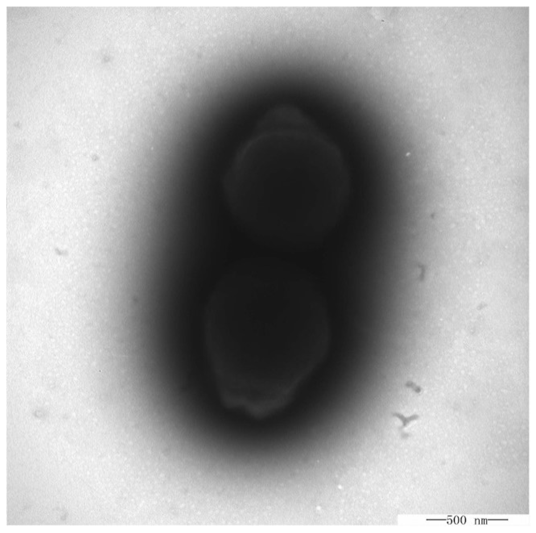
3.1. Morphological, Physiologial, and Biochemical Characteristics

Transmission electron microscope (TEM) observation of OS4T indicated that OS4T was non-flagella, which was similar to the relatively closed strains J. helgolandensis 14858T and J. aquimarina GSW-M26T (Figure 2) [7,18]. The Gram-staining result indicates that OS4T is Gram-negative, which is consistent with species in the genus Jannaschia. The TEM showed that OS4T was oval with a length of 0.8–2.0 µm and a width of 0.5–0.8 µm. The growth was poor at 20 °C, ranging to 37 °C, with an optimum temperature of 25 °C. Poor growth was observed in the absence of sea salts. The pH range tolerated for growth was 7.0–8.5 (with 8.0 as optimum pH) (Table 1). Strain OS4T showed the ability to hydrolyze alginate and casein but not starch and tween (20, 40, 60, and 80) (Table 1). According to the API ZYM results, strain OS4T and its relative species showed positive reactions for alkaline phosphatase, esterase (C4), and esterase lipase (C8), and only OS4T has lipase (C14), leucine arylamidase, valine arylamidase, cystine arylamidase, and acid phosphatase activities. The antibiotic susceptibility results showed that strain OS4T was susceptible to clarithromycin, ceftriaxone, penicillin, erythromycin, cefotaxime, rifampicin, norfloxacin, polymyxin B, vancomycin, and lincomycin but resistant to kanamycin, carbenicillin, neomycin, chloramphenicol, gentamycin, ofloxacin, oxytetracycline, streptomycin, ampicillin, tobramycin, tetracycline, and clindamycin (Table S1). Broad-spectrum profiles of ARG abundance were conducted with the assembled genome by CARD GRI. Including ompA, adeL,S, tetA,B, AAC(6′)-Iak, lptD, bcr-1, and optrA, 86 hits belonging to 10 ARG types were detected in OS4T, which is consistent with the phenotypic antibiotic resistance measurements (Table S2). The detailed phenotypical features and species description are shown in Table 1.

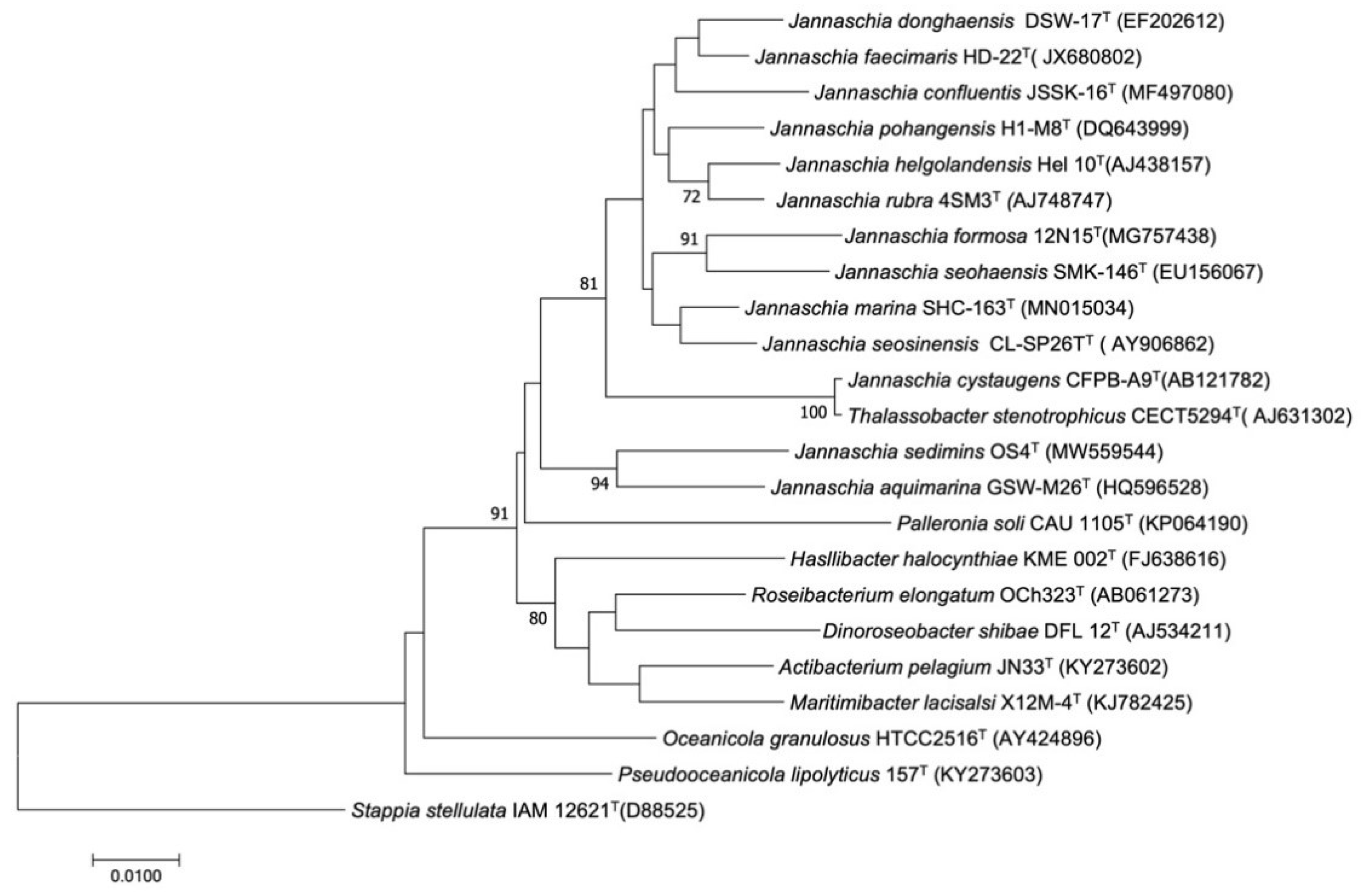
3.2. Phylogenetic Analysis

A near-complete 16S rRNA gene sequence (1500 bp) was obtained from the PCR amplification and sequencing. Similarity analysis on the 16S rRNA gene sequence revealed that OS4T shared the highest similarity with J. aquimarina GSW-M26T (96.04%). Moreover, OS4T shared 94.98% similarity with the type species of genus Jannaschia-strain J. helgolandensis 14858T on the 16S rRNA gene sequence. The ANIs of OS4T to 14858T and GSW-M26T were 77.69% and 78.93%, respectively. In silico DDH analysis showed that the estimated DDHs on a generalized linear model were 20.10% and 19.20%, respectively. Phylogenetic analysis showed that strain OS4T was placed nearest to J. aquimarina GSW-M26T within the genus Jannaschia (Figure 3 and Figure S1). Altogether, both the 16S rRNA comparison analysis and the phylogenetic relationships indicate that strain OS4T is more likely a novel species of the genus Jannaschia. The G + C content was determined to be 72.7 mol% for strain OS4T.

3.3. Chemotaxonomic Characteristics

The major fatty acids (>1%) of strain OS4T were C18:1 ω6c/ω7c (73.3%), C18:0 (9.9%), C10:0 3OH (5.4%), C20:1 ω7c (2.9%), and C16:0 (1.6%) which were different in composition to those of other species in genus Jannaschia (Table 2). The main differences in fatty acids were listed in Table 2, where strains J. helgolandensis 14858T and J. aquimarina GSW-M26T contained branched chain fatty acids (i.e., iso-C15:0, anteiso-C15:0, iso-C16:0, iso-C17:0, and anteiso-C17:0), which were not detected in strain OS4T. Analysis of the respiratory quinone of strains OS4T, 14858T, and GSW-M26T indicated that Q-10 predominated in those strains [7,18]. The polar lipid composition of OS4T was virtually identified (Figure S2), with the phospholipids mainly comprising phosphatidylcholine (PC), phosphatidylethanolamine (PE), and phosphatidylglycerol (PG). Two unknown amino-lipids (i.e., aminolipids (AL) and aminophospholipid (APL)) and four unknown glycerolipids (i.e., glycerolipids 1–4) were also being detected in OS4T (Figure S2). Taken together, both respiratory quinone and polar lipid of OS4T were in line with the previous reports on the genus Jannaschia, and the major fatty acids clearly distinguished OS4T from other Jannaschia strains (e.g., 14858T and GSW-M26T).

3.4. Description of Jannaschia Sediminis sp. nov.

Jannaschia sediminis (se.di’mi.nis. L. gen. n. sediminis, of sediment). The cells of strain OS4T are oval, Gram-stain-negative, facultatively aerobic, non-motile, and approximate with a length of 0.8–2.0 µm and a width of 0.5–0.8 µm. The colonies on MA are circular, pink, and smooth and approximately (<1 mm) in diameter after 3 days of incubation at 25 °C. Growth occurs at 20–37 °C (optimal 25 °C), at pH 7.0–8.5 (optimal pH 8.0), in 0–5.0% (w/v) NaCl (optimal 2.0%). The cells are positive for oxidase and catalase activity. Positive results for the hydrolysis of alginate, casein and negative for hydrolyzing starch and tweens (20, 40, 60, and 80) were observed. Strain OS4T shows activities of alkaline phosphatase, esterase (C4) and esterase lipase (C8), lipase (C14), leucine arylamidase, valine arylamidase, cystine arylamidase, and acid phosphatase. In the carbon source oxidation test, positive results were obtained for sucrose. The major fatty acids are C18:1 ω6c/ω7c, C18:0, C10:0 3OH, C20:1 ω7c, and C16:0. The sole respiratory quinone is Q-10. The polar lipids consist of three phospholipids (i.e., phosphatidylcholine (PC), phosphatidylethanolamine (PE), phosphatidylglycerol (PG)), two unknown amino-lipids (i.e., aminolipids (AL) and aminophospholipid (APL)), and four unknown glycerolipids. The DNA G + C content of OS4T is 72.7 mol%. The type strain, OS4T (=KCTC 82508T = MCCC 1K03755T), was isolated from a marine sediment sample obtained from the Golden Bay of Weihai, China (E122.057274, N 37.541204). The GenBank accession number for the 16S rRNA gene sequence of strain OS4T is MW559544, and the draft genome data have been deposited in GenBank under the accession number JAFCAR000000000.
In addition, strain OS4T is resistant to a wide range of antibiotics (i.e., kanamycin, carbenicillin, neomycin, chloramphenicol, gentamycin, ofloxacin, oxytetracycline, streptomycin, ampicillin, tobramycin, tetracycline, and clindamycin). The wide range of antimicrobial resistant genes identified in OS4T highlights that those aquatic systems could potentially serve as a natural reservoir for ARGs and should be raising concerns about controlling the transfer and spread of antibiotic resistance from ocean non-pathogenic bacteria to pathogens.

Supplementary Materials

The following are available online at https://www.mdpi.com/article/10.3390/app12062883/s1, Table S1: antimicrobial susceptibility of strain OS4T and the related Jannaschia sAPLcies, Table S2: broad-spectrum profiles of ARG abundance predicted with OS4T whole genome, Figure S1: maximal likelihood phylogenetic tree of strain OS4T and other related species, Figure S2: the polar lipid composition of OS4T.

Author Contributions

Conceptualization, S.S. and G.C.; methodology, S.S., L.Z., S.L. and G.C.; software, J.Z.; validation, S.S., L.Z., S.L. and J.Z.; formal analysis, S.S., L.Z. and S.L.; investigation, S.S. and L.Z.; writing—original draft preparation, S.S.; writing—review and editing, S.S., L.Z., S.L., J.Z. and G.C.; visualization, L.Z.; supervision, G.C.; project administration, G.C.; funding acquisition, G.C. All authors have read and agreed to the published version of the manuscript.

Funding

This research was funded by the Shandong Provincial Key Research and Development Program, grant number 2019GSF109007.

Institutional Review Board Statement

Not applicable.

Informed Consent Statement

Not applicable.

Data Availability Statement

The draft genome data have been deposited in GenBank under the accession number JAFCAR000000000.

Acknowledgments

We thank Zongjun Du and his group members for the technical and resources support.

Conflicts of Interest

The authors declare no conflict of interest.

References

  1. Prestinaci, F.; Pezzotti, P.; Pantosti, A. Antimicrobial resistance: A global multifaceted phenomenon. Pathog. Glob. Health 2015, 109, 309–318. [Google Scholar] [CrossRef] [PubMed] [Green Version]
  2. Fair, R.J.; Tor, Y. Antibiotics and bacterial resistance in the 21st century. Perspect. Med. Chem. 2014, 6, S14459. [Google Scholar] [CrossRef] [PubMed] [Green Version]
  3. Miethke, M.; Pieroni, M.; Weber, T.; Brönstrup, M.; Hammann, P.; Halby, L.; Arimondo, P.B.; Glaser, P.; Aigle, B.; Bode, H.B. Towards the sustainable discovery and development of new antibiotics. Nat. Rev. Chem. 2021, 5, 726–749. [Google Scholar] [CrossRef] [PubMed]
  4. Davies, J.; Davies, D. Origins and evolution of antibiotic resistance. Microbiol. Mol. Biol. Rev. 2010, 74, 417–433. [Google Scholar] [CrossRef] [PubMed] [Green Version]
  5. Suzuki, S.; Pruden, A.; Virta, M.; Zhang, T. Antibiotic resistance in aquatic systems. Front. Microbiol. 2017, 8, 14. [Google Scholar] [CrossRef] [PubMed] [Green Version]
  6. Michael, I.; Rizzo, L.; McArdell, C.; Manaia, C.; Merlin, C.; Schwartz, T.; Dagot, C.; Fatta-Kassinos, D. Urban wastewater treatment plants as hotspots for the release of antibiotics in the environment: A review. Water Res. 2013, 47, 957–995. [Google Scholar] [CrossRef] [PubMed] [Green Version]
  7. Wagner-Döbler, I.; Rheims, H.; Felske, A.; Pukall, R.; Tindall, B.J. Jannaschia helgolandensis gen. nov., sp. nov., a novel abundant member of the marine Roseobacter clade from the North Sea. Int. J. Syst. Evol. Microbiol. 2003, 53, 731–738. [Google Scholar] [CrossRef] [PubMed]
  8. Adachi, M.; Kanno, T.; Okamoto, R.; Shinozaki, A.; Fujikawa-Adachi, K.; Nishijima, T. Jannaschia cystaugens sp. nov., an Alexandrium (Dinophyceae) cyst formation-promoting bacterium from Hiroshima Bay, Japan. Int. J. Syst. Evol. Microbiol. 2004, 54, 1687–1692. [Google Scholar] [CrossRef] [PubMed]
  9. Macián, M.; Arahal, D.; Garay, E.; Ludwig, W.; Schleifer, K.; Pujalte, M. Jannaschia rubra sp. nov., a red-pigmented bacterium isolated from sea water. Int. J. Syst. Evol. Microbiol. 2005, 55, 649–653. [Google Scholar] [CrossRef] [Green Version]
  10. Choi, D.H.; Yi, H.; Chun, J.; Cho, B.C. Jannaschia seosinensis sp. nov., isolated from hypersaline water of a solar saltern in Korea. Int. J. Syst. Evol. Microbiol. 2006, 56, 45–49. [Google Scholar] [CrossRef] [Green Version]
  11. Jung, Y.-T.; Yoon, J.-H. Jannaschia faecimaris sp. nov., isolated from a tidal flat sediment. Int. J. Syst. Evol. Microbiol. 2014, 64, 945–951. [Google Scholar] [CrossRef] [PubMed]
  12. Chen, S.; He, M.; Lai, Q.; Xu, Y.; Shang, C. Jannaschia marina sp. nov., isolated from the gut of a gastropod, Onchidium reevesii. Int. J. Syst. Evol. Microbiol. 2021, 71, 004756. [Google Scholar] [CrossRef] [PubMed]
  13. Zhang, R.; Wang, C.; Wang, X.-T.; Mu, D.-S.; Du, Z.-J. Jannaschia formosa sp. nov., isolated from marine saltern sediment. Int. J. Syst. Evol. Microbiol. 2019, 69, 2037–2042. [Google Scholar] [CrossRef] [PubMed]
  14. Park, S.; Choi, S.J.; Won, S.-M.; Yoon, J.-H. Jannaschia confluentis sp. nov., isolated from the junction between the ocean and a freshwater spring. Int. J. Syst. Evol. Microbiol. 2018, 68, 669–674. [Google Scholar] [CrossRef] [PubMed]
  15. Yoon, J.-H.; Kang, S.-J.; Park, S.; Oh, T.-K. Jannaschia donghaensis sp. nov., isolated from seawater of the East Sea, Korea. Int. J. Syst. Evol. Microbiol. 2007, 57, 2132–2136. [Google Scholar] [CrossRef] [PubMed]
  16. Kim, B.-Y.; Yoo, S.-H.; Weon, H.-Y.; Jeon, Y.-A.; Hong, S.-B.; Go, S.-J.; Stackebrandt, E.; Kwon, S.-W. Jannaschia pohangensis sp. nov., isolated from seashore sand in Korea. Int. J. Syst. Evol. Microbiol. 2008, 58, 496–499. [Google Scholar] [CrossRef] [PubMed] [Green Version]
  17. Yoon, J.-H.; Kang, S.-J.; Park, S.; Oh, K.-H.; Oh, T.-K. Jannaschia seohaensis sp. nov., isolated from a tidal flat sediment. Int. J. Syst. Evol. Microbiol. 2010, 60, 191–195. [Google Scholar] [CrossRef] [PubMed]
  18. Park, S.; Yoon, J.-H. Jannaschia aquimarina sp. nov., isolated from seawater. Int. J. Syst. Evol. Microbiol. 2012, 62, 2631–2636. [Google Scholar] [CrossRef] [PubMed]
  19. Zhao, J.-X.; Liu, Q.-Q.; Zhou, Y.-X.; Chen, G.-J.; Du, Z.-J. Alkalimarinus sediminis gen. nov., sp. nov., isolated from marine sediment. Int. J. Syst. Evol. Microbiol. 2015, 65, 3511–3516. [Google Scholar] [CrossRef] [PubMed]
  20. Smibert, R. Phenotypic characterization. Methods Gen. Mol. Bacteriol. 1994, 607–654. [Google Scholar]
  21. Bowman, J.P. Description of Cellulophaga algicola sp. nov., isolated from the surfaces of Antarctic algae, and reclassification of Cytophaga uliginosa (ZoBell and Upham 1944) Reichenbach 1989 as Cellulophaga uliginosa comb. nov. Int. J. Syst. Evol. Microbiol. 2000, 50, 1861–1868. [Google Scholar] [CrossRef] [PubMed] [Green Version]
  22. Clinical and Laboratory Standards Institute. Performance Standards for Antimicrobial Susceptibility Testing, 30th ed.; CLSI Supplement M100; Clinical and Laboratory Standards Institute: Malvern, PA, USA, 2020. [Google Scholar]
  23. Du, Z.-J.; Wang, Y.; Dunlap, C.; Rooney, A.P.; Chen, G.-J. Draconibacterium orientale gen. nov., sp. nov., isolated from two distinct marine environments, and proposal of Draconibacteriaceae fam. nov. Int. J. Syst. Evol. Microbiol. 2014, 64, 1690–1696. [Google Scholar] [CrossRef] [PubMed] [Green Version]
  24. Stackebrandt, E.; Goodfellow, M. Nucleic Acid Techniques in Bacterial Systematics; Wiley: Hoboken, NJ, USA, 1991. [Google Scholar]
  25. Yoon, S.-H.; Ha, S.-M.; Kwon, S.; Lim, J.; Kim, Y.; Seo, H.; Chun, J. Introducing EzBioCloud: A taxonomically united database of 16S rRNA gene sequences and whole-genome assemblies. Int. J. Syst. Evol. Microbiol. 2017, 67, 1613. [Google Scholar] [CrossRef] [PubMed]
  26. Thompson, J.D.; Gibson, T.J.; Plewniak, F.; Jeanmougin, F.; Higgins, D.G. The CLUSTAL_X windows interface: Flexible strategies for multiple sequence alignment aided by quality analysis tools. Nucleic Acids Res. 1997, 25, 4876–4882. [Google Scholar] [CrossRef] [PubMed] [Green Version]
  27. Kumar, S.; Stecher, G.; Tamura, K. MEGA7: Molecular evolutionary genetics analysis version 7.0 for bigger datasets. Mol. Biol. Evol. 2016, 33, 1870–1874. [Google Scholar] [CrossRef] [PubMed] [Green Version]
  28. Bolger, A.M.; Lohse, M.; Usadel, B. Trimmomatic: A flexible trimmer for Illumina sequence data. Bioinformatics 2014, 30, 2114–2120. [Google Scholar] [CrossRef] [PubMed] [Green Version]
  29. Prjibelski, A.; Antipov, D.; Meleshko, D.; Lapidus, A.; Korobeynikov, A. Using SPAdes de novo assembler. Curr. Protoc. Bioinform. 2020, 70, e102. [Google Scholar] [CrossRef]
  30. Jain, C.; Rodriguez-R, L.M.; Phillippy, A.M.; Konstantinidis, K.T.; Aluru, S. High throughput ANI analysis of 90K prokaryotic genomes reveals clear species boundaries. Nat. Commun. 2018, 9, 5114. [Google Scholar] [CrossRef] [Green Version]
  31. Alcock, B.P.; Raphenya, A.R.; Lau, T.T.; Tsang, K.K.; Bouchard, M.; Edalatmand, A.; Huynh, W.; Nguyen, A.-L.V.; Cheng, A.A.; Liu, S. CARD 2020: Antibiotic resistome surveillance with the comprehensive antibiotic resistance database. Nucleic Acids Res. 2020, 48, D517–D525. [Google Scholar] [CrossRef]
  32. Sasser, M. Identification of Bacteria by Gas Chromatography of Cellular Fatty Acids; MIDI, Inc.: Newark, NY, USA, 1990; Available online: http://natasha.eng.usf.edu/gilbert/courses/Biotransport%20Phenomena/pdf/bacteria_gc_1.pdf (accessed on 11 February 2022).
  33. Tindall, B.J.; Sikorski, J.; Smibert, R.A.; Krieg, N.R. Phenotypic characterization and the principles of comparative systematics. Methods Gen. Mol. Microbiol. 2007, 330–393. [Google Scholar] [CrossRef]
  34. Kroppenstedt, R.M. Separation of bacterial menaquinones by HPLC using reverse phase (RP18) and a silver loaded ion exchanger as stationary phases. J. Liq. Chromatogr. 1982, 5, 2359–2367. [Google Scholar] [CrossRef]
  35. Minnikin, D.; O’donnell, A.; Goodfellow, M.; Alderson, G.; Athalye, M.; Schaal, A.; Parlett, J. An integrated procedure for the extraction of bacterial isoprenoid quinones and polar lipids. J. Microbiol. Methods 1984, 2, 233–241. [Google Scholar] [CrossRef]
Figure 1. Map showing the sampling location in Weihai, eastern China.
Applsci 12 02883 g001
Figure 2. Transmission electron microscope (bar, 500 nm) images of cells of strain OS4T.
Applsci 12 02883 g002
Figure 3. Phylogenetic relationship of strain OS4T and other related species. Neighbor-joining phylogenetic tree based on 16S rRNA gene sequences, showing the relationships of strain OS4T and related species. Bootstrap values ≥ 70% (based on 1000 replications) are shown at branching nodes. Stappia stellulata IAM12621T (GenBank accession number, D88525) was used as an outgroup. Bar, 0.01 substitutions per nucleotide position.
Applsci 12 02883 g003
Table 1. Differential phenotypic characteristics of strain OS4T and its related species.
CharacteristicsOS4T14858TGSW-M26T
Cell size (µm)0.5–0.8 × 0.8–2.00.7–1.1 × 1.9–3.20.5–0.7 × 0.5–4.0
pH range for growth7.0–8.5 (8.0)7.0–8.0 (7.5)5.5–8.0 (7.0–8.0)
NaCl range (%)0–5.0 (2.0)1–7.0 (2.0)0–7.0 (2)
Growth temperature (°C)20–37 (25)15–30 (25)15–37 (30)
Catalase+++
Oxidase+++
Hydrolysis of
Starch-++
Alginate+--
Tween (20, 40, 60, 80)--+
Casein+--
Utilization of:
Sucrose+-+
yesD-xylose-++
D-glucose--+
D-fructose ++
Aesculinw--
D-mannose--+
5-ketogluconate-++
Enzyme activity
alkaline phosphatase+++
esterase(C4)+++
esterase lipase (C8)+W+
lipase (C14)+-+
leucine arylamidase+-+
valine arylamidase+-+
cystine arylamidaseW-W
Trypsin---
α-chymotrypsin---
acid phosphatase+-+
naphtol-AS-BI-phosphohydrolase--W
α-galactosidase--+
β-galactosidase---
β-glucuronidase---
α-glucosidase+-+
β-glucosidase--+
Values in bracket represent the optimal temperature, pH, and NaCl for growth. All the data were obtained from this study unless otherwise indicated +, Positive; -, negative; w, weakly positive.
Table 2. Cellular fatty acid contents (%) of strain OS4T and the related Jannaschia species.
Fatty AcidOS4T14858TGSW-M26T
Saturated
C16:01.61.5-
C18:09.9--
Branched chain
iso-C10:0---
iso-C14:0-1.1-
iso-C15:0-47.94.9
anteiso-C15:0-35.350.7
iso-C16:0-1.920.5
iso-C17:0-21.9
anteiso-C17:0-3.619.4
Hydroxy
C10:0 3OH5.4--
Monounsaturated
C20:1 ω7c2.9--
Summed features
C18:1 ω6c/ω7c73.3--
Publisher’s Note: MDPI stays neutral with regard to jurisdictional claims in published maps and institutional affiliations.

