Article

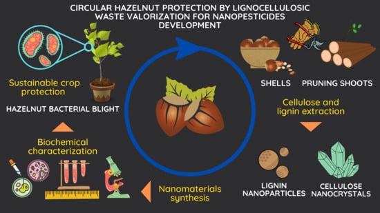
Circular Hazelnut Protection by Lignocellulosic Waste Valorization for Nanopesticides Development

1
Department of Agriculture and Forest Sciences (DAFNE), University of Tuscia, Via San Camillo de Lellis snc, 01100 Viterbo, Italy
2
Department of Pharmaceutical Sciences (DSF), University of Perugia, Via del Liceo 1, 06123 Perugia, Italy
*
Authors to whom correspondence should be addressed.
Appl. Sci. 2022, 12(5), 2604; https://doi.org/10.3390/app12052604
Submission received: 1 February 2022 / Revised: 23 February 2022 / Accepted: 28 February 2022 / Published: 2 March 2022
(This article belongs to the Special Issue Nanotechnology in Agriculture: New Opportunities and Perspectives)

Abstract

:

Featured Application

bacterial plant disease control; hazelnut lignocellulosic waste valorization to obtain nanomaterials.

Abstract

Hazelnut represents a relevant agro-food supply chain in many countries worldwide. Several biological adversities threaten hazelnut cultivation, but among them bacterial blight is one of the most feared and pernicious since its control can be achieved only by prevention through the observation of good agricultural practices and the use of cupric salts. The aim of this work was to evaluate the lignocellulosic biomasses obtained from hazelnut pruning and shelling residues as a renewable source of cellulose nanocrystals and lignin nanoparticles and to investigate their antimicrobial properties against hazelnut bacterial blight. Cellulose nanocrystals were obtained through an acid hydrolysis after a chemical bleaching, while lignin nanoparticles were synthesized by a solvent–antisolvent method after an enzymatic digestion. Both collected nanomaterials were chemically and morphologically characterized before being tested for their in vitro and in vivo antibacterial activity and biocompatibility on hazelnut plants. Results indicated the selected biomasses as a promising starting material for lignocellulosic nanocarriers synthesis, confirming at the same time the potential of cellulose nanocrystals and lignin nanoparticles as innovative tools to control hazelnut bacterial blight infections without showing any detrimental effects on the biological development of treated hazelnut plants.

Graphical Abstract

1. Introduction

Hazelnut (Corylus avellana L.) is one of the most important crops belonging to the food sector of dried fruit. According to the Food and Agriculture Organization, in 2018 the global hazelnut sown area was 660,000 hectares, with an average annual production of 835,000 t of nuts. Most of the production is focused in the Mediterranean area (Turkey and Italy represent, respectively, the first and the second world producer), but this trend, in terms of soil occupation and nuts production, has been constantly growing in recent years, driven by the confectionery industry. Many countries all over the world have now become promising hazelnut investors, such as the USA, Azerbaijan, Chile, Spain, China, and France [1]. However, the rise of hazelnut cultivation could be slowed down by the increasingly present biological adversities. Among them, bacterial blight is one of the most dangerous in hazelnut crops. This disease is provoked by the Gram negative bacterium Xanthomonas arboricola pv. corylina (Xac), which is extremely dangerous, especially for young orchards [2,3]. Due to its harmfulness, this pathogen is classified by the European and Mediterranean Plant-protection Organization (EPPO) as an A2 quarantine pest [4]. Xac was originally first described on C. maxima but now is classified as a hazelnut pathogen that has spread worldwide [5,6]. Xac is able to provoke symptoms on almost every organ of the plant; however, the most susceptible ones are buds, twigs, leaves and (sometimes) fruits. Symptoms appear as cankers, blackening, and oily necrosis and can lead the plant to death in the most severe cases. Xac in field can be spread by rain or infected tools among plants; it penetrates the host through the stomata of leaves, buds and new shoots [7,8]. Infected buds represent the major source of inoculum during spring; the microorganism is able to overwinter only on infected plants, not in soil, although its ability of surviving on fallen leaves for several months has been reported. The main risks are for seedlings and young plants, while adult ones rarely are killed by Xac, even though the dieback of new shoots and the cankers can heavily damage the production [6,9]. The counteracting and spread of control measurements of hazelnut bacterial blight rely on prevention: the use of healthy and disease-free plant material in nurseries is the most relevant strategy, while in-field disinfecting pruning tools and destroying symptomatic branches are equally important as using cupric salts to prevent infections [10,11]. Copper and copper-based products, as for other bacterial plant diseases, nowadays still play an important role in crop-protection strategies, but recently copper was enlisted by EU as a candidate for substitution [12,13]. Due to the environmental risks associated with its use in field, European Commission has recently limited the amount of usable copper to 4 kg per hectare per year (maximum 28 kg per hectare in seven years) (Executive Regulation 2018/1981 of 13 December 2018) [14,15]. Finding alternative compounds to copper and its derivatives is an increasingly urgent priority [16,17]. The implementation of sustainable approaches in the plant-protection sector has in fact become a major research topic over the last years. Several aspects of agriculture system can become more innovative and efficient by relying, for example, on nanotechnology tools [18,19]. Indeed, nanotechnology combined with circular economy can represent a green alternative to conventional plant-protection strategies by ensuring food security in a sustainable way [20,21]. This novel approach allows one to obtain highly technological nanometric materials, such as nanopesticides, starting from agro-industrial wastes [22,23,24]. The residues of a supply chain may become part of the strategy for the sustainable growth and protection of the supply chain itself. The combination of bio-based additives, as active ingredients, and functional nanostructures, as carriers, is one of the most promising tools [25]. Nanoscale structures such as organic nanomaterials can be utilized in the smart delivery of plant-protection inputs, for the encapsulation and entrapment of agrochemicals such as fertilizers, pesticides, herbicides, plant growth regulators, and other active substances, allowing for the slow uptake of active ingredients and reducing the amount of agrochemical applications by minimizing the input and waste [26,27]. Lignocellulosic biomass can represent an importance source of organic nano-compounds [28,29]. Between the different types, the lignocellulosic biomass counts as the most abundant group representing almost 70% of the total plant biomass. Lignocellulosic biomass can originate from diverse sources such as agricultural and forestry residues, municipal wastes and several crops [30]. Hazelnut is a crop of economic importance for the Mediterranean region. During the hazelnut supply chain, huge amounts of lignocellulosic biomasses come from pruning residues and from nut-processing factories (shelling). The chemical composition of shoots (taken as a hardwood) and shells are as follows, respectively: 40–55% and 25–30% cellulose, 25–40% and 25–30% hemicellulose and 15–25% and 30–40% lignin, representing an attractive source of organic compounds of interest [31,32]. Indeed, these lignocellulosic residues can be valorized through the extraction of organic fractions, such as polyphenols and other secondary metabolites, and converted into high value-added compounds, following the circular economy concept [33,34,35]. Cellulose and lignin can be extracted too and further converted in their nanoscale form, exhibiting many more exploitable features with respect to their bulk counterpart [36]. Cellulose nanocrystals (CNC) have some different interesting properties: high stiffness, low density (approximately 1.57 g cm−3), very low thermal expansion coefficient, high elastic modulus around 150 GPa; moreover, in the last few years, attention has been paid to the surface modifications of CNC, specifically to the abundant hydroxyl groups present on their surfaces. Chemical functionalizations have been investigated in order to use CNC in the biomedicine, pharmaceutical, and agri-food sectors for the release of specific drugs and active substances, especially since their application seems to do not show cytotoxicity or any other harmful effects on human health [37,38,39,40,41]. Very recently, lignin-based polymers have also attracted the interest of the research community: indeed, lignin can be used as a low-cost, eco-friendly reinforcement to prepare high-performance nanocomposites [42,43,44]. Different nanoscale lignin compounds can be synthesized by different chemical/physical approaches [45,46,47]. Lignin nanoparticles (LNP), thanks to their antioxidant activity and other characteristics, can find applications both as active ingredients and as a nanocarrier structure [48,49]. Lignin and LNP, as well as for cellulose and CNC, can be isolated and extracted from agricultural waste, adding value to the whole supply-chain [50,51,52]. In this context, the proposed research gives an original and innovative perspective on the valorization of the agricultural biomasses as starting matrices for organic nanomaterials synthesis, investigating at the same time their potential role as nanopesticides. Indeed, the possibility of using hazelnut lignocellulosic waste as a novel source for synthetizing CNC and LNP was investigated for the first time in this study. Obtained nanomaterials were chemically and morphologically characterized and assayed for their antimicrobial properties respect to Xac. As far as we know, the results represent the first report of CNC antimicrobial activity on a plant pathogenic bacterium in the literature. These studies highlight the potential of hazelnut CNC and LNP as active nanomaterials and plant-compatible nanocarriers, perfectly meeting the concept of circular economy and sustainable plant-protection strategies.

2. Materials and Methods

2.1. Materials

The hazelnut tree pruning residues (HP) and the hazelnut shells (HS) were obtained from local organic farmers of the area (Lazio region, Viterbo province) after the harvesting season in 2020. All the chemical reagents used in this work were supplied by Sigma-Aldrich, Inc. (Taufkirchen, Germany).

2.2. Nanomaterialssynthesis

2.2.1. Cellulose Extraction and CNC Synthesis

Hazelnut tree pruning shoots (HP) and hazelnut shells (HS) were used for cellulose extraction and purification. HP were firstly washed in distilled water several times in order to remove the powder and environmental contaminations. Subsequently, the pruning residues were treated for 72 h at room temperature (RT) with 1% w/v of sodium hydroxide (NaOH) (purity ≥ 98%, pellets, anhydrous, CAS No: 1310-73-2) solution and then for 2 h at 98 °C with 5% w/v of NaOH solution to remove the external lignin components. The soaked lignocellulosic materials were chopped with a mechanic shredder in order to obtain individualized fibers (length around 10–15 mm). The fibers were dried at 60 °C for 24 h. Afterwards, the bleaching treatment has been performed: the fibers were bleached with 5% w/v of sodium chlorite (NaClO2) (purity ≥ 80%, powder, CAS No: 7758-19-2) (liquor ratio 1:50) at 80 °C for 120 min [53]. The solution pH was adjusted to 3.5 by means acetic acid before the addition of the biomass. At the end of bleaching treatment, the material was washed several times and treated with sodium bisulfate (NaHSO4) (technical grade, powder, powder, CAS No: 7681-38-1) solution at 5% w/v for 30 min at RT (liquor ratio 1:40), washed again with deionized water, and treated with NaOH solution at 17.5% w/v for 20 min at RT (liquor ratio 1:50) in order to remove lignin and hemicellulose components [22]. The resulting material was finally dried at 60° C for 24 h. Dried and cleaned HS were used for cellulose extraction. HS were milled in a bench-scale mill to make the biomass more prone/accessible to chemical pretreatments and sifted with a 500-mesh sieve to maintain the size uniformity of powdered shells. These powdered HS were subjected to chemical treatment for cellulose purification as previously reported [54,55]. HS were first subjected to dewaxing by using a boiling solution of benzene:methanol (80:20 ratio) (purity ≥ 99.8%, liquid, anhydrous, CAS No: 71-43-2) (purity ≥ 99.8%, liquid, anhydrous, CAS No: 67-56-1) for 6 h with magnetic stirring (powder-solution ratio 3:10). This step was carried out to remove the surface wax, to allow the solvents to better penetrate the matrix [56]. The dewaxed shells were treated with 1 M NaOH solution under magnetic stirring at 85°C for 4 h to first remove the excess of lignin and hemicelluloses (powder-solution ratio 1:10). This alkali treatment was repeated for 3–4 times, and the final product was washed with distilled water at each step. The resulting material was subjected to the bleaching step with 1.7% sodium chlorite solution at 80 °C for 6 h, to further remove lignin (powder-solution ratio 1:20). The pH was adjusted to 3.5 by means of glacial acetic acid (purity ≥ 99%, liquid, CAS No: 64-19-7). This process was repeated for 3–4 times too, and the obtained product was filtered and dried for 24 h at 60 °C. The final dried products obtained from pruning and shells residue are considered as chemically purified cellulose (CPC-HP and CPC-HS). The cellulose nanocrystals were synthesized from chemical purified cellulose extracted from both pruning shoots and hazelnut shells by following the acid hydrolysis method. Twenty grams of CPC-HP and CPC-HS were treated with 175 mL of 64 wt% sulphuric acid (purity ≥ 95–98%, liquid, CAS No: 7664-93-9) solution for 30 min at 45 °C under vigorous stirring. The reaction was stopped by pouring the hydrolyzed cellulose in distilled water. The aqueous suspension of hydrolyzed nanocellulose had been left to precipitate for 2 days. The supernatant was discarded, while the remaining aqueous CNCs suspension was dialyzed with nitrocellulose membranes against distilled water for several days, until the pH reached 6–7. Finally, the resultant cellulose nanocrystals aqueous suspension was ultrasonicated by means of a tip sonicator for 8 min [22]. The final yield was calculated according to the method reported as UNI EN ISO 638:2009. Data were obtained from three independent replicates.

2.2.2. Lignin Extraction and LNP Synthesis

Hazelnut tree pruning shoots (HP) and hazelnut shells (HS) were used for the synthesis of lignin nanoparticles (LNP). The lignin fractions were extracted, and LNP were synthesized from lignin-rich residues from both pruning shoots and hazelnut shells by using a solvent–antisolvent method [57,58]. HP were collected and mechanically treated with a shredder in order to obtain a more homogeneous and defibrated matrix. Dried and cleaned HS were milled and sieved to maintain the size uniformity of powdered shells. A second physical treatment has been performed at high pressure and high temperature to make the HP and HS matrices more accessible to the following treatments: 100 g of defibrated HP and HS were firstly washed to remove the powder and environmental contaminations and put in 200 mL of distillated water and subjected to a pressure of 1.5 atm and a temperature of 121 °C for 30 min, by using an autoclave. The described step represents a steam explosion-like process, allowing for the opening of the biomass fibers and making them more accessible to the enzyme action. The steamed matrices (S-HP and S-HS) were dried at 60 °C for 24 h.
Dried steamed matrices of HP and HS have been subjected to the enzymatic pre-treatment in order to obtain a lignin-rich biomass. Batches of 10 g of S-HP and S-HS were incubated in 100 mL of 50 mM sodium citrate buffer with a dosage of 10% and 20% w/w enzyme/dried pulp, respectively. The enzymatic cocktail used was a blend of cellulases, β-glucosidases and hemicellulases (Cellic Ctec2) [59]. The enzymatic-driven depolymerization has been conducted for 96 h at 50 °C and 150 to 100 rpm. The action of the enzymatic cocktail is aimed to selectively depolymerize accessible reducing sugars such as cellulose and hemicellulose, and the selective release of those components allows one to obtain a residue that is rich in lignin (even though still presenting fractions of cellulose and hemicellulose). The enzymatic-treated matrices (E-HP and E-HS) were then filtered, washed from the buffer in excess and dried at 60 °C for 24 h. The final dried products obtained from pruning and shells residues enzymatically treated are considered as starting lignin-rich residues. The lignin fractions were extracted, and LNP were synthesized from lignin-rich residues from both pruning shoots and hazelnut shells by following the solvent–antisolvent method.
The lignin-rich residues E-HP and E-HS were processed according to the protocol taken from Ma et al. (2020) to obtain three different lignin fractions [60]. At first, batches of 60 g dried E-HP and E-HS were mixed with 600 mL 100% ethanol (purity ≥ 99.8%, liquid, CAS No: 64-17-5) under constant stirring (300 rpm, 2 h) at room temperature. Subsequently, the insoluble and soluble fractions were separated by vacuum filtration: the insoluble residue was washed with 20 mL 100% ethanol three times, then dried, to be further processed; the soluble fraction was denoted as the L100 (dissolved in 100% ethanol) fraction, respectively, L100-HP and L100-HS. The insoluble residues were further processed by mixing with 70% ethanol, under the conditions of 300 rpm, 2 h, and room temperature. The insoluble and soluble fractions were again separated by vacuum filtration: the insoluble residue was washed with 20 mL 70% ethanol for three times, then dried, to be further processed, while the soluble fraction was denoted as the L70 (dissolved in 70% ethanol) fraction, respectively, L70-HP and L70-HS. The dried insoluble residues from the dissolution of 70% ethanol solution, denoted as the LF-HP and LF-HS fraction, were again further processed by mixing with 70% ethanol, under the conditions of 300 rpm, 2 h, and room temperature, then concentrated and directly used for the preparation of LNP, as detailed below. The ethanol used was recovered and reused for further fractionations.
LNP were prepared based on the solvent–antisolvent method, which was described in previous works [60,61]. Briefly, the soluble fractions L100-HP, L100-HS, L70-HP and L70-HS were concentrated and freeze-dried. Afterwards, 1 g of each dried lignin fraction was weighted and dissolved in a 500 mL of a pure ethanol solution by constant stirring (800 rpm, 1 h) at room temperature. The last soluble fractions obtained from the processing of LF-HP and LF-HS with 70% ethanol were filtered, ethanol was added up to 500 mL and the mixture was used as such. Subsequently, a volume of 3.5× of water was added dropwise (2.33 mL min−1) to the lignin/ethanol mixture at room temperature, with continuous stirring (800 rpm, 2 h) provided, resulting in the gradual formation of LNP. Finally, the LNP were obtained by dialyzing the LNP containing ethanol/water suspension with deionized water (periodically replaced with fresh deionized water) for one week. The three lignin samples, namely, L100, L70, and LF, were converted to LNP, which were named LNP 100, LNP 70, and LNP F, respectively. The final yield was calculated according to the method reported as UNI EN ISO 638:2009. Data were obtained from three independent replicates.

2.3. Nanomaterials Characterization
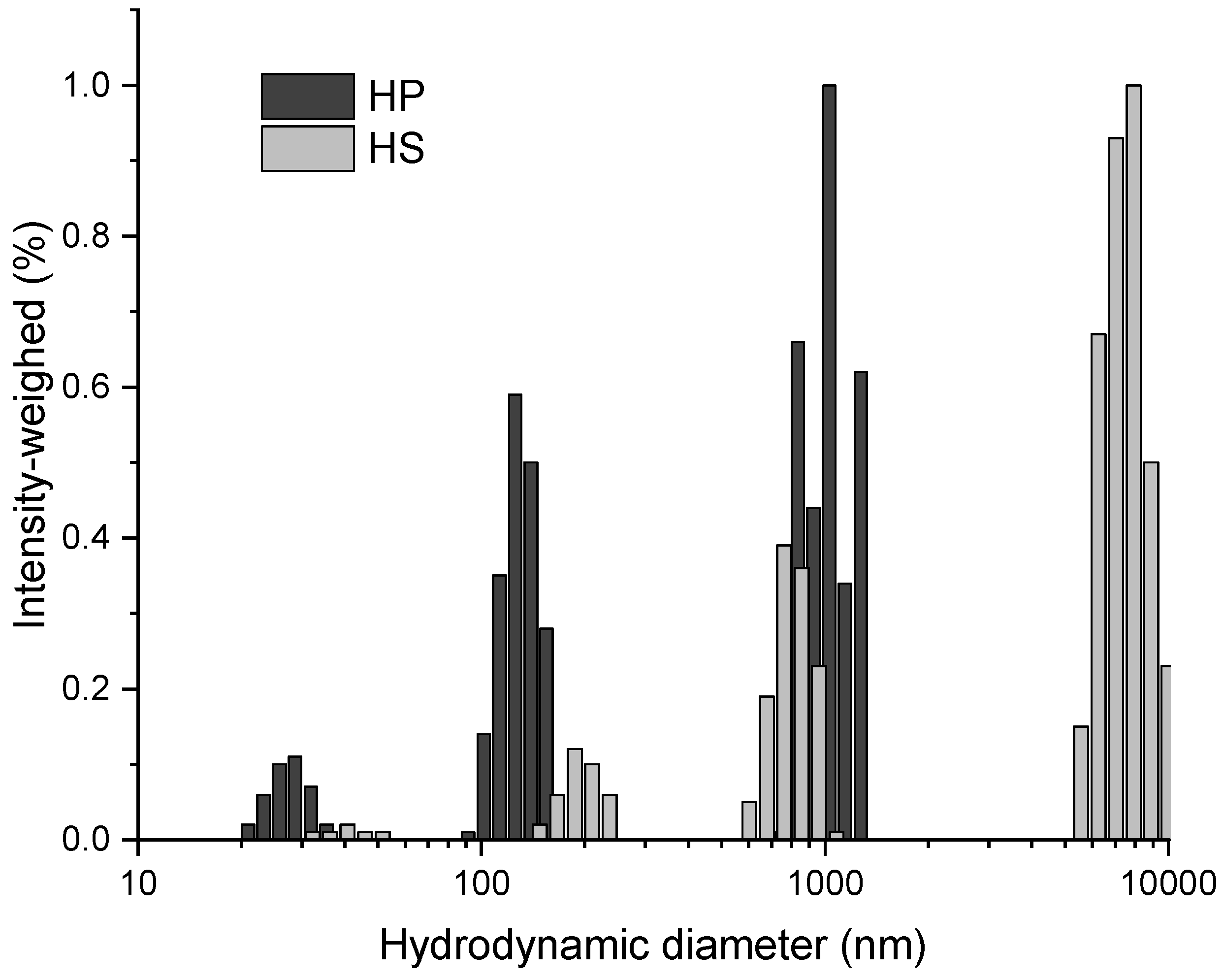
2.3.1. CNC Size

The size distributions of extracted hazelnut CNC samples were determined by dynamic light scattering measurements. A Nicomp 380 ZLS photocorrelator (PSS, Santa Barbara, CA, USA) equipped with a 55 mW He-Ne Coherent Innova 70-3 Laser source (λ = 654 nm) and APD detector was employed. Samples were opportunely diluted with water and analyzed at 20 °C. Gaussian and Nicomp algorithms were employed for fitting the autocorrelation function time decay data. Analyses were run to convergence of the fitting algorithms, and the respective mean diameters were expressed as Gaussian and Nicomp mean hydrodynamic diameters (GMHD and NMHD, respectively).

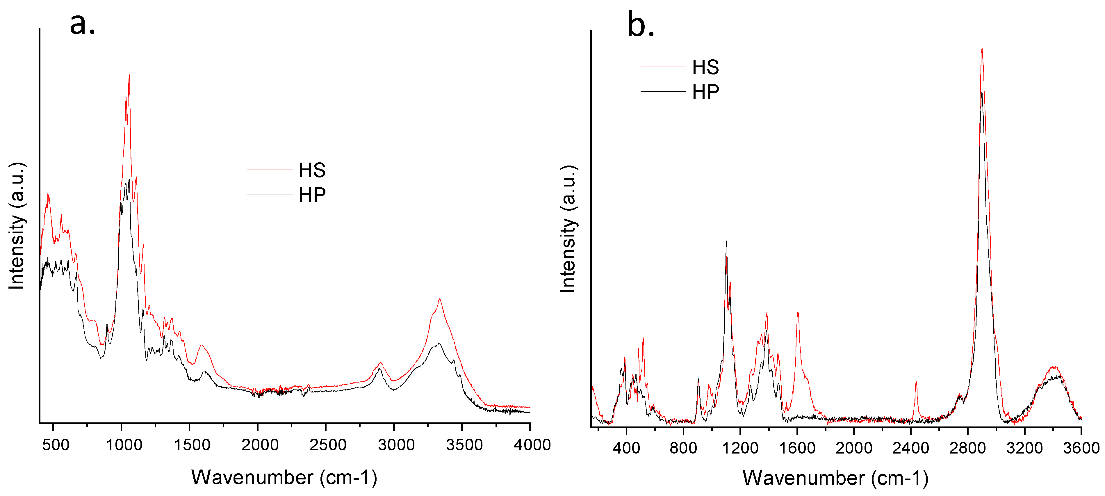
2.3.2. IR and Raman Spectroscopy
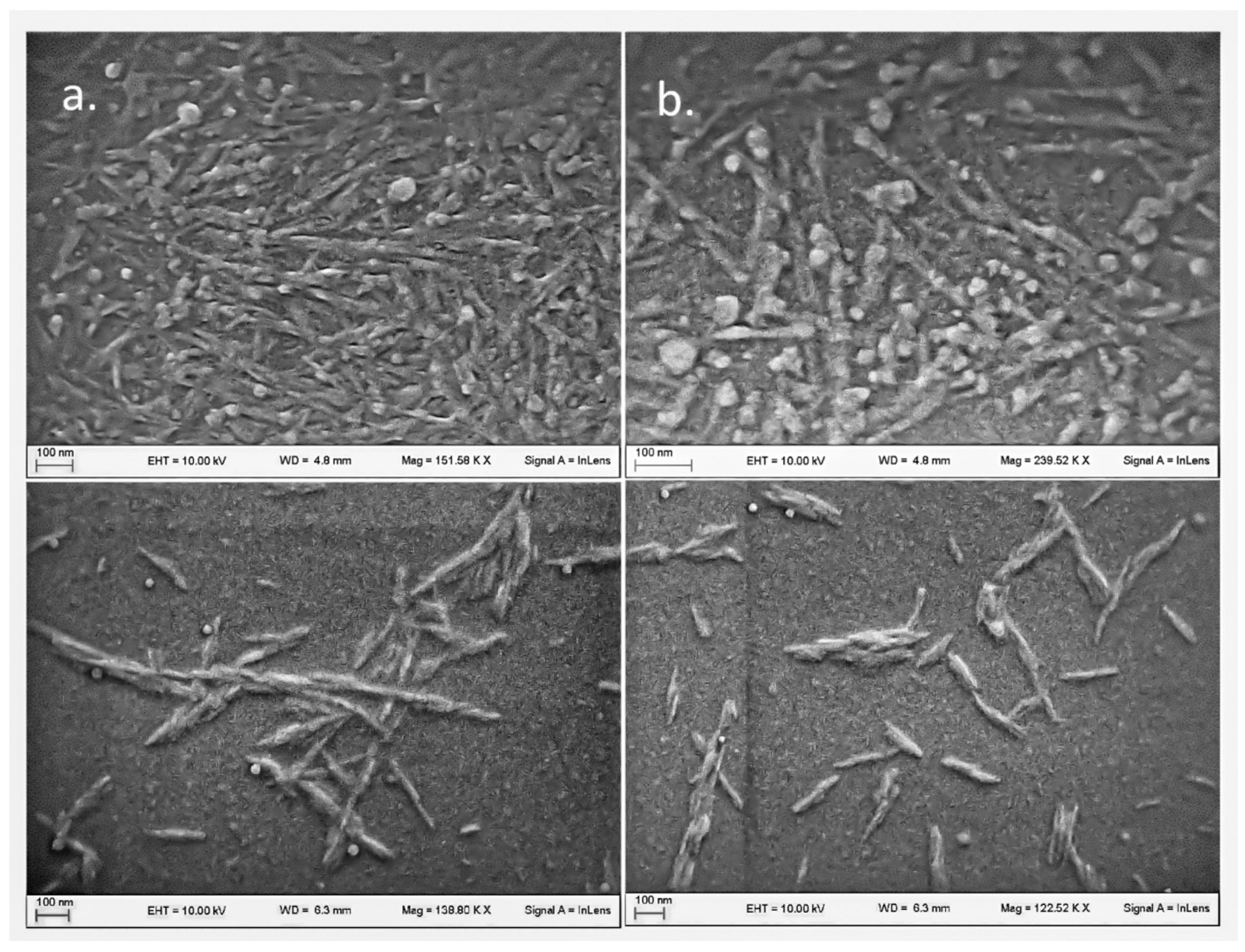
Fourier Transform Infrared spectra with attenuated total reflection (internal reflection accessory, ATR-FTIR) were recorded using an IRSpirit Infrared Spectrophotometer (Shimadzu, Kyoto, Japan) in the spectral range of 400–5000 cm−1 and with a resolution of 4 cm−1 at 50 scans. A small quantity of the powdered samples was deposited directly on the diamond ATR crystal plate with no sample processing. Background spectrum was acquired on the empty crystal plate.
Raman spectra were acquired by using a WITec Raman alpha300RA confocal microscope (WITec, Ulm, Germany). Powder samples were compressed into tablet and directly placed on a microscopy glass specimen at r.t. Accumulation of 40 scans at 2 s integration time was carried out to record spectra in the range 200–3600 cm−1. Spectra were elaborated by WITec Control FIVE software.

2.3.3. Morphology

Sample morphological analysis was performed by using a field-emission microscope Zeiss LEO 1525 equipped with a GEMINI column, (Oberkochen, Germany). Samples were prepared by depositing diluted CNC suspensions onto round glass coverslips attached to an aluminum specimen stub with a double-sided carbon tape. Samples were sputter coated with chromium (8 nm layer thickness) for 15 s at 240 mA before imaging (Quorum Q150T ES East Grinstead, West Sussex, UK).

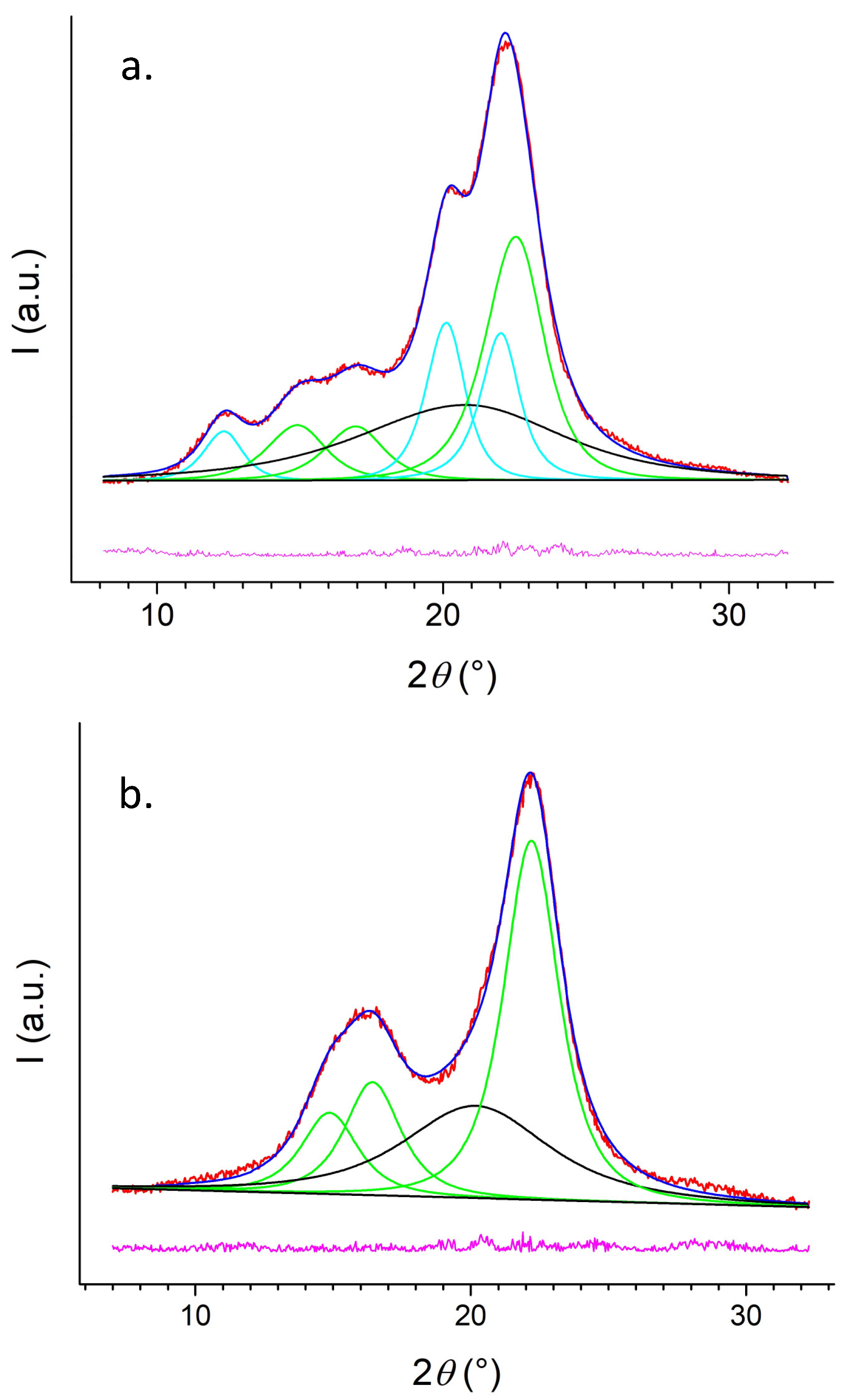
2.3.4. Diffractometric Analysis

X-ray powder diffraction (XRD) data collection was performed with a Bruker D8 Advance diffractometer in Bragg–Brentano geometry, equipped with a Lynxeye XE-T fast detector, using the Cu-Kα radiation (40 kV, 40 mA). Sample 1, being finely powdered, was directly put on a zero-background sample holder, while the fibrous sample 2 was not possible to reduce in a fine powder without avoiding the modification of its microstructural features; therefore, it was pressed in a pellet of 1 cm od diameter and 0.5 mm thick and placed on the sample holder for data collection. Data were collected in the 4–80° 2θ range using a 0.017° step scan and 200 s counting time. The patterns were decomposed in the contributions of cellulose Iβ and cellulose II reflections, and the amorphous content by means of a profile fit procedure with pseudo-Voigt functions, in the 5–32° 2θ range, using the Philips ProFit software, following the method described by Garvey et al. (2005) [62]. Cellulose Iβ and cellulose II peak positions were first set up from Nishiyama et al. (2003) and Langan et al. (2001), respectively, and then refined [63,64]. The relative contributions, in wt%, were assumed to be proportional to the total integrated intensities per each phase, while the crystallinity index was estimated as the wt% of the crystalline phases.

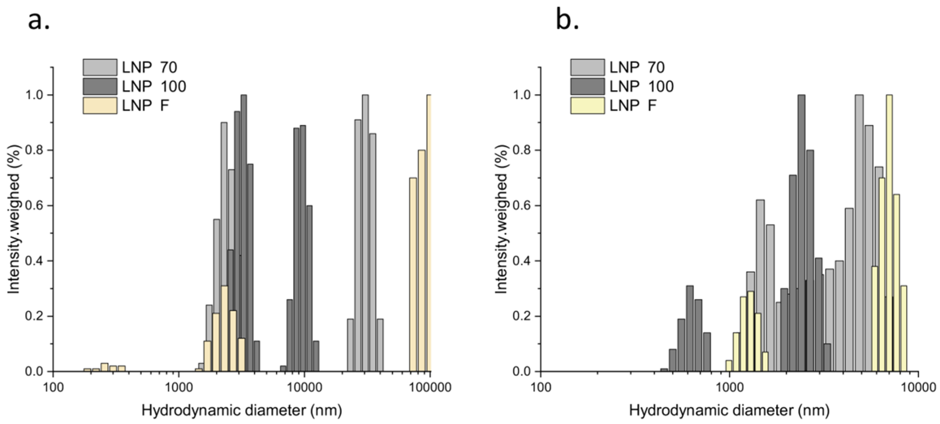
2.3.5. LNP Morphology and Dimension

LNP morphology was investigated by negative staining technique in Transmission Electron Microscopy (TEM). Droplets of sample suspensions (10 µL) were placed on formvar-carbon coated grids and allowed to adsorb for 60 s. The adsorbed specimen was then processed for negative-staining by first washing the specimen grid on a drop of negative stain (2% uranyl acetate in distilled water) and then blotting and repeating this step once more, this time leaving the specimen grid for 60 s on a new drop of negative stain solution. Samples were observed at a JEOL 1200 EX II electron microscope. Micrographs were acquired by the Olympus SIS VELETA CCD camera, equipped with iTEM software. LNP dimensions were studied following the same method previously described in Section 2.3.1.

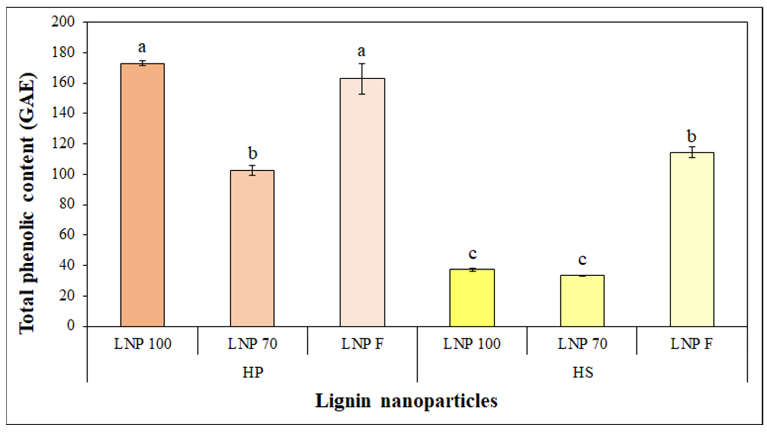
2.3.6. LNP Total Phenolic Content

The total phenolic content (TPC) of the extract was assessed according to the Folin-Ciocalteu method, using gallic acid as a standard. The basic mechanism was an oxidation/reduction reaction and, therefore, the assay can be considered an antioxidant (AOX) method [65]. A 10-fold dilution of the Folin-Ciocalteu reagent was firstly made with deionized water, and 0.1 mL of LNP samples were mixed with 0.75 mL of the diluted Folin-Ciocalteu reagent and incubated for 10 min at room temperature. Finally, a volume of 0.75 mL of a 2% w/v sodium carbonate (Na2CO3) (purity ≥ 99.5%, powder, CAS No: 497-19-8) aqueous solution was added, and the mixture was kept in the dark for 3 h, before measuring the absorbance at 765 nm. The absorbance was measured at 765 nm, and the analyses were performed in triplicate. The results of the TPC determination were expressed in gallic acid equivalents (GAE) using a gallic acid (purity ≥ 99.5%, powder, CAS No: 149-91-7) calibration curve (range from 25 to 250 mg L−1 concentration and R2 = 0.9941).

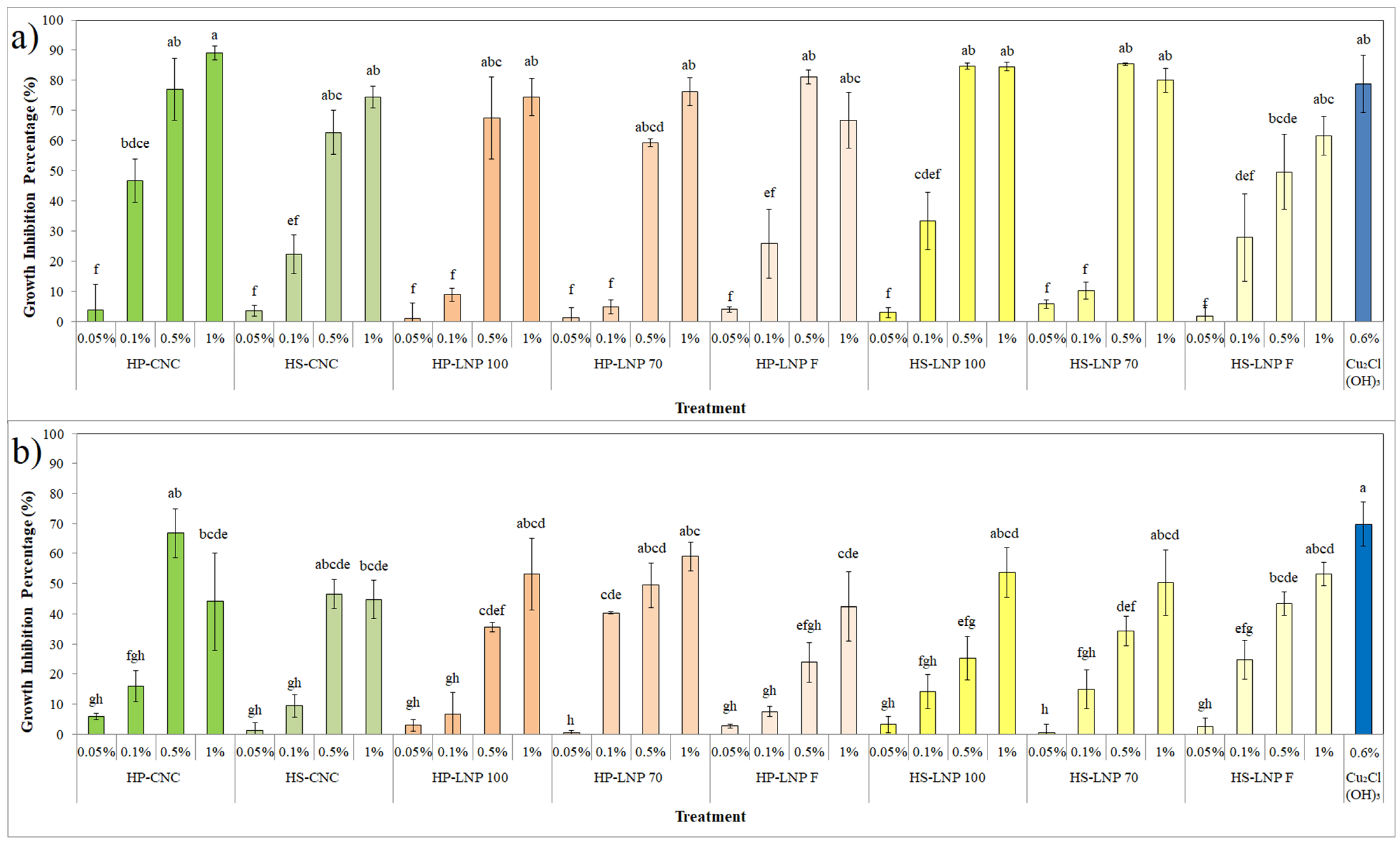
2.4. In Vitro Antibacterial Activity

Obtained CNC and LNP were characterized for their antimicrobial activity on Xac through a microdilution method [66,67]. Xac strain LMG 688 were cultured on YGCA medium for 48 h at 27 °C. A bacterial suspension containing 1 × 10⁸ CFU mL−1 was made in sterile deionized water by a spectrophotometer (OD600 = 0.2) [9]. Several dilutions were made in order to obtain a final concentration of 1 × 10⁴ CFU mL−1, and then 20 μL was added to 180 μL of previously prepared suspensions containing LB Broth and CNC or LNP at concentrations of 1, 0.5, 0.1 and 0.05% w/v in a 96 multiwells microplate. LB Broth alone and amended with copper oxychloride at 0.6% w/v (average field dose) were used, respectively, as negative and positive control. The plates were incubated at 27 °C, and the absorbance at 600 nm was measured using a spectrophotometer after 24 and 48 h. Four replicates were made for each concentration. The inhibition of bacterial growth was calculated as percentage after subtracting the blank as below:
Inhibition (%) = [(Neg. ctrl. abs.-Tested substance abs.) × Neg. ctrl. abs.−1] × 100

2.5. In Vivo Antibacterial Activity

The most effective concentrations of CNC and LNP coming from hazelnut pruning and shelling residues in terms of antibacterial inhibition were chosen from the previous in vitro assay to be in vivo tested. One year old micropropagated Tonda Gentile Romana hazelnut seedlings were placed in a growth chamber with the following climatic parameters: 16/8 day/night photoperiod, 60% air humidity and 22/16 °C day/night thermo-period. Each thesis was composed of ten plants. Plants were foliar treated with a 1% w/v CNC suspension until a uniform coating was reached. Sterile water and copper oxychloride at field dose were used as controls. After, 24 h a 1 × 10⁶ CFU mL−1 Xac suspension was obtained from 48 h fresh culture and homogeneously sprayed on the leaves [68]. Air humidity was raised to 80% for 24 h after the artificial inoculation. Plants were daily checked for symptoms appearance. One day after the first leaf necrosis displayed, data concerning the disease indices were collected: disease incidence (DI) was reported as the percentage of symptomatic leaves (presenting at least one point-like necrosis on the upper surface) on the total plant leaves; disease severity (DS) was expressed as the ratio between the number of point-like necroses and the total surface of the symptomatic leaves per plant [69,70]. Experiment was repeated thrice.

2.6. In Vivo Phytobiological Compatibility

In order to study the effects of the obtained CNC and LNP on the development of hazelnut seedlings, an in vivo assay was held by treating the plants as described in the previous paragraph. The same growth conditions were set up, and several biological parameters were monitored at 1, 7, 14 and 21 days post treatment (dpt). For each time point, two leaves per plant were casually harvested to measure their area through the software ImageJ (version 1.51j8) (NIH, Bethesda, MD, USA) (accessed on Windows 10) (Microsoft, Redmond, WA, USA), while at the same time two measurements were randomly taken on the canopy of each plant to assess the chlorophyll and the flavonol content by a non-destructive method, using a leaf clip sensor (Dualex 4 Scientific, FORCE-A, Orsay Cedex, France) [71]. From the ratio between these two values, the nitrogen balance index (NBI) was calculated [72]. Sterile water and copper oxychloride at field dose were used as controls. Each thesis was composed of ten plants. The experiment was repeated thrice.

2.7. Statistical Ananlysis

Collected data were analyzed using one-way analysis of variance (ANOVA). Statistical significance of means was studied with Fisher’s LSD post hoc test. p-values less than 0.05 were considered significant, while p-values of less than 0.01 were considered highly significant. ANOVA tables are shown in the Supplementary Materials.

3. Results and Discussion

3.1. CNC and LNP Synthesis

Previously described protocols turned out to be adequate to synthesize CNC from both HP and HS (Figure 1). In terms of yield (calculated as percentage ratio between the weights of final cellulosic masses and initial raw materials) obtained, CPC-HP was about 28.2 ± 1.7%, while CPC-HS showed a final yield of 23.9 ± 2.3%. The acid hydrolysis yield, calculated using the method reported as UNI EN ISO 638:2009, was, respectively, 31.8 ± 0.9% and 8.3 ± 1.5% for HP CNC and HS CNC. While the obtained results for the pruning shoots seem to match the cellulose content in hazelnut branches and suckers, highlighting the appropriateness of the adopted protocol to isolate cellulose from the raw materials in terms of purity and quantity, the CNC yield is much lower when using the lignocellulosic biomasses coming from hazelnut shelling. To explain these results, we should first consider the different lignocellulosic composition of the shells, which have a smaller content of cellulose than the pruning shoots. Furthermore, the presence of a recalcitrant component mainly made of waxes could have played an import role in making the matrix more inaccessible to the chemical reagents. There is evidence for this in the characterization analysis of CNC, which have demonstrated a more relevant presence of impurities and foreign substances in HS samples than in HP ones, as described in the following paragraph. In spite of everything, it is important to say that the reported approaches can be considered a successful first step to obtain cellulose and its crystalline nanoform from both hazelnut pruning and shelling waste. More improvements are being studied to enhance the CNC extraction and yield, such as including a more effective dewaxing treatment for HS, as it happened for other nuts residues, or diminishing the amount of used chemical reagents, in order to develop a more sustainable process.
LNPs were synthesized according to the extraction protocols described in the Section 2.2.2 (Figure 2). The isolated lignin fractions showed a yield (calculated as percentage ratio between the weights of final ligninic masses and initial raw materials) of 4.1 ± 0.8% and 3.1 ± 0.5%, respectively, for HS and HP, while the final LNP yield (calculated as reported in the method UNI EN ISO 638:2009, after adding the final weights of the different LNP extractions) was about 79.9 ± 1.8% and 39.9 ± 1.7% for HP and HS, respectively. The proposed approaches of using an enzymatic pretreatment to isolate the lignin component from the lignocellulosic waste seems to be a very promising one since most common LNP synthesis protocols are based on large consumption of chemical reagents [46,73,74]. Nevertheless, more work is needed to enhance yield of the extraction process. The solvent–antisolvent method is based on the use of water and ethanol allowed to synthesize LNP without using heavy chemical reagents, with acceptable yields. The one concerning HS is much lower than HP since the starting matrix is composed of waxes and other lipid fractions that lower the efficacy of the proposed treatments [75,76]. In this sense, a much more effective dewaxing pretreatment is required in order to be able to exploit all the ligninic fraction in shells.

3.2. CNC and LNP Characterization

The obtained HS and HP CNC showed broad particle size distributions that can be explained by considering the tendency of the nanofibrils to cluster and the presence of residues due to the extraction method employed (Figure 3). In particular, either HS or HP extracts displayed colloidal populations between 150 and 200 nm, compatible with reported CNC fibrils size and smaller populations at around 30–50 nm possibly ascribable to fragments or impurities [22,77,78]. Moreover, micrometric clusters are observed in both extracts, with a prevalence of HS.
Spectroscopic analysis confirmed the cellulosic nature for all extracts since both IR and Raman spectra displayed typical signals. However, the comparison of HS and HP profiles highlighted the higher presence of foreign substances in HS CNC. In fact, while IR profiles were identical, with evidence of water stretching and bending signals at 3300 and 1640 cm−1, respectively (Figure 4a), Raman spectra pointed to the presence of impurities in HS CNC, as shown by the band between 1600 and 1650 cm−1, which are likely due to C-N stretching of amide compounds (Figure 4b).
The spectroscopic observations were confirmed by morphological analysis, from which the presence of impurities was depicted (Figure 5). SEM images clearly show the typical elongated structure of cellulose nanofibrils, with size slightly higher than 100 nm, which is compliant with DLS measurements. No relevant macroscopic differences emerged from SEM observations because HS and HP CNC were apparently similar in shape and size.
On the other hand, X-rays analysis evidenced structural differences in the inner crystal organization and nature of the nanofibrils. Insightful investigation of HS and HP CNC crystalline nature is displayed in Figure 6 and Table 1. The X-rays profiles of HP CNC revealed two cellulose crystalline phases Iβ and II, whereas HS CNC were characterized by the Iβ phase only.
In particular, the Iβ and II phases contributed to HP CNC crystallinity for the 42% and 28%, respectively (Table 1). Crystalline cellulose is known to exist as cellulose I, II, III, and IV crystal forms [79]. Cellulose II is the most stable, while cellulose I is known to occur as a mixture of a triclinic Iα and monoclinic Iβ crystalline forms, being the Iβ prevalent in higher plants. Cellulose II can form upon treatment of cellulose I with strong bases or by its solubilization and precipitation [80]. Therefore, the presence of a certain amount of form II in HP CNC, compared to the 100% of Iβ crystalline form of HS CNC, may be the result of the extraction conditions employed, which may have favored the Iβ to II transition, even though different initial crystalline compositions between the two source materials cannot be ruled out.
Moreover, because crystallinity index of both HP and HS CNC is nearly identical, the two extracts had a rather consistent calculated amorphous content of about 30% (Table 1). The obtained LNP were first observed by TEM. The negative staining process revealed an imperfectly spherical form for each LNP extraction step, with inconstant dimensions ranging from 50 to 200 nm (Figure 7). Microscopic observations also highlighted the presence of impurities and other substances that could play a role in promoting LNP aggregation when suspended after some time.
As a confirmation, LNPs were also characterized in terms of size distribution by DLS. In general, LNPs were only partially colloidal due to a strong tendency to aggregate into large clusters. This is evident from the distributions in Figure 8, where in all cases aggregation produced micrometric populations and only LNP F from HP and LNP 100 from HS displayed nanosized populations. Such behavior seems uncorrelated with the production process itself, but it could rather be the result of the intrinsic instability of lignin particles that were not added with proper stabilizers or preservatives.
Regarding the TPC of synthesized LNP, the Folin-Ciocalteu method revealed the following highly statistically significant (p < 0.01) results: 173, 102.6 and 162.8 GAE for the LNP, coming, respectively, from the first, the second and the final extraction step of HP lignin, while LNP 100, LNP 70 and LNP F obtained from HS showed, respectively, 37.2, 33.5 and 114.3 GAE (Figure 9). The reported trend in TPC values for HP LNP is not linear with the extraction process: the most relevant TPC was noted in the first and in the third step. This behavior could be due to the complex composition of lignin, which is constantly rearranged and shaped by the different chemical and physical treatments. On the contrary, HS LNP showed a growing TPC: in fact, the LNP produced by the last extraction step had a higher content in polyphenols. Unlike the pruning residues, hazelnut shells present a much more complex structure, due to the high presence of waxes and other lipid components [75,76]. Such components, as it was assumed for CNC extraction, could play an important role in hampering the access of chemical reagents to the lignin fraction. Thus, the TPC increase in the last HS fraction may be explained by the reaction of a significant lignin fraction in which most of the impurities were moved away in the previous steps. Other assays are scheduled to study the antioxidant activity of LNP since these values drastically change not only the consideration of the extraction process but also the quantification method [45,49,81].

3.3. In Vitro Antibacterial Activity

Antibacterial activity of the obtained CNC and LNP from both HP and HS were tested on Xac at different concentrations and at different times by using a microdilution method. After 24 h, the most interesting and highly statistical significant results were noted for each sample tested at the concentration of 0.5 and 1% w/v: both CNC and LNP showed an inhibition activity comparable to the one displayed by copper oxychloride (Figure 10a). After 48 h, a general reduction of growth inhibition was observed for all tested substances, except for copper, although a highly statistical difference could be still appreciated (Figure 10b). A general, strong dose-dependent activity was noted. The antibacterial mechanisms of cellulose nanoforms are still largely unknown. Several works have reported the cellulose nanofibrils’ ability to disturb bacteria swimming motility in several foodborne pathogens (E. coli, L. monocytogenes, B. aureus and S. typhimurium) or, as shown for E. coli and P. aeruginosa, the CNC capacity to prevent bacterial cell adhesion and biofilm formation [82,83,84]. Noteworthily, Noronha et al. (2021) linked bacterial growth inhibition by CNC to the loss of cell membrane integrity in E. coli bacterial cells [85]. This effect was ascribed to the disrupting action of CNC on the membrane phospholipid structure. Other authors suggest that one of the antibacterial modes of action of CNC could be explained through the depletion effect recorded on P. aeruginosa and P. fluorescens. In these works, the flocculation of the bacterial cells was observed in CNC suspension that impaired cell adhesion capacity and thus biofilm formation [84,86]. Naturally, a simultaneous effect of all previously cited antimicrobial mechanisms cannot be ruled out. In this context, more studies are needed to clarify the prevalent mode of action of CNC on Xac.
To the best of our knowledge, this report is the first describing the CNC antimicrobial activity on a plant pathogenic bacterium.
Lignin antimicrobial properties are instead well known: several works highlight the possibility of using LNPs as promising nanopesticides or nanocarriers against different pathogenic bacteria [87,88]. However, the hypothetical antimicrobial modes of action of LNP against bacterial cells are multiple. LNP are supposed to penetrate the cell wall provoking membrane lysis, reacting with ROS species and inducing oxidative stress, generating a depletion in ATP and a decrease of intracellular pH, resulting in cell death [89,90,91]. Most of these action mechanisms are related to LNP size and composition. The final amount of polyphenols seems to represent a major driver for LNP antimicrobial activity [92,93]. However, our results indicate lack of correlation between LNP antibacterial action and TPC amount, suggesting that different modes of action may concur to LNP antibacterial properties.

3.4. In Vivo Antibacterial Activity

In order to study the effect of the obtained CNC and LNP on plant–pathogen interactions, the concentrations that showed, in the in vitro assay, the most promising antibacterial activity and the most relevant TPC were selected to perform an in vivo test. Hazelnut plants were treated with a 1% w/v suspension of HS CNC and HP CNC and with a 1% w/v suspension of HP LNP 100 and HS LNP F. After 24 h, plants were spray-inoculated with a Xac suspension. Symptoms started to appear on leaves 20 days after foliar inoculation. At 21 dpi, the disease indices (incidence and severity) were calculated as previously described. Results showed a high statistical significance (p < 0.01) in terms of DI. Each of the tested substances was able to diminish the appearance of symptoms in a way comparable to copper oxychloride when used at the field dose. Regarding the DS, even if the recorded values were in almost all cases lower than the one recorded in water-treated plants, a statistical difference (p < 0.05) was noted only in copper-treated plant (Table 2). This behavior could be explained assuming that CNC and LNP antimicrobial effects could have affected the initial adhesion and epiphytical survival of the bacterial cells on the leaves, or prevent them by entering through the stomata, thus reducing the first symptoms display. While copper and its derivatives are well known bactericidals, this study may help advancing the knowledge on the antimicrobial properties of CNC and LNP [15,94,95,96]. The proposed nanomaterials, as also inferred from the in vitro assay results, had prevalent inhibition activity. Presumably, once the inhibition effect is over, bacterial cells may recover the ability to reproduce and infect plant tissues. Further studies are scheduled in order to better clarify the span of the inhibition effect on leaves since the phyllosphere is a very complex environment where organic substances interact with a large number of biological and climatic variables, which could influence even their composition during time [97,98].

3.5. Phytobiological Compatibility

To evaluate any possible effect on plant development, several biological parameters linked to basal metabolism were recorded on hazelnut seedlings after being foliar-treated with the same suspension used in the in vivo antibacterial assay. At 1, 7, 14 and 21 dpt, the leaf area, the chlorophyll and flavonol content in leaves, and the nitrogen balance index, calculated from the ratio between these last two values, can be seen in Table 3. From the statistical analysis, the full compatibility of tested CNC and LNP was confirmed. No detrimental effect was noted on the canopy of treated plants that was comparable with the used controls (water and copper oxychloride at field dose). These results suggest that the proposed nanomaterials could be easily used in field alone or as nanocarriers for other active substances, without any harmful effect on the growth of the plants. In this sense, according also with previous results, it is possible to asses a future role for CNC and LNP in a sustainable hazelnut plant-protection strategy.

4. Conclusions

This work investigated, for the first time, the possibility of exploiting both hazelnut pruning and shelling wastes as innovative sources to obtain cellulose nanocrystals and lignin nanoparticles, for reuse as sustainable tool for the control of hazelnut bacterial blight. The proposed lignocellulosic starting materials and the extraction protocols turned out to be a very promising starting point to increase the value of the whole hazelnut supply chain, in a context of circular economy. Cellulose nanocrystals were successfully synthesized by an acid hydrolysis after isolating the cellulosic component from hazelnut pruning shoots and shells through a chemical bleaching, while lignin nanoparticles were obtained thanks to a solvent–antisolvent method after a fractionation of the ligninic component. In this work, the antimicrobial properties of the proposed lignocellulosic nanocarriers were investigated too. The conducted assays proved the ability of both cellulose nanocrystals and lignin nanoparticles of in vitro inhibiting the bacterial growth of Xanthomonas arboricola pv. corylina and, moreover, when in vivo applied, they were able to diminish the disease incidence in a comparable way to copper oxychloride. These results, together with the confirmed phytobiological compatibility, suggest the innovative use of the tested nanomaterials as sustainable nanopesticides development. Furthermore, other studies are already underway to better understand the biological mechanisms behind the promising bacterial inhibition activity shown in this work, giving to farmers of different chains sustainable alternatives to traditional agrochemicals.

Supplementary Materials

The following supporting information can be downloaded at: https://www.mdpi.com/article/10.3390/app12052604/s1, File S1: Anova tables.

Author Contributions

Conceptualization, D.S. and G.M.B.; methodology, D.S., V.D.L., S.G. and E.C.; validation, G.M.B., S.G. and E.C.; formal analysis, D.S., R.R. and R.V.; investigation, D.S., V.D.L., M.S., C.P., R.R. and R.V.; resources, G.M.B., S.G. and E.C.; data curation, D.S., V.D.L., R.R.; writing—original draft preparation, D.S., V.D.L. and R.R.; writing—review and editing, G.M.B., S.G. and E.C.; supervision, G.M.B., S.G. and E.C. All authors have read and agreed to the published version of the manuscript.

Funding

This research was funded by MUR (Italian Ministry for University and Research) (Law 232/2016, Departments of Excellence) and PON Ricerca e Innovazione 2014–2020. Nanotecnologie chimiche green per la protezione sostenibile delle piante (NEMESI) ARS01_01002 CUP: F36C1800018000X).

Institutional Review Board Statement

Not applicable.

Informed Consent Statement

Not applicable.

Acknowledgments

The authors gratefully thank Anna Rita Taddei for her scientific support in TEM analyses.

Conflicts of Interest

The authors declare no conflict of interest. The funders had no role in the design of the study; in the collection, analyses, or interpretation of data; in the writing of the manuscript; or in the decision to publish the results.

References

  1. Silvestri, C.; Bacchetta, L.; Bellincontro, A.; Cristofori, V. Advances in cultivar choice, hazelnut orchard management, and nut storage to enhance product quality and safety: An overview. J. Sci. Food Agric. 2021, 101, 27–43. [Google Scholar] [CrossRef]
  2. Vauterin, L.; Hoste, B.; Kersters, K.; Swings, J. Reclassification of Xanthomonas. Int. J. Syst. Bacteriol. 1995, 45, 472–489. [Google Scholar] [CrossRef]
  3. Scortichini, M.; Rossi, M.P.; Marchesi, U. Genetic, phenotypic and pathogenic diversity of Xanthomonas arboricola pv. corylina strains question the representative nature of the type strain. Plant Pathol. 2002, 51, 374–381. [Google Scholar] [CrossRef]
  4. EPPO Data Sheets on Quarantine Pests: Xanthomonas arboricola pv. corylina. EPPO Bull. 2004, 34, 179–181. [CrossRef]
  5. Puławska, J.; Kałuzna, M.; Kołodziejska, A.; Sobiczewski, P. Identification and characterization of Xanthomonas arboricola pv. corylina causing bacterial blight of hazelnut: A new disease in Poland. J. Plant. Pathol. 2010, 92, 803–806. [Google Scholar]
  6. Webber, J.B.; Putnam, M.; Serdani, M.; Pscheidt, J.W.; Wiman, N.G.; Stockwell, V.O. Characterization of isolates of Xanthomonas arboricola pv. corylina, the causal agent of bacterial blight, from Oregon hazelnut orchards. J. Plant Pathol. 2020, 102, 799–812. [Google Scholar] [CrossRef]
  7. Kałużna, M.; Fischer-Le Saux, M.; Pothier, J.F.; Jacques, M.A.; Obradović, A.; Tavares, F.; Stefani, E. Xanthomonas arboricola pv. juglandis and pv. corylina: Brothers or distant relatives? Genetic clues, epidemiology, and insights for disease management. Mol. Plant Pathol. 2021, 22, 1481–1499. [Google Scholar] [CrossRef]
  8. Lamichhane, J.R.; Fabi, A.; Varvaro, L. Summer heat and low soil organic matter influence severity of hazelnut Cytospora canker. Phytopathology 2014, 104, 387–395. [Google Scholar] [CrossRef]
  9. Lamichhane, J.R.; Fabi, A.; Ridolfi, R.; Varvaro, L. Epidemiological Study of Hazelnut Bacterial Blight in Central Italy by Using Laboratory Analysis and Geostatistics. PLoS ONE 2013, 8, e56298. [Google Scholar] [CrossRef]
  10. Pisetta, M.; Albertin, I.; Petriccione, M.; Scortichini, M. Effects of hot water treatment to control Xanthomonas arboricola pv. corylina on hazelnut (Corylus avellana L.) propagative material. Sci. Hortic. 2016, 211, 187–193. [Google Scholar] [CrossRef]
  11. Lamichhane, J.R.; Varvaro, L. Xanthomonas arboricola disease of hazelnut: Current status and future perspectives for its management. Plant Pathol. 2014, 63, 243–254. [Google Scholar] [CrossRef]
  12. Lamichhane, J.R.; Osdaghi, E.; Behlau, F.; Köhl, J.; Jones, J.B.; Aubertot, J.N. Thirteen decades of antimicrobial copper compounds applied in agriculture. A review. Agron. Sustain. Dev. 2018, 38, 28. [Google Scholar] [CrossRef]
  13. La Torre, A.; Iovino, V.; Caradonia, F. Copper in plant protection: Current situation and prospects. Phytopathol. Mediterr. 2018, 57, 201–236. [Google Scholar] [CrossRef]
  14. Flemming, C.A.; Trevors, J.T. Copper toxicity and chemistry in the environment: A review. Water Air Soil Pollut. 1989, 44, 143–158. [Google Scholar] [CrossRef]
  15. Varympopi, A.; Dimopoulou, A.; Theologidis, I.; Karamanidou, T.; Kaldeli Kerou, A.; Vlachou, A.; Karfaridis, D.; Papafotis, D.; Hatzinikolaou, D.G.; Tsouknidas, A.; et al. Bactericides Based on Copper Nanoparticles Restrain Growth of Important Plant Pathogens. Pathogens 2020, 9, 1024. [Google Scholar] [CrossRef]
  16. Abrahamian, P.; Jones, J.B.; Vallad, G.E. Efficacy of copper and copper alternatives for management of bacterial spot on tomato under transplant and field production. Crop Prot. 2019, 126, 104919. [Google Scholar] [CrossRef]
  17. Goto, M.; Hikota, T.; Nakajima, M.; Takikawa, Y.; Tsuyumu, S. Occurrence and Properties of Copper-resistance in Plant Pathogenic Bacteria. Jpn. J. Phytopathol. 1994, 60, 147–153. [Google Scholar] [CrossRef]
  18. Prasad, R.; Bhattacharyya, A.; Nguyen, Q.D. Nanotechnology in sustainable agriculture: Recent developments, challenges, and perspectives. Front. Microbiol. 2017, 8, 1014. [Google Scholar] [CrossRef]
  19. Usman, M.; Farooq, M.; Wakeel, A.; Nawaz, A.; Cheema, S.A.; ur Rehman, H.; Ashraf, I.; Sanaullah, M. Nanotechnology in agriculture: Current status, challenges and future opportunities. Sci. Total Environ. 2020, 721, 137778. [Google Scholar] [CrossRef]
  20. Chen, H.; Yada, R. Nanotechnologies in agriculture: New tools for sustainable development. Trends Food Sci. Technol. 2011, 22, 585–594. [Google Scholar] [CrossRef]
  21. Puglia, D.; Pezzolla, D.; Gigliotti, G.; Torre, L.; Bartucca, M.L.; Del Buono, D. The opportunity of valorizing agricultural waste, through its conversion into biostimulants, biofertilizers, and biopolymers. Sustainability 2021, 13, 2710. [Google Scholar] [CrossRef]
  22. Luzi, F.; Fortunati, E.; Giovanale, G.; Mazzaglia, A.; Torre, L.; Balestra, G.M. Cellulose nanocrystals from Actinidia deliciosa pruning residues combined with carvacrol in PVA_CH films with antioxidant/antimicrobial properties for packaging applications. Int. J. Biol. Macromol. 2017, 104, 43–55. [Google Scholar] [CrossRef]
  23. Barana, D.; Salanti, A.; Orlandi, M.; Ali, D.S.; Zoia, L. Biorefinery process for the simultaneous recovery of lignin, hemicelluloses, cellulose nanocrystals and silica from rice husk and Arundo donax. Ind. Crops Prod. 2016, 86, 31–39. [Google Scholar] [CrossRef]
  24. Hamed, O.A.; Fouad, Y.; Hamed, E.M.; Al-Hajj, N. Cellulose powder from olive industry solid waste. BioResources 2012, 7, 4190–4201. [Google Scholar] [CrossRef]
  25. Schiavi, D.; Balbi, R.; Giovagnoli, S.; Camaioni, E.; Botticella, E.; Sestili, F.; Balestra, G.M. A green nanostructured pesticide to control tomato bacterial speck disease. Nanomaterials 2021, 11, 1852. [Google Scholar] [CrossRef]
  26. Balestra, G.M.; Fortunati, E. Nanotechnology-Based Sustainable Alternatives for the Managements of Plant Diseases; Balestra, G.M., Fortunati, E., Eds.; Elsevier: Amsterdam, The Netherlands, 2021; ISBN 9780128233948. [Google Scholar]
  27. Fraceto, L.F.; Grillo, R.; de Medeiros, G.A.; Scognamiglio, V.; Rea, G.; Bartolucci, C. Nanotechnology in agriculture: Which innovation potential does it have? Front. Environ. Sci. 2016, 4, 20. [Google Scholar] [CrossRef]
  28. Fortunati, E.; Verma, D.; Luzi, F.; Mazzaglia, A.; Torre, L.; Balestra, G.M. Novel nanoscaled materials from lignocellulosic sources: Potential applications in the agricultural sector. Handb. Ecomater. 2019, 4, 2657–2679. [Google Scholar] [CrossRef]
  29. García, A.; Gandini, A.; Labidi, J.; Belgacem, N.; Bras, J. Industrial and crop wastes: A new source for nanocellulose biorefinery. Ind. Crops Prod. 2016, 93, 26–38. [Google Scholar] [CrossRef]
  30. de Hoyos-Martínez, P.L.; Erdocia, X.; Charrier-El Bouhtoury, F.; Prado, R.; Labidi, J. Multistage treatment of almonds waste biomass: Characterization and assessment of the potential applications of raw material and products. Waste Manag. 2018, 80, 40–50. [Google Scholar] [CrossRef]
  31. Crus Lopes, L.P.; Martins, J.; Esteves, B.; Texeira De Lemos, L. New Products from Hazelnut Shell. In Proceedings of the Ecowood 2012—5th International Conferences on Environmentally-Compatible Forest Products, Porto, Portugal, 5–7 September 2012; pp. 83–90. [Google Scholar]
  32. Demirbaş, A. Estimating of structural composition of wood and non-wood biomass samples. Energy Sources 2005, 27, 761–767. [Google Scholar] [CrossRef]
  33. Di Michele, A.; Pagano, C.; Allegrini, A.; Blasi, F.; Cossignani, L.; Di Raimo, E.; Faieta, M.; Oliva, E.; Pittia, P.; Primavilla, S.; et al. Hazelnut Shells as Source of Active Ingredients: Extracts Preparation and Characterization. Molecules 2021, 26, 6607. [Google Scholar] [CrossRef] [PubMed]
  34. Lelli, V.; Molinari, R.; Merendino, N.; Timperio, A.M. Detection and comparison of bioactive compounds in different extracts of two hazelnut skin varieties, tonda gentile romana and tonda di giffoni, using a metabolomics approach. Metabolites 2021, 11, 296. [Google Scholar] [CrossRef] [PubMed]
  35. Contini, M.; Baccelloni, S.; Massantini, R.; Anelli, G. Extraction of natural antioxidants from hazelnut (Corylus avellana L.) shell and skin wastes by long maceration at room temperature. Food Chem. 2008, 110, 659–669. [Google Scholar] [CrossRef]
  36. Fortunati, E.; Balestra, G.M. Lignocellulosic Materials as Novel Carriers, Also at Nanoscale, of Organic Active Principles for Agri-Food Applications; Elsevier Ltd.: Amsterdam, The Netherlands, 2019; ISBN 9780081024263. [Google Scholar]
  37. Kamel, R.; El-Wakil, N.A.; Dufresne, A.; Elkasabgy, N.A. Nanocellulose: From an agricultural waste to a valuable pharmaceutical ingredient. Int. J. Biol. Macromol. 2020, 163, 1579–1590. [Google Scholar] [CrossRef]
  38. La Ferla, B.; Zoia, L.; Bigini, P.; Di Gennaro, P. Cellulose nanocrystals as promising nano-devices in the biomedical field. AIP Conf. Proc. 2018, 1990, 020019. [Google Scholar] [CrossRef]
  39. George, J.; Sabapathi, S.N. Cellulose nanocrystals: Synthesis, functional properties, and applications. Nanotechnol. Sci. Appl. 2015, 8, 45–54. [Google Scholar] [CrossRef]
  40. Coelho, C.C.S.; Michelin, M.; Cerqueira, M.A.; Gonçalves, C.; Tonon, R.V.; Pastrana, L.M.; Freitas-Silva, O.; Vicente, A.A.; Cabral, L.M.C.; Teixeira, J.A. Cellulose nanocrystals from grape pomace: Production, properties and cytotoxicity assessment. Carbohydr. Polym. 2018, 192, 327–336. [Google Scholar] [CrossRef]
  41. Roman, M. Toxicity of cellulose nanocrystals: A review. Ind. Biotechnol. 2015, 11, 25–33. [Google Scholar] [CrossRef]
  42. Fortunati, E.; Yang, W.; Luzi, F.; Kenny, J.; Torre, L.; Puglia, D. Lignocellulosic nanostructures as reinforcement in extruded and solvent casted polymeric nanocomposites: An overview. Eur. Polym. J. 2016, 80, 295–316. [Google Scholar] [CrossRef]
  43. Ngo, T.D.; Ho, T. Lignocellulosic biomasses, sustainable platform for bio-based materials with polypropylene. In Polypropylene: Properties, Uses and Benefits; Garcia, P., Ed.; Nova Science Publishers, Inc.: Hauppauge, NY, USA, 2016; pp. 175–210. [Google Scholar]
  44. Gu, H.; Zhang, Y.; Li, X.; Li, W. Lignin improves release behavior of slow-release fertilizers with high content of urea. J. Appl. Polym. Sci. 2019, 136, 48238. [Google Scholar] [CrossRef]
  45. Tang, Q.; Qian, Y.; Yang, D.; Qiu, X.; Qin, Y.; Zhou, M. Lignin-based nanoparticles: A review on their preparations and applications. Polymers 2020, 12, 2471. [Google Scholar] [CrossRef] [PubMed]
  46. Cailotto, S.; Gigli, M.; Bonini, M.; Rigoni, F.; Crestini, C. Sustainable Strategies in the Synthesis of Lignin Nanoparticles for the Release of Active Compounds: A Comparison. ChemSusChem 2020, 13, 4759–4767. [Google Scholar] [CrossRef] [PubMed]
  47. Beisl, S.; Friedl, A.; Miltner, A. Lignin from micro- to nanosize: Applications. Int. J. Mol. Sci. 2017, 18, 2367. [Google Scholar] [CrossRef] [PubMed]
  48. Zikeli, F.; Vinciguerra, V.; Sennato, S.; Scarascia Mugnozza, G.; Romagnoli, M. Preparation of Lignin Nanoparticles with Entrapped Essential Oil as a Bio-Based Biocide Delivery System. ACS Omega 2020, 5, 358–368. [Google Scholar] [CrossRef]
  49. Yang, W.; Owczarek, J.S.; Fortunati, E.; Kozanecki, M.; Mazzaglia, A.; Balestra, G.M.; Kenny, J.M.; Torre, L.; Puglia, D. Antioxidant and antibacterial lignin nanoparticles in polyvinyl alcohol/chitosan films for active packaging. Ind. Crops Prod. 2016, 94, 800–811. [Google Scholar] [CrossRef]
  50. Zikeli, F.; Vinciguerra, V.; D’Annibale, A.; Capitani, D.; Romagnoli, M.; Mugnozza, G.S. Preparation of lignin nanoparticles from wood waste for wood surface treatment. Nanomaterials 2019, 9, 281. [Google Scholar] [CrossRef]
  51. Yang, W.; Kenny, J.M.; Puglia, D. Structure and properties of biodegradable wheat gluten bionanocomposites containing lignin nanoparticles. Ind. Crops Prod. 2015, 74, 348–356. [Google Scholar] [CrossRef]
  52. Cotana, F.; Cavalaglio, G.; Nicolini, A.; Gelosia, M.; Coccia, V.; Petrozzi, A.; Brinchi, L. Lignin as co-product of second generation bioethanol production from ligno-cellulosic biomass. Energy Procedia 2014, 45, 52–60. [Google Scholar] [CrossRef]
  53. Fortunati, E.; Luzi, F.; Puglia, D.; Petrucci, R.; Kenny, J.M.; Torre, L. Processing of PLA nanocomposites with cellulose nanocrystals extracted from Posidonia oceanica waste: Innovative reuse of coastal plant. Ind. Crops Prod. 2015, 67, 439–447. [Google Scholar] [CrossRef]
  54. Kumar, A.; Singh Negi, Y.; Choudhary, V.; Kant Bhardwaj, N. Characterization of Cellulose Nanocrystals Produced by Acid-Hydrolysis from Sugarcane Bagasse as Agro-Waste. J. Mater. Phys. Chem. 2020, 2, 1–8. [Google Scholar] [CrossRef]
  55. Bano, S.; Negi, Y.S. Studies on cellulose nanocrystals isolated from groundnut shells. Carbohydr. Polym. 2017, 157, 1041–1049. [Google Scholar] [CrossRef] [PubMed]
  56. Hemmati, F.; Jafari, S.M.; Kashaninejad, M.; Barani Motlagh, M. Synthesis and characterization of cellulose nanocrystals derived from walnut shell agricultural residues. Int. J. Biol. Macromol. 2018, 120, 1216–1224. [Google Scholar] [CrossRef] [PubMed]
  57. Xing, Q.; Buono, P.; Ruch, D.; Dubois, P.; Wu, L.; Wang, W.J. Biodegradable UV-Blocking Films through Core-Shell Lignin-Melanin Nanoparticles in Poly(butylene adipate- co-terephthalate). ACS Sustain. Chem. Eng. 2019, 7, 4147–4157. [Google Scholar] [CrossRef]
  58. Sipponen, M.H.; Lange, H.; Crestini, C.; Henn, A.; Österberg, M. Lignin for Nano- and Microscaled Carrier Systems: Applications, Trends, and Challenges. ChemSusChem 2019, 12, 2038. [Google Scholar] [CrossRef]
  59. Rodrigues, A.C.; Haven, M.Ø.; Lindedam, J.; Felby, C.; Gama, M. Celluclast and Cellic® CTec2: Saccharification/fermentation of wheat straw, solid-liquid partition and potential of enzyme recycling by alkaline washing. Enzyme Microb. Technol. 2015, 79–80, 70–77. [Google Scholar] [CrossRef]
  60. Ma, M.; Dai, L.; Xu, J.; Liu, Z.; Ni, Y. A simple and effective approach to fabricate lignin nanoparticles with tunable sizes based on lignin fractionation. Green Chem. 2020, 22, 2011–2017. [Google Scholar] [CrossRef]
  61. Li, Y.; Yang, D.; Lu, S.; Qiu, X.; Qian, Y.; Li, P. Encapsulating TiO2 in Lignin-Based Colloidal Spheres for High Sunscreen Performance and Weak Photocatalytic Activity. ACS Sustain. Chem. Eng. 2019, 7, 6234–6242. [Google Scholar] [CrossRef]
  62. Garvey, C.J.; Parker, I.H.; Simon, G.P. On the Interpretation of X-Ray Diffraction Powder Patterns in Terms of the Nanostructure of Cellulose I Fibres. Macromol. Chem. Phys. 2005, 1568–1575. [Google Scholar] [CrossRef]
  63. Nishiyama, Y.; Sugiyama, J.; Chanzy, H.; Langan, P. Crystal Structure and Hydrogen Bonding System in Cellulose Iα from Synchrotron X-ray and Neutron Fiber Diffraction. J. Am. Chem. Soc. 2003, 125, 14300–14306. [Google Scholar] [CrossRef]
  64. Langan, P.; Nishiyama, Y.; Chanzy, H. X-ray structure of mercerized cellulose II at 1 Å resolution. Biomacromolecules 2001, 2, 410–416. [Google Scholar] [CrossRef]
  65. Prior, R.L.; Wu, X.; Schaich, K. Standardized Methods for the Determination of Antioxidant Capacity and Phenolics in Foods and Dietary Supplements. J. Agric. Food Chem. 2005, 53, 4290–4302. [Google Scholar] [CrossRef] [PubMed]
  66. Zgoda, J.R.; Porter, J.R. A convenient microdilution method for screening natural products against bacteria and fungi. Pharm. Biol. 2001, 39, 221–225. [Google Scholar] [CrossRef]
  67. Giacometti, A.; Cirioni, O.; Barchiesi, F.; Del Prete, M.S.; Fortuna, M.; Caselli, F.; Scalise, G. In vitro susceptibility tests for cationic peptides: Comparison of broth microdilution methods for bacteria that grow aerobically. Antimicrob. Agents Chemother. 2000, 44, 1694–1696. [Google Scholar] [CrossRef] [PubMed]
  68. Lamichhane, J.R.; Fabi, A.; Varvaro, L. Severe Outbreak of Bacterial Blight Caused by Xanthomonas arboricola pv. corylina on Hazelnut cv. Tonda di Giffoni in Central Italy. Plant Dis. 2012, 96, 15–17. [Google Scholar] [CrossRef]
  69. Quattrucci, A.; Ovidi, E.; Tiezzi, A.; Vinciguerra, V.; Balestra, G.M. Biological control of tomato bacterial speck using Punica granatum fruit peel extract. Crop Prot. 2013, 46, 18–22. [Google Scholar] [CrossRef]
  70. Canzoniere, P.; Francesconi, S.; Giovando, S.; Balestra, G.M. Antibacterial activity of tannins towards Pseudomonas syringae pv. tomato, and their potential as biostimulants on tomato plants. Phytopathol. Mediterr. 2021, 60, 23–36. [Google Scholar] [CrossRef]
  71. Gao, J.-C.; Guo, G.-J.; Guo, Y.-M. Measuring Plant Leaf Area by Scanner and ImageJ Software. China Veg. 2011, 1, 73–77. [Google Scholar]
  72. Cerovic, Z.G.; Masdoumier, G.; Ghozlen, N.B.; Latouche, G. A new optical leaf-clip meter for simultaneous non-destructive assessment of leaf chlorophyll and epidermal flavonoids. Physiol. Plant. 2012, 146, 251–260. [Google Scholar] [CrossRef]
  73. Lievonen, M.; Valle-Delgado, J.J.; Mattinen, M.L.; Hult, E.L.; Lintinen, K.; Kostiainen, M.A.; Paananen, A.; Szilvay, G.R.; Setälä, H.; Österberg, M. A simple process for lignin nanoparticle preparation. Green Chem. 2016, 18, 1416–1422. [Google Scholar] [CrossRef]
  74. Mushtaq, M.; Sultana, B.; Anwar, F.; Adnan, A.; Rizvi, S.S.H. Enzyme-assisted supercritical fluid extraction of phenolic antioxidants from pomegranate peel. J. Supercrit. Fluids 2015, 104, 122–131. [Google Scholar] [CrossRef]
  75. Fuso, A.; Risso, D.; Rosso, G.; Rosso, F.; Manini, F.; Manera, I.; Caligiani, A. Potential valorization of hazelnut shells through extraction, purification and structural characterization of prebiotic compounds: A critical review. Foods 2021, 10, 1197. [Google Scholar] [CrossRef] [PubMed]
  76. Herrera, R.; Hemming, J.; Smeds, A.; Gordobil, O.; Willför, S.; Labidi, J. Recovery of Bioactive Compounds from Hazelnuts and Walnuts Shells: Quantitative–Qualitative Analysis and Chromatographic Purification. Biomolecules 2020, 10, 1363. [Google Scholar] [CrossRef] [PubMed]
  77. Sankhla, S.; Sardar, H.H.; Neogi, S. Greener extraction of highly crystalline and thermally stable cellulose micro-fibers from sugarcane bagasse for cellulose nano-fibrils preparation. Carbohydr. Polym. 2021, 251, 117030. [Google Scholar] [CrossRef] [PubMed]
  78. Singh, T.; Atanasio, P.; Schiavi, D.; Lorenzo, V.D.; Scaramuzzo, F.A.; Passeri, D.; Balestra, G.M.; Rossi, M. Preliminary results towards the mechanical characterization of cellulose nanofibers using HarmoniX mode atomic force microscopy Preliminary Results Towards the Mechanical Characterization of Cellulose Nanofibers Using HarmoniX Mode Atomic Force Microscopy. AIP Conf. Proc. 2021, 2416, 020011. [Google Scholar] [CrossRef]
  79. Park, S.; Baker, J.O.; Himmel, M.E.; Parilla, P.A.; Johnson, D.K. Cellulose crystallinity index: Measurement techniques and their impact on interpreting cellulase performance. Biotechnol. Biofuels 2010, 3, 10. [Google Scholar] [CrossRef]
  80. Klemm, D.; Heublein, B.; Fink, H.P.; Bohn, A. Cellulose: Fascinating biopolymer and sustainable raw material. Angew. Chem.-Int. Ed. 2005, 44, 3358–3393. [Google Scholar] [CrossRef]
  81. Yang, W.; Fortunati, E.; Dominici, F.; Giovanale, G.; Mazzaglia, A.; Balestra, G.M.; Kenny, J.M.; Puglia, D. Effect of cellulose and lignin on disintegration, antimicrobial and antioxidant properties of PLA active films. Int. J. Biol. Macromol. 2016, 89, 360–368. [Google Scholar] [CrossRef] [PubMed]
  82. Silva, F.; Gracia, N.; McDonagh, B.H.; Domingues, F.C.; Nerín, C.; Chinga-Carrasco, G. Antimicrobial activity of biocomposite films containing cellulose nanofibrils and ethyl lauroyl arginate. J. Mater. Sci. 2019, 54, 12159–12170. [Google Scholar] [CrossRef]
  83. D’Orazio, G.; Munizza, L.; Zampolli, J.; Forcella, M.; Zoia, L.; Fusi, P.; Di Gennaro, P.; La Ferla, B. Cellulose nanocrystals are effective in inhibiting host cell bacterial adhesion. J. Mater. Chem. B 2017, 5, 7018–7020. [Google Scholar] [CrossRef]
  84. Sun, X.; Danumah, C.; Liu, Y.; Boluk, Y. Flocculation of bacteria by depletion interactions due to rod-shaped cellulose nanocrystals. Chem. Eng. J. 2012, 198–199, 476–481. [Google Scholar] [CrossRef]
  85. Noronha, V.T.; Camargos, C.H.M.; Jackson, J.C.; Souza Filho, A.G.; Paula, A.J.; Rezende, C.A.; Faria, A.F. Physical membrane-stress-mediated antimicrobial properties of cellulose nanocrystals. ACS Sustain. Chem. Eng. 2021, 9, 3203–3212. [Google Scholar] [CrossRef]
  86. Sun, X.; Lu, Q.; Boluk, Y.; Liu, Y. The impact of cellulose nanocrystals on the aggregation and initial adhesion of Pseudomonas fluorescens bacteria. Soft Matter 2014, 10, 8923–8931. [Google Scholar] [CrossRef] [PubMed]
  87. Yang, W.; Fortunati, E.; Gao, D.; Balestra, G.M.; Giovanale, G.; He, X.; Torre, L.; Kenny, J.M.; Puglia, D. Valorization of Acid Isolated High Yield Lignin Nanoparticles as Innovative Antioxidant/Antimicrobial Organic Materials. ACS Sustain. Chem. Eng. 2018, 6, 3502–3514. [Google Scholar] [CrossRef]
  88. Dong, X.; Dong, M.; Lu, Y.; Turley, A.; Jin, T.; Wu, C. Antimicrobial and antioxidant activities of lignin from residue of corn stover to ethanol production. Ind. Crops Prod. 2011, 34, 1629–1634. [Google Scholar] [CrossRef]
  89. Zhang, Z.; Terrasson, V.; Guénin, E. Lignin nanoparticles and their nanocomposites. Nanomaterials 2021, 11, 1336. [Google Scholar] [CrossRef] [PubMed]
  90. Alzagameem, A.; Klein, S.E.; Bergs, M.; Do, X.T.; Korte, I.; Dohlen, S.; Hüwe, C.; Kreyenschmidt, J.; Kamm, B.; Larkins, M.; et al. Antimicrobial activity of lignin and lignin-derived cellulose and chitosan composites against selected pathogenic and spoilage microorganisms. Polymers 2019, 11, 670. [Google Scholar] [CrossRef]
  91. Borges, A.; Ferreira, C.; Saavedra, M.J.; Simões, M. Antibacterial activity and mode of action of ferulic and gallic acids against pathogenic bacteria. Microb. Drug Resist. 2013, 19, 256–265. [Google Scholar] [CrossRef]
  92. Maddox, C.E.; Laur, L.M.; Tian, L. Antibacterial activity of phenolic compounds against the phytopathogen Xylella fastidiosa. Curr. Microbiol. 2010, 60, 53–58. [Google Scholar] [CrossRef]
  93. Sedem Dzah, C.; Duan, Y.; Zhang, H.; Wen, C.; Zhang, J. The effects of ultrasound assisted extraction on yield, antioxidant, anticancer and antimicrobial activity of polyphenol extracts: A review. Food Biosci. 2020, 35, 100547. [Google Scholar] [CrossRef]
  94. Carvalho, R.; Duman, K.; Jones, J.B.; Paret, M.L. Bactericidal Activity of Copper-Zinc Hybrid Nanoparticles on Copper-Tolerant Xanthomonas perforans. Sci. Rep. 2019, 9, 20124. [Google Scholar] [CrossRef]
  95. de Lima, P.H.C.; Antunes, D.R.; de L. Forini, M.M.; da S. Pontes, M.; Mattos, B.D.; Grillo, R. Recent Advances on Lignocellulosic-Based Nanopesticides for Agricultural Applications. Front. Nanotechnol. 2021, 3, 809329. [Google Scholar] [CrossRef]
  96. Di Lorenzo, V.; Schiavi, D.; Petrucci, C.; Balestra, G.M. Organic Nanoparticles from Lignocellulosic Agricultural Residues for Plant Protection Applications. AIP Conf. Proc. 2021, 2416, 020013. [Google Scholar] [CrossRef]
  97. Doan, H.K.; Ngassam, V.N.; Gilmore, S.F.; Tecon, R.; Parikh, A.N.; Leveau, J.H.J. Topography-Driven Shape, Spread, and Retention of Leaf Surface Water Impacts Microbial Dispersion and Activity in the Phyllosphere. Phytobiomes J. 2020, 4, 268–280. [Google Scholar] [CrossRef]
  98. Arya, G.C.; Harel, A. Phyllosphere and Its Potential Role in Sustainable Agriculture. In Microbial genomics in sustainable Agroecosystems; Springer: Singapore, 2019; pp. 39–65. [Google Scholar] [CrossRef]
Figure 1. CNC production scheme from different hazelnut lignocellulosic biomasses: (a) pruning shoots; (b) shells.
Applsci 12 02604 g001
Figure 2. LNP production scheme from different hazelnut lignocellulosic biomasses: (a) pruning shoots; (b) shells.
Applsci 12 02604 g002
Figure 3. Size distributions of CNC extracted from HS and HP, obtained by light scattering analysis. HP extracts show colloidal populations compatible with classical CNC size. HS extracts show a higher degree of aggregated populations, which are partially due to impurities.
Applsci 12 02604 g003
Figure 4. IR-ATR (a) and Raman (b) spectra of CNC extracted from HS and HP. Extracts were freeze-dried and directly analyzed. The Raman profiles highlighted the presence of impurities in HS compared to HP extracts.
Applsci 12 02604 g004
Figure 5. SEM microphotographs of CNC extracted from (a) HS and (b) HP. Samples were prepared by diluting the extracts ×1000 in water and overnight drying at room temperature. Typical elongated shape and size of CNC is highlighted. A higher presence of impurities in HS extracts is depicted. Bars = 100 nm.
Applsci 12 02604 g005
Figure 6. X-ray powder diffraction profiles of CNC extracted from HP (a) and HS (b), in which the contributions of Iβ (green) and II (cyan) cellulose phases are shown. The amorphous contribution is also displayed (black line). The experimental profile is in red, the calculated one in blue, and the residual is shown at the bottom (pink). Extracts were freeze-dried and directly analyzed. While CNC from HS show patterns typical of a dominant Iβ crystalline phase, CNC from HP is a mixture of Iβ and II phases (see Table 1).
Applsci 12 02604 g006
Figure 7. TEM observation of different fractions of extracted LNP: (A) HP LNP 100; (B) HP LNP 70; (C) HP LNP F; (D) HS LNP 100; (E) HS LNP 70; and (F) HS LNP F.
Applsci 12 02604 g007
Figure 8. Size distributions obtained by light scattering analysis of LPNs extracted from (a) HP and (b) HS using the three methods described. HP extracts show slightly higher colloidal populations than HS extracts. HS extracts show a higher degree of aggregated populations.
Applsci 12 02604 g008
Figure 9. Total phenolic content of obtained LNP, measured by the Folin–Ciocalteu method. Results are expressed as gallic acid equivalents (GAE). Data are represented as the mean and SD, and different letters show significantly different values (p < 0.01) upon one-way ANOVA followed by Fisher’s LSD post hoc test.
Applsci 12 02604 g009
Figure 10. In vitro Xanthomonas arboricola pv. corylina growth inhibition calculated at 24 (a) and 48 (b) hours through a microdilution method. Results are expressed as percentage (%). Data are represented as the mean and SD, and different letters show significantly different values after one-way ANOVA (p < 0.01) followed by Fisher’s LSD post hoc test.
Applsci 12 02604 g010
Table 1. Calculated crystalline and amorphous contents in CNC extracted from HP and HS. Data was obtained by fitting of the profiles in Figure 6.
CNC ExtractCrystalline PhaseMain Peaks (°2θ)Crystalline Content (%)Amorphous Content (%)
HP14.8
16.5
20.2
4230
II12.3
20.1
22.0
28
HS14.9
16.4
22.2
7129
Table 2. Disease incidence (DI) was calculated as percentage ratio between symptomatic and total leaves per plant. Disease severity (DS) was calculated as the number of necrosis per cm2 on symptomatic leaves. Data are represented as the mean and SD, and different letters show significantly different values after one-way ANOVA followed by Fisher’s LSD post hoc test were performed.
Treatment21 dpi
Disease Incidence
(%) (p < 0.01)
Disease Severity
(Necrosis cm−2) (p < 0.05)
Water48.8 ± 6.0 a1.02 ± 0.08 a
Copper oxychloride24.3 ± 4.1 b0.67 ± 0.06 b
HP CNC29.9 ± 4.2 b0.90 ± 0.04 a
HS CNC25.7 ± 1.6 b0.90 ± 0.03 a
HP LNP30.0 ± 2.5 b0.92 ± 0.10 a
HS LNP24.8 ± 2.1 b1.08 ± 0.09 a
Table 3. Hazelnut seedlings’ biological parameters measurements for each treatment at different times. Two leaves per plant were collected to assess mean area using a scanning software. Five measurements for each plant were performed using a leafclip sensor to assess chlorophyll and flavonols content, whose ratio was used to calculate nitrogen balance index. Values are, respectively, reported as cm2 and Dualex Unity (DU). Means and SD are reported after one-way ANOVA was performed.
TreatmentBiological ParametersDays post Treatment
1 dpt7 dpt14 dpt21 dpt
WaterLeaf Area (cm2)42.9 ± 4.460.7 ± 3.271.2 ± 1.771.6 ± 3.1
Chlorophyll Content (DU)26.9 ± 1.422.0 ± 0.823.9 ± 1.223.0 ± 0.7
Flavonols Content (DU)0.46 ± 0.030.49 ± 0.020.46 ± 0.010.40 ± 0.02
Nitrogen Balance Index 59.3 ± 2.346.2 ± 2.852.3 ± 1.960.3 ± 4.0
Copper oxychlorideLeaf Area (cm2)43.6 ± 5.260.5 ± 4.563.8 ± 1.263.0 ± 4.1
Chlorophyll Content (DU)26.8 ± 1.021.6 ± 0.824.3 ± 1.622.7 ± 0.7
Flavonols Content (DU)0.47 ± 0.020.48 ± 0.020.45 ± 0.020.41 ± 0.03
Nitrogen Balance Index 58.7 ± 2.546.2 ± 2.454.5 ± 3.261.4 ± 5.1
HP CNCLeaf Area (cm2)54.1 ± 4.769.4 ± 5.374.6 ± 4.667.2 ± 5.5
Chlorophyll Content (DU)21.4 ± 1.124.5 ± 1.026.5 ± 1.024.5 ± 1.2
Flavonols Content (DU)0.43 ± 0.030.43 ± 0.010.37 ± 0.010.42 ± 0.03
Nitrogen Balance Index 53.3 ± 3.658.1 ± 2.674.4 ± 4.563.5 ± 4.4
HS CNCLeaf Area (cm2)43.1 ± 0.158.1 ± 3.964.8 ± 4.165.5 ± 5.7
Chlorophyll Content (DU)23.8± 0.624.6 ± 0.823.1 ± 0.822.6 ± 1.0
Flavonols Content (DU)0.40 ± 0.010.38 ± 0.030.39 ± 0.020.40 ± 0.02
Nitrogen Balance Index 61.0 ± 2.770.0 ± 4.262.5 ± 4.058.1 ± 3.4
HP LNPLeaf Area (cm2)36.8 ± 3.6 56.8 ± 4.078.6 ± 6.065.0 ± 5.
Chlorophyll Content (DU)27.1 ± 2.020.0 ± 0.921.3 ± 0.923.2 ± 1.1
Flavonols Content (DU)0.58 ± 0.050.53 ± 0.030.47 ± 0.010.46 ± 0.01
Nitrogen Balance Index 57.5 ± 8.639.9 ±3.046.0 ± 2.150.9 ± 2.9
HS LNPLeaf Area (cm2)55.0 ± 4.971.3 ± 4.969.1 ± 4.161.4 ± 3.8
Chlorophyll Content (DU)22.8 ± 0.719.5 ± 0.920.9 ± 0.721.8 ± 1.0
Flavonols Content (DU)0.57 ± 0.050.50 ± 0.020.46 ± 0.020.42 ± 0.02
Nitrogen Balance Index 46.8 ± 4.039.9 ±2.846.9 ± 2.954.1 ± 3.6
Publisher’s Note: MDPI stays neutral with regard to jurisdictional claims in published maps and institutional affiliations.

