Article

A New Era for the Early and Non-Invasive Diagnosis of Giant Cell Arteritis: The Use of Fast-Track Ultrasound in Clinical Practice

by
Sara Monti
1,*,
Paolo Delvino
1,2 and
Carlomaurizio Montecucco
1
1
Rheumatology Department, IRCCS Policlinico S. Matteo Fondazione, University of Pavia, 27100 Pavia, Italy
2
Experimental Medicine, University of Pavia, 27100 Pavia, Italy
*
Author to whom correspondence should be addressed.
Appl. Sci. 2022, 12(3), 1621; https://doi.org/10.3390/app12031621
Submission received: 1 October 2021 / Revised: 22 December 2021 / Accepted: 30 December 2021 / Published: 3 February 2022

Abstract

:

Featured Application

Ultrasound for the diagnosis and monitoring of giant cell arteritis (GCA) is becoming increasingly used in clinical practice. The fast-track assessment of GCA ensures early, non-invasive diagnosis for patients with suspected vasculitis and significantly improves the prognosis of GCA.

Abstract

Background: The assessment of giant cell arteritis (GCA) in fast-track assessment clinics (FTA) including the use of ultrasound (US) is becoming the preferred practice in specialized centers. Methods: Patients with suspected GCA referred to the FTA of the Rheumatology Department, University of Pavia, Italy, between 2016 and 2021 were included to analyze the clinical and US findings. Results: A total of 553 US examinations were performed on 347 patients. A total of 246 were female (71%), and the mean age was 73 ± 12. Of these, 287 US on newly referred patients led to a confirmed diagnosis of GCA in 111 (39%). The sensitivity of US was 81.98% (95% CI 73.55–88.63%), and the specificity 99.43% (95% CI 96.88–99.99%). Only 4 patients required temporal artery biopsy. The most specific symptoms to inform the pre-test probability of GCA and differentiate from patients with other conditions were: jaw or tongue claudication, scalp tenderness, and bilateral visual loss. Headache was not reported in 33% of patients. Systemic symptoms were significantly more frequent in GCA (42.3%), together with combinations of cranial, systemic, and/or polymyalgia rheumatica symptoms. Out of 88 patients, there were 52% with a confirmed relapse. Of these, 67% had a positive US. Conclusion: The use of FTA in clinical practice ensures an early diagnosis, avoiding invasive procedures for the patient. Our data support the increasingly recognized adjunctive role of US in the monitoring of GCA.

1. Introduction

Giant cell arteritis (GCA) is the most common form of systemic vasculitis in patients >50 years of age. If not promptly recognized and treated, GCA can lead to the most-feared complications of the disease, namely ischemic manifestations such as permanent visual loss and stroke. Moreover, GCA is often associated with polymyalgia rheumatica and frequently presents with systemic symptoms, including fever of unknown origin, making this condition particularly relevant across a number of different medical specialties [1,2]. Raising the awareness of this condition is of utmost importance to ensure early diagnosis and ameliorate its prognosis. In recent years, increasing interest on the use of non-invasive tools to diagnose large vessel vasculitis (LVV) and the spreading of training programs to achieve adequate expertise in the field, together with the achievement of international consensus and standardization on the management of LVV have led to a whole new era, especially in the diagnostic approach to GCA [3,4,5]. Since 2015 and 2016, the first reports of the introduction of a fast-track approach (FTA) for the early diagnosis of GCA have demonstrated a significant reduction of blindness and proven the cost-effectiveness of this practice [6,7,8]. FTA consists of dedicated outpatient clinics with preferential referral (within 24 h) of patients with suspected GCA undergoing clinical, laboratoristic and ultrasonographic (US) evaluation. The core assessment of GCA by US requires the bilateral evaluation of the superficial temporal artery (TA) (with its common, frontal and parietal branches) and the axillary arteries (AX) [9,10]. The assessment of the whole length of the TA and of extra-cranial arteries significantly increases the sensitivity of US, which is now known to be higher than the traditional, gold-standard tool to diagnose GCA: TA biopsy (TAB) [4]. FTA and the use of US require a high level of expertise and are currently routinely used for the assessment of LVV in specialized referral centers. On the other hand, the use of imaging to monitor disease activity in LVV has only recently been demonstrated [11] and is still being evaluated. In this study, we report on the routine use of US in clinical practice as part of a long-standing FTA in order to assess the characteristics of referred patients, the US findings and the improvements brought to the diagnostic process in these patients.

2. Materials and Methods

Patients with suspected GCA referred to the FTA clinic of the Rheumatology Department, IRCCS Policlinico S. Matteo Fondazione, University of Pavia, Italy, from November 2016 until July 2021 were included in this observational prospective study. Patients can be referred to the FTA by general practitioners and primary care or from other specialist within or beyond our hospital facility. Ethical approval (ref. E 2016 0031606) was obtained, and all patients provided written informed consent. Inclusion criteria were: all consecutive patients referred to the FTA clinic for suspected GCA. There were no specific exclusion criteria as long as the inclusion ones were satisfied. New referrals were defined as patients referred for the first time to the FTA clinic to rule out or confirm a diagnosis of GCA. The diagnosis of GCA was confirmed by an expert rheumatologist based on typical symptoms and signs, laboratory tests, and imaging [US, fluorodeoxyglucose 18 (18FDG) PET-CT] or temporal artery biopsy (TAB). The same rheumatologists (S.M., P.D.) performed the clinical and US assessments. US was performed by the same rheumatologists, with expertise in the assessment of LVV. Information on relapses, including clinical and US findings, were recorded. Relapse was defined as the recurrence of signs and symptoms of GCA, with or without elevation of the inflammatory markers, with the need to increase the dosage of glucocorticoids (GC) or to modify immunosuppressive treatments. Patients with a confirmed diagnosis of GCA were treated according to European recommendations for the management of patients with GCA, with the initial treatment being glucocorticoids 40–60 mg/day, with adjunctive immunosuppressive treatments mainly in relapsing patients.
All patients were assessed clinically for signs or symptoms suggestive of LVV, and with US of the TA and AX arteries. US settings and procedures have been described previously [8,12]. Briefly, a MyLab Seven (Esaote, Genova, Italy) US machine was used with a high-frequency linear transducer (18–6 MHz). Focus was set at 5 mm for TA and 3 cm for AX. A Doppler frequency of 10 MHz was used. Pulse repetition frequency was set at 2–3 KHz for TA and 3–4 KHz for AX. The color box was adjusted to obtain an angle steer correction ≤60 degrees. The common, parietal, and frontal branches of the TA and the AX were assessed in longitudinal and transverse views. The presence of a halo was defined in accordance with the standard definitions as a homogeneous, hypoechoic wall thickening, well-delineated towards the luminal side, visible in both longitudinal and transverse planes. The compression sign was assessed in transverse views to confirm the findings in case of halo detection [3]. US was defined as positive in case a halo was found at the level of at least one branch of the TA or at least one AX artery. The minimum, routine assessment performed on all patients included TA and AX ultrasound. In selected cases, according to clinical presentation and US findings on TA and AX, other cranial or extra-cranial arteries could be explored to assess for halo signs.

Statistical Analysis

Statistical analysis included the description of categorical variables as numbers and percentages; continuous variables were presented as means and standard deviations (SD). For comparison between groups, the independent samples were used.
The t-test for continuous variables and the chi-squared test for categorical variables were applied. The N-1 Chi-squared tests was used; for smaller samples, the Fisher’s exact tests was used. Accounting for an expected US positivity of at least 70% in patients with GCA compared to <5% in patients without GCA, a power of 80%, and a 2-sided type I error of 5%, the number of patients included in the study was sufficient to show a difference between the two groups. The sensitivity and specificity of US were calculated based on the number of true positives (positive US in patients with a final diagnosis of GCA), true negatives (negative US in patients without GCA), false positives (positive US but without a final diagnosis of GCA), and false negatives (negative US in patients with a final diagnosis of GCA). p < 0.05 were considered to be significant. The MedCalc software v. 20.027 was used for statistical analysis.

3. Results

A total of 553 US examinations were performed on 347 single patients; 246 were female (71%), mean age 73 ± 12. As part of the FTA US requests, 287 (52%) were performed on new referrals with suspected GCA; 177 (32%) were monitoring follow-up scans of asymptomatic patients with confirmed GCA, and 89 (16%) were performed on patients with a suspicion of relapsing disease.
Amongst the newly referred patients, there were 111 patients in whom GCA was confirmed, while 176 did not have a final confirmed diagnosis of LVV. A flowchart of the reasons for US referral and the results of the FTA evaluation is presented in Figure 1.

3.1. Clinical Presentation of Patients Referred to the Fast-Track Clinic

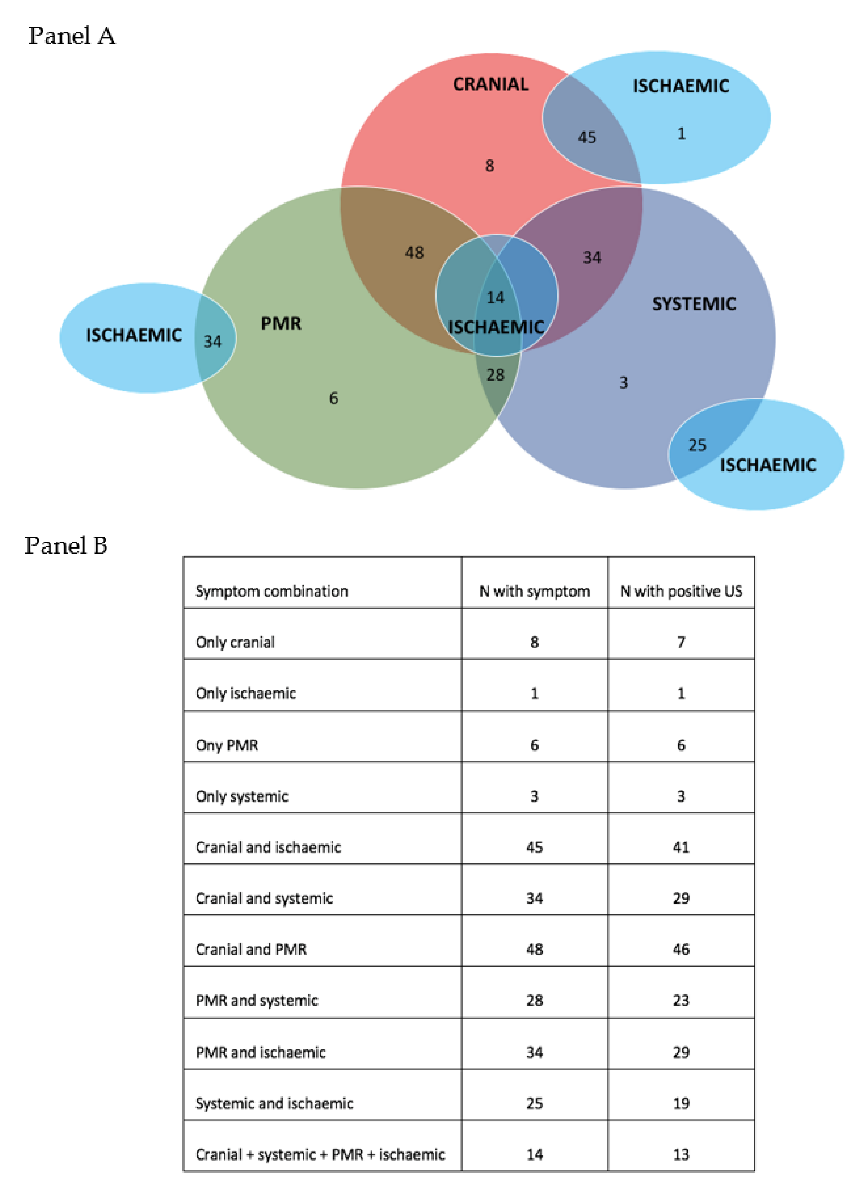
The detailed description of symptoms leading to referral to the FTA are shown in Table 1. Headache was the most frequent symptom, followed by PMR, and systemic symptoms. The following manifestations were the most characteristics to distinguish patients with a final confirmed diagnosis of GCA compared to those who received an alternative diagnosis after FTA assessment: jaw claudication, scalp tenderness, bilateral blindness. Systemic symptoms such as fever and weight loss, albeit non-specific, were significantly more frequent in patients with confirmed GCA. Figure 2 (panel A and B) represents the intersection and overlap of presenting symptoms in relation to US findings among patients referred to the FTA who received a confirmed diagnosis of GCA.
Visual loss occurred in 10.8% of patients with a final diagnosis of GCA. Visual loss not attributable to a final diagnosis of GCA was recorded in 4.5% of patients (p = 0.041). Bilateral visual loss occurred in 2.7% of patients with GCA.
PMR had been previously diagnosed and treated prior to the occurrence of a new clinical suspicion leading to the referral to the FTA in 8 (7.2%) patients with GCA. Ongoing new-onset PMR symptoms were recorded for 61 (54.9%) patients receiving a final diagnosis of GCA. On the other hand, 72 (40.9%) referred patients without GCA had a previous diagnosis of PMR and were referred after the occurrence of new symptoms such as headache or systemic symptoms, but these were finally not attributed to a diagnosis of GCA. Peripheral arthritis was found in 4.5% of patients with GCA. Other rarer manifestations such as non-productive cough were recorded in 2.7% of patients.

3.2. US Findings in First Referrals

US results were positive in 91 (82%) patients with a final confirmed diagnosis of GCA, negative in 175 (99.4%) patients without a confirmed diagnosis of GCA, and dubious in one patient without GCA. The sensitivity of US was 81.98% (95% CI 73.55–88.63%), and the specificity 99.43% (95% CI 96.88–99.99%).
The diagnosis of GCA was not supported by a positive US in 20 (18%) patients; of these, 4 patients underwent TAB (2 with histologic findings confirming GCA), one patient refused to receive TAB, and 5 patients had confirmed LV-GCA at subsequent PET-CT. The 11 (9.9%) patients with a clinical diagnosis all had very typical clinical presentation, laboratory findings, and response to treatment, with a diagnosis of GCA that was also confirmed at follow-up visits.
US findings were found at the level of the TA in 85 patients and AX in 18 patients. The mean number of sites with halos at the level of the TA was 2 ± 1.6 (minimum 0; maximum 6). Bilateral halos were found in 46 (55%) patients with TA involvement and 4 (22%) with AX involvement. Seventy-one patients (77%) had cranial involvement only; 5 (5%) had isolated extra-cranial involvement, and 15 (16%) had both TA and AX halo signs. Two patients had isolated carotid involvement and one patient occipital arteries involvement. Facial arteries were involved in 2 patients but never as isolated.

3.3. Relapsing Disease

The clinical manifestations of the 88 patients assessed with US due to possible signs or symptoms of relapsing disease are represented in Table 2. In patients with a confirmed relapse of GCA, the most frequent manifestations were PMR (32%) and headache (28%). Major relapses including ischemic symptoms such as jaw claudication were really rare (2.2%). There were no cases of visual loss during relapsing disease. Relapses were characterized by an isolated rise in inflammatory markers in 10 patients; nevertheless, inflammatory markers were within the normal ranges in 21.7% of relapsing cases.
US was positive in 31 (67%) of patients with a confirmed relapse. Mean disease duration of patients with a confirmed relapse was 65 ± 79 months. US was negative in all patients without a confirmed relapse of GCA.
US findings were found at the level of the TA in 21 relapsing patients and at the level of the AX in 12 patients. Two had both TA and AX halo signs. Bilateral halos were found in 13 (62%) patients with TA involvement and 3 (25%) with AX involvement.

4. Discussion

Over the last decade, the growing evidence supporting the use of US has led to a radical revolution in the management of patients with GCA [13]. According to the 2018 update of the EULAR recommendations for the management of LVV, a diagnosis of GCA should be confirmed by either imaging techniques or histologic examination in order to increase the diagnostic accuracy [14,15]. In specialized centers provided with the adequate machine equipment and appropriate expertise, TA and AX US has become the first-line imaging modality in case of suspected GCA. In recent years, the application of US as part of FTA clinics has progressively reduced the need for TAB in patients presenting with signs and symptoms suggestive of GCA. In our study, we have confirmed this trend, with only four patients needing an invasive tool to confirm the diagnosis, with obvious advantages for patients and in terms of costs [4,6]. US has been demonstrated to further improve the diagnostic yield given its significantly higher sensitivity compared to TAB [16]. Indeed, US has the advantage of being non-invasive, allowing a thorough assessment of vessels in their whole length, and permits extending the exam to other cranial or extra-cranial arteries if needed. Moreover, US allows access to the results of the examination instantly to guide further decisions on diagnosis and treatment [4,17]. In our study, a high sensitivity of US reaching 81.98% (95% CI 73.55–88.63%) and specificity of 99.43% (95% CI 96.88–99.99%) was demonstrated. Although our study confirmed a high specificity of US for the diagnosis of GCA, it must be kept in mind that the halo sign has been described in patients with TA involvement due to other diseases, including anti-neutrophil cytoplasm antibodies (ANCA)-associated vasculitis, amyloidosis, and neoplasms, therefore, the US findings must in all cases be correlated with the clinical scenario [18,19,20]. The early referral of patients with suspected GCA to a FTA clinic is critical to prevent acute complications, such as permanent visual loss [6,7,21]. In a recent study, we showed that FTA combined with the US of TA and AX significantly reduced the incidence of blindness, mainly due to a decreased diagnostic delay and earlier treatment initiation [8]. In the current study, visual loss occurred in 10.8% of patients with a final diagnosis of GCA, representing a significantly lower frequency compared to the standard care assessment of the disease. The FTA clinics have without doubt changed the prognosis of GCA and the irreversible ischemic damage. Nonetheless, the fear of severe complications such as visual loss and the non-specific nature of the majority of symptoms of LVV also leads to a relevant proportion of patients being referred to the FTA who do not receive a final diagnosis of GCA. In our cohort, only 39% of first referrals had a final confirmed diagnosis of GCA. We analyzed the presenting symptoms leading to the referral to the FTA to identify relevant clinical features to help improve our evaluation of the pre-test probability of GCA and distinguish patients without vasculitis. We demonstrated that jaw claudication, tongue claudication, scalp tenderness, and bilateral visual loss are the most specific symptoms to distinguish GCA from other conditions. Interestingly, headache is not reported in at least 33% of patients with a final diagnosis of GCA, underlying the need to keep a high level of suspicion even in patients without the most frequent cranial symptoms of the disease. Especially for patients with extra-cranial GCA, the referral to FTA clinics might be further delayed by the non-specific nature of the reported symptoms. Systemic manifestations, including ≥2 general symptoms, although non-specific, were significantly more frequent in the GCA group. Furthermore, patients with a final diagnosis of GCA far more frequently had a combination of different cranial or extra-cranial symptoms. Overall, only 18 patients with GCA had isolated symptoms among headache, PMR, systemic symptoms or ischemic manifestations. Recently, the potential difficulties related to the assessment of the probability of a GCA diagnosis prior to the use of further diagnostic tests has been addressed, and a GCA probability score has been proposed to be used in clinical practice [22]. Moreover, dedicated educational programs are of utmost importance in raising the awareness of this condition among primary care practitioners and other specialists. Even though the role of US in the diagnosis of LVV is becoming well-recognized, also with the support of these educational programs, the role of US during follow-up and in the recognition of relapsing disease is still being investigated. Recently, in a multicentric international cohort, we demonstrated the role of US (particularly at the level of the TA) as a monitoring tool for GCA and with a good correlation with parameters of active disease of GCA. In our cohort, we assessed 88 patients with a suspicion of relapse, in whom only 46 (52%) had a confirmed relapsing disease leading to a change in treatment. These findings highlight the difficulties encountered, even by expert rheumatologists, in managing GCA based on clinical and laboratory findings (such as ESR and CRP) that are not disease-specific. Yet, the severe complications associated with GCA impose a low threshold for referral to avoid the underestimation of the clinical presentation and delaying in assessing a patient with a potential recurrence of the disease. Headache (often due to other reasons) and isolated increase in inflammatory markers (not related to LVV activity) were the most frequent manifestations in patients being assessed for a possible relapse which was then not confirmed. Importantly, US was positive in approximately 70% of patients with a relapse and in none of the patients without a confirmed relapse. These findings further clarify and support a possible role for US in the follow-up of patients with LVV.
The study has some limitations. Even though the FTA ensures the assessment of the patient within 24-working hours from referral, we did not have the exact interval between the onset of symptoms and the first assessment of the patient by a primary care physician or another specialist then referring the patient to the FTA. This delay in seeking medical attention by the patient might influence the clinical presentation and possibly the complication rate of the disease. Nevertheless, the FTA still proves to significantly improve the frequency of ischemic sequelae despite this possible delay which can only be improved by increasing the awareness on GCA. Furthermore, the physician performing the US was not blinded to the clinical history of the patient. Nonetheless, in order to reduce the potential bias, the physician always performed the US prior to collecting the anamnestic data of the patient.
In conclusion, US as part of FTA for the diagnosis of GCA is a well-recognized and standardized diagnostic tool that significantly improves the diagnostic sensibility. The clinical presentation of GCA is often non-specific and deserves specialist evaluation whenever suspected. We have provided a thorough description of the clinical characteristics of a large, long-standing FTA clinic that can be helpful in understanding the more frequent, together with the less typical, onset manifestations to improve the early referral of patients. Moreover, the role of US in the management of patients with GCA experiencing a possible relapse is reported.

Author Contributions

Conceptualization, S.M. and C.M.; methodology, S.M. and P.D.; software, S.M.; validation, S.M., P.D. and C.M.; formal analysis, S.M.; investigation, S.M. and P.D.; resources, C.M.; data curation, S.M.; writing—original draft preparation, S.M. and P.D.; writing—review and editing, S.M.; visualization, S.M. and P.D.; supervision, C.M.; project administration, S.M.; funding acquisition, not applicable. All authors have read and agreed to the published version of the manuscript.

Funding

This research received no external funding.

Institutional Review Board Statement

The study was conducted according to the guidelines of the Declaration of Helsinki and approved by the Ethics Committee of Policlinico S. Matteo IRCCS Fondazione, University of Pavia (Reference number E_2016 0031606 of 24 November 2016).

Informed Consent Statement

Informed consent was obtained from all subjects involved in the study. Written informed consent has been obtained from the patient(s) to publish this paper.

Data Availability Statement

Data are securely stored at Policlinico S. Matteo, IRCCS Fondazione, University of Pavia and are available upon request.

Conflicts of Interest

The authors declare no conflict of interest.

References

  1. Salvarani, C.; Cantini, F.; Boiardi, L.; Hunder, G.G. Polymyalgia rheumatica and giant-cell arteritis. N. Engl. J Med. 2002, 347, 261–271. [Google Scholar] [CrossRef] [PubMed]
  2. Salvarani, C.; Cantini, F.; Hunder, G.G. Polymyalgia rheumatica and giant-cell arteritis. Lancet 2008, 372, 234–245. [Google Scholar] [CrossRef]
  3. Chrysidis, S.; Duftner, C.; Dejaco, C.; Schäfer, V.S.; Ramiro, S.; Carrara, G.; Scirè, C.A.; Hocevar, A.; Diamantopoulos, A.P.; Iagnocco, A.; et al. Definitions and reliability assessment of elementary ultrasound lesions in giant cell arteritis: A study from the OMERACT Large Vessel Vasculitis Ultrasound Working Group. RMD Open 2018, 4, e000598. [Google Scholar] [CrossRef] [PubMed] [Green Version]
  4. Luqmani, R.; Lee, E.; Singh, S.; Gillett, M.; Schmidt, W.A.; Bradburn, M.; Dasgupta, B.; Diamantopoulos, A.P.; Forrester-Barker, W.; Hamilton, W.; et al. The role of ultrasound compared to biopsy of temporal arteries in the diagnosis and treatment of giant cell arteritis (TABUL): A diagnostic accuracy and cost-effectiveness study. Heal. Technol. Assess. 2016, 20, 1–238. [Google Scholar] [CrossRef] [PubMed] [Green Version]
  5. Dejaco, C.; Ramiro, S.; Duftner, C.; Besson, F.; Bley, T.A.; Blockmans, D.; Brouwer, E.; Cimmino, M.A.; Clark, E.; Dasgupta, B.; et al. EULAR recommendations for the use of imaging in large vessel vasculitis in clinical practice. Ann. Rheum. Dis. 2018, 77, 636–643. [Google Scholar] [CrossRef]
  6. Diamantopoulos, A.P.; Haugeberg, G.; Lindland, A.; Myklebust, G. The fast-track ultrasound clinic for early diagnosis of giant cell arteritis significantly reduces permanent visual impairment: Towards a more effective strategy to improve clinical outcome in giant cell arteritis? Rheumatology 2016, 55, 66–70. [Google Scholar] [CrossRef] [PubMed] [Green Version]
  7. Patil, P.; Williams, M.; Maw, W.W.; Achilleos, K.; Elsideeg, S.; Dejaco, C.; Borg, F.; Gupta, S.; Dasgupta, B. Fast track pathway reduces sight loss in giant cell arteritis: Results of a longitudinal observational cohort study. Clin. Exp. Rheumatol. 2015, 33 (Suppl. S89), S103–S106. [Google Scholar]
  8. Monti, S.; Bartoletti, A.; Bellis, E.; Delvino, P.; Montecucco, C. Fast-track ultrasound clinic for the diagnosis of giant cell arteritis changes the prognosis of the disease but not the risk of future relapse. Front. Med. 2020, 7, 589794. [Google Scholar] [CrossRef]
  9. Schmidt, W.A. Ultrasound in vasculitis. Clin. Exp. Rheumatol. 2014, 32 (Suppl. S80), S71–S77. [Google Scholar]
  10. Schmidt, W.A.; Seifert, A.; Gromnica-Ihle, E.; Krause, A.; Natusch, A. Ultrasound of proximal upper extremity arteries to increase the diagnostic yield in large-vessel giant cell arteritis. Rheumatology 2008, 47, 96–101. [Google Scholar] [CrossRef] [Green Version]
  11. Ponte, C.; Monti, S.; Scirè, C.A.; Delvino, P.; Khmelinskii, N.; Milanesi, A.; Teixeira, V.; Brandolino, F.; Diamantino Saraiva, F.M.; Montecucco, C.; et al. Ultrasound halo sign as a potential monitoring tool for patients with giant cell arteritis: A prospective analysis. Ann. Rheum. Dis. 2021, 80, 1475–1482. [Google Scholar] [CrossRef] [PubMed]
  12. Monti, S.; Floris, A.; Ponte, C.; Schmidt, W.A.; Diamantopoulos, A.P.; Pereira, C.; Piper, J.; Luqmani, R. The use of ultrasound to assess giant cell arteritis: Review of the current evidence and practical guide for the rheumatologist. Rheumatology 2017, 57, 227–235. [Google Scholar] [CrossRef] [PubMed] [Green Version]
  13. Schäfer, V.S.; Jin, L.; Schmidt, W.A. Imaging for diagnosis, monitoring, and outcome prediction of large vessel vasculitides. Curr. Rheumatol. Rep. 2020, 22, 76. [Google Scholar] [CrossRef]
  14. Hellmich, B.; Agueda, A.; Monti, S.; Buttgereit, F.; de Boysson, H.; Brouwer, E.; Cassie, R.; Cid, M.C.; Dasgupta, B.; Dejaco, C.; et al. 2018 Update of the EULAR recommendations for the management of large vessel vasculitis. Ann. Rheum. Dis. 2020, 79, 19–30. [Google Scholar] [CrossRef] [Green Version]
  15. Mackie, S.L.; Dejaco, C.; Appenzeller, S.; Camellino, D.; Duftner, C.; Gonzalez-Chiappe, S.; Mahr, A.; Mukhtyar, C.; Reynolds, G.; de Sousa, A.W.S.; et al. British Society for Rheumatology guideline on diagnosis and treatment of giant cell arteritis. Rheumatology 2020, 59, e1–e23. [Google Scholar] [CrossRef] [Green Version]
  16. Duftner, C.; Dejaco, C.; Sepriano, A.; Falzon, L.; Schmidt, W.A.; Ramiro, S. Imaging in diagnosis, outcome prediction and monitoring of large vessel vasculitis: A systematic literature review and meta-analysis informing the EULAR recommendations. RMD Open 2018, 4, e000612. [Google Scholar] [CrossRef] [PubMed] [Green Version]
  17. Monti, S.; Águeda, A.F.; Luqmani, R. The use of ultrasound in the management of large-vessel vasculitis: An evolving concept. Clin. Exp. Rheumatol. 2018, 36 (Suppl. S114), 96–102. [Google Scholar]
  18. Fernández-Fernández, E.; Monjo-Henry, I.; Bonilla, G.; Plasencia, C.; Miranda-Carús, M.E.; Balsa, A.; De Miguel, E. False positives in the ultrasound diagnosis of giant cell arteritis: Some diseases can also show the halo sign. Rheumatology 2020, 59, 2443–2447. [Google Scholar] [CrossRef]
  19. Kanaan, M.Z.; Lorenzi, A.R.; Thampy, N.; Pandit, R.; Dayan, M. Bilateral non-arteritic anterior ischaemic optic neuropathy as the presentation of systemic amyloidosis. Neuro Ophthalmol. 2017, 41, 330–334. [Google Scholar] [CrossRef]
  20. Chrysidis, S.; Lewinski, M.; Schmidt, W.A. Temporal arteritis with ultrasound halo sign in eosinophilic granulomatosis with polyangiitis. Rheumatology 2019, 58, 2069–2071. [Google Scholar] [CrossRef]
  21. Monti, S.; Delvino, P.; Bellis, E.; Milanesi, A.; Brandolino, F.; Montecucco, C. Impact of delayed diagnoses at the time of COVID-19: Increased rate of preventable bilateral blindness in giant cell arteritis. Ann. Rheum. Dis. 2020, 79, 1658–1659. [Google Scholar] [CrossRef]
  22. Sebastian, A.; Tomelleri, A.; Kayani, A.; Prieto-Pena, D.; Ranasinghe, C.; Dasgupta, B. Probability-based algorithm using ultrasound and additional tests for suspected GCA in a fast-track clinic. RMD Open 2020, 6, e001297. [Google Scholar] [CrossRef]
Figure 1. Flowchart of the fast-track reasons for referral and findings. US: ultrasound; GCA: giant cell arteritis; NOT GCA: diagnosis of GCA not confirmed; pos: positive.
Applsci 12 01621 g001
Figure 2. Clinical presentation, symptoms overlap, and corresponding ultrasound findings in patients with GCA. Panel A: overlap of presenting symptoms in patients referred to the fast-track clinic and receiving a final diagnosis of GCA. Panel B: number of patients with a positive ultrasound according to the different types of clinical presentation.
Applsci 12 01621 g002
Table 1. Clinical presentation of newly referred patients to the fast-track clinic.
GCA (N= 111)NOT GCA (N = 176)Overall (N = 287)p
Headache74 (66.6%)65 (36.9%)138 (47.9%)<0.0001
Jaw claudication40 (36%)4 (2.3%)50 (17.3%)<0.0001
Tongue claudication11 (9.9%)011 (3.8%)<0.0001
Blurred vision6 (5.4%)4 (2.3%)10 (3.5%)0.165
Visual loss12 (10.8%)8 (4.5%)20 (6.9%)0.041
Bilateral visual loss3 (2.7%)05 (1.7%)0.029
Scalp tenderness25 (22.5%)1 (0.56%)26 (9%)<0.0001
Polymyalgia rheumatica61 (54.9%)66 (37.5%)127 (44%)0.039
Systemic symptoms47 (42.3%)32 (18.1%)79 (27.4%)<0.0001
≥2 systemic symptoms10 (9%)4 (2.3%)14 (4.9%)0.011
Isolated raised inflammatory markers1 (0.9%)9 (5.1%)10 (3.5%)0.058
Normal inflammatory markers2 (1.8%)45 (25.6%)47 (16.3%)<0.0001
Fever29 (26.1%)13 (7.4%)42 (24.5%)<0.0001
Weight loss33 (29.7%)19 (10.7%)52 (18%)<0.0001
Arthritis5 (4.5%)7 (3.9%)12 (4.2%)0.804
Non-productive cough3 (2.7%)2 (1.1%)5 (1.7%)0.312
Mean ESR value (mm/h)59 ± 3558 ± 3558 ± 350.814
Mean CRP value (mg/L)43 ± 5042 ± 5042 ± 500.869
Mean prednisone-equivalent dose on the day of US scan (mg/day)18 ± 6417 ± 6417 ± 640.897
Not treated with glucocorticoids on the day of US scan21 (19%)66 (38%)87 (30%)0.0007
Table 2. Clinical presentation of patients with possible relapse assessed at the fast-track clinic.
Relapse Confirmed
(N = 46)
Relapse Not Confirmed
(N = 42)
Overall (N = 88)p
Headache13 (28%)19 (45.2%)32 (36%)0.095
Jaw claudication1 (2.2%)01 (1.1%)0.0336
Tongue claudication000na
Blurred vision1 (2.2%)2 (4.7%)3 (3.4%)0.520
Visual loss000na
Bilateral visual loss000na
Scalp tenderness01 (2.3%)1 (1.1%)0.304
Polymyalgia rheumatica15 (32%)015 (17%)0.0001
Systemic symptoms5 (11%)4 (9.5%)9 (10%)0.818
≥2 systemic symptoms2 (4.3%)1 (2.3%)3 (3.4%)0.604
Isolated raised inflammatory markers10 (21.7%)13 (30.9%)23 (25.8%)0.329
Normal inflammatory markers10 (21.7%)7 (16.6%)17 (19.1%)0.547
Fever2 (4.3%)2 (4.7%)4 (2.3%)0.928
Weight loss4 (8.6%)2 (4.7%)6 (6.7%)0.418
Arthritis/arthralgia1 (2.2%)3 (7.1%)4 (2.3%)0.273
Non-productive cough01 (2.3%)1 (1.1%)0.304
Mean ESR value (mm/h)36 ± 2437 ± 3036 ± 240.863
Publisher’s Note: MDPI stays neutral with regard to jurisdictional claims in published maps and institutional affiliations.