Article

Periodontal Tissue Reaction Consecutive Implantation of Endodontic Materials and Subsequent Integration of Complex Oral Rehabilitation Treatments

by
Liana Aminov
1,
Aurelian Sorin Pasca
2,*,
Eusebiu Viorel Sindilar
3,
Maria Antonela Beldiman
4,
Bogdan Petru Bulancea
4,
Ovidiu Stamatin
4,*,
Iulian Costin Lupu
4,
Irina Croitoru
5,
Silvia Teslaru
1,*,
Sorina Mihaela Solomon
1,
Georgeta Liliana Foia
6 and
Laura Elisabeta Checherita
1
1
2nd Dental Medicine Department, “Grigore T. Popa” University of Medicine and Pharmacy, 700115 Iasi, Romania
2
Department of Pathology, Faculty of Veterinary Medicine, “Ion Ionescu de la Brad” University of Life Sciences, 700490 Iasi, Romania
3
Department of Surgery, Faculty of Veterinary Medicine, “Ion Ionescu de la Brad” University of Life Sciences, 700490 Iasi, Romania
4
3rd Dental Medicine Department, “Grigore T. Popa” University of Medicine and Pharmacy, 700115 Iasi, Romania
5
Department of Preventive Medicine and Interdisciplinarity, “Grigore T. Popa” University of Medicine and Pharmacy, 700115 Iasi, Romania
6
1st Dental Medicine Department, “Grigore T. Popa” University of Medicine and Pharmacy, 700115 Iasi, Romania
*
Authors to whom correspondence should be addressed.
Appl. Sci. 2022, 12(3), 1464; https://doi.org/10.3390/app12031464
Submission received: 9 November 2021 / Revised: 21 January 2022 / Accepted: 24 January 2022 / Published: 29 January 2022
(This article belongs to the Special Issue Diagnosis and Treatment of Periodontal Diseases)

Abstract

:
Oral rehabilitation is a main branch of dentistry focused on diagnosing the patient’s problem and creating a treatment plan to restore aesthetics, recondition morphologically all components, and recover the functionality of the oral cavity. Biological compatibility of the materials used has a major importance, due to the direct contact with essential tissues, such as the soft and hard tissue of the periodontium and the potential influence on the outcome of the treatment. The present material aims to assess the inflammatory response after subcutaneous implantation of three materials frequently used in endodontics (Mineral Trioxide Aggregate—MTA, DiaRoot BioAggregate, and Sealapex). The evaluation of the reparative tissue reaction after 7, 30, and 60 days, respectively, subsequent to in vivo implantation, was carried out through electron microscopy imaging. Moreover, evaluation of the dynamics of the osteogenesis process was an indicator for the maintenance of internal homeostasis in the context of complex intraoral rehabilitation treatments that include fixed prosthodontics correlated with the particular periodontal-aesthetic aspects and completed by cranio-mandibular repositioning. Our study showed increased absolute values of alkaline phosphatase in all material-implanted cases (more pronounced in MTA and Bio Aggregate), highlighting that this enzyme could be an effective indicator of bone formation, which takes place after the material implantation, with the most significant elevated values at 30 days postoperatively.

1. Introduction

Intraoral complex rehabilitation treatments are intended to restore the aesthetics and functionality at the dental-level through prosthetics or substitution as dental implant, in accordance with the surrounding tissue homeostasis, and in connection with the related muscular and temporo-mandibular joint (TMJ) components of the stomatognathic system (SS), complementary to facial rehabilitation.
Endodontic treatment, in context of oral complex rehabilitation treatment is a conspicuous, precise, sometimes very complicated, painstaking intervention that requires multiple skills, care, and training.
The purpose of endodontics, in general, include non-surgical treatment of teeth management of traumatic injuries, reintervention in teeth previously treated endodontically, and treatment procedures related to coronal restorations by means of posts and/or cores involving the root canal space. Despite major advances in endodontic therapy in recent decades, clinicians are still confronted with a complex root canal anatomy and a wide selection of endodontic filling materials, being guided by a consistent biochemical stability and newly formed biochemical bonds that provide the valid support required in the reconstructions needed, amongst which dentists can choose according to the case and needs [1].
In the pathophysiological context, one can resume the idea that the newly created connections are based on the inflammatory response that is commonly thought to operate during homeostasis impairments, such as infection, tissue injury, and the presence of foreign bodies or irritants. More generally, an inflammatory response is presumably triggered whenever tissue malfunctions are detected. These types of inflammatory response are likely to be more common but of lower magnitude than the classic inflammatory responses induced by infection or injury. The nature and the degree of tissue malfunction will influence whether the inflammatory responses are detectable by using common biomarkers. A generic inflammatory ‘pathway’ consists of inducers, sensors, mediators, and effectors, with each component determining a certain type of inflammatory response. The inducers of the specific inflammation during our research have been of exogenous, non-microbial origin [2,3]. Exogenous inducers of inflammation that are of non-microbial origin include allergens, irritants, foreign bodies, and toxic compounds. Certain allergens are detected because they mimic the virulence activity of parasites; others can act as irritants on the mucosal epithelia. The inflammatory response induced by both types of allergens is largely similar because defense against parasites and environmental irritants relies on expulsion and clearance mediated by the mucosal epithelia. Foreign bodies are indigestible particles that either are too large to be phagocytosed or can cause phagosome membrane damage in macrophages; in this situation, the encapsulation of foreign objects is an ancient defensive strategy [4,5].
Within their limit amalgam, gutta-percha, zinc oxide and eugenol (ZOE), zinc oxide eugenol reinforced with polymethacrylate (IRM), zinc oxide eugenol reinforced with ethoxy benzoic (Super EBA), glass ionomer cement, composite, and mineral trioxide aggregate (MTA) have all been used for being nontoxic, non-mutagenic, biocompatible, and insoluble, root-end filling materials, closely considered to the ideal one’s, each of them, at the time of their use as top materials.
Incrementally with the specific evolution of the times in most scientific fields, these materials—as our study follows—other new as well, with higher impact upon tissue regeneration, but which due to certain limitations by different socio-economic criteria and specific condition have not yet been studied by us, being mentioned only.
Bioactive bioceramic (BA) materials used in endodontic treatment promote pulpal and periapical tissue healing and are easy to use. They are dental materials used extensively for vital pulp therapy, protecting scaffolds during regenerative endodontic procedures, apical barriers in teeth with necrotic pulps and open apices, perforation repairs as well as root canal filling, and root-end filling during surgical endodontics [6].
These materials can be placed in contact with both soft and hard vital tissues, influencing the final result of the treatment and, therefore, their biocompatibility and regenerative properties are of critical importance [7,8]. The biological properties of root canal sealers and the degree of cytotoxicity or stimulation they manifest on different cells, including fibroblasts, osteoblasts, and cementoblasts must be evaluated in vivo as well as in vitro. The main characteristic properties of MTA include superior sealing ability, biocompatibility, antimicrobial effect, radiopacity, dimensional stability, and tolerance to moisture over other dental materials such as IRM, amalgam, Ca(OH)2, super EBA, or ZOE. Its composition was described as primarily a mixture of calcium silicates comprised of calcium oxide (CaO) and silicon dioxide (SiO2). MTA restoratives are typically tested with a mixture of tests from multiple standards. As the setting of MTA is dependent upon hydration, the results of various MTA restoratives and sealers are dependent upon the healing approach. Since its design, MTA has been tested under laboratory conditions, in animal studies and in clinical trials as well [9].
Another product that is not marketed as MTA but is chemically similar is the BioAggregate (BA-Innovative BioCeramix, Inc., Vancouver, BC, Canada), a biocompatible pure white powder composed of ceramic nanoparticle upon mixing DiaRoot with BioA. It differs from MTA, being aluminum-free and containing calcium phosphate monobasic and tantalum pentoxide. BA also includes hydroxyapatite and amorphous silicon oxide. Hydroxyapatite and amorphous silicon oxide are added by the manufacturer to reduce the levels of the calcium hydroxide, which does not contribute to the strength of the material.
Polymeric calcium hydroxide root canal sealer named Sealapex, the original non-eugenol, calcium hydroxide polymeric root canal sealant produces rapid healing and hard tissue formation issue confirmed by some histopathologichal analysis studies that showed it was the best sealer that permitted the deposition of mineralized tissue at the apical level and also the only sealer that provided complete sealing with no inflammatory infiltrate occurring and no reabsorption of mineralize tissues [10]. Several studies in the literature report various results regarding the tested sealers, cell type, time of cell exposure to materials, extract dilutions, and specimen size [11]. However, there are few studies that evaluate the cytotoxic effect of root canal sealers in an in vivo condition and, also in a reproducible model that comes close to the real response to the area of exposure of the apical foramen diameter without using animal models [12].
In most of clinical situations, the endodontic treatment is followed by an unidental or bridge component prosthetic restoration. As a matter of theory issue, this stage framed of the treatment plan—in non-specific preprosthetic treatments part, is considered as a premise of restoring the prosthetic support that needs an increased resistance considering the height power of masticatory forces that require an optimally functionality in the stomatognathic system areal.

Aim of Study

The aim of this study was the comparative evaluation of the reparative response of three materials used in endodontic treatment, through implantation in experimental animals and the evaluation of the endo—periodontal complex tissue response at different time periods. The specific objectives of the research regarded the analysis of the bone tissue response and osteogenic effect after subcutaneous implantation of three materials used in endodontics, displaying different chemical composition, in bone proximity. The importance of conducting the in vivo biocompatibility and integration study stems from the need to validate temporal behavior from the perspective of a subsequent clinical application in the prosthetic rehabilitation complex treatment phases and endo-periodontal aesthetics aspects as well.

2. Materials and Methods

2.1. The Experimental Procedures

The experimental procedures performed in this study have been carried out according to the protocol revised and approved by the animal research ethics commission of the Faculty of Veterinary Medicine “Ion Ionescu de la Brad”, Iasi, in accordance with international principles of biomedical research on animal’s experience and with the approval of the Ministry of Education, Research and Innovation [13]. The biomaterials that have been implanted are not dangerous, and the performed method is recognized by the international scientific community, the character of bio and cytocompatibility being endorsed [14].
Since the biomaterials used in root fillings and repairs come into direct contact with living tissue, a correct assessment of the response of these tissues was attempted by implanting the materials in the connective tissue in rabbits. The irritating and stimulating effect of biomaterials was achieved by electron microscopy examination of the implant regions and evaluation of the extent of the inflammatory process of the subcutaneous connective tissue around the implants.
The materials used were as follows:
  • MTA (Mineral Trioxide Aggregate, Dentsply, Tulsa Dental, Johnson City, TN, USA), an alkaline powder consisting of fine hydrophilic particles that set in the presence of moisture, promotes bone formation, and facilitates the regeneration of the periodontal ligament, with a high antibacterial effect [15,16,17]. It is made of calcium hydroxide, bismuth oxide (Bi2O3), calcium sulfate (CaSO4), tricalcium silicate ((CaO)3·SiO2), dicalcium silicate ((CaO)2·SiO2), and tricalcium aluminate ((CaO)3·Al2O3).
  • DiaRoot BioAggregate (Innovative BioCeramix, Inc., Vancouver, BC, Canada) is a material similar in structure to MTA that additionally contains ceramic nanoparticles. It has proven antiseptic proprieties and at the same time stimulates cementogenesis [18,19]. The chemical composition includes calcium silicate, calcium hydroxide, hydroxyapatite, and tantalum oxygen (Ta2O5).
  • Sealapex (Kerr, Switzerland) is a calcium hydroxide-based cement with good compatibility used for root canal sealing [20], but with a weak leakage resistance [21]. It has the following chemical composition: barium sulfate, titanium dioxide, zinc oxide, calcium hydroxide, butylbenzene, sulfonamide, zinc stearate.
The used endodontic materials were prepared according to the instructions of the manufacturer and were subsequently introduced in 10 mm in length and 2 mm in diameter polyethylene tubes.

2.1.1. In Vivo Experiment

Thirty rabbits, aged 6 months and weighing 3.5 kg (±50 g), raised and fed in identical conditions (food and water ad libitum), were used.
The rabbits were divided into 4 groups:
  • Group 1—9 rabbits—receiving MTA implants;
  • Group 2—9 rabbits—receiving DiaRoot implants;
  • Group 3—9 rabbits—receiving Sealapex implants;
  • Group 4 (control)—3 rabbits—receiving empty polyethylene tube implants.
The experimental period lasted 60 days, while the rabbits were kept in similar conditions and were fed identical food, except 24 h prior to the dental implant surgery when they received water, but no food. All rabbits were fed a normal diet until the end of the study.

Surgical and Post-Operative Protocol

The surgical interventions have been accomplished under general anesthesia and aseptic conditions. Prior to anesthesia, rabbits received atropine premedication (0.02 mg/kg; Atropine, Pasteur Institute, Bucharest, Romania). Anesthesia was induced with xylazine (0.1 mg/kg i.m, Xylazine Bio 2%, Bioveta, Czech Republic) and ketamine (10 mg/kg i.m., Ketaminol® 10, Intervet International GmbH, Neufahrn bei Freising, Germany). Preoperatively, the lateral thoracic regions were shaved and disinfected with antiseptic solution 96% (Videne; Adams Healthcare Ltd., Birmingham, UK). The rabbits were first placed in the left and then in the right lateral decubitus position. Skin incisions were made for implanting the tubes, which were inserted in depth of the subcutaneous connective tissue, in contact with the bone.
In the end, the surgical wound was sutured with non-absorbable suture thread.
For each animal, blood samples were collected prior to implantation, following the dynamic variation of some parameters corresponding to the osteogenesis processes. Alkaline phosphatase values were evaluated in each case.
For 3 postoperative days, each rabbit was given an analgesic (carprofen 4 mg/kg i.sc; Rimadyl®, Pfizer, Tadworth, UK) and prophylactic antibiotic (7.5 mg/kg amoxicillin; VEYX® YL LA 200, Veyx-Pharma GmbH, Schwarzenborn, Germany).
At 7, 30, and 60 days after implantation, 3 rabbits from groups 1, 2, and 3 and one rabbit in the control group (4) have been euthanized using T61 solution (2 mL/kg, i.p., MSD Animal Health GmbH, Cuxhaven, Germany). Briefly, post sampling, all specimens were fixed in 10% buffered formalin and embedded in paraffin with tissue processor Leica TP1020 tissue processor (Leica Microsystems GmbH, Wetzlar, Germany). Sections of 5 μm thickness were obtained with a Microtome SLEE CUT 6062 (SLEE Medical GmbH, Mainz, Germany).
The evaluation of the reparative tissue reaction at 7, 30, respectively 60 days after implantation have been performed through electron microscopy images and the evaluation of osteogenesis dynamics indicators and maintenance of internal homeostasis by means of comparative analysis of the results.
The part with applicability for our experiment lies in respecting the curative and functional principles that restore both, morphology and functionality, through complex rehabilitation treatments; in this case, prosthetic treatments with complex endo-periodontal support require first an integration and a prior biocompatibility of the materials applied to the subjects from the study, an issue that has been interpreted by us as an objective tissue response of the biological interactions.
The present study reflects an interdisciplinarity collaboration between the Clinical Dental Education Base, “Mihail Kogalniceanu”—Grigore T. Popa University of Medicine and Pharmacy from Iasi, Romania among the disciplines of Endodontics, Periodontology, Fixed and Removable Prosthesis associated with Prosthesis Technology and Orthodontics specialties and Apollonia University from Iasi. The study included 28 patients presented between 2015–2020, subjects that followed endodontic treatments with the materials used in our study. The subjects that were urban residents all proceeded to the informed consent, beneficiated from rigorous clinical observation and prosthetic rehabilitation of unidental or pluridental restauration treatments, as inclusion criteria, allergic patients and uncooperative ones being excluded.
The demographic distribution of patients was as follows: 16 men between 21 and 45 years old, 10 urban and 6 rural, and 12 women between 18 and 38 years old, of which 10 urban and 2 rural (Figure 1a).
The need for endodontic and prosthetic reintervention during the study, according to the clinical rehabilitation plan, with the methods used: pre- and pro-prosthetic stages, post-prosthetic one, with the primary and secondary adaptation, the type of prosthesis, the type of material and the supporting paraclinical investigations in patient’s diagnosis, constitute a complex and important level to manage [22,23].
Analog Visual Scale assessment questionnaires were used, which could quantify the success of prosthetic treatment in oral rehabilitation (Figure 1b).

3. Results

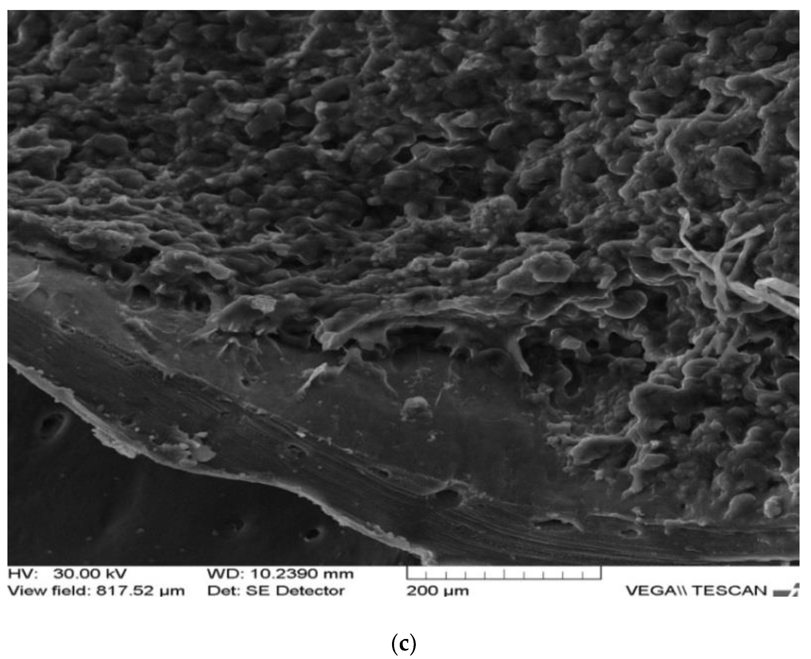
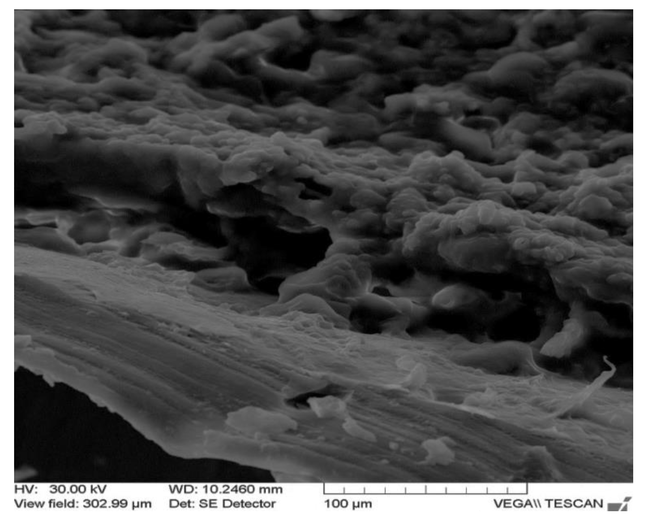
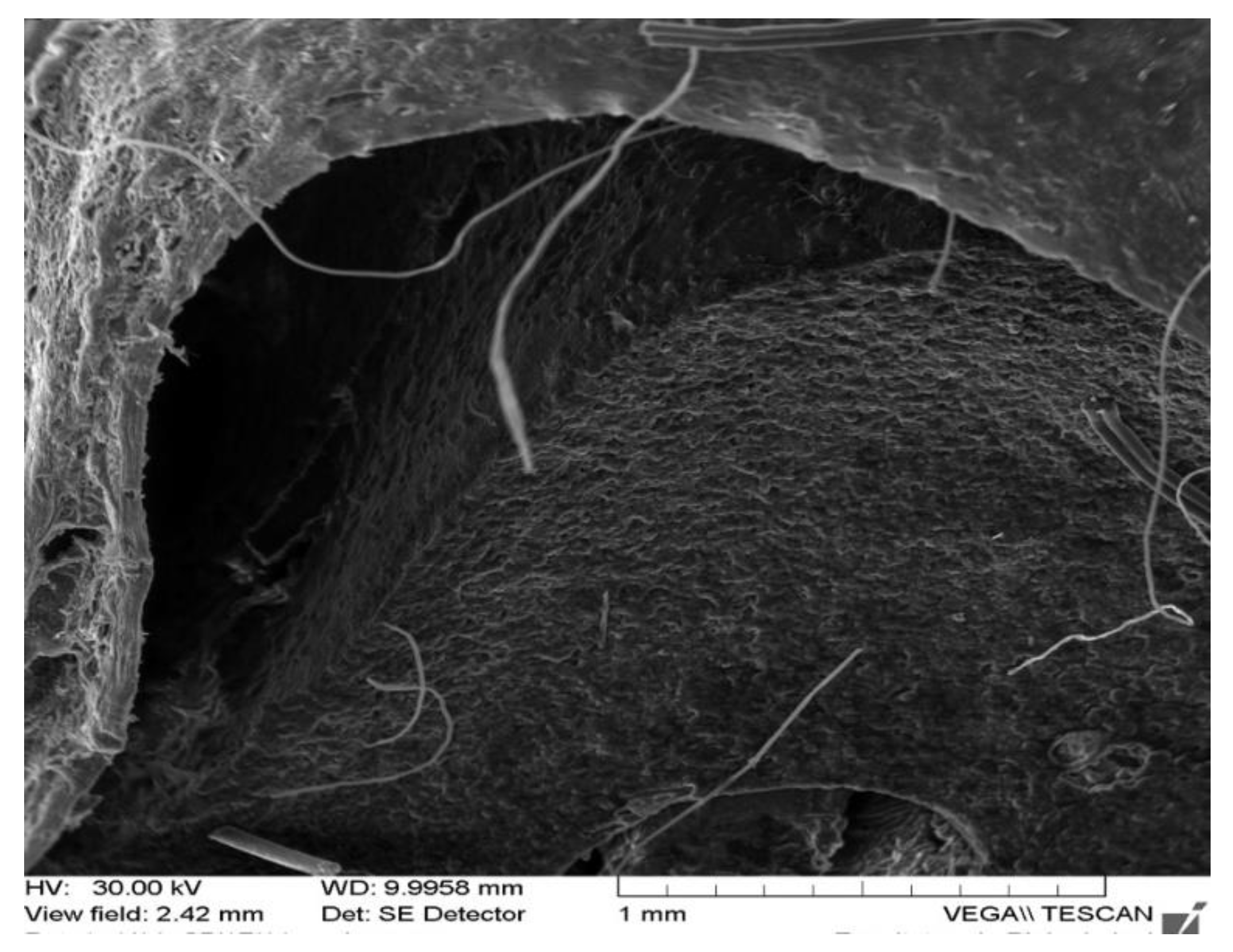
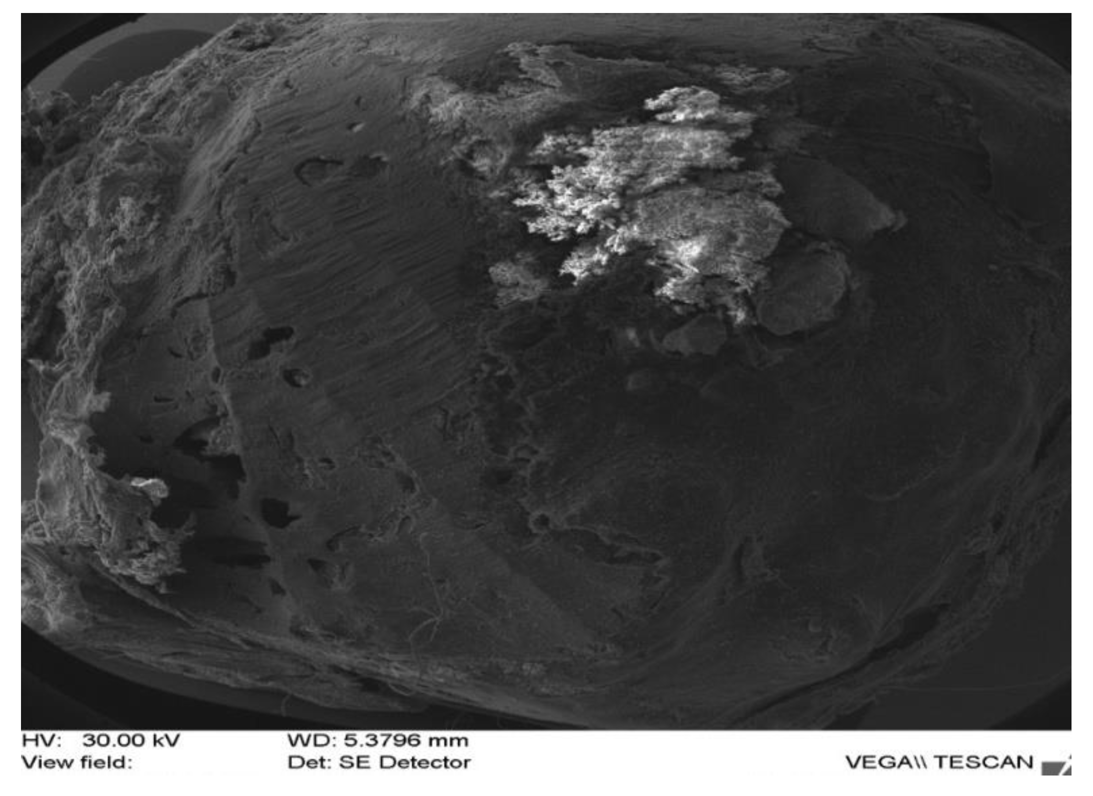
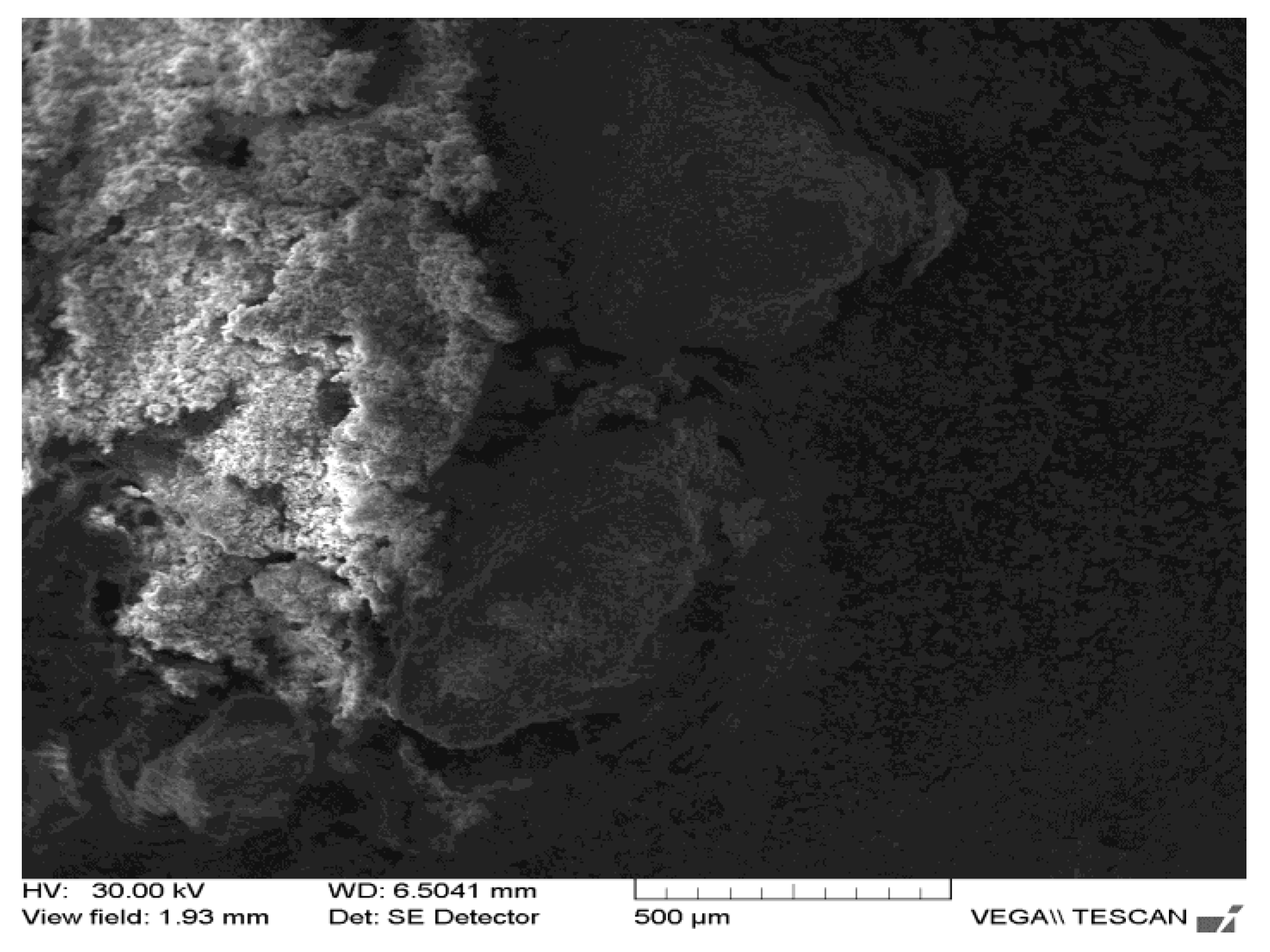
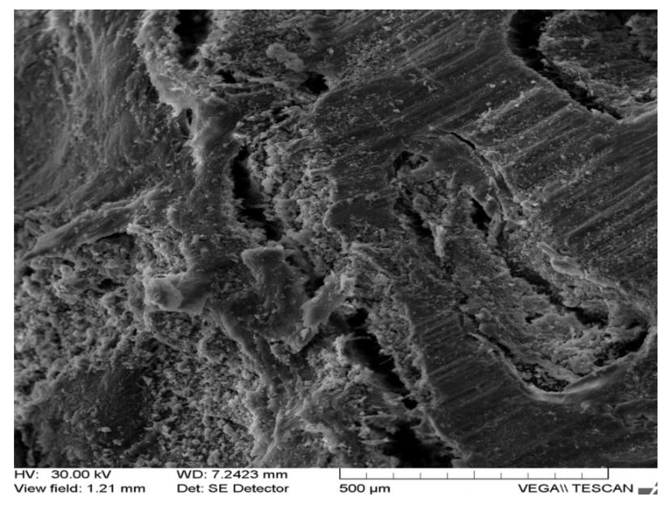
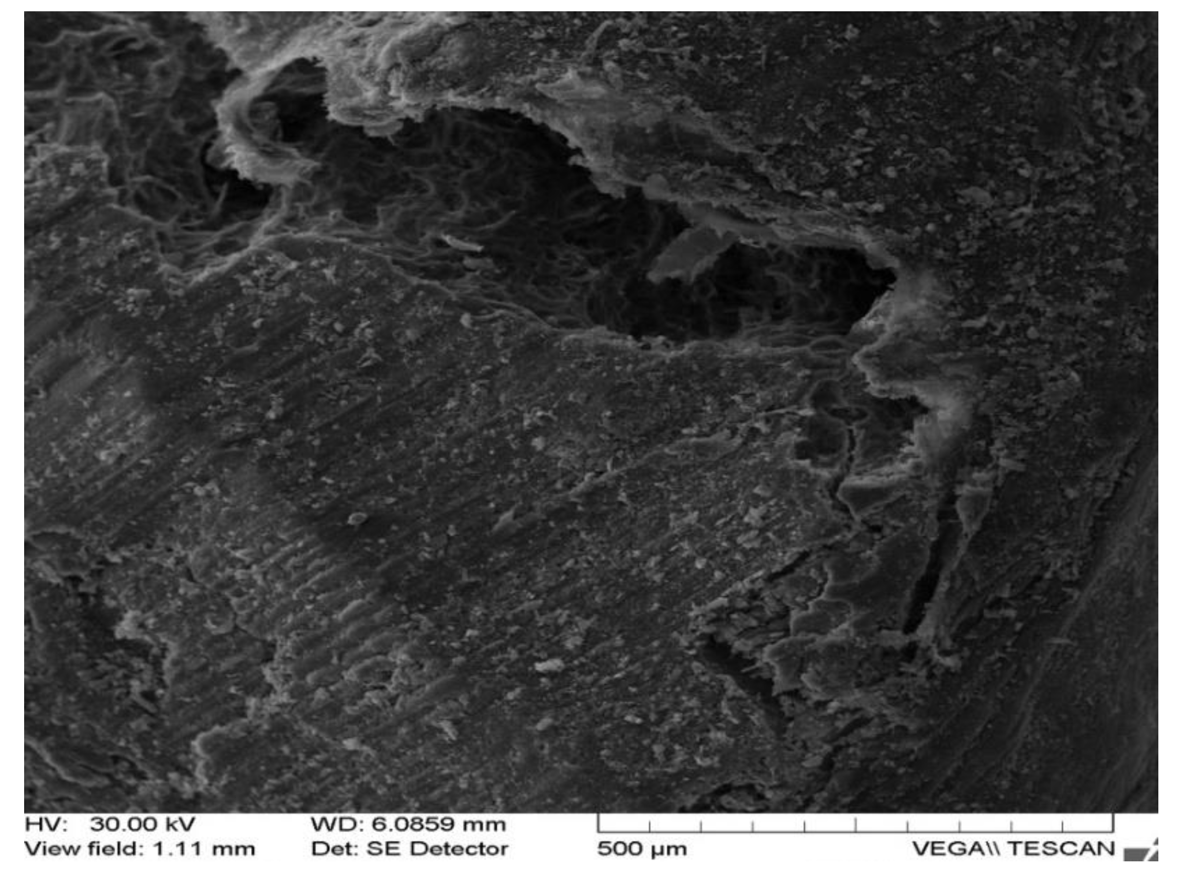
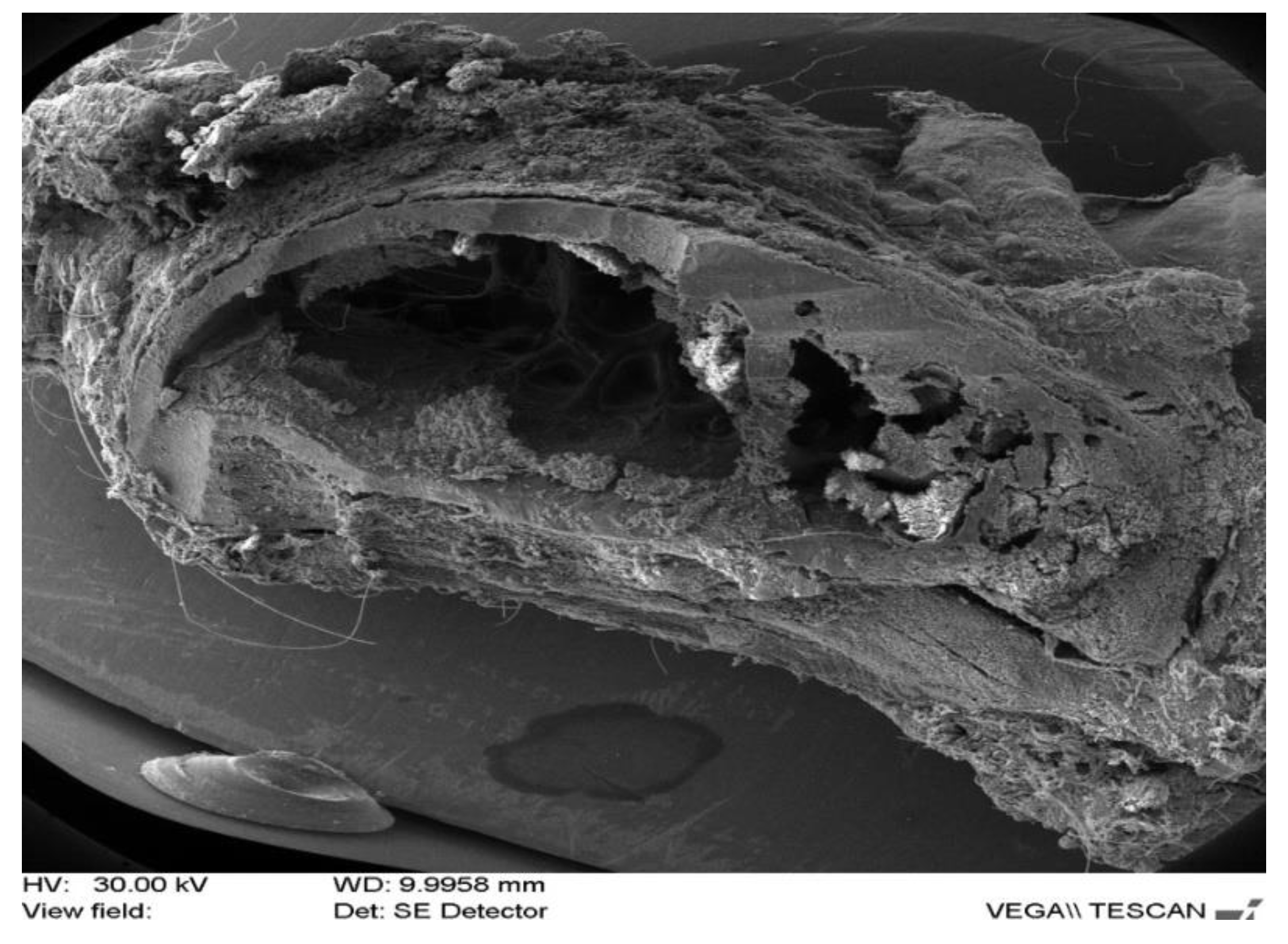
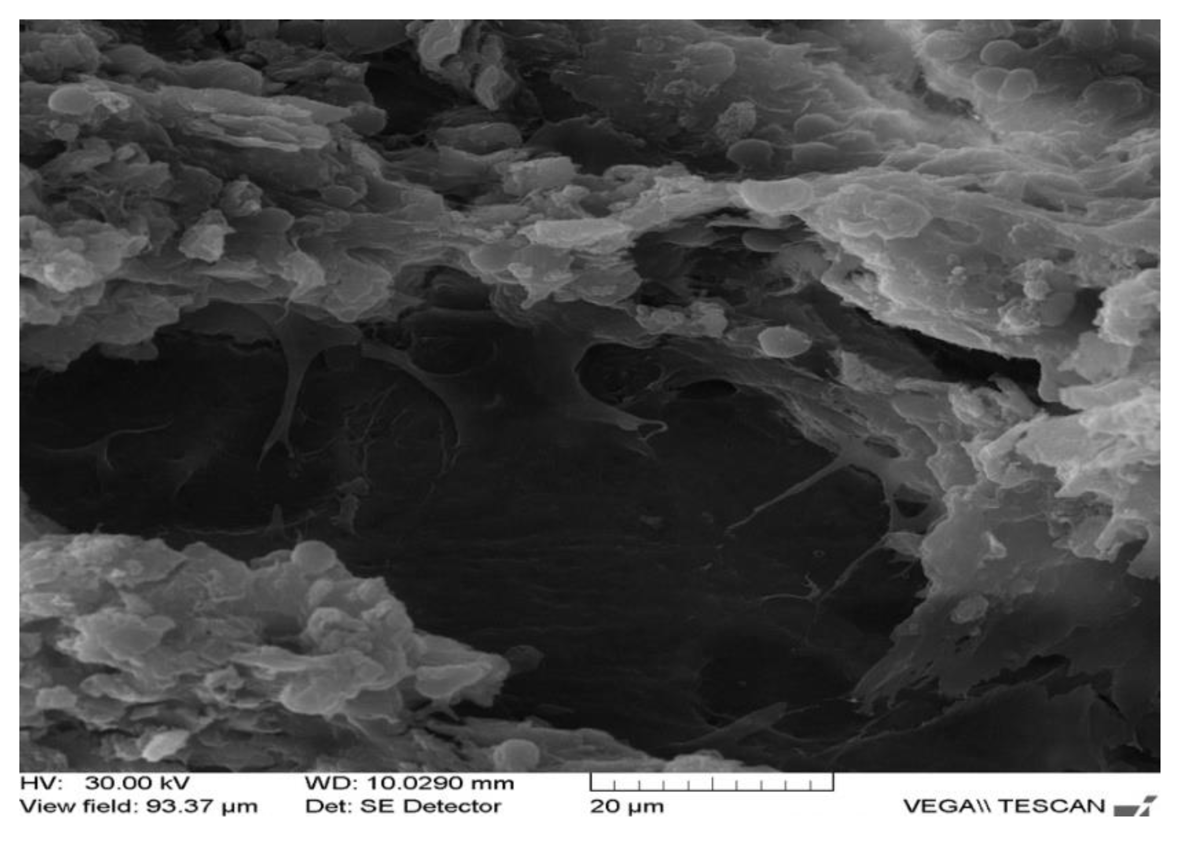
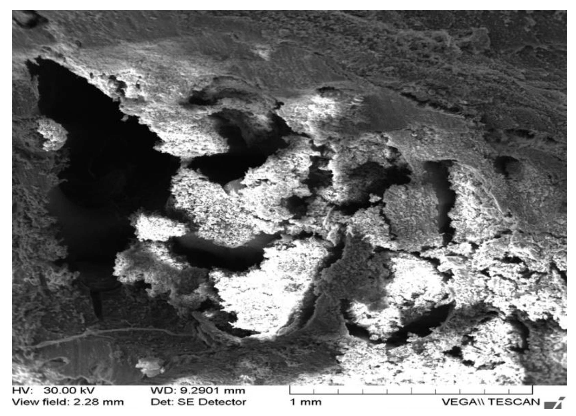
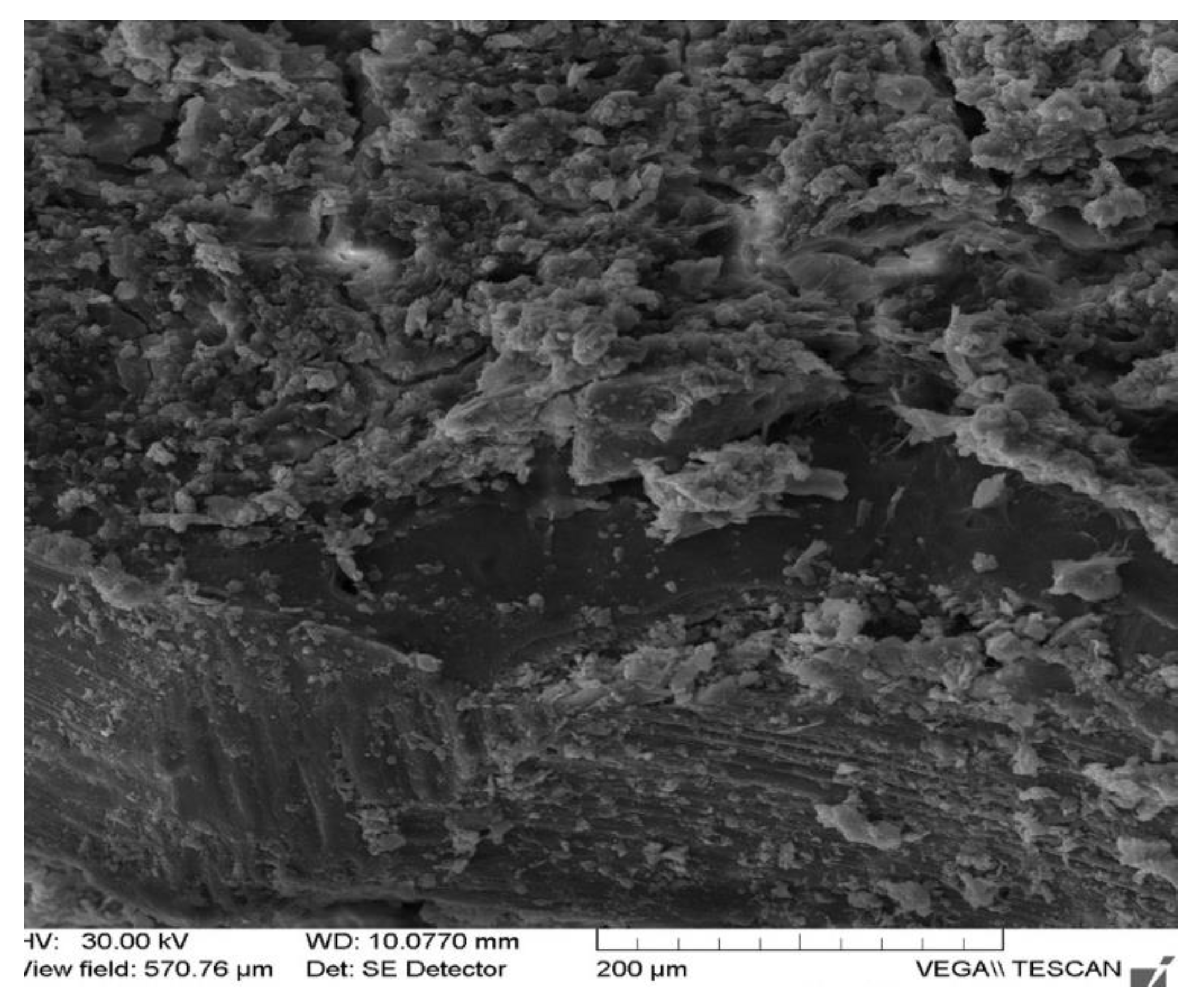
Scanning electron microscopy images collected 30 days after implantation point out the presence of numerous osteoblasts, included in ossein sequestrations, with a tendency to organize in osteoid structure, in order to generate the pre-osseous substance oriented in the traverses (Figure 1c and Figure 2). There are also Howship gaps in which active osteoclasts are present, involved in bone remodeling and which by phagocytosis eliminate the excessive bone (Figure 3). Moreover, multiple areas of deposition of excess valuable substance, in the form of agglomerations and smaller areas of rarefied bone, due to the monocytic macrophage system (osteoclasts) have been identified as well (Figure 4 and Figure 5). In the areas adjacent to the excess osteine deposition, gaps in which osteoclasts are present have also been detected. The new bone bands that delimit these gaps are obvious and have numerous osteocytes in their structure (Figure 6 and Figure 7). There are also typical osteoclasts, with a brushed edge, which attach to the dense material formed by the precious substance elaborated in excess by osteoblasts (Figure 8). In a part of the newly deposited bone substance, a clear tendency to organize in the guideways could be noticed, in the vicinity of the external fundamental system (Figure 9). Osteoblasts with intense osteogenic activity are present in the bone bands, indicating bone regeneration process around the implanted material (Figure 10 and Figure 11).
The following analysis acquaint the biochemical profile of the implanted animals at 7, 30, and 60 days postoperatively for alkaline phosphatase, reported at the operative moment with initial biochemical parameters in all analyzed cases, corresponding to the three materials (Table 1, Table 2 and Table 3). The initial value in the control rabbit = 163.6 IU/L.
Subsequent to analysis of the biochemical profile of implanted animals at intervals set: 7, 30, and 60 days after surgery, the results pointed out significant elevation of alkaline phosphatase values reported within seven days at operation, growth that is extended up to 30 days, thus reaching significantly higher levels than the control group. After 30 days postoperatively, alkaline phosphatase values decreased significantly, the results exhibiting the treatment in all three cases studied. The baseline value in control group rabbits = 163.6 IU/L (Table 4).
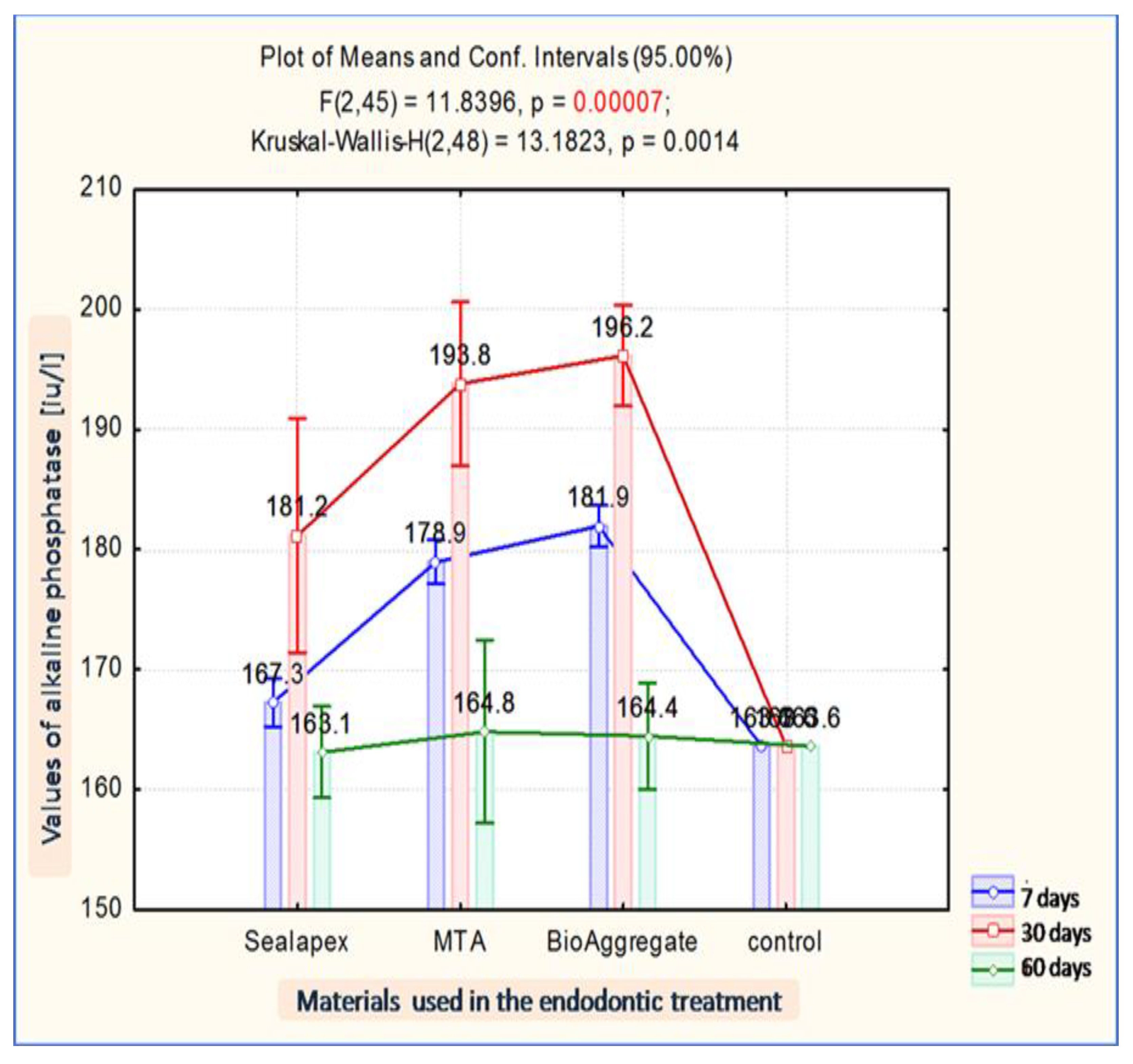
As depicted in Figure 12, the graphic indicator that reveals the results of the local research, a clear distinction can be noticed for Bioaggregates (BA) at 30 days 196.2 IU/L, followed closely by MTA 193.8 IU/L and the last Sealapex 181.2 IU/L.
Our results point to an average time to stabilize the reaction of 30 days, with a clear tolerance and self-adaptation mediated by the biochemical reaction of tissue responsiveness, favorably framing the prognosis of the subsequent situation at 60 days, when the MTA has the final response of 164.8 IU/L, slight difference as 164.4 IU/L in Bio Aggregate (BA) followed by Sealapex at 163.1 IU/L.
The aspects recorded at seven days capture the biochemical events when the cascade of reaction of the inflammatory response, allergen type reaction, continues with the particularities corresponding to the stage of intercellular interaction upon material type, a fact iterated by the obtained values statistically interpreted as MTA 178, 9 IU/L, Bioaggregate 181.9 IU/L, Sealapex 167.3 IU/L.
Through the above-mentioned procedures, we tried to validate the information, using rehabilitation treatments methodology, based on various prosthetic devices and all kinds of possible, biocompatible, materials used in prosthesis technology, with regard to the electronic-microscopically studied root filling materials, in order to be used in dental practice, especially in the prosthetic stability rehabilitation accompanied by aesthetic response of periodontal tissue support therapy.
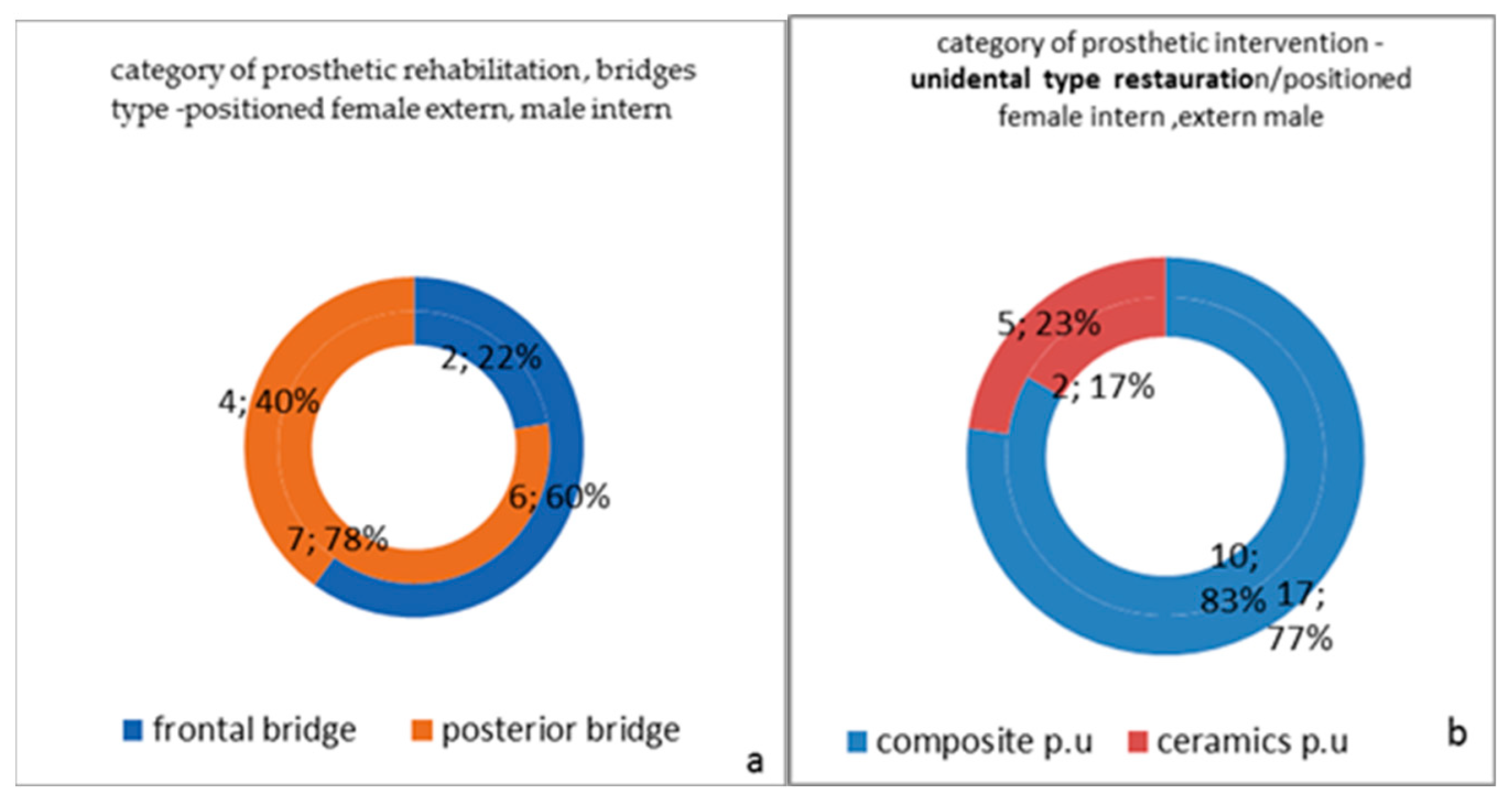
Therefore, we applied endodontic treatments to the patients using the mentioned materials, followed by prosthetic restorations, special fixed restorations: composite crowns in the lateral arch, premolar area, in ten women (35.71%) and seventeen men (60.71%) as well as ceramic restorations placed in the frontal area in two women (7.14%) and five men (17.85%). In the posterior arch area, metal ceramic prosthetic devices were applied bilaterally in seven women (28%), and in the frontal area for two women (7.14%), while for men there were six (21.42%) metal-ceramic prosthetic devices in the frontal area and four (14.28%) prosthetic appliances in the posterior area.
Periodontal cure has been used in twelve cases (42.85%) through etiological curettage, surfacing also followed by gingivectomy interventions and subsequent occlusal interventions, orthodontic treatments, and mouthguard appliances.
Considering the aspects revealed by the individual pathologies, it is absolutely necessary to take into account the more complex situations that would require a special approach. Thus, following the pain triggered by the effects of malocclusions and manifest cranio-mandibular mal-relationships, treatments involving joint repositioning, stress therapies, muscle rebalancing that require drug combinations are mandatory, as a need for complex rehabilitation, all completing the clinical picture of the study group subjects.
Not only prescription of myorelaxants, but also the patients’ restoration and integration by inducing psychical rebalancing using adjuvant therapies in order to establish homeostasis parameters are of critical importance (Figure 13a,b).
As we consider representative and suggestive the following images from rehabilitation treatments clinical cases (Figure 14a–d).
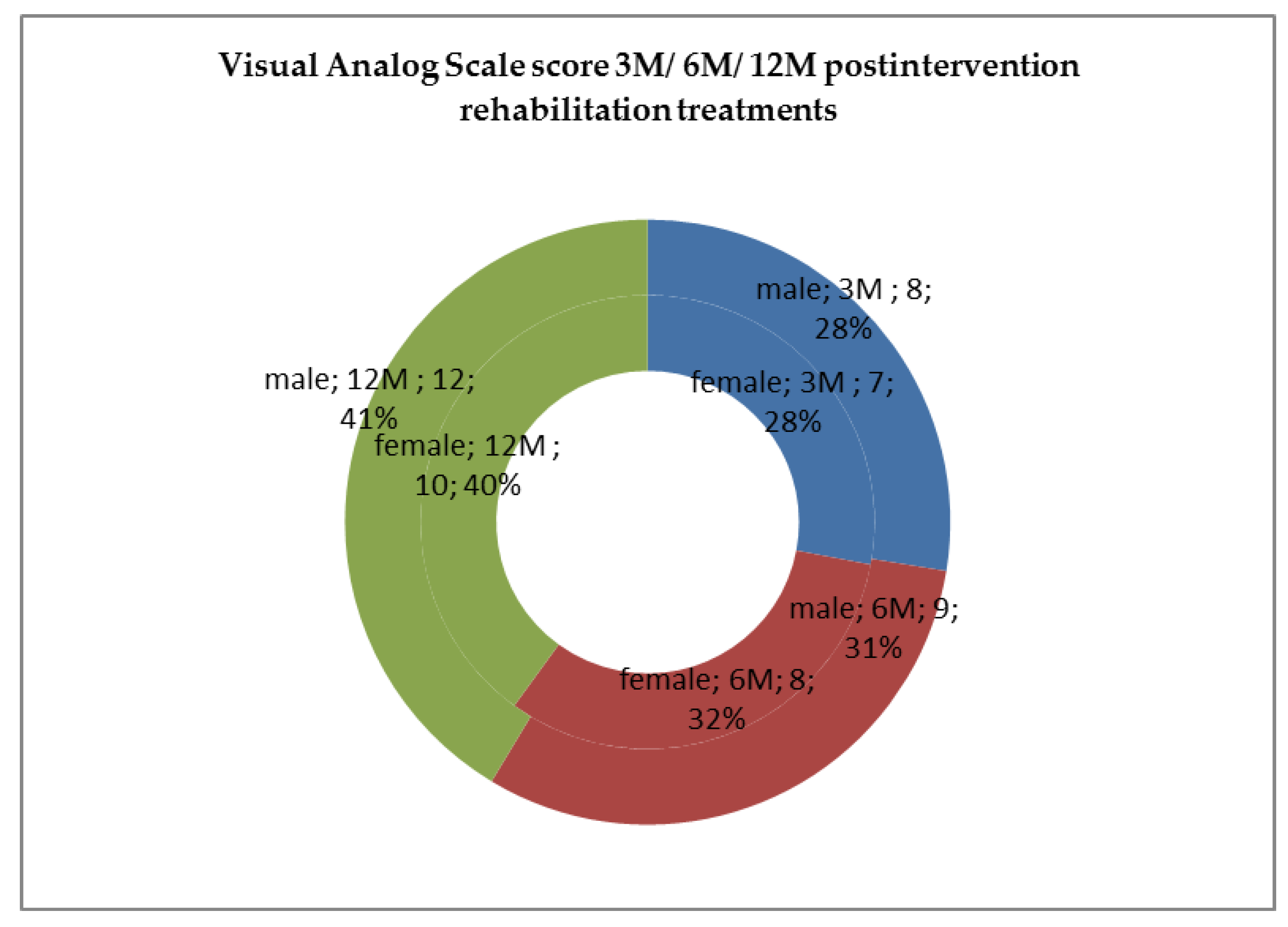
The score obtained after applying the Visual Analog Scale is a subjective but important record, because the patient’s perception is the one that validates our rehabilitation complex treatments and SDSS interferences(Figure 15), hence, we noticed by Visual Analog Scale diagnostic tools that male study subgroup presented a superior rate of recovering and good response to rehabilitation treatment than women, which was less, but in the same positive way at 3, 6, and 12 months of the evaluation time period (Figure 16).

4. Discussion

The success of any type of treatment can be translated by the lack of destructive inflammatory reactions in time. This idea is frequently repeated in the literature by presenting the explosion of new biomaterials and tissue reactions in contiguity, observed through contact between them during integration, with limitations and permissions based on socio-economic and biological criteria (patient grounds).
The limitation of our experiment can be related to the applicability skills-inherent factors to the endodontic procedures such as perforations, instrument breakage, calcifications, and anatomic anomalies, as all can lead to treatment failure connected with anatomy that may require microscopic interventions, the non-observance of the indications, not following the instructions received, and delay for proper cure plan, not using rubber dam isolation, inefficiently performed cleaning and shaping of the root canal system or variability of the implant material surfaces.
Other similar studies in this field highlight the role played by budding extracellular vesicles in mesenchymal stem cells (MSCs). This approach is considered to have great potential in clinical applications by disseminating valuable biological information, allowing the establishment of many functional connections related to cell therapy. The regenerative and immunomodulatory characteristics of MSCs have benefits in a multitude of clinical situations, including the regeneration of traumatic injuries.
Exosomes have the same characteristics as the cells they came from, such as the transport of proteins, miRNA, mRNA, and other soluble factors implied by MSCs’ functions, including immunomodulation and tissue regeneration. For the treatment of osteochondral defects, a scientist proposed a ‘cell-free’ therapeutic approach, demonstrating the ability of human embryonic MSC-derived exosomes to repair cartilage and subchondral bone injuries [24]. Mineral Trioxide Aggregate (MTA) was recommended as a repair material for root perforations a long time ago [25]. It was developed by Loma Linda University in 1990 and was originally introduced as a retrograde filling material. This has been shown to cause a much lower percolation than other materials in contact with the surrounding tissues compared to most used raw materials. For these reasons, MTA has been considered a material of choice for root perforation repair, as it has not only demonstrated biocompatibility with peri-radicular tissues (minimal inflammatory reaction, but also the ability to allow regeneration of hard tissue such as bone or cementum), thus facilitating regeneration of the periodontal support tissues, which is of utmost importance in the overall stability of teeth [26,27]. In a research model on human osteoblasts, MTA stimulated the increase in cytokines, such as interleukin-1 α, interleukin-1β, and interleukin-6, which are involved in bone turnover [28].
The cytotoxicity of Bio Aggregate, the second material used in the study, was also evaluated compared to that of MTA on mesenchymal cell cultures (p > 0.05). DiaRoot Bio Aggregate showed in vitro compatibility comparable to MTA [29].
Sealapex is a sealant based on calcium hydroxide that is widely used in endodontics, whose bio stimulatory properties and cytotoxicity in contact with peri radicular tissues are widely discussed in the literature [30]. Overall, Sealapex demonstrated a reduced inflammatory response compared to other endodontic sealants showing moderate inflammation at 48 h, which decreased in subsequent periods. Other sealants, based on zinc oxide-eugenol, were more toxic at 48 h and seven days, respectively. This toxicity gradually decreased in later time periods [31].
In the study, seven days after implantation, the more pronounced inflammatory reactions as well as the presence of multinucleated giant cells in contact with the enclaves of remaining Sealapex and MTA material was noted histologically, while in the case of Bio Aggregate samples, a considerably lower reaction, with the absence of these cells and a reduced inflammatory cell infiltrate could be registered. This aspect indicates a higher biocompatibility of the last material compared to the live subcutaneous tissue, compared to the first two. The inflammatory reaction and the presence of thicker bands of necrotic tissue on contact with implanted materials (thicker at MTA compared to Sealapex and Bioaggregate) in the first seven days may be due to both the different chemical composition of the cements and the stronger basic pH of the MTA: the immediately post mixing pH is 10.2 and rises to 12.5 in 3 h, after which it remains constant, the pH of Sealapex is up to 9.1, and for Bio Aggregate pH~12 after the setting of the material [32,33,34].
The presence of large numbers of active osteoblasts and osteoclasts located in specific gaps and the organizing of the new bone tracts 30 days after material implantation, demonstrates an intense activity of bone regeneration and reorganization in the proximity of the implanted material, most evident in MTA and Bio Aggregate [35,36,37].
According to some studies from the literature, it may be revealed that MTA, Super EBA, and IRM had similar histopathological results and presented a better performance than ZOE in a puttylike consistency, and only MTA stimulated deposition of neo-formed cementum in direct contact with the retro filling material [38]. Completing the present study, there are a number of other materials that increase the motivation of our study and offer a dimension closer to the ideal that will optimize the results through a modern biomaterial that must meet several requirements, even involving their biological and structural characteristics. The literature points out that graphene has revealed interesting antibacterial and physical peculiarities, but it has also shown limitations. Black phosphorus has structural and biochemical properties that make it ideal for biomedical applications: 2D sheets of black phosphorus are called Black Phosphorene (BP), and it could replace graphene in the coming years.
Scientists have taken smart two-dimensional materials (2D materials) into consideration, as the allotropic forms of many such materials have shown distinct characteristics that may be usable in several applications. The interface between cells and biomaterials should be biocompatible and bioactive: 2D materials are tunable on the nanometric-scale, which can be used to improve the connection between such materials and human tissue. An important evaluation regarding the advantages of BP, compared to graphene is related to its lower cytotoxicity, advanced in vivo biodegradability and its tendency to release few nanoparticles in the human body [39].
By means of prosthetic treatments application, our research gives an objective answer of the biocompatibility and functionality that are appreciated as having a response of 88% tolerability and integration, and which are in correlation with compliance with the required conditions and of course the quality of structural components and presentation to clinical treatment, similar to other studies in the literature [40,41,42,43,44,45,46,47,48].
Another issue that must be accounted for is the biocompatibility and the integration capabilities of materials used in the context of systemic diseases, such as tumors, fractures, vitamin deficiencies, liver pathology, and autoimmune diseases [49,50,51,52,53,54]. Although in the majority of cases, such issues do not critically alter the treatment course, still, precautions must be taken in order to ensure the best possible outcome [55,56]. Moreover, the complexity of the phenomena that may interfere with simple or complicated endodontic pathology as a differential diagnosis, namely the dysfunctional syndrome of the stomatognathic system, can be intercepted during detailed diagnostic procedures and addressed by specific elaborate treatment [57,58,59,60,61], which was also used in the present study. The cases were treated by splints in 29% of patients, 12% of them with muscle relaxants, also 98% of subjects with occlusal parameters restoration, namely: occlusal areas, composite fillings, and amalgam, but also occlusal imbalances of existing components of gnato-prosthetic devices in 57% or their total replacement, as well as orthodontic adjustments in some cases.
In our study, objective scores were obtained from histomorphological examination in order to classify the tested materials from optimum to least favorable, by analyzing the presence and width of cementum covering the root-end filling material surface. Furthermore, the incidence of well-organized fibrous capsules without inflammatory cells were also observed. In addition, it is observed that the root canals of the patients included in the study that needed a better sealing of the root fillings to aid the healing process were in a position to simulate a clinical-surgical situation in which the canal could be recovered by MTA, BA, or SELAPEX, and depending on the result, according to our study more pronounced in the MTA and Bio Aggregate. However, the complex rehabilitation clinical situations will have to be further validated also in the context of resistance to the masticatory forces in time. The tolerability of the endo-periodontal system can be a desideratum in the aesthetic and functional spectrum of long-term oral rehabilitation.
Also, we considered that modern materials studied have a major influence in the overall homeostatis of the oral cavity and create a stable and functional framework. This creates a foundation for the continuity and longevity of prosthetic treatments, in terms of dental support, as a resistant pillar of future oral rehabilitation constructions through dental prostheses. In combined prosthetic approaches, they are regarded as a unitary prosthetic restoration, and in particular, in the case of our study, as plural constructions—namely, the type of gnathoprosthetic devices with three elements, which react quite well in this case, as a whole. As our results demonstrated, they are accompanied and validated by optimal periodontal response, especially when considering the broader context of the rehabilitation of aspects from the associated dysfunctional syndrome of the stomatognathic system.

5. Conclusions

Our results showed increased absolute values of alkaline phosphatase in all material-implanted cases (more pronounced in the MTA and Bio Aggregate), highlighting that this enzyme could be an effective indicator of bone formation, which takes place after the material implantation, with the most significant elevated values at 30 days postoperatively. This suggests—in the absence of other existing associated pathologies—an intense reparative and restorative bone activity in the body that is favorable and necessary to the optimum subsequent stages of prosthetic management in the context of a complex oral rehabilitation approach therapy.
This study opens the path for future research regarding dental materials biocompatibility, not only from a strictly local endo-periodontal biological context, but from a wider systemic integration perspective.

Author Contributions

Conceptualization, L.A., A.S.P. and G.L.F.; Methodology, A.S.P. and E.V.S.; Software, M.A.B. and B.P.B.; Validation, O.S., S.T. and L.E.C.; Formal analysis, L.E.C., S.T. and E.V.S.; Investigation, L.A., G.L.F. and E.V.S.; Resources, B.P.B. and M.A.B.; Data curation, I.C.L. and O.S.; Writing—original draft preparation, L.A. and L.E.C.; Writing—review and editing, O.S., A.S.P. and E.V.S.; Visualization, M.A.B., S.M.S. and I.C.; Supervision I.C.L., S.T., S.M.S. and I.C.; Project administration, I.C.L.,I.C., S.M.S., G.L.F. and B.P.B. All authors contributed scientifical equally to this work. All authors have read and agreed to the published version of the manuscript.

Funding

This research received no external funding.

Institutional Review Board Statement

The study was conducted according with national and international regulations on animal welfare, identification, control, and elimination factors causing physiological and behavioral disorders: Directive EC86/609 EU; Government Ordinance no. 37/2002, approved by Law no. 471/2002; Law 205/2004 on animal protection, amended and supplemented by Law no. 9/2008. The study was approved by the Ethics Committee of Faculty of Veterinary Medicine Iasi (Protocol Code No. 533/22.04.2020) and by the Ethics Committee of UMF, Grigore T. Popa”’ Iasi (Protocol Code No. 547.10012012). The clinical investigations were carried out following the rules of the Declaration of Helsinki of. All subjects gave their informed consent for inclusion before they participated in the study.

Informed Consent Statement

Informed consent was obtained from all subjects and for rehabilitation treatments involved in the study.

Data Availability Statement

Not applicable.

Conflicts of Interest

The authors declare no conflict of interest.

Abbreviations

SSstomatognathic system
ZOEzinc oxide eugenol
Super-EBAzinc oxide eugenol cement reinforced with ethoxy benzoic acid (EBA)
IRMzinc oxide eugenol-based cement reinforced with polymethacrylate
SDSS/TMDsdysfunctional syndrome of the stomatognathic system
TMJtemporo-mandibular joint
PMMAPoly(methyl methacrylate)
Ca(OH)2calcium dioxide
BAmaterial similar in structure to MTA (mineral trioxide aggregate), that additionally contains ceramic nanoparticles.
MTAmineral trioxide aggregate,
Sealapexcalcium hydroxide-based cement
SiO2silicon dioxide

References

  1. Mohapatra, S.; Patro, S.; Mishra, S. Bioactive Materials in Endodontics: An Evolving Component of Clinical Dentistry. Compend. Contin. Educ. Dent. 2016, 38, 376–381, quiz 382. [Google Scholar] [PubMed]
  2. Medzhitov, R. Origin and physiological roles of inflammation. Nature 2008, 454, 428–435. [Google Scholar] [CrossRef] [PubMed]
  3. Di Alberti, L.; Rossetto, A.; Albanese, M.; D’Agostino, A.; De Santis, D.; Bertossi, D.; Nocini, P.F. Expression of Vascular Endothelial Growth Factor (VEGF) mRNA in healthy bone tissue around implants and in peri-implantitis. Minerva Stomatol. 2013, 62 (Suppl. 1), 1–7. [Google Scholar] [CrossRef] [PubMed]
  4. Matarese, G.; Isola, G.; Anastasi, G.P.; Favaloro, A.; Milardi, D.; Vermiglio, G.; Vita, G.; Cordasco, G.; Cutroneo, G. Immunohistochemical analysis of TGF-β1 and VEGF in gingival and periodontal tissues: A role of these biomarkers in the pathogenesis of scleroderma and periodontal disease. Int. J. Mol. Med. 2012, 30, 502–508. [Google Scholar] [CrossRef] [PubMed] [Green Version]
  5. Bressan, E.; Ferroni, L.; Gardin, C.; Bellin, G.; Sbricoli, L.; Sivolella, S.; Brunello, G.; Schwartz-Arad, D.; Mijiritsky, E.; Penarrocha, M.; et al. Metal Nanoparticles Released from Dental Implant Surfaces: Potential Contribution to Chronic Inflammation and Peri-Implant Bone Loss. Materials 2019, 12, 2036. [Google Scholar] [CrossRef] [Green Version]
  6. Eliyas, S.; Jalili, J.; Martin, N. Restoration of the root canal treated tooth. Br. Dent. J. 2015, 218, 2–7. [Google Scholar] [CrossRef]
  7. Gupta, S.K. A study to assess the methodological quality of in vivo animal experiments published in Indian journal of pharmacology: A retrospective, cross-sectional, observational study. Indian J. Pharmacol. 2019, 51, 11–16. [Google Scholar] [CrossRef]
  8. De-Deus, G.; Canabarro, A.; Alves, G.; Linhares, A.; Isabel Senne, M.; Granjeiro, J.M. Optimal cytocompatibility of a bioceramic nanoparticulate cement in primary human mesenchymal cells. J. Endod. 2009, 35, 1387–1390. [Google Scholar] [CrossRef]
  9. Camps, J.; About, I. Cytotoxicity testing of endodontic sealers: A new method. J. Endod. 2003, 29, 583–586. [Google Scholar] [CrossRef]
  10. Banomyong, D.; Ongchavalit, L.; Yanpiset, K. Cytotoxicity evaluatin of newly developed bi-functional, oligomer-based sealers and a methacrylate resin-based root canal sealer. M Dent. J. 2016, 36, 89–98. [Google Scholar]
  11. Estrela, C.; Bammann, L.L.; Estrela, C.R.d.A.; Silva, R.S.d.; Pecora, J.D. Antimicrobial and Chemical Study of MTA, Portland Cement, Calcium Hydroxide Paste, Sealapex and Dycal. Braz. Dent. J. 2000, 11, 3–9. [Google Scholar] [PubMed]
  12. Teixeira, L.; Gonçalves Basso, F.; Hebling, J.; Costa, C.A.; Garrido Mori, G.; Yara Silva-Sousa, T.C.; Oliveira, C.F. Cytotoxicity Evaluation of Root Canal Sealers Using an In Vitro Experimental Model with Roots. Braz. Dent. J. 2017, 28, 165–171. [Google Scholar] [CrossRef] [PubMed] [Green Version]
  13. European Parliament. Council of the European Union. Caring for animals aiming for better science. Directive 2010/63/EU of the European Parliament and of the council of 22 September 2010 on the protection of animals used for scientific purposes. Off. J. Eur. Union 2010, L276, 33–79. [Google Scholar]
  14. Kopper, P.M. Evaluation of bone tissue response to a sealer containing mineral trioxide aggregate. J. Endod. 2015, 41, 62–66. [Google Scholar]
  15. Roberts, H.W.; Toth, J.M.; Berzins, D.W.; Charlton, D.G. Mineral trioxide aggregate material use in endodontic treatment: A review of the literature. Dent. Mater. 2008, 24, 149–164. [Google Scholar] [CrossRef] [PubMed] [Green Version]
  16. Holland, R.; de Souza, V.; Nery, M.J.; Otoboni Filho, J.A.; Bernabe, P.F.; Dezan Junior, E. Reaction of rat connective tissue to implanted dentin tubes filled with mineral trioxide aggregate or calcium hydroxide. J. Endod. 1999, 25, 161–166. [Google Scholar] [CrossRef]
  17. Chacko, V.; Kurikose, S. Human pulpal response to mineral trioxide aggregate (MTA): A histologic study. J. Clin. Pediatr. Dent. 2006, 30, 203–209. [Google Scholar] [CrossRef]
  18. Khalil, W.A.; Eid, N.F. Biocompatibility of BioAggregate and mineral trioxide aggregate on the liver and kidney. Int. Endod. J. 2013, 46, 730–737. [Google Scholar] [CrossRef]
  19. Assadian, H.; Hamzelouei Moghaddam, E.; Amini, A.; Nazari Moghaddam, K.; Hashemzehi, M. A Review of Endodontic Bioceramics. J. Islamic Dent. Assoc. Iran. 2016, 28, 20–33. [Google Scholar] [CrossRef]
  20. Chang, S.-W.; Lee, S.-Y.; Kang, S.-K.; Kum, K.-Y.; Kim, E.-C. In Vitro Biocompatibility, Inflammatory Response, and Osteogenic Potential of 4 Root Canal Sealers: Sealapex, Sankin Apatite Root Sealer, MTA Fillapex, and iRoot SP Root Canal Sealer. J. Endod. 2014, 40, 1642–1648. [Google Scholar] [CrossRef]
  21. Huang, F.M.; Tai, K.W.; Chou, M.Y.; Chang, Y.C. Cytotoxicity of resin-, zinc oxide-eugenol, and calcium hydroxide-based root canal sealers on human periodontal ligament cells and permanent V79 cells. Int. Endod. J. 2002, 35, 153–158. [Google Scholar] [CrossRef] [PubMed]
  22. Lüthy, H.; Filser, F.; Loeffel, O.; Schumacher, M.; Gauckler, L.J.; Hämmerle, C.H. Strength and reliability of four-unit all-ceramic posterior bridges. Dent. Mater. 2005, 21, 930–937. [Google Scholar] [CrossRef] [PubMed]
  23. Groten, M.; Huttig, F. The performance of zirconium dioxide crowns: A clinical follow-up. Int. J. Prosthodont. 2010, 23, 429–431. [Google Scholar]
  24. Codispoti, B.; Marrelli, M.; Paduano, F.; Tatullo, M. NANOmetric BIO-Banked MSC-Derived Exosome (NANOBIOME) as a Novel Approach to Regenerative Medicine. J. Clin. Med. 2018, 7, 357. [Google Scholar] [CrossRef] [PubMed] [Green Version]
  25. Conrad, H.J.; Seong, W.J.; Pesun, I.J. Current ceramic materials and systems with clinical recommendations: A systematic review. J. Prosthet. Dent. 2007, 98, 389–404. [Google Scholar] [CrossRef]
  26. Martu, M.-A.; Surlin, P.; Lazar, L.; Maftei, G.A.; Luchian, I.; Gheorghe, D.-N.; Rezus, E.; Toma, V.; Foia, L.-G. Evaluation of Oxidative Stress before and after Using Laser and Photoactivation Therapy as Adjuvant of Non-Surgical Periodontal Treatment in Patients with Rheumatoid Arthritis. Antioxidants 2021, 10, 226. [Google Scholar] [CrossRef] [PubMed]
  27. Teodorescu, A.C.; Martu, I.; Teslaru, S.; Kappenberg-Nitescu, D.C.; Goriuc, A.; Luchian, I.; Martu, M.A.; Solomon, S.M.; Mârțu, S. Assessment of salivary levels of RANKL and OPG in aggressive versus chronic periodontitis. J. Immunol. Res. 2019, 28, 2019. [Google Scholar] [CrossRef] [Green Version]
  28. Gabbert, O.; Ohlmann, B.; Schmitter, M.; Gilde, H.; Ruef, T.; Rammelsberg, P. Fracture behavior of zirconia ceramic cantilever fixed dental prostheses in vitro. Acta Odontol. Scand. 2008, 66, 200–206. [Google Scholar] [CrossRef] [PubMed]
  29. Moseley, G.L.; Butler, D.S. Fifteen Years of Explaining Pain: The Past, Present, and Future. J. Pain 2015, 16, 807–813. [Google Scholar] [CrossRef] [Green Version]
  30. Van der Meer, H.A.; Speksnijder, C.M.; Engelbert, R.H.H.; Lobbezoo, F.; Nijhuis-van der Sanden, M.W.G.; Visscher, C.M. The association between headaches and temporomandibular disorders is confounded by bruxism and somatic complaints. Clin. J. Pain 2017, 33, 835–843. [Google Scholar] [CrossRef]
  31. Jenei, Á.; Sándor, J.; Hegedűs, C.; Bágyi, K.; Nagy, L.; Kiss, C.; Szabó, G.; Márton, I.J. Oral health-related quality of life after prosthetic rehabilitation: A longitudinal study with the OHIP questionnaire. Health Qual. Life Outcomes 2015, 13, 99–101. [Google Scholar] [CrossRef] [PubMed] [Green Version]
  32. Al-Shammari, K.F.; Al-Khabbaz, A.K.; Al-Ansari, J.M.; Neiva, R.; Wang, H.L. Risk indicators for tooth loss due to periodontal disease. J. Periodontol. 2005, 76, 1910–1918. [Google Scholar] [CrossRef] [PubMed]
  33. Armitage, G.C.; Cullinan, M.P. Comparison of the clinical features of chronic and aggressive periodontitis. Periodontology 2000 2010, 53, 12–27. [Google Scholar] [CrossRef] [PubMed]
  34. Thyvalikakath, T.; Song, M.; Schleyer, T. Perceptions and attitudes toward performing risk assessment for periodontal disease: A focus group exploration. BMC Oral Health 2018, 18, 90–95. [Google Scholar] [CrossRef] [Green Version]
  35. Hategan, S.I.; Kamer, A.R.; Sinescu, C.; Craig, R.G.; Jivanescu, A.; Gavrilovici, A.M.; Negrutiu, M.-L. Periodontal disease in a young Romanian convenience sample: Radiographic assessment. BMC Oral Health 2019, 19, 94–98. [Google Scholar] [CrossRef]
  36. Satheesh Kumar, P.; Satheesh, K.K.; John, J.; Patil, G.; Patel, R. Force Transfer and Stress Distribution in an Implant-Supported Overdenture Retained with a Hader Bar Attachment: A Finite Element Analysis. ISRN Dent. 2013, 2013, 21–27. [Google Scholar] [CrossRef]
  37. Vitale-Brovarone, C.; Baino, F.; Tallia, F.; Gervasio, C.; Verné, E. Bioactive glass-derived trabecular coating: A smart solution for enhancing osteointegration of prosthetic elements. J. Mater. Sci. Mater. Med. 2012, 23, 2369–2380. [Google Scholar] [CrossRef] [Green Version]
  38. Bernabé, P.F.E.; Holland, R.; Morandi, R.; Souza, V.d.; Nery, M.J.; Otoboni Filho, J.A.; Dezan Junior, E.; Gomes-Filho, J.E. Comparative Study of MTA and other materials in retrofilling of pulpless dogs’ teeth. Braz. Dent. J. 2005, 16, 151–155. [Google Scholar] [CrossRef]
  39. Tatullo, M.; Genovese, F.; Aiello, E.; Amantea, M.; Makeeva, I.; Zavan, B.; Rengo, S.; Fortunato, L. Phosphorene is the new Graphene in Biomedical Applications. Materials 2019, 12, 2301. [Google Scholar] [CrossRef] [Green Version]
  40. Scalera, F.; Esposito Corcione, C.; Montagna, F.; Sannino, A.; Maffezzoli, A. Development and characterization of UV curable epoxy/hydroxyapatite suspensions for stereolithography applied to bone tissue engineering. Ceram. Int. 2014, 40, 15455–15462. [Google Scholar] [CrossRef]
  41. Solomon, S.M.; Timpu, D.; Forna, D.A.; Stefanache, M.A.M.; Martu, S.; Stoleriu, S. AFM comparative study of root surface morphology after three methods of scaling. Mater. Plast. 2016, 53, 546–549. [Google Scholar]
  42. Rokaya, D.; Srimaneepong, V.; Sapkota, J.; Qin, J.; Siraleartmukul, K.; Siriwongrungson, V. Polymeric materials and films in dentistry: An overview. J. Adv. Res. 2018, 14, 25–34. [Google Scholar] [CrossRef] [PubMed]
  43. Popa, C.G.; Luchian, I.; Ioanid, N.; Goriuc, A.; Martu, I.; Bosinceanu, D.; Martu, M.A.; Tirca, T.; Martu, S. ELISA Evaluation of RANKL Levels in Gingival Fluid in Patients with Periodontitis and Occlusal Trauma. Rev. Chim. 2018, 69, 1578–1580. [Google Scholar] [CrossRef]
  44. Nicolae, V.; Neamtu, B.; Picu, O.; Stefanache, M.A.M.; Cioranu, V.S.I. The comparitive evaluation of salivary biomarkers (calcium, phosphate, salivary pH) in the cario-resistance versus cario-activity. Rev. Chim. 2016, 68, 821–824. [Google Scholar]
  45. Taraboanta, I.; Stoleriu, S.; Nica, I.; Georgescu, A.; Gamen, A.C.; Maftei, G.A.; Andrian, S. Roughness variation of a nonhybrid composite resin submitted to acid and abrasive challenges. Int. J. Med. Dent. 2020, 24, 182–187. [Google Scholar]
  46. Martu, M.A.; Maftei, G.A.; Luchian, I.; Popa, C.; Filioreanu, A.M.; Tatarciuc, D.; Nichitean, G.; Hurjui, L.-L.; Foia, L.-G. Wound healing of periodontal and oral tissues: Part II—Patho-phisiological conditions and metabolic diseases. Rom. J. Oral Rehabil. 2020, 12, 30–40. [Google Scholar]
  47. Doyle, S.L.; Hodges, J.S.; Pesun, I.J.; Law, A.S.; Bowles, W.R. Retrospective cross sectional comparison of initial nonsurgical endodontic treatment and single-tooth implants. J. Endod. 2006, 32, 822–827. [Google Scholar] [CrossRef]
  48. Iqbal, M.K.; Kim, S. For teeth requiring endodontic treatment, what are the differences in outcomes of restored endodontically treated teeth compared to implant-supported restorations? Int. J. Oral Maxillofac. Implant 2007, 22, 96–116. [Google Scholar]
  49. Filip, A.; Badulescu, O.V.; Sirbu, P.D.; Cojocaru, E.; Filip, N.; Puha, G.; Trandafir, L.; Iancu, C.; Trandafirescu, M.F.; Alexa, O. Serum Homocysteine and Reactive Species Levels in Fragility Fractures of the Pelvis. Rev. Chim. 2019, 70, 3216–3219. [Google Scholar] [CrossRef]
  50. Cojocaru, E.; Mihaila, D.; Leon-Constantin, M.M.; Cobzaru, R.G.; Ripa, C.V.; Trandafirescu, M.F.; Trandafir, L.M. Update in pediatric primary brain tumors—From histology to genetically defined tumors. Rom. J. Morphol. Embryol. 2019, 60, 761–767. [Google Scholar] [PubMed]
  51. Filip, A.; Alexa, O.; Sirbu, P.D.; Filip, C.; Cojocaru, E.; Puha, G.; Trandafirescu, M.F.; Badulescu, O.V. Calcium and vitamin D involvement in the fragility fracture of the pelvis. Rev. Chim. 2019, 70, 3674–3677. [Google Scholar] [CrossRef]
  52. Luca, A.C.; Miron, I.C.; Trandafir, L.M.; Cojocaru, E.; Pădureț, I.A.; Trandafirescu, M.F.; Iordache, A.C.; Țarcă, E. Morphological, genetic and clinical correlations in infantile hemangiomas and their mimics. Rom. J. Morphol. Embryol. 2020, 61, 687–695. [Google Scholar] [CrossRef] [PubMed]
  53. Trandafir, L.M.; Frăsinariu, O.; Leon-Constantin, M.M.; Chiriac, S.; Trandafirescu, M.F.; Miron, I.C.; Luca, A.C.; Iordache, A.C.; Cojocaru, E. Pediatric nonalcoholic fatty liver disease—A changing diagnostic paradigm. Rom. J. Morphol. Embryol. 2020, 61, 1023–1031. [Google Scholar] [CrossRef] [PubMed]
  54. Martu, M.A.; Solomon, S.M.; Toma, V.; Maftei, G.A.; Iovan, A.; Gamen, A.; Hurjui, L.; Rezus, E.; Foia, L.; Forna, N.C. The importance of cytokines in periodontal disease and rheumatoid arthritis. Review. Rom. J. Oral Rehabil. 2019, 11, 220–240. [Google Scholar]
  55. Cojocaru, E.; Magdalena Leon-Constantin, M.; Ungureanu, C.; Trandafirescu, M.F.; Maștaleru, A.; Mihaela Trandafir, L.; Dumitru Petrariu, F.; Viola Bădulescu, O.; Filip, N. Hypolipemiant Actions and Possible Cardioprotective Effects of Valine and Leucine: An Experimental Study. Medicina 2021, 57, 239. [Google Scholar] [CrossRef] [PubMed]
  56. Trandafirescu, M.F.; Agop-Forna, D.; Clim, A.; Bețiu, C.; Clim, I.A.; Cojocariu, S.; Pînzariu, A.C. Oral manifestations and management of patients with chronic kidney disease. Rom. J. Oral Rehabil. 2020, 12, 212–219. [Google Scholar]
  57. Nam, J.; Tokutomi, H. Using zirconia-based prosthesis in a complete-mouth reconstruction treatment for worn dentition with the altered vertical dimension of occlusion. J. Prosthet. Dent. 2015, 113, 81–85. [Google Scholar] [CrossRef] [PubMed]
  58. Tatarciuc, M.; Maftei, G.A.; Vitalariu, A.; Luchian, I.; Martu, I.; Diaconu-Popa, D. Inlay-Retained Dental Bridges—A Finite Element Analysis. Appl. Sci. 2021, 11, 3770. [Google Scholar] [CrossRef]
  59. Singh, R.G.; Sinha, P. Functional and Aesthetic Full Mouth Rehabilitation of a Severely Worn Dentition to Restore Vertical Dimension: A Case Report. J. Indian Prosthodont. Soc. 2014, 14, 210–214. [Google Scholar] [CrossRef]
  60. Şahin, O.; Doğan, D.Ö.; Fatih, S. Aesthetic full-mouth oral rehabilitation of a young adult with amelogenesis imperfecta: A clinical report. Cumhur. Dent. J. 2014, 17, 308–315. [Google Scholar] [CrossRef]
  61. Mocanu, R.C.; Martu, M.-A.; Luchian, I.; Sufaru, I.G.; Maftei, G.A.; Ioanid, N.; Martu, S.; Tatarciuc, M. Microbiologic Profiles of Patients with Dental Prosthetic Treatment and Periodontitis before and after Photoactivation Therapy—Randomized Clinical Trial. Microorganisms 2021, 9, 713. [Google Scholar] [CrossRef] [PubMed]
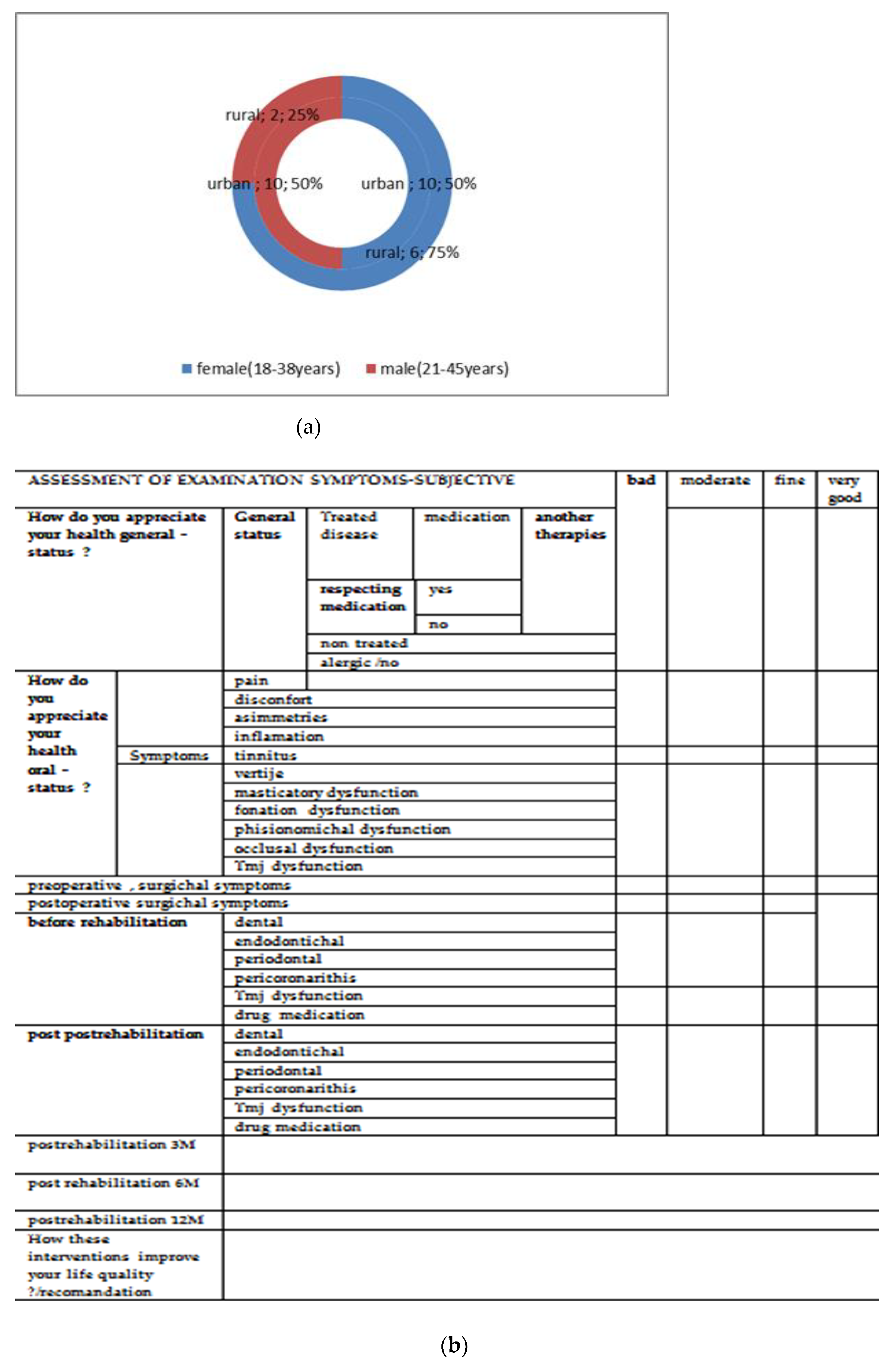
Figure 1. (a) Distribution of the patients from Clinical Dental Education Base, “Mihail Kogalniceanu”—Grigore T. Popa University of Medicine and Pharmacy from Iasi, Romania participants in the study. (b) Visual Analog Scale used for making score objectivation by patients’ assessment. (c) Presence of numerous osteoblasts included in ossein sequestrations, with a tendency to organize in osteoid matrix (Sealapex).
Applsci 12 01464 g001aApplsci 12 01464 g001b
Figure 2. Detail of the previous image (Figure 1c), with increased magnification: the osteoblasts seized in the osteine oriented in traverses (Sealapex) are noteworthy.
Applsci 12 01464 g002
Figure 3. Presence of a Howship gap in an osteoclast involved in bone remodeling (MTA).
Applsci 12 01464 g003
Figure 4. Excessive pre-osseous deposition in the form of agglomerations and smaller areas of less dense bone by phagocytic activity (MTA).
Applsci 12 01464 g004
Figure 5. Detail of the previous image (Figure 4) that highlights the area adjacent to the excess bone deposition, with gaps in which osteoclasts (MTA) are present.
Applsci 12 01464 g005
Figure 6. Area with new bone spans delimiting the gaps: numerous osteocytes (MTA) are present in their structure.
Applsci 12 01464 g006
Figure 7. Presence of a Howship Gap in which typical osteoclasts with a brushed edge that attach to excess, newly formed dense material (MTA).
Applsci 12 01464 g007
Figure 8. Pre-osseous substance deposited in excess in the area adjacent to the implanted material (Bio Aggregate).
Applsci 12 01464 g008
Figure 9. Detail of the previous image (Figure 8) in which the tendency of organization in steering paths in the vicinity of the fundamental external system of the bone is noticed (Bio Aggregate).
Applsci 12 01464 g009
Figure 10. Presence of newly formed bone bands near the implanted material (Bio Aggregate).
Applsci 12 01464 g010
Figure 11. Rounded bone deposits: osteoblasts with intense osteogenic activity are present in the bone traverse (Bio Aggregate).
Applsci 12 01464 g011
Figure 12. Statistical indicators of alkaline phosphatase according to the assessing moment and material used.
Applsci 12 01464 g012
Figure 13. (a) Distribution of single restauration and (b) prosthetics devices—composites and ceramics placed in anterior or posterior arch area.
Applsci 12 01464 g013
Figure 14. (ad) Rehabilitation treatments through prosthetic modalities by reinforced composite, Poly(methyl methacrylate) (PMMA) composite, unidental and pluridental localization, and ceramic materials pluridental prosthesis.
Applsci 12 01464 g014
Figure 15. Distribution of rehabilitation treatments at dysfunctional syndrome—SDSS interferences.
Applsci 12 01464 g015
Figure 16. The score obtained from the results of Visual Analog Scale 3 M 6 M 12 M, postintervention.
Applsci 12 01464 g016
Table 1. Alkaline phosphatase values at 7 days postoperatively (IU/L).
Rabbit No.Control RabbitImplanted Material
SealapexMTABioAggregate
1163.6164.3176.5182.4
2168.8178.4180.1
3165.6181.3179.8
4167.4179.5182.3
5169.1180.2183.2
6168.3177.8183.8
7167.6175.6179.6
8165.7176.6181.5
9168.7180.2182.4
Table 2. Alkaline phosphatase values at 30 days postoperatively (IU/L).
Rabbit No.Control RabbitImplanted Material
SealapexMTABioAggregate
1163.6181.3196.5199.4
2173.9198.4193.1
3180.6189.8196.8
4188.9190.5195.3
5182.3191.3196.2
6181.2193.5190.2
Table 3. Alkaline phosphatase values at 30 days postoperatively (IU/L).
Rabbit No.Control RabbitImplanted Material
SealapexMTABioAggregate
1163.6162.8164.2164.8
2163.4165.4164.1
3165.2167.5166.4
Table 4. Statistical indicators of alkaline phosphatase in the study group according to the biomaterial used and time of determination.
Implanted MaterialEvaluation MomentMean
FA
AverageStd Dev.Std Er.Min.Max.Median
−95%+95%
Sealapex7 days167.25165.24169.261.920.78164.30169.10167.85
Sealapex30 days181.18171.41190.946.143.07173.90188.90180.95
Sealapex60 days163.10159.29166.910.420.30162.80163.40163.10
MTA7 days178.95177.13180.771.730.71176.50181.30178.95
MTA30 days193.80186.97200.634.292.15189.80198.40193.50
MTA60 days164.80157.18172.420.850.60164.20165.40164.80
BioAggregate7 days181.93180.22183.651.630.67179.80183.80182.35
BioAggregate30 days196.15191.94200.362.651.32193.10199.40196.05
BioAggregate60 days164.45160.00168.900.490.35164.10164.80164.45
control 163.60 163.60163.60163.60
Publisher’s Note: MDPI stays neutral with regard to jurisdictional claims in published maps and institutional affiliations.