Article

Multi-Technique Diagnostic Investigation in View of the Restoration of “The Glory of St. Barbara” Painting by Mattia Preti

1
Department of Mathematical and Computer Sciences, Physical Sciences and Earth Sciences, University of Messina, Viale Ferdinando Stagno D’Alcontres 31, 98166 Messina, Italy
2
Department of Biology, Ecology and Earth Science, University of Calabria, Via Pietro Bucci, Cubo 12B, Arcavacata, 87036 di Rende, Italy
3
Department of Chemical, Biological, Pharmaceutical and Environmental Sciences, University of Messina, Viale Ferdinando Stagno D’Alcontres 31, 98166 Messina, Italy
4
Department of Literature and Philosophy, University of Trento, Via Tommaso Gar, 14, 38122 Trento, Italy
5
Giuseppe Mantella Restauro Opere D’Arte, Circonvallazione Paparo 25, 88060 Isca sullo Ionio, Italy
6
Department of Earth and Sea Sciences, University of Palermo, Via Archirafi, 26, 90123 Palermo, Italy
*
Author to whom correspondence should be addressed.
First Author.
Last Author.
Appl. Sci. 2022, 12(3), 1385; https://doi.org/10.3390/app12031385
Submission received: 5 January 2022 / Revised: 24 January 2022 / Accepted: 25 January 2022 / Published: 27 January 2022

Abstract

:
The present paper illustrates the results of a diagnostic investigation performed on the oil on canvas painting “The Glory of St. Barbara” (1680–1688) by Mattia Preti. The painting is located inside the St. Barbara Church in Taverna (Catanzaro, Italy), the city that gave birth to the artist. In situ, non-invasive studies, by applying X-ray fluorescence (XRF) spectroscopy, along with laboratory micro-destructive analytical investigations, by employing electron probe microanalyses (EPMA) coupled with energy-dispersive spectrometry (EDS) and micro-Raman (µ-Raman) spectroscopy, were combined to retrieve the color palette and identify the painting technique and style of the famous master. As a result of this multi-scale characterization, an extraordinary pictorial technique was revealed, enriching knowledge about one of the oldest pictorial traditions outlined by Mattia Preti, and solving doubts still existing about many of his investigated artworks. Moreover, the achieved results represent useful and essential tools to address management issues of the artwork, by providing valuable information for planning and monitoring future restoration interventions of the canvas.

1. Introduction

The investigation of ancient artworks is an attractive issue for scientists working in the cultural heritage field. Conservation strategies of ancient paintings still represent current challenges, especially for restorers and conservators who approach the study of artworks to safeguard them from the common processes of deterioration and ageing, mainly due to the passage of time.
To this end, acquiring information on the painting technique and the raw materials (i.e., pigments, preparatory layers, etc.) is, firstly, decisive to understand the choices and style of the author and, secondly, to develop a proper intervention strategy, dependent on the state of conservation of the asset.
This paper shows the results of a non-invasive or, at least, micro-invasive, multi-scale analytical approach to the study of an ancient painted canvas, with particular focus on the composition of pigments and preparatory layers, as well as their alteration products. Specifically, the canvas painting “The Glory of St. Barbara” by Mattia Preti was investigated and its constituent materials were analyzed in order to reconstruct the artist’s pictorial palette useful for carrying out the restoration. According to literature, the “Calabrian Knight”, as the artist was called, apparently painted directly from brain to canvas, without elaborate preparations. In this sense, the use of preparatory sketches, although it was a rather diffuse practice for the realization of more or less complex pictorial artworks, was never adopted by Mattia Preti, who actually preferred to develop his creations in his mind and then paint them directly on the canvas. For this reason, as a matter of fact, no two pictures of Mattia Preti are alike, and concerning the mixing and relations of colors, no one will ever know them with absolute certainty [1,2].
“The Glory of St. Barbara” painting (Figure 1a), an oil on canvas (460 × 305 cm) realized in Malta in 1688, is an altarpiece representing one of Mattia Preti’s masterpieces of the 1680s, the last decade completed by the artist before his death in 1699. The painting is now located in Taverna, a small town in the Calabria region, Southern Italy (Figure 1b), inside the St. Barbara Church (Figure 1c). With a very simple architecture, the St. Barbara Church was built in 1427 at the behest of the Franciscan friars, housing nowadays several artworks by Mattia Preti, with the canvas under study kept in the main altar.
The painting, a masterpiece of late Baroque art, represents Santa Barbara welcomed into heaven by Christ seated on clouds. Two putti, behind the kneeling saint, hold up her tower, the place of her imprisonment, and the sword with which she was beheaded. In the lower part, four saints are facing the celestial vision, ichnographically representing, from left to right, St. Clemente, St. Eustorgio, St. Mercurio and St. Vicario.
It is a rather delicate painting with an articulated history that needs particular insights to improve its conservation and the reading of the composition. The canvas has in fact been the subject of various conservation interventions, the first of which documented dates back to 1909. During the last restoration, promoted and carried out by the Superintendency of Cosenza and completed in 1993, they were unveiled a series of repentances in the sky above the figure of Barbara which brought out an angel-just sketched-holding a crown in the act of crowning the saint; a detail that Preti himself had amended in the final version.
Aimed at the material knowledge of the work, as a necessary specific in-depth study in order to plan a consequent and delicate conservative intervention capable of restoring a correct reading of it, scientific investigations became mandatory. These can represent at the same time a profitable opportunity for further verification but also of the extension of numerous repaintings, the result of previous conservation interventions. Such investigations, in addition, can support the evaluation of the current state of conservation of the painting itself.
In particular, the study presented here was carried out by following a multi-analytical approach, involving the use of geochemical techniques and non-invasive (or minimally invasive) spectroscopic methods. In order to maximize the information and data obtainable from the artefact micro-analytical techniques were first used, by operating in situ, and then, on the same areas, a careful sampling of micro-fragments was conducted.
Specifically, a preliminary in-situ investigation through portable X-ray fluorescence spectroscopy (XRF) allowed for the identification, at the elemental scale, of the composition of representative areas of the painted canvas, in terms of major and minor constituents. Then, a sampling campaign was performed on the same areas, investigated by the above mentioned non-destructive technique, in order to assess the morphology of the samples and better reconstruct the painting palette, giving weight to the detection of any additional compounds and raw materials used by the artist to make the painting. Electron probe microanalyses (EPMA) coupled with energy-dispersive spectrometry (EDS) and micro-Raman (µ-Raman) spectroscopy were performed in order to further analyze materials and to investigate the stratigraphy of the samples.
The planned interventions on “The Glory of St. Barbara” will complete what has recently been investigated, on the initiative of the Superintendency of Cosenza, on the other four paintings by Mattia Preti preserved in the same church of Taverna, as well as the canvas depicting “The SS. Trinity”, and “St. Barbara and Holy Martyrs”, whose restoration has been recently completed.
The reported study is carried out in the framework of well-established multi-technique archaeometric research co-operation involving the University of Messina, the University of Calabria, and the University of Malta, coordinated by the Restorers, aimed at investigating the artist’s autograph works present in the Maltese and Calabrian territory and, after that, at taking into consideration, with a better set of data and insights, the works present in the Italian territory always in relation to the production of the Maltese years. The final goal is to contribute to the creation of a database that collects the technical aspects of the Maltese period of Preti in order to complete the understanding of the number of works present in many museums in Italy and abroad.

2. Materials and Methods

2.1. Sampling

The entire sampling activity was performed during the latest survey campaign carried out in April 2021 with the assistance of the restorers in charge of the conservative intervention of the artwork, to select areas and collect representative samples whose study would have been crucial to choose the most appropriate intervention strategies.
A total of 28 points of the pictorial surface were analyzed, both in situ and in the laboratory, representative of different colors and tonalities (Figure 2).
In the case of laboratory measurements, micro-fragments having sizes smaller than ~3 mm2 were chopped-off from the painted canvas following minimally invasive or rather micro-invasive procedures, by using suitable stainless steel tools (i.e., small tweezers, scalpels and micro-scalpels) and a 5× magnification for proper observation of the artefact. In particular, micro-fragments of the pictorial surface were sampled close to the already-existing lacunae, and in areas where normal ageing processes were visible. Table 1 reports details about sampling, together with all the analytical techniques employed.

2.2. In Situ X-ray Fluorescence (XRF) Measurements

A portable XRF “Alpha 4000” (Innov-X systems, Inc., Woburn, MA, USA) analyzer was used. The equipment consisted of a Ta anode X-ray tube excitation source, and a Si PIN diode detector with an active area of 170 mm2 (full width at half maximum < 220 eV at 5.95 keV for Mn Kα line). The analyzer allowed the detection of elements ranging from phosphorus (Z = 15) to lead (Z = 82). The instrument operated in “soil” mode, a Compton normalization algorithm designed in order to achieve a limit of detection (LOD) as low as possible, i.e., trace concentrations (ppm levels) for soil and bulk samples. The “soil” mode was employed together with the “Environmental” elements suite, using the following element list: P, S, Cl, K, Ca, Ti, Cr, Mn, Fe, Co, Ni, Cu, Zn, As, Se, Br, Rb, Sr, Zr, Mo, Ag, Cd, Sn, Sb, I, Ba, Au, Hg, and Pb. Each area was investigated by two sequential tests, using operating conditions of 40 kV and 7 μA for the first run (60 s collection time) and 15 kV and 5 μA for the second one (60 s collection time). The instrument management and the data storage were guaranteed by a Hewlett-Packard iPAQ Pocket PC. The calibration was performed by means of soil LEAP (Light Element Analysis Program) II and verified using alloy certified reference materials produced by Analytical Reference Materials International. When detected, peaks at ~8.15 keV and ~9.34 keV have to be ascribed to Lα and Lβ transitions of Ta anode.

2.3. Laboratory Electron Probe Microanalyses–Energy-Dispersive Spectroscopy (EPMA-EDS) Measurements

Electron probe microanalyses (EPMA) coupled with energy-dispersive spectroscopy (EDS) were performed on the painted samples’ surface to obtain information about the chemical composition (in term of major elements) by a JEOL JXA 8230 equipment coupled with a JEOL EX-94310FaL1Q silicon drift type EDS. The measurements were performed on samples coated with a thin and highly conductive graphite film.

2.4. Laboratory Micro-Raman Measurements

Micro-Raman measurements were conducted by using a portable ‘BTR 111 Mini-RamTM’ (B&W Tek, Inc., Newark, NJ, USA) spectrometer with an excitation wavelength of 785 nm (diode laser), 280 mW maximum laser power at the excitation port, and a charge-coupled device (CCD) detector (thermoelectric cooled, TE). It is possible to continuously adjust the laser output power in order to obtain the best signal-to-noise ratio in the minimum integration time. The 62–3153 cm−1 spectral range was investigated, with a resolution of 10 cm−1, and an acquisition time of 10 s × 32 scans. The optimal performance of the instrument was guaranteed by a calibration procedure before each measurement using the peak at 520.6 cm−1 of a silicon chip. The system was equipped with a BAC151B Raman microscope. An 80×\40× objective was used, with a working distance of 1.25 mm\3.98 mm and laser beam spot size 26 µm\50 µm. The maximum power at the samples was ~15 mW. The identification of the peaks was obtained by comparing the experimental spectra with those reported in various databases and in the literature [3,4,5].

3. Results and Discussion

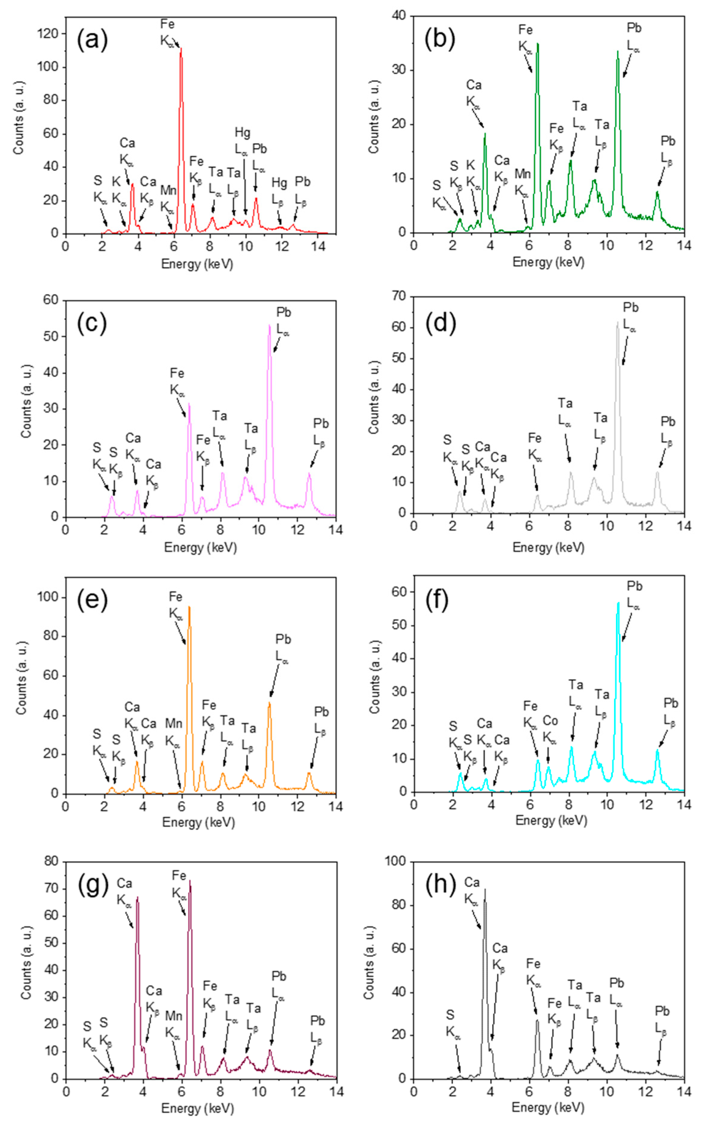
3.1. In Situ XRF Results

Table 2 reports the elemental composition, as obtained by XRF measurements, for all the investigated areas.
In addition, Figure 3 shows some XRF spectra collected on representative areas of the painting, corresponding to the red from St. Clemente’s cloak (S1, Figure 3a), the green from St. Vicario’s cloak (S4, Figure 3b), the flesh-toned from St. Clemente’s cheek (S10, Figure 3c), the grayish-white from the cloud under St. Barbara’s sandal (S12, Figure 3d), the yellow-orange from St. Barbara’s cloak (S13, Figure 3e), the light blue from the cerulean shades under the angel’s feet (S15, Figure 3f), the blackish-brown from Christ’s hair (S20, Figure 3g), and the left side of the canvas, at the bottom (S28, Figure 3h).
Results are discussed by color type, highlighting how similar-looking color tones may arise from the use of different pigmenting compounds, thereby characterizing the artist’s palette.
As far as red areas are concerned (S1, S2) the observation of Hg and Fe can suggest, in agreement with previous literature [6,7,8,9], the use of vermilion (HgS) mixed with red ochre (Fe2O3). The detection of Pb in both cases could be ascribed, other than to the preparatory layer, to the use of Pb-based compounds, reasonably white lead (2PbCO3·Pb(OH)2) or minium (Pb3O4), in order to obtain the desired reddish nuance. In particular, in the case of point S2, Ti is also observed, indicating the employment of a few amounts of a Ti-based compound, such as the white pigment TiO2, in order to give whiteness and opacity.
Orange, yellow-orange, and yellow colors (S3, S7, S13, S24, S26, S27) seem to be obtained by a Pb-based or Pb/Sb-based compounds, i.e., massicot (PbO) or “Naples yellow” (Pb2Sb2O7), mixed with a Fe-based compound, i.e., yellow ochre (α-FeO(OH)), according to what already reported in [7,8,9,10].
A mixture of a Fe-based pigmenting agent, reasonably green earth (K[(Al,Fe3+),(Fe2+,Mg](AlSi3,Si4)O10(OH)2), with a Cu-based compound, that could be malachite (Cu2(OH)2CO3), lightened by white lead, was used for the green tonalities (S4, S5, S8, S9).
The elemental composition of flesh-toned areas (S6, S10, S19, S22, S23, S25) is characterized by the presence of Pb in quite large amount (according to the relative intensity of the corresponding transition lines with respect to those of the other elements). For all fragments, with the exception of S22, Hg and Fe are also observed. Starting from that, the application of a similar receipt, composed by red Pb-, Hg- and Fe-based compounds, such as minium, vermilion and red ochre, respectively, lightened by a white Pb-based pigmenting agent, such as white lead, can be hypothesized.
It is worth noting that Hg and Fe were not detected in the composition of point S22. This can be justified taking into account its whitish coloration, presumably based on white lead.
Blackish/brown areas, as well as grayish areas (S11, S12, S14, S16, S17, S18, S20), are characterized by high-intensity peaks associated to Fe and Ca. This suggests the use by the artist of a red/brown ochre mixed with some white/red lead, together with a carbon-based black pigment, whose key-element cannot be directly identified by XRF due to the low atomic number, like charcoal and/or black bone (C + Ca3(PO4)2).
In the case of the light blue areas of the painting (S15, S21), the elemental composition obtained does not reveal any chromophore element that would allow the suggestion of a specific blue pigmenting agent of mineral provenance. Hence, the possible application of an organic-based blue pigment, most likely of plant origin, can be reasonably hypothesized. Unfortunately, as in the case of black colorations, no further suggestions can be proposed, due to the low atomic number of the corresponding elements.
It is worth noting, however, that in the case of sample S15 a not-negligible amount of cobalt can be observed, suggesting the possible addition of a Co-based blue pigments, i.e., cobalt-blue (CoAl2O4) or a smalt, in the blue-bluish recipes of Mattia Preti. Furthermore, the high concentration of Pb detected in both areas could be justified in terms of the application of white Pb-based pigmenting agent, such as white lead, used to achieve the desired nuance.
Finally, the elemental composition associated to a portion of the canvas (S28) revealed, as main result, the presence of S, Ca and Fe in large amount. Their simultaneous occurrence suggests a preparatory layer mostly made up of a mixture of Ca-based and S-based compounds, such as calcium carbonate (CaCO3) and gypsum (CaSO4·2H2O), in combination with a Fe-based oxide (i.e., red ochre). The high amount of Pb can possibly be due to the presence of a lightening agent rich in lead or to a Pb-based dryer for the binder, in agreement with what was found in reference [11].

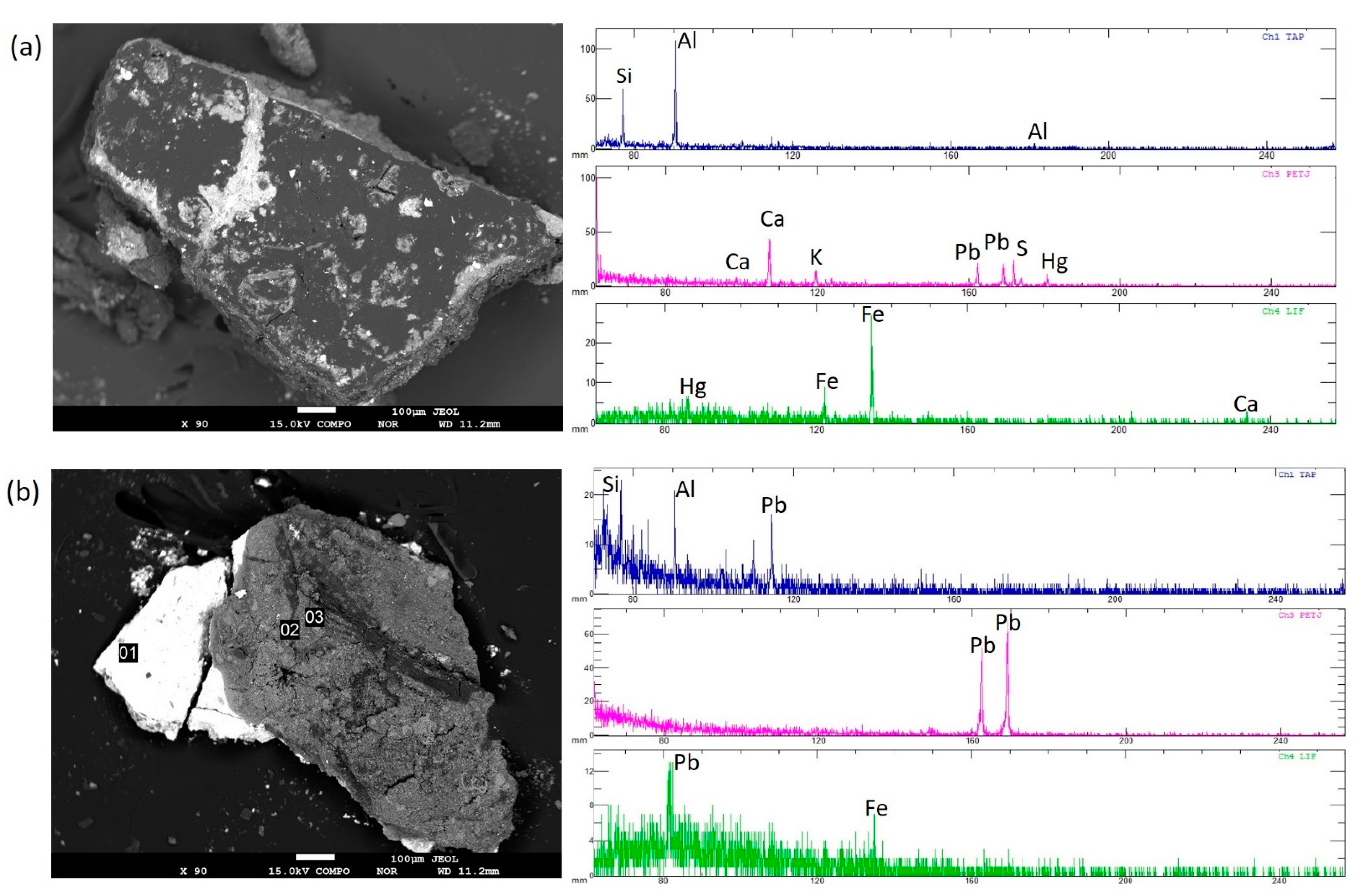
3.2. Laboratory EPMA-EDS Results

EPMA-EDS analyses were performed on all micro-fragments (Table 1) investigating the superficial pictorial film.
Table 3 provides a summary of the data, where the main elemental composition of paint layers is given for each sample.
Although EDS analyses have a semi-quantitative nature, they can provide information about pigments by the identification of the key chemical element responsible for the color. Moreover, due to the surface alteration and to detached and damaged portions of the pictorial film, in some samples the contribution of the composition of the underlying layers was evident during the investigations. In particular, considerable contents of Pb, Ca and iron oxide with aluminosilicates were detected in all the examined samples, most likely attributable to the composition of the preparatory layers. In particular, the significant Ca and Pb content suggest that the preparatory layer could contain species linked to these compounds, such as calcium carbonate and white lead. These data are in agreement with the results coming from other studies carried out on Mattia Preti’s masterpieces which report the use of such compounds for the preparatory layers especially during the Maltese period of the artist [6,8,12,13,14].
As for the two reddish samples (S1, S2), high amounts of S and Hg were both detected, leaving the hypothesis that the artist used a red mercury sulfide (HgS, vermilion) for the reddish shades. The further addition of earths is not to be excluded given the presence of Al, Ca, K, Si, and Fe which are chemical elements originating from accessory minerals, typical of the natural earth pigments from iron oxide deposits.
Samples with orange-salmon tones (S3, S7), and samples with pink shades typically used to make skin colors (S6, S10, S19, S22, S23, S25) show high concentrations of Pb followed by lower quantities of Ca and further chemical elements from earths (Si, Al, Mg, K, Fe, Na, Ti).
In Figure 4 some representative EPMA images collected on sample S2 and S10, along with relative EDX spectra, are reported as examples.
As for the high Pb content, in addition to its use in the preparatory layer, as mentioned above, it could also be associated to the use of a mixture of Pb-based pigments having a reddish coloration, such as the red lead (i.e., minium), and white color, such as the white lead. As far as the detection of chemical elements attributable to earths (Si, Al, Mg, K, Fe, Na, Ti) is concerned, another guess is the use of a mixture made of white lead added to red lead and green earth, all compounds used by the artist for flesh undertones. In fact, the green earths neutralized the effect of the pinks and reds of the flesh colors. Only S23, from a chemical point of view, slightly differs from all others due to the additional detection by EDS of Hg and S, suggesting the presence of vermilion. The sample S23 was in fact taken from the face of the angel, at the height of the cheek. The addition of a Hg-based red was likely intentional to make cheek tones more reddish.
As for the samples with yellowish tonalities (S13, S24, S26, S27), the EDS spectra reveal high amounts of Pb followed by Sb and smaller amounts of Si, Ca, Na, K, Al, Mg, Cl, Fe, S and Ti. The detection of high Pb contents, accompanied by Sb, pointed to possible use of a lead-based pigment, presumably a lead-antimony yellow (“Naples yellow”). The use of such a yellow pigment is in agreement with most used yellow shades in Mattia Preti’s palette [8,10,12]. Furthermore, traces of mercury and sulfur were also detected in S24. In fact, the S24 sample taken from the angel’s hair shows yellow-gold tones, which suggests that the artist has mixed yellow pigment with a red one to obtain more golden shades of yellow.
The EDS elemental analysis performed on green samples (S4, S5, S8, S9) led us to hypothesize a probable use of green earth as a mixture of aluminosilicate minerals (i.e., hydrous alumino-silicate of Mg, Fe and K), where the green color of the clays is caused by the presence of green Fe-containing minerals belonging to the group of clay micas (i.e., celadonite or glauconite) [10]. Obviously, it is evident that EDS results cannot complete the identification of such components and the contribution of vibrational spectroscopy techniques could be crucial for their identification. Also the sample S9, with yellowish-green hue, shows similar chemical composition with respect to previous samples, further suggesting that although green has warmer yellowish tones, chemical elements detected still imply the use of green earths as a pigment. This is in agreement with the literature, according to which green earth colors used in paint range from neutral yellow-green to pale greenish-gray to dark matte olive green [15,16,17].
Going on, a comparable elemental composition, mainly based on Pb, Ca, Si, Al, Mg, Na, Fe, K, S, Cl and Ti, can be observed for blackish (S11, S16) and grayish (S12, S14, S17, S18) samples. However, these elements are not sufficient to hypothesize the use of specific pigmenting substances, testifying to a similar method used by Mattia Preti for these shades. The major difference is in the detection of Pb, which exhibits a higher concentration in samples with greyish tones. Therefore, it is possible to assume the use of a white lead to blend the blacker colors. The brownish sample S20 shows high concentrations of Ca and Pb followed by Al, Si, Mg, and Fe, and low concentrations of Mn, P, K, Na, Ti, leading to the hypothesis that the artist used natural earth and ochers pigments for the browns, and probably a Sienna [6,9,18], a pigment made from clay, mainly composed of iron oxide and manganese oxide, and other minerals common in soils. Given the relevant Pb content, partly attributable to the preparatory layers, the addition of white lead to soften the brownish tones has not to be excluded.
Finally, regarding the two samples with blue shades (S15, S21), a different chemical composition was detected. According to EDS data, sample S15 could be a smalt, a blue potash glass pigment where the blue color is due to the presence of Co [19,20,21,22]. Actually, the elemental chemical composition of sample S15 corresponds to that of the smalt pigment, with high Si, K and Co contents, followed by elements as impurities surely due to the raw materials used for the manufacturing process of the pigment, which does not allow a precise chemical formula to be proposed [19,22]. In the case of sample S21, it was not possible to assert the use of a specific pigmenting substance. Although the detection of Cu as chemical element contained in various blue pigments (i.e., azurite, Egyptian blue, etc.), the EDS data are not sufficient for an exhaustive characterization and do not coincide with the blue shades of the artist’s palette [6,7,8,12].
The results achieved by XRF and EPMA-EDS techniques, even revealing, on one side, an overall agreement between each other, clearly underline, on the other side, the necessity to apply further analytical techniques to overcome the limitation associated with elemental analysis.

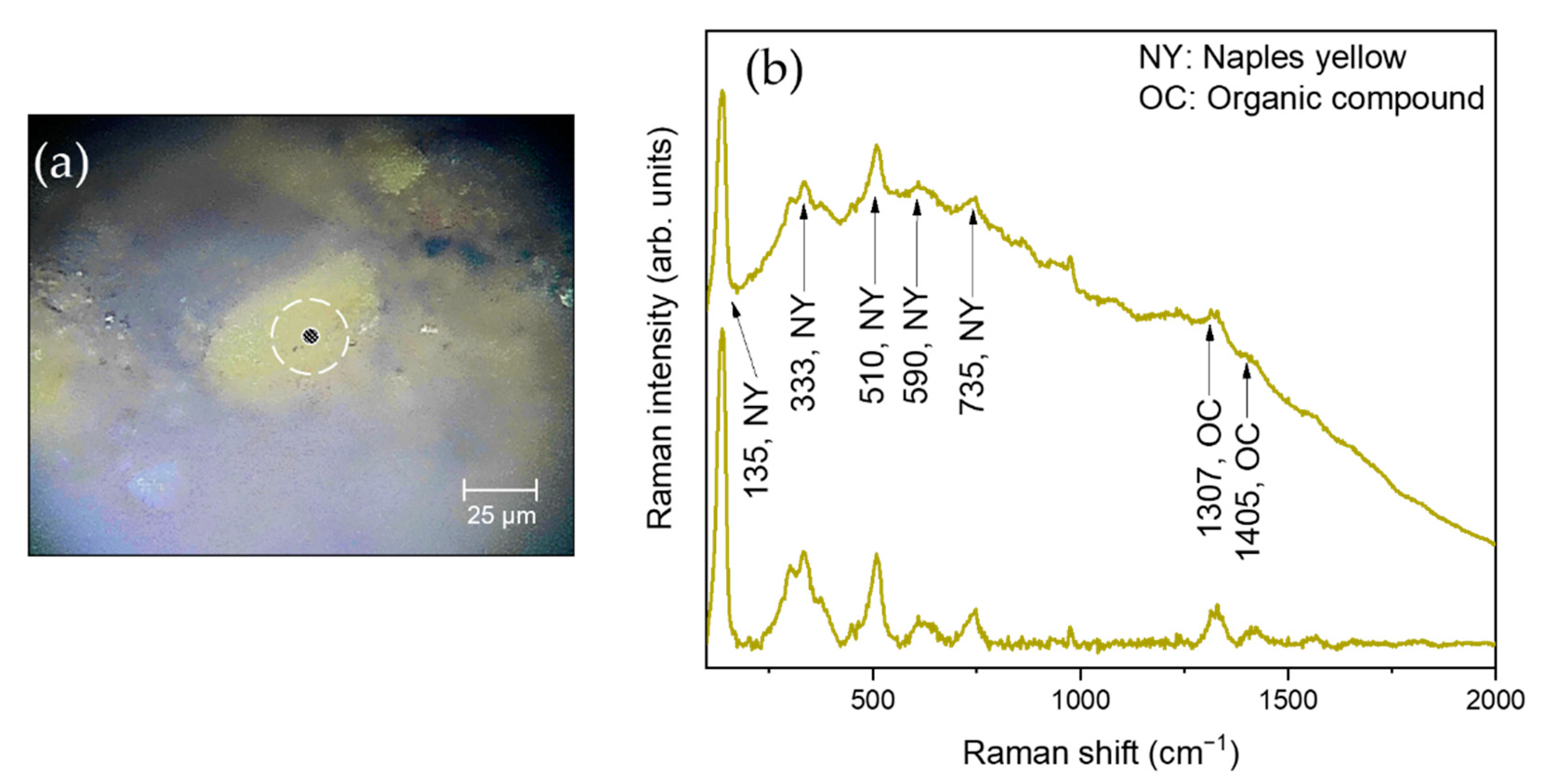
3.3. Laboratory Micro-Raman Results

Raman spectroscopy was employed to unambiguously recognize all the compounds previously hypothesized through XRF and EPMA-EDS techniques. The molecular characterization of different colored areas of the painting allowed us to gain insights not only on the presence of repaintings made by the artist during the production of his artwork, but also on the existence of non-document restoration interventions based on the detection of modern pigmenting compounds.
Concerning the red-colored areas of the painting (points S1, S2), Figure 5 reports a representative photomicrograph, together with the corresponding µ-Raman spectrum, collected on a red micro-fragment (sampled at point S1) of the painting, as example.
Based on a comparison with literature [23,24,25,26], the low-frequency contributions centered at ~252 cm−1 (A1g), ~288 cm−1 (Eg) and ~347 cm−1 (Eg) can be ascribed to the presence of vermilion (HgS), confirming our previous XRF and EPMA-EDS results. Such a compound, obtained by powdering the cinnabar mineral, assumed remarkable relevance as an artist’s red paint since Roman times, sometimes combined with burnt sulfur, despite its high cost and toxicity.
Interestingly, the employment of a red laser permits, in the case of vermilion, the acquisition of the best-quality Raman spectrum, since the laser frequency closely matches that associated with an electronic transition of this compound. Accordingly, resonant or pre-resonant Raman scattering phenomena are likely to occur, strongly enhancing the Raman signal by a factor of 102–106 [27].
It is worth noting that, according to previous published papers [6,7], the employment of vermilion by Mattia Preti, often in mixture with other composites such as iron oxides or red lead [13], turned out to be recurrent in the preparation of his red-reddish recipes. Interestingly, the observation of the band at ~612 cm−1 seems to confirm that vermilion was presumably mixed with poor amount of iron oxides, as also hypothesized by XRF and EPMA-EDS analysis. Furthermore, the presence of an organic compound, through the detection of contributions centered at ~942 cm−1, ~1311 cm−1 and ~1404 cm−1, can be ascribed to an organic-based binding medium, such as linseed oil or animal glue, or to a polishing agent used to enhance both the colors and brilliance of the painted surface. In this sense, we remark that a detailed characterization of organic binders will be the subject of a future investigation.
Finally, it is worth noting that the µ-Raman spectrum of S2 (not reported) almost completely matches that observed in the case of S1, indicating that the aforementioned areas share the same molecular composition.
As far as the orange, yellow-orange, and yellow colors are concerned (points S3, S7, S13, S24, S26, S27), Figure 6 shows a representative photomicrograph, together with the corresponding µ-Raman spectrum, collected on a yellow-orange micro-fragment (sampled at point S13) of the painting.
The detected µ-Raman profile revealed, as a main feature, an intense peak at ~135 cm−1, together with small contributions centered at ~333 cm−1, ~510 cm−1, ~590 cm−1 and ~735 cm−1, all ascribable to Naples yellow (Ny). According to literature [28,29,30,31], the strong contribution at ~135 cm−1, arising from the Pb-O lattice vibrations of the Ny cubic pyrocholore structure (A2B2O6O′), furnishes a clear indication of the employment of powdered bindheimite (Pb2Sb2O6(O,OH)) for the yellow-yellowish colorations. In fact, the presence of modified Ny, commonly characterized by ternary-oxide structures of the Pb2Sb2-xSnxO7-x/2 (Sn-modified Ny) or Pb2(ZnSb)O6 (Zn-modified Ny) type [32] (respectively known since the 17th and 20th centuries), would lead to a shift of the aforementioned peak to ~125 cm−1 or to ~145 cm−1, respectively [31]. Such considerations, corroborated by the fact that, at least, traces of Zn were detected by XRF and EPMA-EDS only in the case of sample S13, lead us to exclude the employment of a modified Ny applied during a possible recent intervention. It is worth remarking that according to previously published papers, the employment of Ny was already extensively evidenced in other paintings by Mattia Preti [8,10,12].
Concerning the green pigment of St. Vicario’s cloak (S4 and S5) and St. Clemente’s cloak and halo (S8 and S9, respectively), no reliable µ-Raman spectra were obtained. This was due to the presence of a prominent fluorescence background that almost completely covered the corresponding Raman profiles, making the molecular characterization of such samples neither feasible nor accurate.
The µ-Raman analysis of the flesh-toned micro-fragments (sampled at points S6, S10, S19, S22, S23, S25) furnished evidence of a complex molecular composition characterized by a mixture of several compounds. In particular, Figure 7 reports three representative photomicrographs and corresponding µ-Raman spectra, collected on different flesh-toned micro-fragments (sampled at point S6, S10 and S22) of the painting, as example. More in detail, the µ-Raman profile collected on S6 (Figure 7d) indicates the presence of minium, through the detection of its main peak centered at ~121 cm−1 (due to O–Pb–O angle deformations), along with other contributions falling at ~151 cm−1, ~311 cm−1, ~390 cm−1 and ~548 cm−1. Furthermore, in the case of the µ-Raman spectrum collected on S10 (Figure 7e), vermilion can be distinguished, by its typical triplet of low-frequency peaks centered at ~252 cm−1 (A1g), ~288 cm−1 (Eg) and ~347 cm−1 (Eg). The µ-Raman spectrum of sample S22 (Figure 7f) shows, among other features, a strong and sharp Raman band at ~1051 cm−1, indicating the application of a white lead pigment. Generally, the occurrence of such peak is considered to be related to degradation phenomena involving lead-rich materials of the original portion of the canvas [33,34], consequently the prolonged exposure of the painting with air and biodeteriogenic agents (i.e., bacteria and fungi). Under such circumstances, the production of lead sulfide (PbS) and lead dioxide (plattnerite, PbO2) onto the pictorial layer is highly favored, resulting in a blackening of the original white coloration in favor of a pinkish nuance. Based on the aforementioned results, the flesh-toned colorations were reasonably realized by mixing minium with dilute amount of vermilion and white lead in different proportion, depending on the desired pinkish shades, in good agreement with the previous analysis of the pink layer by XRF and EPMA-EDS and literature results [8,10,12].
It is worth noting that in all micro-fragments the presence of an organic compound was identified, through the observation of contributions falling at ~942 cm−1, ~1308 cm−1 and ~1406 cm−1 (Figure 7d–f), supporting the hypothesis that it is in fact distributed uniformly onto the whole surface of the artwork.
As far as the blackish-brown and grayish areas (points S11, S12, S14, S16, S17, S18, S20) are concerned, Figure 8 reports two representative photomicrographs and corresponding µ-Raman spectra, collected on a blackish and grayish micro-fragments (sampled at point S11 and S12) of the painting, respectively.
Concerning the µ-Raman spectrum of sample S11 (Figure 8c), it shows two major contributions centered at ~1312 cm−1 and ~1567 cm−1, associated with the D (disorder) and G (graphitic) bands of amorphous carbon. This result indicates the use by Mattia Preti of a typical carbon-based black pigment of organic provenance. Unfortunately, an accurate identification of the carbon source cannot be accomplished through Raman spectroscopy. Nevertheless, considering the high concentration of Ca observed in the corresponding area through XRF and EPMA-EDS, several hypotheses can be still made. In particular, the detected amorphous carbon could be either of vegetal origin, such as vine black, usually attained by carbonizing plants or wood, or of animal origin, such as bone black (~85%: hydroxyapatite; ~10%: elemental carbon; ~5%: calcite and/or magnesium phosphate) or black ivory (~80%: calcite mixed with calcium phosphate; ~20%: elemental carbon). In our case, the observation of a band falling at ~959 cm−1, associated to the stretching mode of the phosphate ion [PO4]3−, along with the main peak of calcite at ~1081 cm−1, led us to conclude that the black pigment used was most likely of animal origin (bone black or ivory black), thus excluding any vegetal recipe for the black-brownish colorations [6,7,8,10,12].
The µ-Raman spectrum of the grayish micro-fragment (sample S12, Figure 8d) seems almost similar to that of sample S11, with the only exception of the appearance of a well-visible peak at ~1051 cm−1 due to the presence of white lead. Accordingly, it is reasonable to assume that a mixture of an animal-based carbon black and white lead was probably employed for the preparation of the grayish nuances, following black discoloration procedures. Such a consideration is in full agreement with both the XRF and EPMA-EDS results, which revealed a similar elemental composition for both the blackish and grayish areas of the painting except for the Pb content, which was found to be higher in gray areas than in black ones. The aforementioned considerations are further validated by the fact that the application of a carbon-based black pigment, often in mixture with white lead or calcite, was already testified in other paintings by Mattia Preti, including the “Penitent St. Peter” [6], the “Madonna of Sorrows” [6], the “St. Michael defeating Evil” [9], the “Dinner of Emmaus” [7] and the “Conversion of St. Paul” [13].
Figure 9 reports the collected photomicrographs and µ-Raman spectra of the blue pigmented micro-fragments (sampled at points S15 and S21), representative of the cerulean and light blue areas of the painting. The µ-Raman profiles revealed, as a main result, the characteristic bands of indigo (C16H10N2O2), centered at ~545 cm−1 and ~1573 cm−1. Indigo is an organic compound in the form of a dark blue crystalline powder usually attained from plants of the genus Indigofera (typical of tropical regions) since the Middle Ages. In European countries, indigo was usually extracted from woad (Isatis tinctoria), which contains a high amount of indican (precursor of indigo), a water-soluble/colorless secondary-products of the amino acid tryptophan [35,36]. The employment of blue pigmenting agents based on indigo was already well-documented in other oil-on-canvas paintings by Mattia Preti [6,7], although also lapis, blue smalt and copper-based compounds were repeatedly employed [8].
Interestingly, for both sample S15 and S21, the cerulean/light blue colorations were reasonably achieved by mixing pure indigo (characterized by a natural dark blue appearance) with a very dilute amount of white lead (recognized by its main contribution at ~1051 cm−1, Figure 9c,d), in order to lighten the dark blue color in favor of lighter tones. Although this result is in agreement with the high content of Pb previously detected on both areas, it does not explain the high concentration of elemental Co observed in the case of S15 (see Table 2 and Table 3).
In fact, based on the elemental composition of sample S15 (see Table 2 and Table 3), the application of a cobalt blue, usually obtained by mixing cobalt and aluminum oxides, or a smalt can be reasonably hypothesized. It is worth noting, however, that the observation of a mixture of indigo and white lead even in this sample does not exclude the possible addition of one of the aforementioned compounds in the blue-bluish recipes of Mattia Preti. In fact, the absence of reliable Raman features associated to a Co-based composites may be due to the fact that a single color microparticle, mostly composed by indigo, was spatially isolated, due to the high degree of beam-focusing achieved through the microscope. In this sense, considering that the laser-spot size was of ~25/~50 µm, we cannot exclude that in the immediately surrounding zones a different type of blue, likely based on cobalt, could be found.
Finally, even in these two micro-fragments, an organic-based compound can be identified through its main spectral features at ~942 cm−1, ~1308 cm−1 and ~1406 cm−1, interpreted as due to some binder and/or polishing agent.
For all the investigated samples, an overview of the main hypotheses regarding the used pigments and other materials, deduced from the analyses carried out in the work, is reported in Table 4.

4. Conclusions

In this work, a combined multi-analytical approach, involving both in situ and laboratory-based physical/chemical methodologies, was successfully applied on what is considered as one of the Mattia Preti’s masterpieces known as “The Glory of St. Barbara” (oil on canvas (460 × 305 cm)), located inside the St. Barbara Church in Taverna (Calabria region, Italy).
In particular, the simultaneous employment of X-ray fluorescence (XRF) spectrometry, electron probe microanalyses (EPMA) coupled with energy-dispersive spectroscopy (EDS) and µ-Raman spectroscopy, allowed us to successfully characterize, both at the elemental and molecular scales, the composition of the raw materials used by the artist both in terms of preparatory components and pigmenting agents, as well as their stratigraphy and morphology. The results obtained not only provided new useful insights for a better knowledge of the pictorial technique and painting palette used by the famous master, but they were also crucial in establishing the state of conservation of the painting as well as whether possible non-documented restoration interventions took place. Furthermore, the presence of recurring elements/compounds for all the examined samples allowed us to confirm the Maltese origin of “The Glory of St. Barbara” painting, with such elements/compounds similar to those observed in other paintings by Mattia Preti executed during the Maltese period of the artist.
It is worth noting that all the aforementioned aspects, in the broad context of a scientific examination carried out on artworks of high artistic and historical value, can represent a necessary pre-requisite in terms of optimizing the latest restoration protocols and for the correct selection, by conservators and restorers, of the best cleaning and protection strategies to be applied.

Author Contributions

Conceptualization, S.G., G.M., M.F.L.R. and V.V.; methodology, F.C., M.R., G.P., A.D. and L.R.; validation, V.C., D.M., M.F.L.R. and V.V.; formal analysis, F.C., M.R., G.P., A.D. and L.R.; investigation, F.C., M.R., G.P., V.C., A.D., L.R., M.F.L.R. and V.V.; data curation, F.C., M.R., G.P., A.D., L.R., M.F.L.R. and V.V.; writing—original draft preparation, F.C., M.R., G.P., S.G., G.M. and V.V.; writing—review and editing, F.C., M.R., G.P., A.D. and L.R.; visualization, V.C., D.M., M.F.L.R. and V.V.; supervision, M.F.L.R. and V.V. All authors have read and agreed to the published version of the manuscript.

Funding

This research received no external funding.

Institutional Review Board Statement

Not applicable.

Informed Consent Statement

Not applicable.

Conflicts of Interest

The authors declare no conflict of interest.

References

  1. Debono, S.; Valentino, G. Mattia Preti: Faith and Humanity; Misdea Books Ltd.: Valletta, Malta, 2013; ISBN 9789993274360. [Google Scholar]
  2. King, G.G. Mattia Preti. Art Bull. 1936, 18, 371–386. [Google Scholar] [CrossRef]
  3. Lafuente, B.; Downs, R.T.; Yang, H.; Stone, N. The power of databases: The RRUFF project. In Highlights in Mineralogical Crystallography; Armbruster, T., Danisi, R.M., Eds.; De Gruyter: Berlin, Germany, 2015; pp. 1–30. ISBN 9783110417104. [Google Scholar]
  4. Bell, I.M.; Clark, R.J.H.; Gibbs, P.J. Raman spectroscopic library of natural and synthetic pigments (pre- ≈ 1850 AD). Spectrochim. Acta Part A Mol. Biomol. Spectrosc. 1997, 53, 2159–2179. [Google Scholar] [CrossRef]
  5. Caggiani, M.C.; Cosentino, A.; Mangone, A. Pigments Checker version 3.0, a handy set for conservation scientists: A free online Raman spectra database. Microchem. J. 2016, 129, 123–132. [Google Scholar] [CrossRef]
  6. Pelosi, C.; Agresti, G.; Baraldi, P. The ‘slash of light’ in the late religious paintings of Mattia Preti technique and materials. Eur. J. Sci. Theol. 2018, 14, 151–160. [Google Scholar]
  7. Chiavari, G.; Prati, S.; Lanterna, G.; Lalli, C.; Cagnini, A. Diagnostic study of the materials and painting techniques in “The Dinner of Emmaus” by Gregorio (and Mattia?) Preti. Microchim. Acta 2007, 159, 357–362. [Google Scholar] [CrossRef]
  8. Ridolfi, S. Relazione Tecnico-Scientifica Delle Indagini Effettuate Sull’allegoria Dei Cinque Sensi di Mattia e Gregorio Preti; Ars Mensurae: Roma, Italy, 2019. [Google Scholar]
  9. D’Amico, S.; Comite, V.; Paladini, G.; Ricca, M.; Colica, E.; Galone, L.; Guido, S.; Mantella, G.; Crupi, V.; Majolino, D.; et al. Multitechnique diagnostic analysis and 3D surveying prior to the restoration of St. Michael defeating Evil painting by Mattia Preti. Environ. Sci. Pollut. Res. 2021; in press. [Google Scholar] [CrossRef]
  10. Hradil, D.; Hradilová, J.; Lanterna, G.; Galeotti, M.; Holcová, K.; Jaques, V.; Bezdička, P. Clay and alunite-rich materials in painting grounds of prominent Italian masters—Caravaggio and Mattia Preti. Appl. Clay Sci. 2020, 185, 105412. [Google Scholar] [CrossRef]
  11. Venuti, V.; Fazzari, B.; Crupi, V.; Majolino, D.; Paladini, G.; Morabito, G.; Certo, G.; Lamberto, S.; Giacobbe, L. In situ diagnostic analysis of the XVIII century Madonna della Lettera panel painting (Messina, Italy). Spectrochim. Acta Part A Mol. Biomol. Spectrosc. 2020, 228, 117822. [Google Scholar] [CrossRef]
  12. D’Amico, S.; Venuti, V.; Colica, E.; Crupi, V.; Paladini, G.; Guido, S.; Mantella, G.; Majolino, D. A combined 3D surveying, XRF and Raman in situ investigation on The Conversion of St Paul painting (Mdina, Malta) by Mattia Preti. ACTA IMEKO 2021, 10, 173–179. [Google Scholar] [CrossRef]
  13. D’Amico, S.; Venuti, V.; Colica, E.; Crupi, V.; Majolino, D.; Paladini, G.; Guido, S.; Mantella, G.; Zumbo, R. Scientific investigation of the Conversion of St Paul painting (Mdina, Malta). In Proceedings of the 2019 IMEKO TC4 International Conference on Metrology for Archaeology and Cultural Heritage, MetroArchaeo 2019, Florence, Italy, 4–6 December 2019; pp. 330–334. [Google Scholar]
  14. Lalli, C.G.; von Breska-Ficović, N.; Innocenti, F.; Kolić Pustić, M. Le due serie dei quattro “Evangelisti” di Mattia Preti a Dubrovnik: Un progetto multidisciplinare e di collaborazione tra il Laboratorio Scientifico dell’Opificio e l’Istituto Croato di RestauroSource: OPD Restauro. Cent. Di Della Edifimi SRL 2014, 26, 231–248. [Google Scholar]
  15. Feller, R.L. Artists’ Pigments: A Handbook of Their History and Characteristics; Feller, R.L., Ed.; Cambridge University Press: Cambridge, UK, 1986; Volume 1, ISBN 978-1-904982-74-6. [Google Scholar]
  16. Gettens, R.J.; Fitzhugh, E.W. Malachite and Green Verditer. In Artists’ Pigments: A Handbook of Their History and Characteristics; Roy, A., Ed.; Oxford University Press: Oxford, UK, 1993; Volume 2, ISBN 978-1-904982-75-3. [Google Scholar]
  17. Salvadó, N.; Butí, S.; Cotte, M.; Cinque, G.; Pradell, T. Shades of green in 15th century paintings: Combined microanalysis of the materials using synchrotron radiation XRD, FTIR and XRF. Appl. Phys. A 2013, 111, 47–57. [Google Scholar] [CrossRef]
  18. Genestar, C.; Pons, C. Earth pigments in painting: Characterisation and differentiation by means of FTIR spectroscopy and SEM-EDS microanalysis. Anal. Bioanal. Chem. 2005, 382, 269–274. [Google Scholar] [CrossRef] [PubMed]
  19. Cavallo, G.; Riccardi, M.P. Glass-based pigments in painting: Smalt blue and lead–tin yellow type II. Archaeol. Anthropol. Sci. 2021, 13, 199. [Google Scholar] [CrossRef]
  20. Zlámalová Cílová, Z.; Gelnar, M.; Randáková, S. Smalt production in the Ore Mountains: Characterization of samples related to the production of blue pigment in Bohemia. Archaeometry 2020, 62, 1202–1215. [Google Scholar] [CrossRef]
  21. Berrie, B.H. Rethinking the History of Artists’ Pigments Through Chemical Analysis. Annu. Rev. Anal. Chem. 2012, 5, 441–459. [Google Scholar] [CrossRef]
  22. Mühlethaler, B.; Thissen, J. Smalt. In Artists’ Pigments, Vol. 2; Roy, A., Ed.; Oxford University Press: Oxford, UK, 1993; pp. 113–130. [Google Scholar]
  23. Sakellariou, K.; Miliani, C.; Morresi, A.; Ombelli, M. Spectroscopic investigation of yellow majolica glazes. J. Raman Spectrosc. 2004, 35, 61–67. [Google Scholar] [CrossRef]
  24. Borgia, I.; Brunetti, B.G.; Miliani, C.; Ricci, C.; Seccaroni, C.; Sgamellotti, A. The combined use of lead–tin yellow type I and II on a canvas painting by Pietro Perugino. J. Cult. Herit. 2007, 8, 65–68. [Google Scholar] [CrossRef]
  25. Welter, N.; Schüssler, U.; Kiefer, W. Characterisation of inorganic pigments in ancient glass beads by means of Raman microspectroscopy, microprobe analysis and X-ray diffractometry. J. Raman Spectrosc. 2007, 38, 113–121. [Google Scholar] [CrossRef]
  26. Venuti, V.; Crupi, V.; Fazio, B.; Paladini, G.; La Russa, M.F.; Ricca, M.; Rovella, N.; Macchia, A.; Khalilli, F.; Majolino, D. Investigation of glazed pottery fragments (XIX century A. D.) from Agsu site (Azerbaijan) by XRF and Raman techniques. EPJ Web Conf. 2020, 230, 00012. [Google Scholar] [CrossRef] [Green Version]
  27. Efremov, E.V.; Ariese, F.; Gooijer, C. Achievements in resonance Raman spectroscopy. Anal. Chim. Acta 2008, 606, 119–134. [Google Scholar] [CrossRef]
  28. Rosi, F.; Manuali, V.; Grygar, T.; Bezdicka, P.; Brunetti, B.G.; Sgamellotti, A.; Burgio, L.; Seccaroni, C.; Miliani, C. Raman scattering features of lead pyroantimonate compounds: Implication for the non-invasive identification of yellow pigments on ancient ceramics. Part II. In situ characterisation of Renaissance plates by portable micro-Raman and XRF studies. J. Raman Spectrosc. 2011, 42, 407–414. [Google Scholar] [CrossRef]
  29. Sandalinas, C.; Ruiz-Moreno, S.; López-Gil, A.; Miralles, J. Experimental confirmation by Raman spectroscopy of a Pb-Sn-Sb triple oxide yellow pigment in sixteenth-century Italian pottery. J. Raman Spectrosc. 2006, 37, 1146–1153. [Google Scholar] [CrossRef]
  30. Coentro, S.; Mimoso, J.M.; Lima, A.M.; Silva, A.S.; Pais, A.N.; Muralha, V.S.F. Multi-analytical identification of pigments and pigment mixtures used in 17th century Portuguese azulejos. J. Eur. Ceram. Soc. 2012, 32, 37–48. [Google Scholar] [CrossRef] [Green Version]
  31. Rosi, F.; Manuali, V.; Miliani, C.; Brunetti, B.G.; Sgamellotti, A.; Grygar, T.; Hradil, D. Raman scattering features of lead pyroantimonate compounds. Part I: XRD and Raman characterization of Pb2Sb2O7 doped with tin and zinc. J. Raman Spectrosc. 2009, 40, 107–111. [Google Scholar] [CrossRef]
  32. Cascales, C.; Alonso, J.A.; Rasines, I. The new pyrochlores Pb2(MSb)O6.5 (M = Ti, Zr, Sn, Hf). J. Mater. Sci. Lett. 1986, 5, 675–677. [Google Scholar] [CrossRef]
  33. Muralha, V.S.F.; Miguel, C.; Melo, M.J. Micro-Raman study of Medieval Cistercian 12–13th century manuscripts: Santa Maria de Alcobaça, Portugal. J. Raman Spectrosc. 2012, 43, 1737–1746. [Google Scholar] [CrossRef]
  34. Burgio, L.; Clark, R.J.H.; Firth, S. Raman spectroscopy as a means for the identification of plattnerite (PbO2), of lead pigments and of their degradation products. Analyst 2001, 126, 222–227. [Google Scholar] [CrossRef]
  35. Baran, A.; Fiedler, A.; Schulz, H.; Baranska, M. In situ Raman and IR spectroscopic analysis of indigo dye. Anal. Methods 2010, 2, 1372–1376. [Google Scholar] [CrossRef]
  36. Vandenabeele, P.; Moens, L. Micro-Raman spectroscopy of natural and synthetic indigo samples. Analyst 2003, 128, 187–193. [Google Scholar] [CrossRef]
Figure 1. “The Glory of St. Barbara” painting (a); Location of Taverna (yellow spot), in Calabria Region, southern Italy (b); the St. Barbara Church in Taverna (c).
Applsci 12 01385 g001
Figure 2. Analyzed points from the “The Glory of St. Barbara” painting.
Applsci 12 01385 g002
Figure 3. XRF spectra collected on the red area from St. Clemente’s cloak (S1, (a)); the green area from St. Vicario’s cloak (S4, (b)); the flesh-toned area from St. Clemente’s cheek (S10, (c)); the grayish-white area from the cloud under St. Barbara’s sandal (S12, (d)); the yellow-orange area from St. Barbara’s cloak (S13, (e)); the light blue area from the cerulean shades under the angel’s feet (S15, (f)); the blackish-brown area from Christ’s hair (S20, (g)); and the left side of the canvas, at the bottom (S28, (h)).
Applsci 12 01385 g003
Figure 4. Representative electron probe microanalyses (EPMA) images showing: (a) the pictorial film (sample S2, scale bar 100 μm) and (b) the layers below the pictorial film (sample S10, scale bar 100 μm) along with relative EDS spectra. Note: for the sample S10 (b) the EDS spectrum reflects the elemental composition at the point analysis marked as 01.
Applsci 12 01385 g004
Figure 5. (a) Photomicrograph, in white reflected light, and (b) µ-Raman spectrum collected on a red micro-fragment (sampled at point S1) of the “The Glory of St. Barbara” painting.
Applsci 12 01385 g005
Figure 6. (a) Photomicrograph, in white reflected light, and (b) µ-Raman spectrum collected on a yellow-orange micro-fragment (sampled at point S13) of the “The Glory of St. Barbara” painting.
Applsci 12 01385 g006
Figure 7. Photomicrographs, in white reflected light, and µ-Raman spectra collected on flesh-toned micro-fragments sampled at point S6 (a,d), S10 (b,e) and S22 (c,f) of the “The Glory of St. Barbara” painting.
Applsci 12 01385 g007
Figure 8. Photomicrographs, in white reflected light, and µ-Raman spectra collected on a blackish and grayish micro-fragment sampled at point S11 (a,c) and S12 (b,d) of the “The Glory of St. Barbara” painting, respectively.
Applsci 12 01385 g008
Figure 9. Photomicrographs, in white reflected light, and µ-Raman spectra collected on the cerulean and light blue micro-fragment sampled at point S15 (a,c) and S21 (b,d) of the “The Glory of St. Barbara” painting, respectively.
Applsci 12 01385 g009
Table 1. Investigated samples, together with a brief description and the employed techniques.
Analyzed PointSampling Area and Brief DescriptionEmployed Techniques
S1Red micro-fragment from St. Clemente’s cloakXRF, EPMA-EDS, µ-Raman
S2Red micro-fragment from St. Mercurio’s cloakXRF, EPMA-EDS, µ-Raman
S3Orange micro-fragment from St. Vicario’s cloakXRF, EPMA-EDS, µ-Raman
S4Green micro-fragment from St. Vicario’s cloakXRF, EPMA-EDS, µ-Raman
S5Green micro-fragment from St. Vicario’s cloakXRF, EPMA-EDS, µ-Raman
S6Flesh-toned micro-fragment from St. Vicario’s right handXRF, EPMA-EDS, µ-Raman
S7Salmon-orange micro-fragment from St. Clemente’s cloakXRF, EPMA-EDS, µ-Raman
S8Green micro-fragment from St. Clemente’s cloakXRF, EPMA-EDS, µ-Raman
S9Yellowish-green micro-fragment from St. Clemente’s haloXRF, EPMA-EDS, µ-Raman
S10Flesh-toned micro-fragment from St. Clemente’s cheekXRF, EPMA-EDS, µ-Raman
S11Blackish-brown micro-fragment from St. Barbara’s sandalXRF, EPMA-EDS, µ-Raman
S12Grayish-white micro-fragment from the cloud under St. Barbara’s sandalXRF, EPMA-EDS, µ-Raman
S13Yellow-orange micro-fragment from St. Barbara’s cloakXRF, EPMA-EDS, µ-Raman
S14Grayish micro-fragment from the Angel’s wingXRF, EPMA-EDS, µ-Raman
S15Light blue micro-fragment from the cerulean shades under the Angel’s feetXRF, EPMA-EDS, µ-Raman
S16Blackish-brown micro-fragment, from the back of the Angel’s headXRF, EPMA-EDS, µ-Raman
S17Grayish micro-fragment, from the top of the Angel’s swordXRF, EPMA-EDS, µ-Raman
S18Grayish micro-fragment from the greyish shades under the Angel’s wingXRF, EPMA-EDS, µ-Raman
S19Flesh-toned micro-fragment from the Angel’s bellyXRF, EPMA-EDS, µ-Raman
S20Blackish-brown micro-fragment from Christ’s hairXRF, EPMA-EDS, µ-Raman
S21Light blue micro-fragment from behind Christ’s shouldersXRF, EPMA-EDS, µ-Raman
S22Flesh-toned (whitish) micro-fragment from Christ’s armXRF, EPMA-EDS, µ-Raman
S23Flesh-toned micro-fragment from the Angel’s face, under Christ’s handXRF, EPMA-EDS, µ-Raman
S24Golden yellow micro-fragment from the Angel’s hair under Christ’s footXRF, EPMA-EDS, µ-Raman
S25Flesh-toned micro-fragment from the Angel’s leg under Christ’s footXRF, EPMA-EDS, µ-Raman
S26Yellow micro-fragment from St. Vicario’s cloakXRF, EPMA-EDS, µ-Raman
S27Yellow micro-fragment from St. Vicario’s cloakXRF, EPMA-EDS, µ-Raman
S28Left side of the canvas, bottom 1XRF
1 Sample was collected from an already-existing lacunae, with the surface paint layer locally damaged.
Table 2. X-ray fluorescence (XRF) elemental composition. Minor or trace elements reported in brackets.
Analyzed PointElemental Composition by XRF
S1S, Ca, Pb, Fe, Hg, Cl, K, As, (Zn, Se, Mn, Ba, Sr, Rb, Cr, Mo, Zr)
S2Ca, S, Fe, K, Cl, Pb, (I, Hg, As, Ti, Mn, Zn, Ba, Sr, Cr, Mo, Zr, Rb, Se)
S3S, Pb, Ca, Cl, As, Fe, K, (Ti, Se, Hg, Mn, Cd, Zn, Sn, Ba, Rb, Sr)
S4S, Pb, Ca, Cl, As, K, Fe, Co, (Cu, Sb, Ti, Hg, Se, Ni, Mn, Zn, Sr, Rb, Ba, Cd, Cr, Mo)
S5S, Pb, Ca, Cl, As, K, Fe, Co, (Ni, Mn, Cu, Se, Hg, Zn, Sb, Rb, Ba, Sr, Cd, Mo)
S6S, Pb, Cl, As, Ca, Fe, K, Hg, (Se, Ti, Cd, Zn, Mn, Rb, Mo)
S7S, Pb, Ca, Cl, Fe, K, Ti, Fe, Zn, (Se, Sb, Mn, Ba, Cd, Rb, Sr)
S8S, Ca, Pb, Fe, Cl, As, K, Mn, (Hg, Ti, Zn, Se, Ba, Sr, Rb, Cr, Cu, Cd, Mo, Zr)
S9S, Pb, Ca, Cl, As, Fe, K, (Mn, Se, Ti, Zn, Hg, Cu, Rb, Ba, Sr, Cd, Mo)
S10S, Pb, Cl, As, Ca, Fe, Hg, K, (Se, Zn, Ti, Cd, Ba, Mn, Rb)
S11S, Pb, Ca, Cl, As, Fe, K, Hg, (Co, Mn, Se, Zn, Ti, Ni, Rb, Cd, Ba, Sr, Mo)
S12S, Pb, Cl, As, Ca, K, Co, (Fe, Se, Ti, Zn, Cd, Mn)
S13S, Pb, Ca, Fe, Cl, K, As, Sb, Hg, I, (Ti, Mn, Zn, Cd, Ba)
S14S, Pb, Cl, As, Ca, K, Hg, Fe, Co, (Zn, Cd, Sn, Ti, Mn)
S15S, Pb, Cl, As, Ca, K, Co, Fe, (Se, Zn, Ni, Cd, Rb, Mn, Ba)
S16S, Pb, Ca, Cl, As, K, Fe, Co, (Ti, Ni, Se, Cu, Zn, Ba, Mn, Rb)
S17S, Ca, Pb, Cl, As, K, Fe, (Hg, Se, Zn, Sr, Rb, Mn, Cd, Mo)
S18S, Pb, Ca, Cl, As, K, Fe, (Se, Zn, Ti, Cu, Rb, Cd, Sr, Mn, Ba)
S19S, Pb, Ca, Cl, As, Hg, Fe, K, (Ti, Se, Zn, Cu, Rb, Sb, Mn, Sr)
S20Ca, S, Fe, Pb, K, Cl, As, I, (Hg, Mn, Zn, Sr, Ba, Cu, Se, Rb, Mo, Zr)
S21S, Pb, Cl, As, Ca, K, (Fe, Se, Cd, Zn, Ti, Mn)
S22S, Pb, Cl, As, Ca, K, Sn, (Cd, Mn)
S23S, Pb, Cl, As, Ca, Hg, K, Fe, (Se, Zn, Cd, Rb, Mn)
S24S, Ca, Cl, Pb, K, Fe, As, (Hg, Mn, Se, Cu, Sb, Zn, Sr, Rb)
S25S, Pb, Cl, As, Ca, Hg, K, Fe, (Ti, Se, Cd, Zn, Mn, Ag)
S26S, Pb, Ca, Cl, K, As, Fe, Sb, Ti, (Zn, Ba, Se, Cr, Cd, Mn, Sr)
S27S, Ca, Pb, Cl, Fe, As, K, (Sb, Hg, Ti, Se, Mn, Zn, Cu, Sr, Rb, Cd, Mo)
S28Ca, S, Pb, K, Cl, Fe, As, (Zn, Sr, Mn, Cu, Se, Ba, Mo, Rb)
Table 3. Energy-dispersive spectroscopy (EDS) chemical data detected on painted surfaces.
Analyzed PointEDS Data
S1Hg, S, Pb, Fe, Ca, Si, Al, K
S2Hg, S, Pb, Fe, Ca, Si, Al, K, Ti
S3Pb, Fe, Ca, Si, Al, Mg, K
S4Si, Al, Fe, Cl, Mg, K, Na, Pb, Ca
S5Si, Al, Fe, Cl, Mg, K, Na, Pb, Ca
S6Pb, Fe, Ca, Si, Al, K, Na
S7Pb, Fe, Ca, Si, Al, K, Na
S8Si, Al, Fe, Cl, Mg, K, Na, Pb, Ca
S9Si, Al, Fe, Ca, Mg, K, Na, Ba, Pb, Ca
S10Pb, Fe, Ca, Si, Al, Mg, K
S11Si, Al, Mg, Na, Fe, K, S, Cl, Pb, Ca
S12Pb, Ca, Si, Al, Mg, Na, Fe, K, S, Ti
S13Pb, Sb, Si, Ca, Na, K, Al, Mg, S, Fe, Cl
S14Pb, Ca, Si, Al, Mg, Na, Fe, K, S, Ti
S15Si, Co, K, Ca, S, Fe, Al, K, Ti, Pb, Ca
S16Si, Al, Mg, Na, Fe, K, S, Cl, Pb, Ca
S17Pb, Ca, Si, Al, Mg, Na, Fe, K, S, Ti
S18Pb, Ca, Si, Al, Mg, Na, Fe, K, S, Ti
S19Pb, Fe, Ca, Si, Al, Mg, K, Ti
S20Pb, Ca, Si, Al, Mg, Fe, Mn, P, Na, K, Ti
S21Si, Al, Fe, Cu, K, Mg, Fe, Na, Pb, Ca
S22Pb, Fe, Ca, Si, Al, Mg, K, Na
S23Pb, Ca, Si, Al, Mg, K, Fe, S, Na, Hg
S24Pb, Hg, Sb, S, Ca, Si, Na, Al, Mg, K, Cl
S25Pb, Fe, Ca, Si, Al, Mg, K
S26Pb, Sb, Si, Ca, Na, K, Al, Mg, S, Fe, Cl, Ti
S27Pb, Sb, Si, Ca, Na, K, Al, Mg, S, Fe, Cl, Ti
Table 4. Investigated samples, summarized according to the color of analyzed pigments, together with the main hypotheses regarding the used pigments and other materials deduced from all the analyses carried out in the work.
Point of AnalysisColorUsed Pigments and Other Materials
S1, S2RedVermilion, iron oxides, Pb-based compounds, Ti-based compounds, organic compounds
S3, S7, S13, S24, S26, S27Orange/yellow-orange/yellowNaples yellow, Fe-based compounds, Hg-based compounds, organic compounds
S4, S5, S8, S9GreenFe-based compounds
S6, S10, S19, S22, S23, S25Flesh tonesMinium, vermilion, iron oxides, calcite, lead white, organic compounds
S11, S12, S14, S16, S17, S18, S20Blackish-brown/grayishCarbon black (bone black or ivory black), calcite, lead white, Fe-based compounds, Mg-based compounds
S15, S21BlueIndigo, lead white, Co-based compounds, organic compounds
S28CanvasCa-based compounds, S-based compounds, Pb-based compounds
Publisher’s Note: MDPI stays neutral with regard to jurisdictional claims in published maps and institutional affiliations.

