Article

Characterizing the Grating-like Nanostructures Formed on BaF2 Surfaces Exposed to Extreme Ultraviolet Laser Radiation

1
National Key Laboratory of Science and Technology on Tunable Laser, School of Aeronautics, Harbin Institute of Technology, Harbin 150001, China
2
Department of Pulse Plasma Systems, Institute of Plasma Physics, Czech Academy of Sciences, Za Slovankou 1782/3, 182 00 Prague, Czech Republic
3
Department of Radiation and Chemical Physics, Institute of Physics, Czech Academy of Sciences, Na Slovance 1999/2, 182 21 Prague, Czech Republic
*
Author to whom correspondence should be addressed.
Appl. Sci. 2022, 12(3), 1251; https://doi.org/10.3390/app12031251
Submission received: 28 November 2021 / Revised: 6 January 2022 / Accepted: 21 January 2022 / Published: 25 January 2022

Abstract

:

Featured Application

laser surface-processing.

Abstract

Monocrystalline barium fluoride (BaF2) slab targets were irradiated by focused 46.9-nm laser radiation at various fluence levels above the ablation threshold. Well-developed ablation patterns with sharp edges were studied by AFM (atomic force microscopy). Their inner surfaces were uniformly covered by periodic structures. The spatial period of the ripples depends on the laser fluence. When the sample is rotated by 45°, the orientation of the grating-like structure changes accordingly. Thus, the grating vector of the periodic structure seems to be coupled to the crystallographic planes of the single crystal. This means that the XUV-laser induced ripples reported here differ from LIPSS (laser-induced periodic surface structures) associated with interference phenomena occurring on illuminated surfaces. Therefore, other mechanisms are discussed to explain the formation of the periodic nanostructures reported in this article.

1. Introduction

The Ne-like Ar capillary-discharge laser (CDL) represents a compact, handy device that emits intense monochromatic radiation with a wavelength of 46.9 nm delivered in nanosecond pulses [1]. The extreme ultraviolet laser promises great potential for use in the field of holographic imaging [2], microscopy [3] and materials processing [4,5,6,7,8,9,10] including nanostructure manufacturing, benefitting from the short wavelength of its radiation. For numerous applications it is important that even a single photon carries the energy that exceeds the band gaps and the cohesive energies of almost all common materials. It is also important that such energetic photons (their energy is 26.4 eV) are strongly absorbed in the material almost solely by the photoelectric effect. Thus, the intense short-wavelength radiation can create ultra-fine structures via a direct ablation of dielectric materials that would be transparent to long-wavelength lasers, i.e., those working in UV, visible (Vis) and IR spectral ranges.
Patterning of the surface of materials can be performed with intense coherent electromagnetic radiation in two ways. Ripples are formed either spontaneously (so called LIPSS, first observed in the middle sixties [11]—frequently caused by an interference of incoming laser radiation with the front of the pulse scattered on an imperfect surface [12]) or by an externally modulated (e.g., in an interferometer, by reflection/transmission masks) wavefront of the laser beam (for such experiments with XUV-CDL radiation see refs [7,8,9,10]). In this article, we shall concentrate on the spontaneous LIPSS formation on XUV-CDL irradiated surfaces. It has been demonstrated earlier that the LIPSS associated with interference phenomena on the surfaces exposed to XUV radiation (radiation with wavelengths shorter than 100 nm here referred to as XUV [13]; EUV is now usually attributed to 13.5-nm radiation routinely engaged in EUV Lithography—EUVL [14]) occurring only rarely [15,16]. This scarcity is likely caused by the absence of back-reflected XUV radiation under normal incidence conditions. However, LIPSS of other kinds may quite often be found on the XUV-exposed surfaces.
In this article, barium fluoride (BaF2) was exposed to focused XUV-CDL radiation as it is a typical UV-Vis transparent inorganic dielectric material widely used as a scintillator and in the manufacture of various optical elements (for more details on this material see ref [17]). It crystalizes (similarly to the more popular CaF2) in an 8-coordinated fluorite structure [18]. In our initial experiments, reported in ref. [19], we indicated a spontaneous formation of grating-like nanostructures with a spatial period of 400 nm on the BaF2 surfaces exposed to a focused 46.9-nm laser beam delivered by the CDL that was developed and operated at the Harbin Institute of Technology in China. This was quite a surprising finding because of a high abundance of such uniform ripples registered in the ablated area with no external modulation of the laser wavefront. Here, we made use of the CDL device available at the Institute of Plasma Physics in Prague, which is more powerful than the CDL source engaged in the earlier Harbin irradiation experiment, to characterize the grating-like nanostructures under wider irradiation conditions to test the possibility of tailoring their properties and to shed light on their formation mechanisms.

2. Materials and Methods

The experiment was performed using the CAPEX device in Prague, which is a source of intense XUV radiation (with a wavelength of 46.9 nm) with an output pulse energy of 200 μJ. A more detailed description of CAPEX apparatus can be found in papers published earlier [7,20,21]. The XUV laser beam was focused inside a vacuum interaction chamber by a Si/Sc multilayer spherical mirror (R = 2100 mm) with reflectivity 15%. The laser beam was attenuated by free-standing Al filters [21] of different thicknesses to vary the fluence while keeping the distance between the target surface and the beam’s tight focus position unchanged. The BaF2 sample was fixed on a holder together with a gold-coated PMMA substrate. The irradiation was performed under normal incidence conditions. The experiment proceeded as follows: after a couple of periodically fired shots, when laser pulse energy had been proven to be stable, the windowless vacuum photodiode [20] measuring the pulse energy was removed from the beam path, and the gold-coated PMMA substrate was exposed to single shots to characterize the focused beam by the ablation imprint technique [22,23,24,25]. The imprint shapes were directly monitored by a CCD camera equipped with a suitable objective [23,24] and analyzed by well-proven procedures [22,25]. Moving the holder in several steps along axis of the incoming beam (z-scan [22]) we chose the best focus position by looking at the shape and size of the focal spot (midplane between tangential and sagittal foci). When the best focus was found, the holder with both the samples was rotated to place the BaF2 sample into the beam path. Then, the BaF2 samples were exposed to a given number of XUV-CDL pulses. The irradiated samples were removed from the sample holder to be analyzed by an optical profiler (WLI—white light interferometer), Nomarski (DIC—differential interference contrast) microscope and an AFM working in tapping mode.

3. Results and Discussion

3.1. Experimental Results

Figure 1 shows the ablation patterns on BaF2 induced by 25 accumulated XUV-CDL pulses at different fluence levels as detected by the white light interferometer (WLI). The pattern in Figure 1a was ablated by the full laser energy. In Figure 1b,c, the laser was attenuated by Al filters with thicknesses of 200 nm and 400 nm. The corresponding fluence of the XUV laser in Figure 1 was approximately 230 mJ/cm2, 30 mJ/cm2 and 15 mJ/cm2, from left to the right. Ablation craters become smaller as Al foils attenuate the beam, the deepest part of each crater is always located at the crater center.
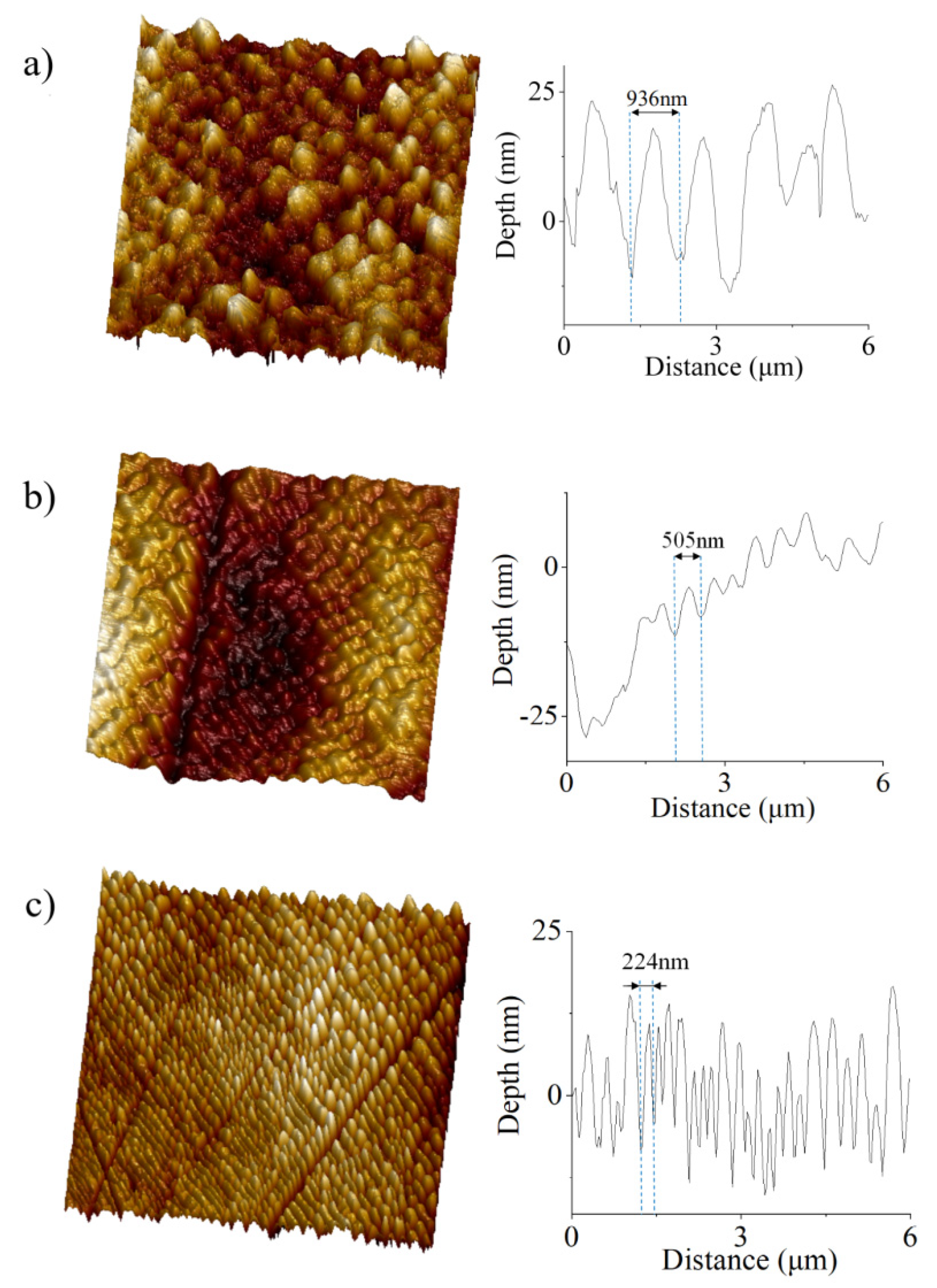
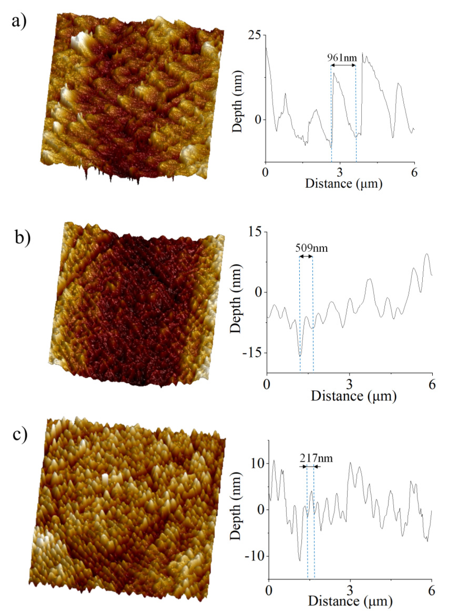
The AFM scanned several 10 μm × 10 μm square areas inside the ablation craters. The corresponding AFM images can be seen in Figure 2. Nanostructures with a uniform morphology have been always registered. The structures exhibit a spatial period decreasing from approximately 1 μm down to 250 nm when the fluence decreases from 230 mJ/cm2 down to 15 mJ/cm2. Figure 3 shows the results obtained at a reduced number of accumulated pulses, i.e., 12 pulses were applied in creating the ablation crater. The spatial periods of the nanostructures remained unchanged with respect to the irradiation by more pulses at the same fluence level. However, the surface inside the ablation crater created by a lower number of pulses shows a higher roughness in comparison to the surfaces processed by more pulses.
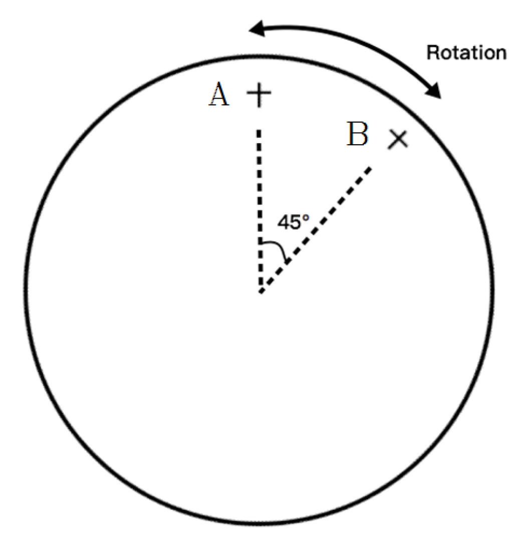
We may see in Figure 2 and Figure 3 that the nanostructures on BaF2 induced by a 46.9 nm laser have a specific orientation. To characterize the effect of the orientation, the BaF2 monocrystalline slab targets were ablated at two different positions with respect to the XUV-CDL beam. Figure 4 provides a sketch of the sample arrangement. First, in position A, the ablation pattern made by 25 laser pulses was obtained. Then, the sample was turned by 45 degrees into position B, where another ablation pattern was created by the same number of laser pulses under identical experimental conditions. Therefore, the only change in this experiment concerns the angle between the tangential plane of the focusing mirror and crystallographic planes of the single crystal of barium fluoride.
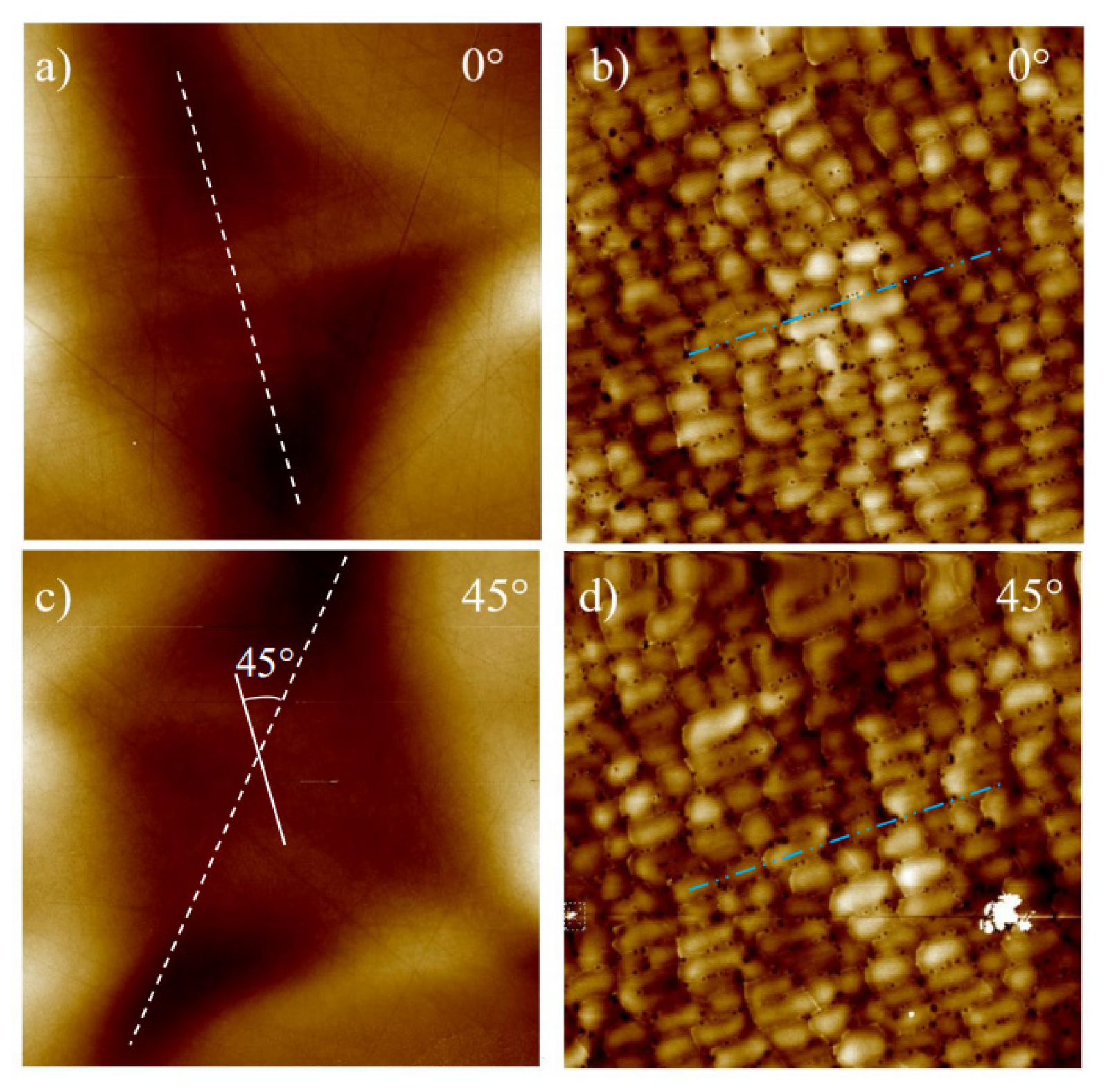
These two ablation patterns were subjected to AFM analysis without moving the sample (this is quite important, because the orientation of the structures observed in the AFM scanning area could change if the sample is moved). In Figure 5a,c, the overview images show ablation craters created in sample positions A and B, respectively. The shapes of these two patterns confirm that the focal spot was rotated by 45°. In Figure 5b,d, the ablated surfaces of samples A and B are visualized by AFM with a higher resolution. It can be clearly seen that, despite the rotation of the focal spot, the orientation of the nanostructures remains the same in both the sample positions. This means that the k-vector of the grating-like structure is coupled to the orientation of crystallographic planes in the single crystal.

3.2. Discussion and Outlook

Alkali earth halides—CaF2 more frequently than BaF2—have been exposed to intense ionizing and non-ionizing radiation many times. The main motivation of these studies lies in the study of damage to optical components and the manufacture of micro/nanostructures. Therefore, we may compare the characteristics of periodic nanostructures observed in our irradiation experiment with the surface damage patterns created under different irradiation conditions.
Grating-like structures (LIPSS) appeared on CaF2 and BaF2 surfaces exposed to picosecond [26] and femtosecond [27,28,29] long-wavelength laser pulses, respectively. In both cases, the periodicity of the ripples was comparable to the laser wavelength and the LIPSS grating vectors depended on the direction of the polarization vector of laser radiation. This contrasts with our finding, where it is the orientation of crystallographic planes which is related to the grating-vector direction. It seems that the crystallographic planes played an important role in the damage to CaF2 induced by nanosecond pulses of UV laser radiation [30,31]. The damage pattern shows linear edges oriented along the crystallographic planes. However, their spacing is at least by one order of magnitude greater than in the case of XUV-CDL irradiation and they often cross each other at certain angles while the lines of the XUV-LIPSS reported here are dominantly parallel. Even nanoparticles emitted from BaF2 irradiated by the nanosecond laser radiation often show a calcite crystal habitus and sharp edges [32]. This all indicates that laser-induced cleavage of alkali-earth halide crystals along the crystallographic planes can be observed in both the cases but with the long-wavelength laser radiation they have not been found to be as regular and uniform as observed on the XUV-CDL-illuminated surfaces in the present study.
Both long- and the short-wavelength radiation may create various defects in the crystal lattice of insulators [33] (similar effects are reported also in wide-gap semiconductors [34]) resulting in irreversible changes of illuminated surfaces. In CaF2, the formation of defects leading to surface metallization was induced by nanosecond pulses of 193-nm [35] and 157-nm [36] excimer laser radiation. However, the calcium-rich nanostructures show an oval shape not a linear one. CaF2 has also been irradiated at a wavelength of 46.9 nm [37] by a similar XUV-CDL source as utilized in the present study. However, only the ablation thresholds and the rates were measured. A detailed characterization of ablated areas has not been performed. As to ionizing radiation, the defect formation was induced in BaF2 and CaF2 by synchrotron radiation [38] and 850-eV [39] electrons, respectively. The results are similar to those obtained with intense UV radiation.
It is clearly visible that the periodic nanostructures observed in the XUV-CDL irradiation experiment differ from the morphology of all the damage patterns observed on alkali-earth-halide surfaces exposed to radiation of various kinds in numerous studies reported earlier [26,27,28,29,30,31,32,33,34,35,36,37,38,39]. We should keep in mind that an attenuation length of 46.9-nm XUV-CDL radiation in BaF2 is very short, i.e., only about 10 nm [40,41]. Thus, energy density gradients are enormous in the near-surface region of the irradiated crystal. Thus, unique thermo-mechanical processes (and their interplays with other phenomena) may occur in a very thin near-surface layer under these irradiation conditions. To distinguish between different possible mechanisms of XUV-LIPSS formation, further XUV-CDL interaction experiments performed in ultra-clean UHV environments are needed. They should be followed by an elemental sensitive imaging of the surface with high spatial resolution to reveal local changes of the Ba:F ratio. In this way, we might elucidate the possible role of crystal lattice defects. For testing the spallation/micro-cracking mechanism, different single crystal cuts should be irradiated on a wider variety of crystallographic plane orientations with respect to the laser beam. Interaction experiments with ultra-short pulses delivered by free-electron lasers tuned at a wavelength of 46.9-nm under different incident angles would help to verify the validity of various LIPSS formation theories.

4. Conclusions

A detailed characterization of periodic, grating-like nanostructures spontaneously formed on the surface of BaF2 single crystals irradiated by nanosecond pulses of 46.9-nm laser radiation was performed. The structures are highly abundant and uniform. Their spatial periods strongly depend on the fluence of the XUV-CDL radiation. An important finding is that their grating vector follows crystallographic planes when the sample is rotated by 45°. In brief, lines of the grating-like structure are also rotated by 45° as the sample rotates by 45°. Therefore, complex processes occurring in a near surface layer of the material associated with the crystallographic planes take place here rather than the standard LIPSS origination mechanism due to the interference of incoming laser light with the laser light scattered or diffracted on the ablating surface.

Author Contributions

Investigation, H.C.; methodology, A.F., J.S. (Jiri Schmidt), J.S. (Jaroslav Straus), T.B., J.C. and V.H.; supervision and interpretation of results, Y.Z., K.K. and L.J. All authors have read and agreed to the published version of the manuscript.

Funding

This work was performed under auspices and with a financial support of the Ministry of Education, Youth, and Sports of the Czech Republic (Grant No. LTT17015 and LTC20061), the Czech Science Foundation (Grant No. 20-08452S), the COST (European Cooperation in Science and Technology) Action TUMIEE (Grant. No. CA 17126), and the National Natural Science Foundation of China (Grant No. 62005066), and the National Natural Science Foundation of China (Grant No. 61875045).

Conflicts of Interest

The authors declare no conflict of interest. The funders had no role in the design of the study; in the collection, analyses, or interpretation of data; in the writing of the manuscript, or in the decision to publish the results.

References

  1. Rocca, J.J. Table-top soft X-ray lasers. Rev. Sci. Instrum. 1999, 70, 3799–3827. [Google Scholar] [CrossRef]
  2. Nejdl, J.; Howlett, I.D.; Carlton, D.; Anderson, E.H.; Chao, W.; Marconi, M.C.; Rocca, J.J.; Menoni, C.S. Image plane holographic microscopy with a table-top soft X-ray laser. IEEE Photonics J. 2015, 7, 1–8. [Google Scholar] [CrossRef]
  3. Sandberg, R.L.; Song, C.; Wachulak, P.W.; Raymondson, D.A.; Paul, A.; Amirbekian, B.; Lee, E.; Sakdinawat, A.E.; La-O-Vorakiat, C.; Marconi, M.C.; et al. High numerical aperture tabletop soft x-ray diffraction microscopy with 70-nm resolution. Proc. Natl. Acad. Sci. USA 2008, 105, 24–27. [Google Scholar] [CrossRef] [PubMed] [Green Version]
  4. Juha, L.; Bittner, M.; Chvostova, D.; Krasa, J.; Otcenasek, Z.; Präg, A.G.; Ullschmied, J.; Pientka, Z.; Krzywinski, J.; Pelka, J.B.; et al. Ablation of organic polymers by 46.9-nm laser radiation. Appl. Phys. Lett. 2005, 86, 034109. [Google Scholar] [CrossRef] [Green Version]
  5. Kolacek, K.; Schmidt, J.; Straus, J.; Frolov, O.; Juha, L.; Chalupsky, J. Interaction of extreme ultraviolet laser radiation with solid surface: Ablation, desorption, nanostructuring. In Proceedings of the SPIE 9255, XX International Symposium on High Power Laser Systems and Applications 2014, Chengdu, China, 3 February 2015; p. 92553U. [Google Scholar]
  6. Vaschenko, G.; Etxarri, A.G.; Menoni, C.S.; Rocca, J.J. Nanometer-scale ablation with a table-top soft X-ray laser. Opt. Lett. 2006, 31, 3615–3617. [Google Scholar] [CrossRef] [PubMed] [Green Version]
  7. Kolacek, K.; Schmidt, J.; Straus, J.; Frolov, O.; Prukner, V.; Melich, R.; Psota, P. Spontaneous and artificial direct nanostructuring of solid surface by extreme ultraviolet laser with nanosecond pulses. Laser Part. Beams 2016, 34, 11–22. [Google Scholar] [CrossRef]
  8. Capeluto, M.G.; Vaschenko, G.; Grisham, M.; Marconi, M.; Luduena, S.; Pietrasanta, L.; Lu, Y.; Parkinson, B.; Menoni, C.; Rocca, J.J. Nanopatterning with interferometric lithography using a compact λ=46.9-nm laser. IEEE Trans. Nanotechnol. 2006, 5, 3–7. [Google Scholar] [CrossRef]
  9. Wachulak, P.W.; Capeluto, M.G.; Marconi, M.C.; Patel, D.; Menoni, C.S.; Rocca, J.J. Nanoscale patterning in high resolution HSQ photoresist by interferometric lithography with tabletop extreme ultraviolet lasers. J. Vac. Sci. Technol. B 2007, 25, 2094–2097. [Google Scholar] [CrossRef]
  10. Wachulak, P.; Grisham, M.; Heinbuch, S.; Martz, D.; Rockward, W.; Hill, D.; Rocca, J.J.; Menoni, C.S.; Anderson, E.; Marconi, M. Interferometric lithography with an amplitude division interferometer and a desktop extreme ultraviolet laser. J. Opt. Soc. Am. B 2008, 25, B104–B107. [Google Scholar] [CrossRef] [Green Version]
  11. Birnbaum, M. Semiconductor surface damage produced by ruby lasers. J. Appl. Phys. 1965, 36, 3688–3689. [Google Scholar] [CrossRef]
  12. Sipe, J.E.; Young, J.F.; Preston, J.S.; Van Driel, H.M. Laser-induced periodic surface structure. I. Theory. Phys. Rev. B 1983, 27, 1141–1154. [Google Scholar] [CrossRef]
  13. Tousey, R. XUV—The extreme ultraviolet. J. Opt. Soc. Am. 1962, 52, 1186–1187. [Google Scholar] [CrossRef]
  14. Bakshi, V. (Ed.) EUV Lithography; SPIE Press-Wiley Interscience: Bellingham, NY, USA, 2009. [Google Scholar]
  15. Steeg, B.; Juha, L.; Feldhaus, J.; Jacobi, S.; Sobierajski, R.; Michaelsen, C.; Andrejczuk, A.; Krzywinski, J. Total reflection amorphous carbon mirrors for vacuum ultraviolet free electron lasers. Appl. Phys. Lett. 2004, 84, 657–659. [Google Scholar] [CrossRef]
  16. Juha, L.; Bittner, M.; Chvostova, D.; Krasa, J.; Kozlov, M.; Pfeifer, M.; Polan, J.; Präg, A.R.; Rus, B.; Stupka, M.; et al. Short-wavelength ablation of molecular solids: Pulse duration and wavelength effects. J. Microlithogr. Microfabr. Microsyst. 2005, 4, 033007. [Google Scholar] [CrossRef] [Green Version]
  17. Hahn, D. Calcium fluoride and barium fluoride crystals in optics: Multispectral optical materials for a wide spectrum of applications. Opt. Photonik 2014, 9, 45–48. [Google Scholar] [CrossRef]
  18. Wels, A.F. Structural Inorganic Chemistry, 3rd ed.; Clarendon Press: Oxford, UK, 1962; p. 337. [Google Scholar]
  19. Zhao, Y.; Cui, H.; Zhang, S.; Zhang, W.; Li, W. Formation of nanostructures induced by capillary-discharge soft X-ray laser on BaF2 surfaces. Appl. Surf. Sci. 2017, 396, 1201–1205. [Google Scholar] [CrossRef]
  20. Kolacek, K.; Schmidt, J.; Straus, J.; Frolov, O. Calibration of windowless photodiode for extreme ultraviolet pulse energy measurement. Appl. Opt. 2015, 54, 10454–10459. [Google Scholar] [CrossRef]
  21. Schmidt, J.; Kolacek, K.; Frolov, O.; Straus, J.; Hoffer, P.; Stelmashuk, V.; Tuholukov, A.; Jiricek, P.; Houdkova, J. Long-term changes in Al thin-film extreme ultraviolet filters. Appl. Opt. 2021, 60, 8766. [Google Scholar] [CrossRef]
  22. Chalupsky, J.; Bohacek, P.; Hajkova, V.; Hau-Riege, S.P.; Heimann, P.A.; Juha, L.; Krzywinski, J.; Messerschmidt, M.; Moeller, S.P.; Nagler, B.; et al. Comparing different approaches to characterization of focused X-ray laser beams. Nucl. Instrum. Methods Phys. Res. A 2011, 631, 130–133. [Google Scholar] [CrossRef]
  23. Gerasimova, N.; Dziarzhytski, S.; Weigelt, H.; Chalupský, J.; Hájková, V.; Vysin, L.; Juha, L. In situ focus characterization by ablation technique to enable optics alignment at an XUV FEL source. Rev. Sci. Instrum. 2013, 84, 65104. [Google Scholar] [CrossRef] [Green Version]
  24. Kolacek, K.; Schmidt, J.; Straus, J.; Frolov, O.; Prukner, V.; Melich, R.; Choukourov, A. A new method of determination of ablation threshold contour in the spot of focused XUV laser beam of nanosecond duration. In Proceedings of the SPIE 777, Damage to VUV, EUV, and X-ray Optics IV; and EUV and X-ray Optics: Synergy between Laboratory and Space II, Prague, Czech Republic, 3 May 2013; p. 87770N. [Google Scholar]
  25. Chalupský, J.; Krzywinski, J.; Juha, L.; Hájková, V.; Cihelka, J.; Burian, T.; Vyšín, L.; Gaudin, J.; Gleeson, A.; Jurek, M.; et al. Spot size characterization of focused non-Gaussian X-ray laser beams. Opt. Express 2010, 18, 27836–27845. [Google Scholar] [CrossRef]
  26. Lee, H. Picosecond mid-IR laser induced surface damage on gallium phosphate (GaP) and calcium fluoride (CaF2). J. Mech. Sci. Technol. 2007, 21, 1077–1082. [Google Scholar] [CrossRef]
  27. Costache, F.; Henyk, M.; Reif, J. Modification of dielectric surfaces with ultra-short laser pulses. Appl. Surf. Sci. 2002, 186, 352–357. [Google Scholar] [CrossRef]
  28. Reif, J.; Costache, F.; Henyk, M.; Pandelov, S.V. Ripples revisited: Non-classical morphology at the bottom of femtosecond laser ablation craters in transparent dielectrics. Appl. Surf. Sci. 2002, 197–198, 891–895. [Google Scholar] [CrossRef]
  29. Costache, F.; Henyk, M.; Reif, J. Surface patterning on insulators upon femtosecond laser ablation. Appl. Surf. Sci. 2003, 208–209, 486–491. [Google Scholar] [CrossRef]
  30. Sils, J.; Reichling, M.; Matthias, E.; Johansen, H. Laser damage and ablation of differently prepared CaF2(111) surfaces. Czechoslov. J. Phys. 1999, 49, 1737–1742. [Google Scholar] [CrossRef]
  31. Reichling, M. Laser ablation in optical components and thin films. Exp. Methods Phys. Sci. 1997, 573–624. [Google Scholar] [CrossRef]
  32. Moses, L.M.; Farnsworth, P.B. Evaluation of particle size distributions produced during ultra-violet nanosecond laser ablation and their relative contributions to ion densities in the inductively coupled plasma. Spectrochim. Acta B 2015, 113, 54–62. [Google Scholar] [CrossRef] [Green Version]
  33. Gao, S.; Duan, Y.Z.; Tian, Z.N.; Zhang, Y.L.; Chen, Q.D.; Gao, B.R.; Sun, H.B. Laser-induced color centers in crystals. Opt. Laser Technol. 2022, 146, 107527. [Google Scholar] [CrossRef]
  34. Saleem, U.; Birowosuto, M.D.; Hou, S.; Maurice, A.; Kang, T.B.; Teo, E.H.T.; Tchernycheva, M.; Gogneau, N.; Wang, H. Light emission from localised point defects induced in GaN crystal by femtosecond-pulsed laser. Opt. Mater. Express 2011, 8, 2703–2712. [Google Scholar] [CrossRef]
  35. Rix, S.; Natura, U.; Loske, F.; Letz, M.; Felser, C.; Reichling, M. Formation of metallic colloids in CaF2 by intense ultraviolet light. Appl. Phys. Lett. 2011, 99, 261909. [Google Scholar] [CrossRef]
  36. Cramer, L.P.; Langford, S.C.; Dickinson, J.T. The formation of metallic nanoparticles in single crystal CaF2 under 157 nm excimer laser irradiation. J. Appl. Phys. 2006, 99, 054305. [Google Scholar] [CrossRef]
  37. Ritucci, A.; Tomassetti, G.; Reale, A.; Arrizza, L.; Zuppella, P.; Reale, L.; Palladino, L.; Flora, F.; Bonfigli, F.; Faenov, A.; et al. Damage and ablation of large bandgap dielectrics induced by a 46.9 nm laser beam. Opt. Lett. 2006, 31, 68–70. [Google Scholar] [CrossRef] [Green Version]
  38. Watanabe, M.; Azuma, J.; Asaka, S.; Tsujibayashi, T.; Arimoto, O.; Nakanishi, S.; Itoh, H.; Kamada, M. Photostimulated detection of defect formation in BaF2 under irradiation of synchrotron radiation. Phys. Status Solidi (b) 2012, 250, 396–401. [Google Scholar] [CrossRef]
  39. Bennewitz, R.; Smith, D.; Reichling, M. Bulk and surface processes in low-energy-electron-induced decomposition of CaF2. Phys. Rev. B 1999, 59, 8237–8246. [Google Scholar] [CrossRef]
  40. Henke, B.L.; Gullikson, E.M.; Davis, J.C. X-ray interactions: Photoabsorption, scattering, transmission, and reflection at E = 50–30,000 eV, Z = 1–92. At. Data Nucl. Data Tables 1993, 54, 181–342. [Google Scholar] [CrossRef] [Green Version]
  41. X-ray Interactions with Matter. Available online: http://henke.lbl.gov/optical_constants/ (accessed on 15 September 2021).
Figure 1. Ablation patterns on the BaF2 sample exposed to 25 CDL pulses at fluences of (a) 230 mJ/cm2, (b) 30 mJ/cm2 and (c) 15 mJ/cm2.
Applsci 12 01251 g001
Figure 2. Nanostructures on BaF2 induced by 25 laser pulses with fluences of (a) 230 mJ/cm2, (b) 30 mJ/cm2 and (c) 15 mJ/cm2.
Applsci 12 01251 g002
Figure 3. Nanostructures on BaF2 surfaces exposed to 12 XUV-CDL pulses at fluences of (a) 230 mJ/cm2, (b) 30 mJ/cm2 and (c) 15 mJ/cm2.
Applsci 12 01251 g003
Figure 4. Schematic diagram of the sample rotation.
Applsci 12 01251 g004
Figure 5. AFM images of ablation craters (a,c) created at a different orientation of crystallographic planes with respect to the tangential plane of the focusing mirror—positions A and B in Figure 4; (b,d) are highly-resolved AFM images measured inside these two craters.
Applsci 12 01251 g005
Publisher’s Note: MDPI stays neutral with regard to jurisdictional claims in published maps and institutional affiliations.