Article

Skeletal, Dental, and Soft Tissue Changes after Applying Active Skeletonized Sutural Distractor (ASSD) for Class III Malocclusion Patients

by
Rozita Hassan
1,
Wael Ahmed Bayomy Mohamed
1,
Norma Ab. Rahman
1,*,
Shaifulizan Ab. Rahman
2 and
Norkhafizah Saddki
3
1
Orthodontic Unit, School of Dental Sciences, Universiti Sains Malaysia, Kubang Kerian 16150, Kelantan, Malaysia
2
Oral and Maxillofacial Surgery Unit, School of Dental Sciences, Universiti Sains Malaysia, Kubang Kerian 16150, Kelantan, Malaysia
3
Dental Public Health Unit, School of Dental Sciences, Universiti Sains Malaysia, Kubang Kerian 16150, Kelantan, Malaysia
*
Author to whom correspondence should be addressed.
Appl. Sci. 2022, 12(3), 1233; https://doi.org/10.3390/app12031233
Submission received: 18 November 2021 / Revised: 19 December 2021 / Accepted: 22 December 2021 / Published: 25 January 2022

Abstract

:
Background: Nonsurgical treatment of Class III malocclusion may reduce the chances for orthognathic surgery in the future. This study aimed to determine the post-treatment skeletal, dental and soft tissue changes in the anteroposterior plane applying active skeletonized sutural distractor (ASSD) in late adolescent Class III malocclusion patients. Materials and Methods: After screening 13,500 students from 18 secondary schools, a sample of 26 subjects was recruited for this study, with a mean age of 15(±1.4) years after considering the inclusion and exclusion criteria. ASSD appliances were inserted into patients, and lateral cephalometric x-rays were taken for pre- and post-treatment analysis. Dental casts of the patients were also taken and scanned using a 3D laser scanner for comparisons. Results: The intraclass correlation coefficients (ICC) for inter-examiner were 0.87 to 0.99 and 0.96 to 0.99 for lateral cephalometric and dental cast measurements, respectively. The difference in pre- and post-treatment of cephalometric measurements of the mandible showed statistically significant changes for mandibular plane angle (p < 0.001) and SNB angle (p = 0.029). The dental cast measurements showed statistically significant changes of inter-molars width (p < 0.001) as well as palatal (p = 0.002). Conclusions: The results showed statistically significant improvement in skeletal, dental, and soft tissue changes after treatment applying ASSD for Class III malocclusion patients.

1. Introduction

Class III malocclusion is caused by either the deficient growth of the maxilla in the downward and forward direction or excessive growth of the mandible and is considered one of the most difficult cases in orthodontics. Nonsurgical treatment of Class III malocclusion remains a challenge. Current literature on the global prevalence varies significantly among different races, ethnic groups, and geographic regions.
In a study conducted by Soh et al. [1] to evaluate the occlusal status of an Asian population in Malaysia among the sample of male army recruits (N 339, age 17–22 years), they found that the highest prevalence of Class III malocclusion was in Malay 26.67% followed by Chinese 22.87% and the Indian was the least with 4.76%.
Orthopedic advancement of the maxilla was not successful in adolescent patients and should be accomplished as early as possible before the patient become older than 10 years [2]. Its effect is limited, although there were some case reports that proved that face masks can still have favorable treatment effects when the patient is over 15 years [3,4]. Nowadays, there are mainly three treatment approaches that can provide successful treatment for Class III malocclusion patients, which involves the use of skeletal anchorage, sutural distraction, and alternate rapid maxillary expansion and contraction [5]. The surgical intervention or orthognathic surgery for correction of maxillary hypoplasia by surgical advancement of the maxillary bone with a Le Fort I osteotomy only can be carried out after the growth had completed. This approach provides a rapid solution that requests limited patient cooperation, direct access for bony correction, treatment of the cross-bite, and makes significant facial morphological changes without any unfavorable dental tipping, which is often accompanied with conventional orthopedic treatment [6]. The surgical approach has some disadvantages, including the need to delay surgery until growth ceased causing patients suffering from the unaesthetic appearance until an older age, expensive and invasive with a time recovery period from 6 to 8 weeks and always be associated with the patients’ discomfort, soft diet, facial swelling, transient paresthesia, and sometimes permanent loss of neuro sensation [7].
It was reported that orthopedic protraction of the maxilla is limited in adolescent patients, and this treatment should only be accomplished at a younger age [2]. Thus, active skeletonized sutural distractor (ASSD) appliance was designed to take advantage of these treatment methods and avoid their limitations.
The objective for this study was to determine the post-treatment skeletal, dental and soft tissue changes in the anteroposterior plane applying ASSD in late adolescent Class III malocclusion patients using lateral cephalometric radiograph.

2. Materials and Methods

After screening 13,500 students from 18 secondary schools, a sample of 26 subjects was recruited for this study, with a mean age of 15(±1.4) years. The ethical approval was obtained from the committee (USM/JEPeM/15120548).
The inclusion criteria for this study were patients who are in late adolescence, which corresponds to the age range between 12 years and 6 months to 16 years in girls and 14 years and 6 months to 17 years in boys [8], angle Class III malocclusion, and skeletal Class III malocclusion with maxillary retrognathism (Wits appraisal is-4 mm or less) and horizontal changes of maxilla at A point: A-N perp is −3.83 mm (+/−2.97) or less [9] with or without mandibular protrusion.

2.1. Applications of the ASSD Appliance

The bands for upper and lower first permanent molars in both sides were fitted, then an alginate impression was taken and poured into stone plaster for fabrication of the working model.
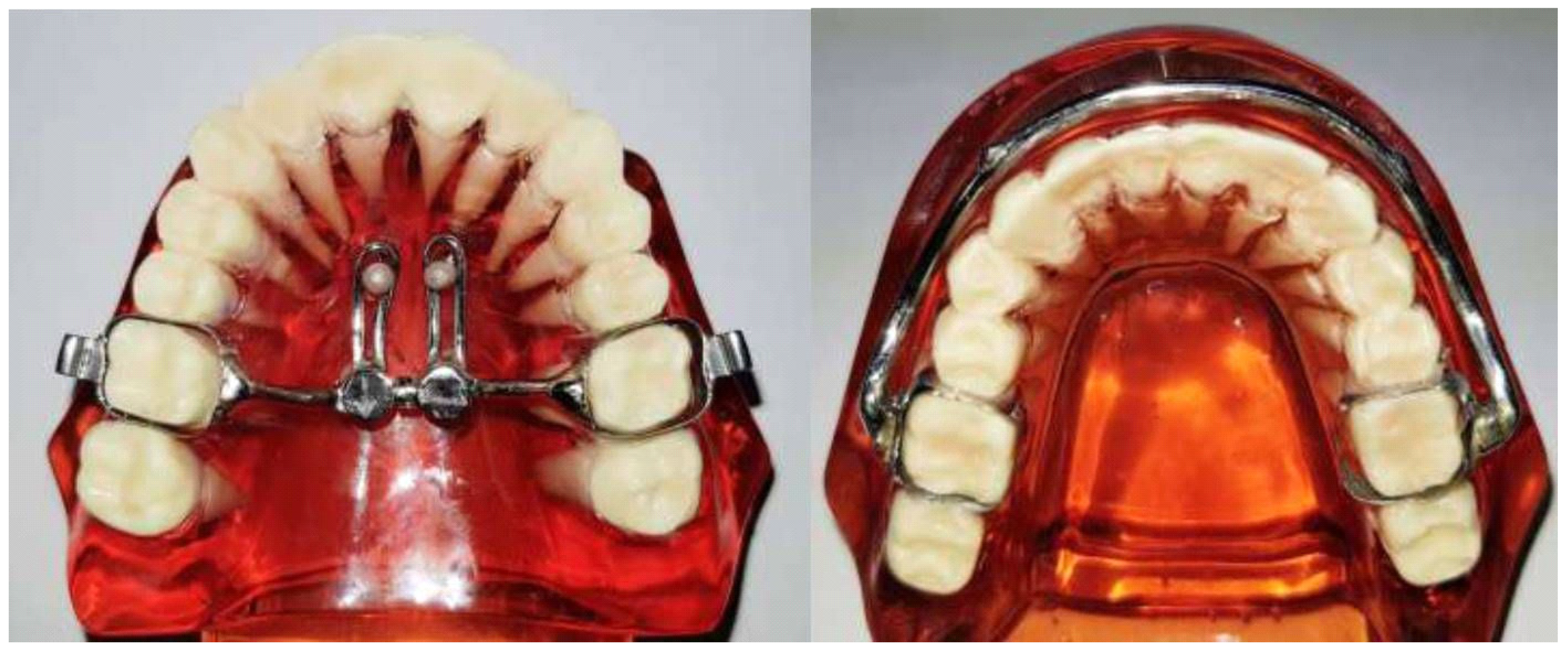
Fabrication of the upper and lower parts of the ASSD (Figure 1).
In the upper jaw, two palatal arms made from 2 mm stainless-steel wire were soldered to the expansion mini-screw, as well as two 1.2 mm stainless-steel wire teardrop loops that are used for insertion of two anterior mini implants at the level of the first premolar connecting it to the upper appliance.
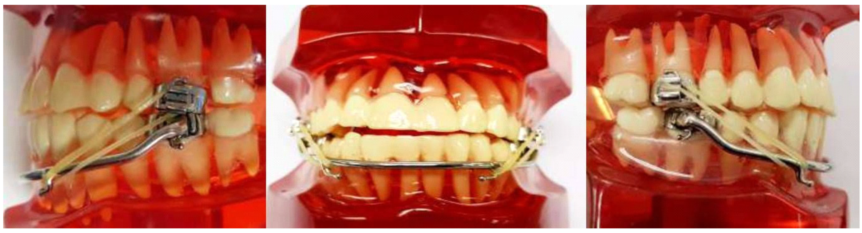
In the lower jaw, a buccal arch made of 2 mm stainless-steel wire was soldered to the first molars band on both sides. Two buccal stainless-steel hooks solder to the buccal arch at the level of the lower canines bilaterally for the attachment of the Class III intraoral elastics (Figure 2).
Insertion of the appliance.
Insertion of the appliance in the patient’s mouth was performed by bonding it to the teeth by glass ionomer cement (RMGI Cements, 3535TK, 3M, Monrovia, CA, USA). After cementation of the appliance, the mini-implant was inserted at the distal end of the two teardrop loops and passively fit over it, and the expansion screw was opened by 1 mm by the operator. The mini-implant was inserted according to the manufacturer’s instructions using the Cope placement protocol. This protocol is considered the least invasive protocol because, in most cases, there is no need for local anesthesia or flap incision or predrilling.
Two mini implants were inserted about 3 mm away from the mid-palatal suture. In the anterior palate bilaterally (1.8 mm width and 10 mm length) at the level of the second premolars and about 8 mm posterior to the incisive foramen, in a position that coincides with the plane of maximum resistance of the maxilla and near to the maxillary center of resistance. The length of the mini-implant was determined according to a previous study [10,11]. The mobility of the mini-implant was checked with the application of 500 gm of force determined by force gauge (Correx, Orthocare, U.K.), where intraoral elastics (3M, Bummer (heavy) 6oz, 3M, Monrovia, USA) were attached in a direction nearly perpendicular to the mini-implant. This test was made at the start of treatment and every month until the end of the active treatment period [12].
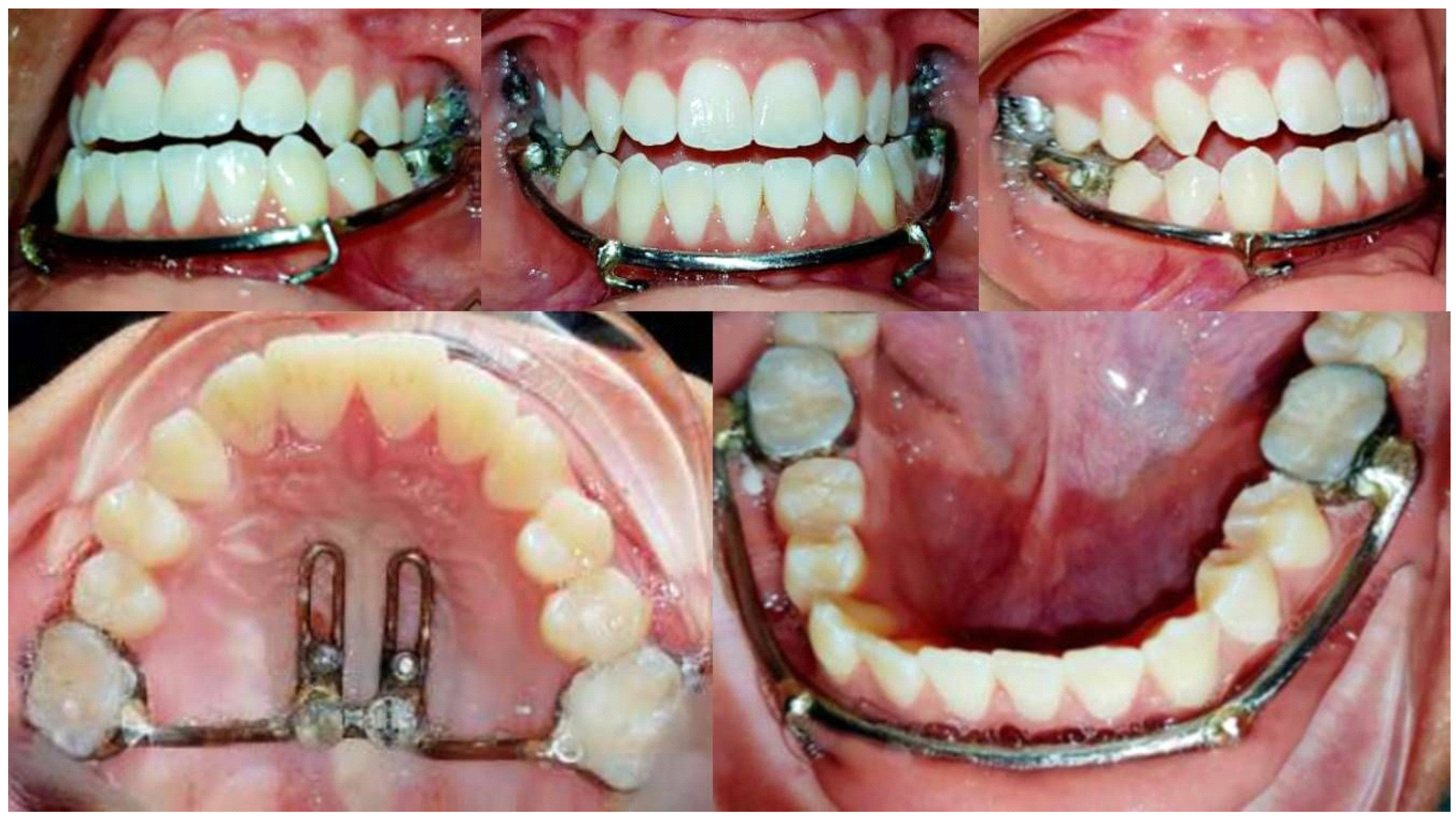
After 24 h from the fixation of the ASSD in the patient mouth (Figure 3), the patient was instructed to perform the alternate rapid maxillary expansion and contraction (Alt.RAMEC) according to the schedule given starting from 4 turns/day of open and close the expansion screw, which corresponds to 1 mm until reaching 28 turn/day, which corresponds to 7 mm until end of the active treatment period [6].
After two weeks of the start of treatment, the patient was asked to wear intraoral elastics (3M, Bummer (heavy) 6oz, 3M, Monrovia, USA), which exerts protraction forces of about 500 gm in each side that was attached to the upper and lower part of the appliance [13]. To decrease the counterclockwise rotation of the upper occlusal plane, the elastic oriented with a 15–30 degree angle to the occlusal plane [9]. The oral hygiene of the patients was monitored throughout the treatment.

2.2. Lateral Cephalometric Analysis

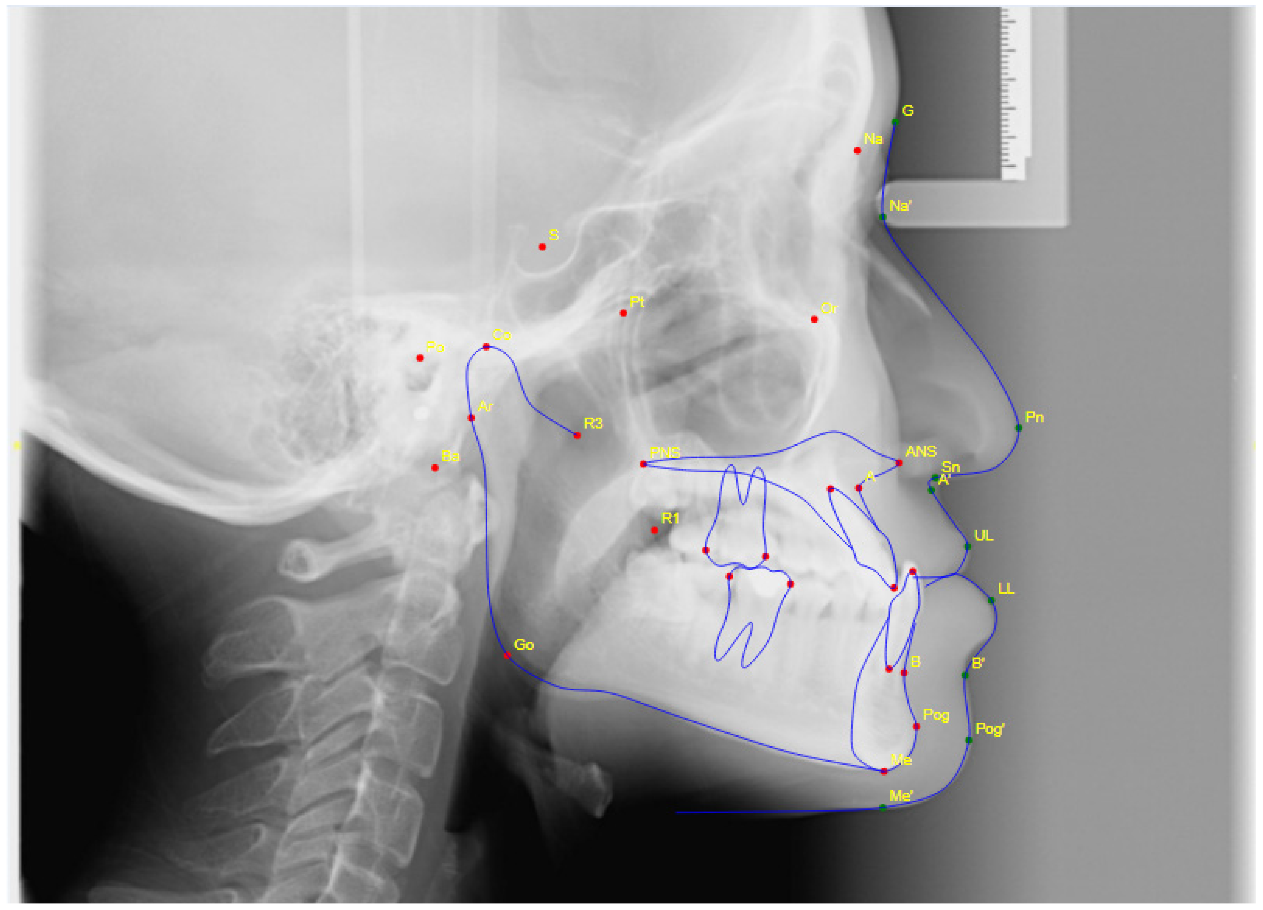
The cephalometric X-ray was taken using lateral cephalometric machine Gendex Orthoralix, Italy, at initial observation (T1) before the start of treatment and (T2) at the end of active treatment. Cephalometric tracing was performed using Planmeca Romexis Software, which is considered a reliable tool for lateral cephalometric measurements [14] and analyzed by a single operator. Tracing and measurements were performed on pre- and post-treatment cephalograms to measure the efficacy of treatment (skeletal, dental, and soft tissue changes) as shown in Figure 4 according to variables in Table 1 [15].

2.3. Upper Study Model Analysis

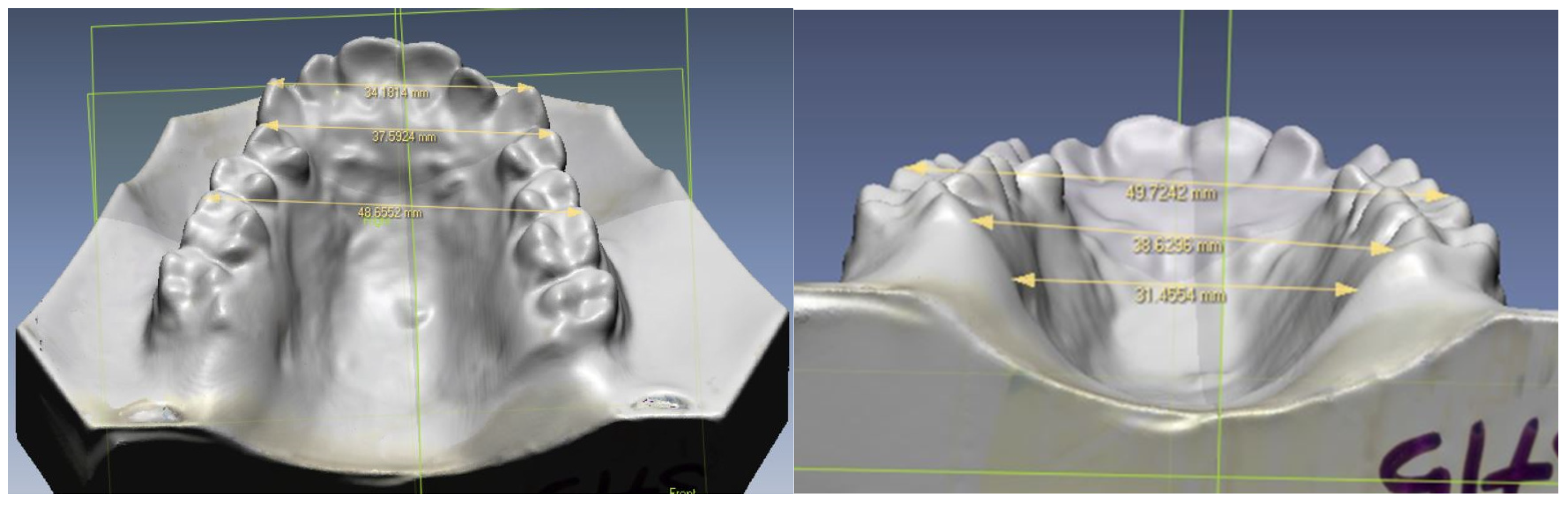
This step was performed using a 3D laser scanner (Next Engine Inc., Model 2020i, Santa Monica, CA, USA) for pre-and post-dental models. Dental casts of the subjects were obtained from alginate impressions before expansion (D1), before start of treatment and after expansion (D2), at the end of treatment. After image acquisition (Figure 5), the landmarks (Table 2) [16] are digitized, and the transverse measurements were recorded. The variables used per identification and measurements were inter-canine widths (ICW), inter-1st premolar widths (IPW), inter-first molar widths (IMW), and palatal width (PW).
All scanned data coordinates (in x, y, z) are transferred into ScanStudio HD software in DXF format to be measured. Spatial linear measurements are measured with a digital caliper (using computer mouse) accurate to 0.5 mm, and the average value is used. For the equipment calibration, the 3D laser scanner and software were autocalibated from time to time, and manual calibration was repeated by comparing the value with the direct measurement from a few study casts to ensure accuracy and reliability. The accuracy of the measurements from a 3D laser scanner is up to 0.05 [17].

2.4. Error Measurement

The reliability of the cephalometric and dental cast measurements was tested by repeating the measurements on randomly selected subjects (20% of total sample size) by two examiners. The reliability of the method was analyzed using Dahlberg’s formula [18].

3. Results

The socio-demographic characteristics for all subjects are shown in Table 3. The study group consisted of 26 children treated with ASSD appliance (11 males, 15 females) with a mean age of 15(±1.4) years. The chief complain of them was mostly from both more forward position of the lower jaw, and more backward position of the upper jaw 39.3% followed by 35.7% of them complained from a more forward position of the lower jaw, and the rest of them complained of more backward position of the upper jaw 25%. The family history of Class III malocclusion was present in 38.5% of them, while 61.5% have not.
Table 4 shows the reproducibility of lateral cephalogram measurements. The result showed that the intraclass correlation coefficients for inter-examiner were 0.87 to 0.99. These values of coefficients were considered suitable.
Table 5 shows the reproducibility of study model measurements. The result showed that the intraclass correlation coefficients for inter-examiner were 0.96 to 0.99. These values of coefficients were considered suitable.

3.1. Skeletal, Dental and Soft Tissue Changes after Treatment with ASSD Appliance

Table 6 shows the difference in pretreatment (T1) and post-treatment (T2) of cephalometric measurements. The results showed that there was an improvement in skeletal, dental, and soft tissue measurements after treatment with the ASSD appliance. The skeletal measurements showed statistically significant changes in the maxilla and the mandible that favor correction of Class III malocclusion. The maxillary position became more forward as the SNA angle increased by 2.73° (p < 0.001), maxillary length (Co-A) increased by 4.62 mm (p < 0.001), and forward movement of A point (A-NB) by 3.69 mm (p < 0.001).
Statistically significant changes were also seen in the mandible. Backward rotation of the mandible manifested by a significant increase in mandibular plane angle (MP-SN) by 2.35° (p < 0.001). In addition to that, the mandible showed a more backward position, as indicated by a significant decrease in SNB angle by 0.71° (p = 0.029).
The inter-arch relationship was also improved shown in Wits appraisal value (AO-BO) by 6.46 mm (p < 0.001), ANB angle by 3.29° (p < 0.001). Besides that, the lower anterior facial height (ANS-Gn) showed an increase of 1.16 mm (p = 0.007). The dental relation was also improved where clinically the overjet was increased. However, the inclination of the upper incisors reduced by 0.19°, and the lower incisors inclination increased by 2.67°.
The soft tissue profile was also showed an improvement where the upper lip thickness (Ls-NB) increased significantly by 2.08 mm (p < 0.001), as well as increased protrusion of the upper lip (UL-EL) by 1.87 mm (p < 0.001). On the contrary, a few unfavorable changes were also noticed, such as increase in mandibular length (Co-Gn) by 1.92 mm (p = 0.004), lower incisor inclination by 2.67° (p = 0.008), and more protrusive position of the lower lip (LL-EL) by 1 mm (p = 0.004).

3.2. Morphological Changes of the Palate and Maxillary Dental Arch after Treatment with ASSD Appliance

Table 7 shows the difference in pretreatment (T1) and post-treatment (T2) transverse variables. The result showed that there were statistically significant changes of inter-molars width (IMW), which was significantly increased after treatment by 5.38 mm (p < 0.001) as well as palatal width (PW) by 0.92 mm (p = 0.002).

4. Discussion

The study was conducted to measure the effectiveness of ASSD appliances in treating severe Class III malocclusion cases that were indicated for surgical treatment. There were some unique characteristics to this study. First, the sample of the study was collected from 18 secondary schools by a screening of 13,500 students. It was considered a very big number in comparison to similar studies, which collect smaller sample sizes from the outpatient clinic, not by screening of the target population. In addition, many studies were carried out to investigate the effects of using rigid skeletal anchorage, Alt.RAMEC and sutural distraction method for treatment of Class III malocclusion, but as far as we are aware, no study had been performed to investigate the effect of combining these three approaches.
Furthermore, we innovated a unique design for the ASSD appliance, which involves a small size maxillary expander that is not presented in any other similar studies making the appliance suitable for any patients who have different palatal shape and height. Next, the effects on the craniofacial skeleton induced by facemask therapy have seldom been investigated in adequate samples, especially in late adolescent subjects, as evident in this study [19].
The result of this study showed that the ASSD was effective in the treatment of Class III malocclusion in late adolescent subjects. Although most of the subjects showed improvement of the Class III malocclusion, the amount of the correction varied greatly among patients, as did their level of cooperation.
Assessment of the results demonstrated significant changes in the maxilla as well as in the mandible that favored correction of Class III malocclusion. The maxilla became more forward due to a significant increase in the maxillary position (SNA), increase in maxillary length (CO-A), and forward movement of A point (A-N perp).
The skeletal point A forward changes by 3.69 mm revealed that maxillary growth was achieved in the ASSD treatment group and that the ASSD effectively facilitated skeletal growth of the maxilla. In spite of differences between ASSD treatment protocols and the other similar studies, a common trend was observed in similar studies for A-point advancement in the horizontal plane as a result of bone-anchored orthopedic treatment.
Morales-Fernandez et al. [20] compared the results of conventional orthopedic appliances with those obtained from recent bone-anchored orthopedics for Class III malocclusion treatment. In those studies, using skeletal anchorage, there was forward movement in the horizontal plane with a mean A-point advancement of 5.2 mm [21,22,23].
In addition, the maxilla became more forward due to an increase in the maxillary position (SNA) by 2.73°, similar to the finding of Nienkemper et al. [24] by 2.4° as well as Katyal et al. [25] by 2° with more increments. These differences may be attributed to the differences in the subject’s characteristics, treatment protocol, and treatment durations (14 months). In addition to that, an increase in maxillary length (Co-A) was significant as well (4.62 mm), and this was in agreement with [6,24,25].
The mandible has a backward rotation manifested by a significant increase in mandibular plane angle (MP/SN) and SNB value. The lower anterior facial height (ANS-Gn) showed a significant increase as a reflection of the backward rotation of the mandible.
The negative change in SNB in the ASSD group shared similar results with previous studies [3,4,21] and was lower than that reported by Nienkemper et al. [24] by 1.7°. This difference may be attributed to limited downward decedents of the maxilla due to using of the skeletal anchorage and hence lower backward rotation of the mandible and lower changes in the SNB angle. The negative numerical value change in SNB indicated that mandible growth is limited and that the forward change at the B point is controlled.
The smaller increase in mandibular plane angle (MP/SN) by 2.35°, occlusal plane angle (OP/SN) by 1.46°, and the lower anterior facial height (ANS-Gn) by 1.6 mm reflected the limited effects of ASSD on increasing the vertical dimension, which is primarily important in treating patients having Class III malocclusion with increased lower facial height. This finding was similar to the finding reported in other studies [6,25].
The inter-arch relationship was also improved as there was an improvement in Wits appraisal value (AO-BO) as well as ANB value. The improvement in Wits appraisal value (AO-BO) was 6.4 mm, and this was much more than the finding of other studies [24,25], where the changes were 4.5 and 3.4 mm, respectively. Moreover, the ANB value improved by 3.29°, which was more than the finding of Nienkemper et al. [24] as well as Katyal et al. [25] as it was 1.9° and 2.4° respectively.
These differences in the improvement of the Wits appraisal as well as the ANB values may be attributed to the differences in the treatment protocol used in the present study from that used in the other studies. As in both studies performed by Nienkemper et al. [24] and Katyal et al. [25], the improvement was lesser than in the present study because they used only skeletal anchorage without performing the repetitive cycles of Alt.RAMEC.
Reports of maxillary orthopedic protraction using skeletal anchorage and intraoral forces are rare in contemporary orthodontic literature. Three-dimensional imaging studies by De Clerck et al. [21] showed that bone-anchored maxillary protraction stimulated forward displacement and modeling of the maxillary and zygomatic bones as well as affecting mandibular shape.
It was evident that the ASSD group in the present study and those of De Clerck et al. [23] were similar in relation to the pretreatment Wits value. However, age at commencement was older in the ASSD group than that in the De Clerck et al. [21] study as the age of the subjects in the present study was in the post-adolescent stage of growth while the subjects were in the pre-pubertal stage.
It was interesting to note that a slightly greater improvement in Wits value of 6.4 mm was found in the present study than that found by De Clerck et al. [21] 4 mm at the end of treatment. It may be argued that the difference in Wits improvement between the two studies is valuable and could be attributed to the unique protocol used in the present study that combined three different treatment approaches in one appliance.
These differences also could have been due to variations in the sample size, demographics, compliance of the patients, or the measurements obtained on three-dimensional images compared with two-dimensional imaging. An advantage of the ASSD protocol over the protocol used by De Clerck et al. [21] and Katyal et al. [25] is that we could only obtain the skeletal anchorage by inserting two mini-screws in the palate and avoiding the invasive surgical placement and removal of the surgical mini-plates that were used with these treatment protocols (four and two mini-plates, respectively).
The dental relation showed a great improvement in increasing overjet clinically due to the improvement of the skeletal relations of both maxilla and mandible. This finding is also supported and is in line with Stephen, 2011 [6] and Katyal et al. 2016 [25]. Dental compensation was recorded due to the lip bumper effect of the lower labial arch, and spacing was noticed between incisors and/or premolars. This finding agreed with a study by Won 2013 [26].
The soft tissue profile also showed an improvement as it became more convex due to a significant increase in the upper lip thickness (Ls-NB) as well as an increase in its protrusion (Ls-EL) [27,28]. A more protrusive position of the lower lip (Li-EL) was found, which may be attributed to the pressure from the labial bow of the lower part of the ASSD appliance.
The limitation encountered in this present study was the patients’ drop-out mainly because of the lack of patient compliance and motivation, the difficulty in following the study protocol in terms of the Alt. RAMEC of the ASSD appliances. In terms of the fabrication of ASSD appliances, there is a need for high technical and very skillful technicians as it is fully custom-made and easy to break at the solder joint.

5. Conclusions

In conclusion, the results showed statistically significant improvement in skeletal, dental, and soft tissue changes after treatment applying active skeletonized sutural distractor (ASSD) for Class III malocclusion patients. Due to the significant effect of increasing intermolar and palatal width during the treatment, the ASSD appliance can provide versatility when rapid palatal expansion is desired and can be performed simultaneously during protraction. ASSD appliance is effective in treating severe Class III malocclusion in adolescent patients and can eliminate the need for orthognathic surgery in the future. The recommendations for future studies may include the following:
To study the effect of the ASSD appliance in different populations, such as adult and cleft lip and palate patients;
To study the relapse after the treatment with ASSD appliance.

Author Contributions

Conceptualization of the project was carried out by R.H. and W.A.B.M.; Methodology, validation, and formal analysis were carried out by W.A.B.M. and N.S.; Investigation, resources, data curation, performed by W.A.B.M.; writing—original draft preparation, writing—review and editing were carried out by N.A.R. and R.H.; visualization; supervision, project administration performed by R.H., S.A.R. and N.A.R.; funding acquisition carried out by N.A.R., R.H., N.S. and S.A.R. All authors have read and agreed to the published version of the manuscript.

Funding

This research was funded by Research University Individual (RUI) (1001/PPSG/8012216).

Institutional Review Board Statement

The study was conducted according to the guidelines of the Declaration of Helsinki and approved by the Institutional Review Board (or Ethics Committee) of Universiti Sains Malaysia committee (USM/JEPeM/15120548).

Informed Consent Statement

Informed consent was obtained from all subjects involved in the study.

Data Availability Statement

The data will be provided upon request.

Acknowledgments

We acknowledge the participants and staff involved in this study.

Conflicts of Interest

The authors declare no conflict of interest.

References

  1. Soh, J.; Sandham, A.; Chan, Y.H. Occlusal status in Asian male adults: Prevalence and ethnic variation. Angle Orthod. 2005, 75, 814–820. [Google Scholar]
  2. Rachmiel, A. Treatment of maxillary cleft palate: Distraction osteogenesis versus orthognathic surgery—part one: Maxillary distraction. J. Oral Maxillofac. Surg. 2007, 65, 753–757. [Google Scholar] [CrossRef]
  3. Hamamci, N.; Basaran, G.; Tumen, E.C.; Ozdemir, E. Nonsurgical orthodontic treatment of an adolescent girl with Class III malocclusion and asymmetric maxillary narrowing. Am. J. Orthod. Dentofac. Orthop. 2008, 134, 309–317. [Google Scholar] [CrossRef]
  4. Leon-Salazar, V.; Janson, G.; de Freitas, M.R.; de Almeida, R.R.; Leon-Salazar, R. Nonextraction treatment of a skeletal Class III malocclusion. Am. J. Orthod. Dentofac. Orthop. 2009, 136, 736–745. [Google Scholar] [CrossRef]
  5. Ngan, P.; Moon, W. Evolution of Class III treatment in orthodontics. Am. J. Orthod. Dentofac. Orthop. 2015, 148, 22–36. [Google Scholar] [CrossRef] [Green Version]
  6. Stephen, Y. Protocol for late maxillary protraction in cleft lip and palate patients at childrens hospital, Loss Angeles. Semin. Orthod. 2011, 17, 138–148. [Google Scholar]
  7. Steinberg, B.; Padwa, B.L.; Boyne, P.; Kaban, L. State of the art in oral and maxillofacial surgery: Treatment of maxillary hypoplasia and anterior palatal and alveolar clefts. Cleft Palate-Craniofacial J. 2000, 36, 283–291. [Google Scholar] [CrossRef] [PubMed]
  8. Baccetti, T.; Reyes, B.C.; McNamara, J.A., Jr. Gender differences in Class III malocclusion. Angle Orthod. 2005, 75, 510–520. [Google Scholar] [PubMed]
  9. Kucukkeles, N.; Nevzatoglu, S.; Koldas, T. Rapid maxillary expansion compared to surgery for assistance in maxillary face mask protraction. Angle Orthod. 2011, 81, 42–49. [Google Scholar] [CrossRef]
  10. Ryu, J.H.; Park, J.H.; Thu, T.V.T.; Bayome, M.; Kim, Y.; Kook, Y.A. Palatal bone thickness compared with cone-beam computed tomography in adolescents and adults for mini-implant placement. Am. J. Orthod. Dentofac. Orthop. 2012, 142, 207–212. [Google Scholar] [CrossRef]
  11. Marquezan, M.; Nojima, L.I.; Freitas, A.O.; Baratieri, C.; Júnior, M.A.; Nojima, M.D.C.G.; Araújo, M.T.D.S. Tomographic mapping of the hard palate and overlying mucosa. Braz. Oral Res. 2012, 26, 36–42. [Google Scholar] [CrossRef] [PubMed] [Green Version]
  12. Wang, Y.-C.; Liou, E.J.W. Comparison of the loading behavior of selfdrilling and predrilled mini screws throughout orthodontic loading. Am. J. Orthod. Dentofac. Orthop. 2008, 133, 38–43. [Google Scholar] [CrossRef]
  13. Liu, C.; Hou, M.; Liang, L.; Huang, X.; Zhang, T.; Zhang, H.; Ma, X.; Song, R. Sutural distraction osteogenesis (SDO) versus osteotomy distraction osteogenesis (ODO) for midfacial advancement: A new technique and primary clinical report. J. Craniofacical Surg. 2005, 16, 537–548. [Google Scholar] [CrossRef]
  14. Rusu, O.; Petcu, A.; Dragan, E.; Haba, D.; Moscalu, M.; Zetu, I. Reliability and accuracy of three different computerized cephalometric analysis software. Med.-Surg. J. 2015, 119, 248–256. [Google Scholar]
  15. Şar, Ç.; Arman-Özçırpıcı, A.; Uçkan, S.; Yazıcı, A. Comparative evaluation of maxillary protraction with or without skeletal anchorage. Am. J. Orthod. Dentofac. Orthop. 2011, 139, 636–649. [Google Scholar] [CrossRef]
  16. Andriana, A.; Mithran SGoon, W. Morphologic changes of the palate after rapid maxillary expansion: A 3-dimensional computed tomography evaluation. Am. J. Orthod. Dentofac. Orthop. 2008, 134, 117–124. [Google Scholar]
  17. Kuroda, T.; Motohashi, N.; Tominaga, R.; Iwata, K. Three-dimensional dental cast analyzing system using laser scanning. Am. J. Orthod. Dentofac. Orthop. 1996, 110, 365–369. [Google Scholar] [CrossRef]
  18. Kim, H.Y. Statistical notes for clinical researchers: Evaluation of measurement error 1, using intraclass correlation coefficients. Restor. Dent. Endod. 2013, 38, 98–102. [Google Scholar] [CrossRef] [PubMed]
  19. Watkinson, S.; Harrison, J.E.; Furness, S.; Worthington, H.V. Orthodontic treatment for prominent lower front teeth (Class III malocclusion) in children. Cochrane Database Syst. Rev. 2013, 30, CD003451. [Google Scholar] [CrossRef] [PubMed] [Green Version]
  20. Morales-Fernandez, M.; Iglesias-Linares, A.; Yanez-Vico, R.M.; Mendoza-Mendoza, A.; Solano-Reina, E. Bone- and dentoalveolar-anchored dentofacial orthopedics for Class III malocclusion: New approaches, similar objectives: A systematic review. Angle Otrhod. 2013, 83, 540–552. [Google Scholar]
  21. De Clerck, H.; Cevidanes, L.; Baccetti, T. Dentofacial effects of bone-anchored maxillary protraction: A controlled study of consecutively treated Class III patients. Am. J. Orthod. Dentofac. Orthop. 2010, 138, 577–581. [Google Scholar] [CrossRef] [Green Version]
  22. Baccetti, T.; De Clerck, H.J.; Cevidanes, L.H.; Franchi, L. Morphometric analysis of treatment effects of bone-anchored maxillary protraction in growing Class III patients. Eur. J. Orthod. 2011, 33, 121–125. [Google Scholar] [CrossRef] [Green Version]
  23. Jamilian, A.; Haraji, A.; Showkatbakhsh, R.; Valaee, N. The effects of miniscrew with Class III traction in growing patients with maxillary deficiency. Int. J. Orthod. Milwaukee. 2011, 22, 25–30. [Google Scholar]
  24. Nienkemper, M.; Wilmes, B.; Pauls, A.; Drescher, D. Maxillary protraction using a hybrid hyrax-facemask combination. Prog. Orthod. 2013, 14, 5. [Google Scholar] [CrossRef] [Green Version]
  25. Katyal, V.; Wilmes, B.; Nienkemper, M.; Darendeliler, M.A.; Sampson, W.; Drescher, D. The efficacy of Hybrid Hyrax-Mentoplate combination in early Class III treatment: A novel approach and pilot study. Aust. Orthod. J. 2016, 32, 88–96. [Google Scholar] [CrossRef]
  26. Won, M. Interview. Dent. Press J. Orthod. 2013, 18, 12–28. [Google Scholar]
  27. McNamara, J.J. An orthopaedic approach to the treatment of Class III malocclusion in young patients. J. Clin. Orthod. 1987, 21, 598–608. [Google Scholar] [PubMed]
  28. Macdonald, K.E.; Kapust, A.J.; Turley, P.K. Cephalometric changes after the correction of Class III malocclusion with maxillary expansion/facemask therapy. Am. J. Orthod. Dentofac. Orthop. 1999, 116, 13–24. [Google Scholar] [CrossRef]
Figure 1. Upper and lower parts of ASSD appliance.
Applsci 12 01233 g001
Figure 2. Application of Class III intraoral elastics to the buccal hooks of the upper and lower appliance.
Applsci 12 01233 g002
Figure 3. Intraoral photographs at the start of treatment with ASSD appliance.
Applsci 12 01233 g003
Figure 4. Cephalometric point identification and measurements.
Applsci 12 01233 g004
Figure 5. Dental cast transverse measurements.
Applsci 12 01233 g005
Table 1. Measurements used to assess the anterioposterior changes.
MeasurementDescription
Maxillary position (SNA)Sella-Nasion to A-point angle
Maxillary length (CO-A)Condylon to A-point distance
Horizontal changes of maxilla at A point (A-N perp)Horizontal distance from A point to nasion perpendicular to Frankfort plane
Occlusal plane inclination (OP/SN)Angle between occlusal plane and Sela-Nasion line
Mandibular position (SNB)Sella-Nasion to B point angle
Mandibular length (CO-GN)Distance from condylon to gnathion point
Horizontal changes of the mandible at Pog point (Pog-NB)Horizontal distance from Pog point to a vertical line dropped from nasion to B point
Mandibular plane inclination (MP/SN)Angle between the line intersecting Gonion-Menton and the Sella-Nasion line
Wits appraisal (AO and BO)Horizontal distance on the occlusal plane between two vertical lines dropped perpendicular from A and B point to occlusal plane
Anterior–posterior relation of maxilla to mandibule (ANB)A point to B point angle at nasion point
Lower facial height (ANS-Gn)Distance from anterior nasal spine to gnathion point
Posterior facial height (CO- MP)Perpendicular distance from condylon point to line connecting Gonion to Menton
Upper incisor inclination (U1/NA)Angle between long axis of upper incisor and line connecting Nasion to A point
Lower incisor inclination (L1/NB)Angle between long axis of lower incisor and line connecting Nasion to B point
Chin thickness (Pogs-NB)Horizontal distance from soft tissue Pog to NB line
Upper lip thickness (Ls-NB)Horizontal distance from upper lip border Ls point to NB line
Upper lip horizontal changes (UL/EL)Distance from upper lip to E line
Lower lip horizontal change (LL/EL)Distance from lower lip to E line
Table 2. The acquisitions of the transverse measurements.
MeasurementDescription
Inter-canine widths (ICW)Distance from cusp tip of one side to the opposing side cusp tip
Inter-1st premolar widths (IPW)Distance from buccal cusp tip to the buccal cusp tip of the contralateral side
Inter-first molar widths (IMW)Distance from the mesiobuccal cusp tip of right side to the left side tip
Palatal width (PW)The cross-sectional distance across the constructed line one-half of the distance from the palatal floor to the IMW line of the first molar tooth on both sides
Table 3. Socio-demographic characteristics (n = 26).
VariablesMean (SD)Frequency (%)
Age (years)15 (1.4)
Sex
        Male 11 (42.3)
        Female 15 (57.7)
Ethnic group
        Malay 11 (42.3)
        Chinese 15 (57.7)
Chief complaint
        Maxillary jaw retrusion 7 (25.0)
        Mandibular jaw protrusion 10 (35.7)
        Both 11 (42.3)
Family history of Class III malocclusion
        Yes 10 (38.5)
        No 16 (61.5)
Table 4. Reproducibility of lateral cephalogram measurements (n = 12).
Measurement (mm)ICC * (Cronbach’s Alpha)
SNA0.995
CO-A0.995
A-N perp0.995
OP/SN0.989
SNB0.997
CO-GN0.991
Pog-NB0.978
MP/SN0.991
AO and BO0.990
ANB0.962
ANS-Gn0.995
CO-MP0.999
U1/NA0.996
L1/NB0.998
PogS-NB0.874
Ls-NB0.987
UL/EL0.985
LL/EL0.983
* Intraclass correlation.
Table 5. Reproducibility of dental model measurements (n = 12).
Measurement (mm)ICC * (Cronbach’s Alpha)
ICW0.984
IPW0.992
IMW0.989
PW0.965
* Intraclass correlation.
Table 6. Difference in pretreatment (T1) and post-treatment (T2) cephalometric variables in ASSD group (n = 26).
VariablesMean (SD)Mean Difference (95% CI)t-Statistics (df = 25)p-Value
T1T2
Skeletal
SNA (°)81.7 (3.36)84.4 (3.88)2.73 (1.63, 3.84)5.09<0.001 *
Co-A (mm)74.3 (4.67)79.0 (5.40)4.62 (2.37, 6.86)4.23<0.001 *
A-Nperp (mm)−5.7 (3.95)−2.0 (3.98)3.69 (2.44, 4.95)6.05<0.001 *
OP/SN (°)14.8 (4.42)13.4 (3.91)1.46 (−2.51, −0.41)−2.870.008 *
SNB (°)87.3 (3.68)86.6 (3.65)0.71 (−1.34, −0.08)−2.320.029 *
Co-Gn (mm)112.2 (5.08)114.1 (5.18)1.92 (0.66, 3.19)3.130.004 *
Pog-NB (mm)0.8 (1.31)0.9 (1.03)0.16 (−0.15, 0.38)0.900.376
MP/SN (mm)34.5 (6.20)36.9 (5.93)2.35 (1.50, 3.19)5.71<0.001 *
AO-BO (mm)−13.8 (3.33)−7.3 (7.89)6.46 (3.41, 9.51)4.36<0.001 *
ANB (°)−5.6 (2.66)−2.3 (2.79)3.29 (2.25, 4.32)6.55<0.001 *
ANS-Gn (mm)63.3 (3.86)64.4 (3.93)1.16 (0.33, 1.90)2.920.007 *
Co-MP (mm)40.8 (5.68)40.6 (5.13)0.23 (−1.29, 0.82)−0.450.656
Dental
UI/NA (°)36.1 (5.68)35.9 (5.90)−0.19 (−2.35, 1.97)−0.180.856
LI/NB (°)21.8 (6.33)24.5 (6.24)2.67 (0.75, 4.60)2.860.008 *
Soft tissue
PogS-NB (mm)11.8 (2.42)11.8 (2.63)0.12 (−0.47, 0.71)0.420.677
Ls-NB (mm)15.0 (3.09)17.1 (3.30)2.08 (1.32, 2.83)5.65<0.001 *
UL/EL (°)−3.9 (2.99)−2.0 (2.47)1.87 (1.07, 2.66)4.81<0.001 *
LL/EL (°)0.8 (2.57)1.8 (2.10)1.00 (0.36, 1.64)3.200.004 *
* p < 0.05.
Table 7. Difference in pretreatment (T1) and post-treatment (T2) transverse variables in ASSD group (n = 26).
VariablesMean (SD)Mean Difference (95% CI)t-Statistics (df = 25)p-Value
T1T2
ICW35.8 (3.31)36.1 (3.38)0.35 (−0.07, 0.76)1.740.095
IPW44.1 (3.38)44.3 (3.47)0.23 (−0.06, 0.52)1.660.110
IMW54.5 (3.10)59.9 (3.41)5.38 (4.72, 6.05)16.63<0.001 *
PW26.0 (3.35)26.9 (2.90)0.92 (0.38, 1.47)3.480.002 *
* p < 0.05.
Publisher’s Note: MDPI stays neutral with regard to jurisdictional claims in published maps and institutional affiliations.

