Article

Oral Immune-Related Adverse Events Associated with PD-1 Inhibitor Treatment: A Case Series

1
Department of Oral and Maxillofacial Sciences, Sapienza University of Rome, Via Caserta 6, 00161 Rome, Italy
2
Department of Radiological, Oncological and Anatomical-Pathological Sciences, Sapienza University of Rome, Viale Del Policlinico 155, 00161 Rome, Italy
*
Author to whom correspondence should be addressed.
Appl. Sci. 2022, 12(24), 12994; https://doi.org/10.3390/app122412994
Submission received: 24 October 2022 / Revised: 8 December 2022 / Accepted: 14 December 2022 / Published: 18 December 2022

Abstract

:
Introduction: Immune Checkpoint Inhibitor (ICI) drugs have led to a revolution in the treatment of different forms of cancer, shifting the target of action from cancer cells to the patient’s immune system, enhancing their responses against the tumor itself. On the other hand, this mechanism can lead to responses against oneself, with the appearance of immune-related adverse events. The aim of the present study was to examine the immune-related adverse events (irAEs) affecting the mucous membranes of the oral cavity and the possible correlation between these and skin toxicities, which are reported in the literature as the most common adverse events. Materials and methods: Thirteen patients treated with anti-Programmed Death (PD-1) drugs (pembrolizumab, nivolumab, and cemiplimab) were selected. The data collected include the general history of the patient and the type of anticancer treatment. The sample was then analyzed by recording the alterations found on the mucous membranes of the oral cavity and on the skin. Finally, the average time that elapsed between the start of immunotherapy and the onset of lesions was analyzed. Results: Patients often had multiple lesions at the same time. Hyperkeratosis was found in three patients, candidiasis (pseudomembranous and median rhomboid glossitis) in two patients, epithelial atrophy in four patients, and ulcerative areas in two patients. One patient reported xerostomia with dysphagia. The anatomical areas most involved were the dorsal tongue and palate. Skin irAEs included skin rash erythema (n = 7) with diffuse redness, the presence of small bubbles with a crusty outcome, and dryness of the skin in the affected areas. Discussion: In the literature, there are few studies that analyze how irAEs affect the mucous membranes of the oral cavity in patients treated with ICI drugs. The most frequently described lesions are lichenoid reactions and xerostomia. Moreover, the development of mucositis, generally of low grade, has been reported. The present study has confirmed the data from the literature and, in addition, reports two cases of candidiasis, an adverse event that has never been shown in the literature. Conclusions: irAEs have the potential to affect any organ. The only way to avoid the occurrence of serious events that is currently available is early interception, which is only possible through the knowledge of these manifestations. It is therefore considered necessary to deepen our knowledge of oral irAEs and their correlation with dermatological toxicities, allowing for a multidisciplinary classification of the patient and a timely diagnosis of any adverse event and avoiding progression to more advanced stages, which could lead to the temporary or permanent suspension of anticancer drugs.

1. Introduction

Immune Checkpoint Inhibitor (ICI) drugs are a new category of anticancer drugs able to act on the patient’s immune system, enhancing its response against cancer cells and restoring the state of immunosurveillance [1,2]. Responses obtained through this approach have led to objective results, such as reduction of tumor mass, and to an increase in survival compared to traditional therapies [1].
Another characteristic that distinguishes immunotherapy is the ability to establish an immunological memory, allowing it to have lasting responses over time, improving the progression-free survival of the disease and overall survival [3].
Nowadays, the most popular drugs in cancer immunotherapy are monoclonal antibodies, which act at two immune checkpoints CTLA-4 (Cytotoxic T-Lymphocyte-Associated Antigen 4) and PD-1/PDL-1 (Programmed Cell Death Protein-1), receptors with immunosuppressive functions. The receptor–antibody binding inhibits the immunosuppressive signal, allowing T-lymphocytes to continue to proliferate and play a defensive role in the tumor microenvironment [4,5]. Although antitumor cytotoxic activity is therefore favored, this mechanism of action may lead to the onset of immune-related adverse events (irAEs) [6]. The organs that are the most involved are the colon, liver, lungs, pituitary gland, thyroid, and skin, although less common adverse events have been described affecting the heart, nervous system, and other organs [7].
There are many studies in the literature that analyze irAEs, but few are related to the oral mucous membranes. In this study, oral manifestations were examined during immunotherapy with ICI drugs, with anti-PD-1 drugs in particular. The aim of the study was to evaluate the type of lesions, the timing with which they occur during treatment, and the response to treatment. In addition, since the reported cutaneous irAEs had the highest incidence rate, a possible correlation between skin changes and intraoral manifestations was examined.

2. Materials and Methods

Thirteen patients treated with anti-PD-1 drugs (pembrolizumab, nivolumab, and cemiplimab) were selected. Two patients were excluded from the study due to discontinuation of immunotherapy following the first administration. Data were collected on the general history of each patient: age, sex, remote pathological history with any drugs taken, and tumor diagnosis. In addition, information related to immunotherapy treatment was collected: type of drug, dosage of the drug, date of first infusion, and frequency of infusions. It was reported whether the patient had undergone combined treatment with chemotherapy or radiotherapy. The sample was then analyzed by recording the alterations found. Finally, the median time between the start of immunotherapy and the onset of lesions on the oral mucous membranes and skin were analyzed in relation to the number of infusions.
Patients were evaluated after the first administration (average duration: 19.3 days) of the immunosuppressant drug. Follow-up visits were carried out following subsequent infusions, up to the 10th infusion. On each visit, any changes to the oral mucous membranes or skin were reported (Table 1).

3. Results

The mean age of patients was 67 years (range: 36–81). Cancer diagnoses included lung adenocarcinoma (n = 6), urothelial cancer (n = 2), oral squamous cell carcinoma (n = 2), parotid cancer (n = 1), squamous skin cancer (n = 1), and melanoma (n = 1). Ten patients were treated with pembrolizumab at a dosage of 200 mg every three weeks; two patients received treatment with nivolumab at a dosage of 240 mg every two weeks; and only one patient was treated with cemiplimab at a dosage of 350 mg every three weeks. In addition, three patients received adjuvant treatment with radiotherapy, and the other three patients received chemotherapy.

3.1. Oral irEAs

Oral immune-related adverse events are reported in Table 2.
Patients often had multiple lesions at the same time. Hyperkeratosis was found in three patients, candidiasis (pseudomembranous and median rhomboid glossitis) in two patients, epithelial atrophy in four patients, and ulcerative areas in two patients. One patient reported xerostomia with dysphagia. The anatomical areas most involved were the dorsal tongue and palate.
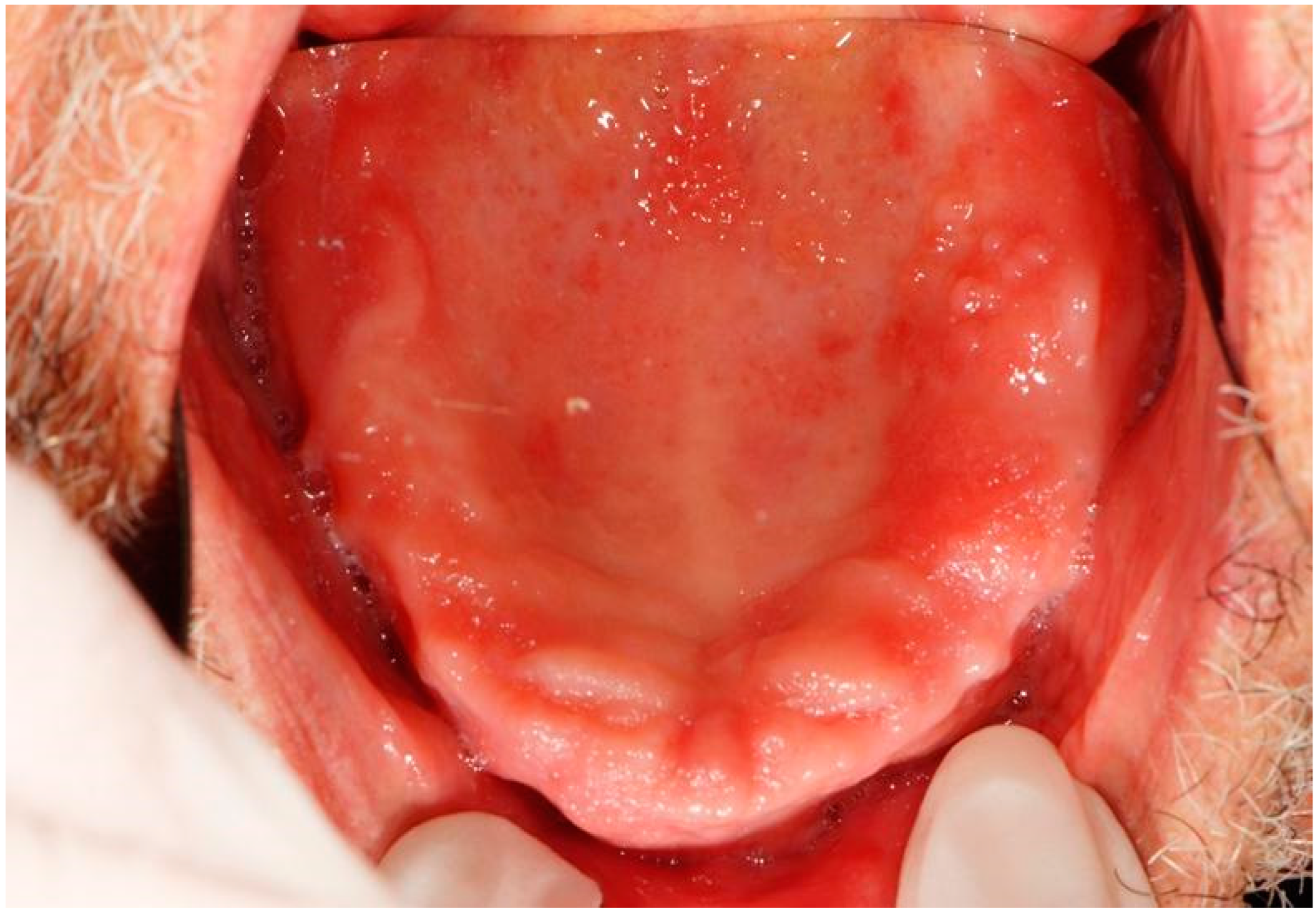
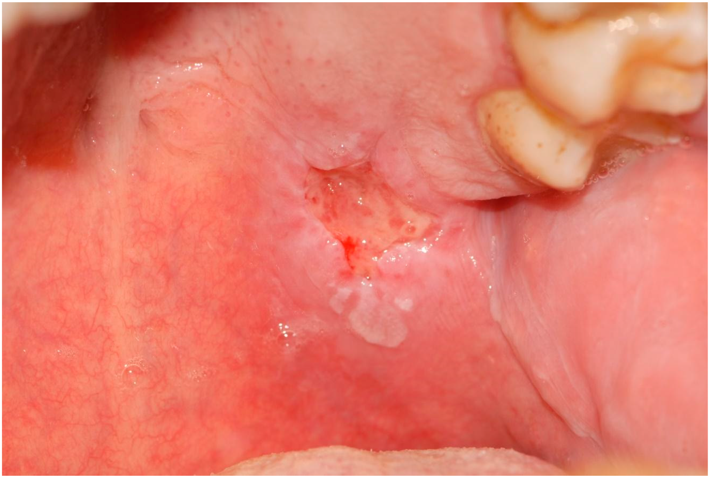
The sites affected by hyperkeratosis included the palate, cheek mucosa, and dorsal tongue (Figure 1). Candidiasis (Figure 2 and Figure 3) presented itself with a pseudomembranous type (Figure 2) or as a median rhomboid glossitis (Figure 3). Erythematous areas (Figure 4), with epithelial atrophy, as well as ulcerative lesions (Figure 5 and Figure 6), were found mainly on the palate.
Generally, oral alterations were reported at a minimum after the second infusion; in only one patient was an atrophic area in the palate reported after the first dose of pembrolizumab. The event with the earliest onset was found to be mucosal erythema; hyperkeratotic lesions developed between the third/fourth infusion of anti-PD-1 inhibitor therapy. The same timing was reported for the development of candidiasis of the oral mucous membranes, while xerostomia was found later, following the ninth infusion.
Oral candidiasis was diagnosed after an oral swab and a culture test in order to report the amount of the infectious load.
All the nonremovable whitish and reddish lesions were subjected to biopsy and histological examination.
The administration of steroids was generally avoided due to the immunological status of the patients. Ulcerative and erythematous lesions showed complete recovery 10–15 days after the topical administration of hyaluronic acid. Candidiasis was treated with the administration of antifungal drugs (nystatin).
Finally, six patients did not show significant oral alterations.

3.2. Cutaneous irAEs

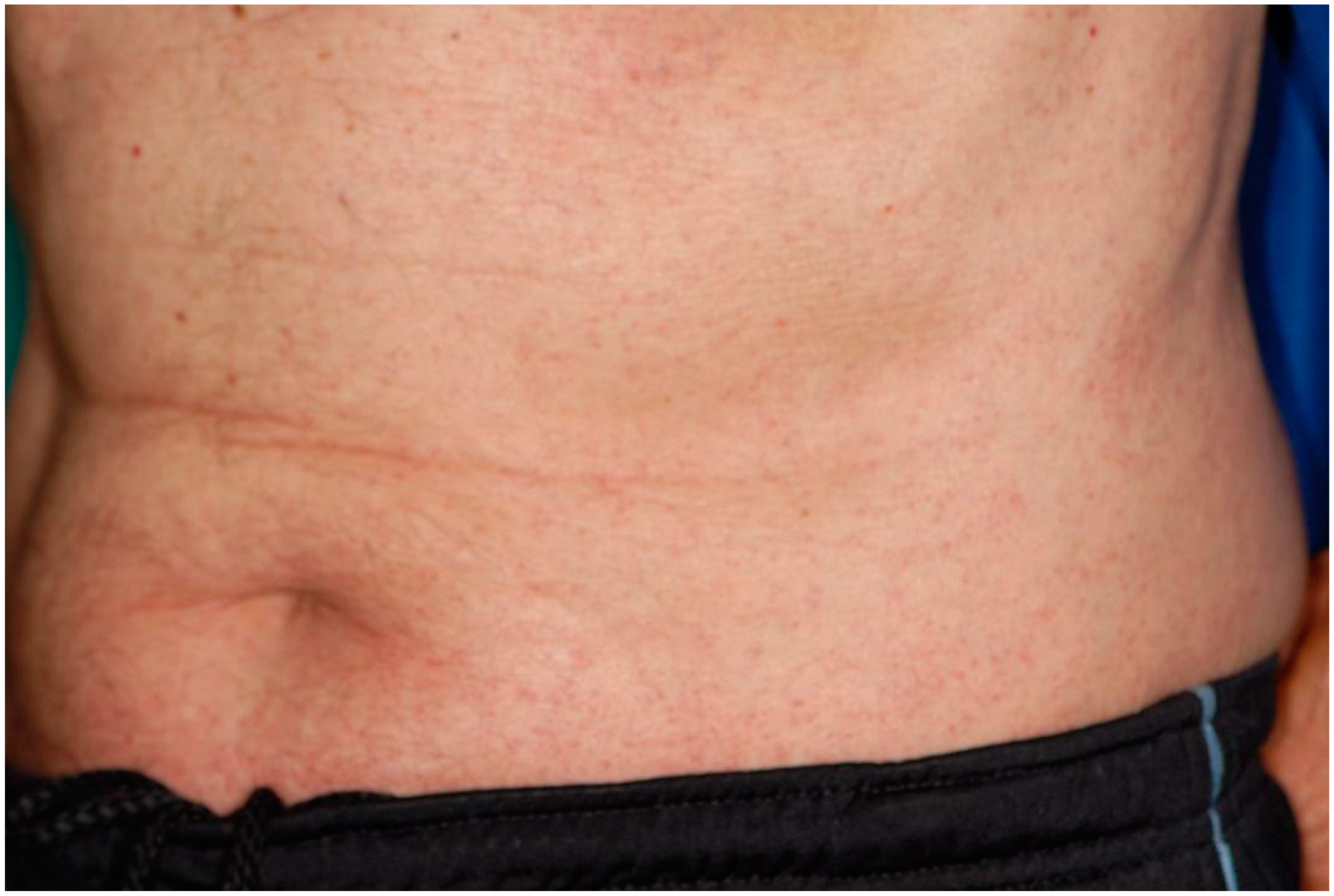
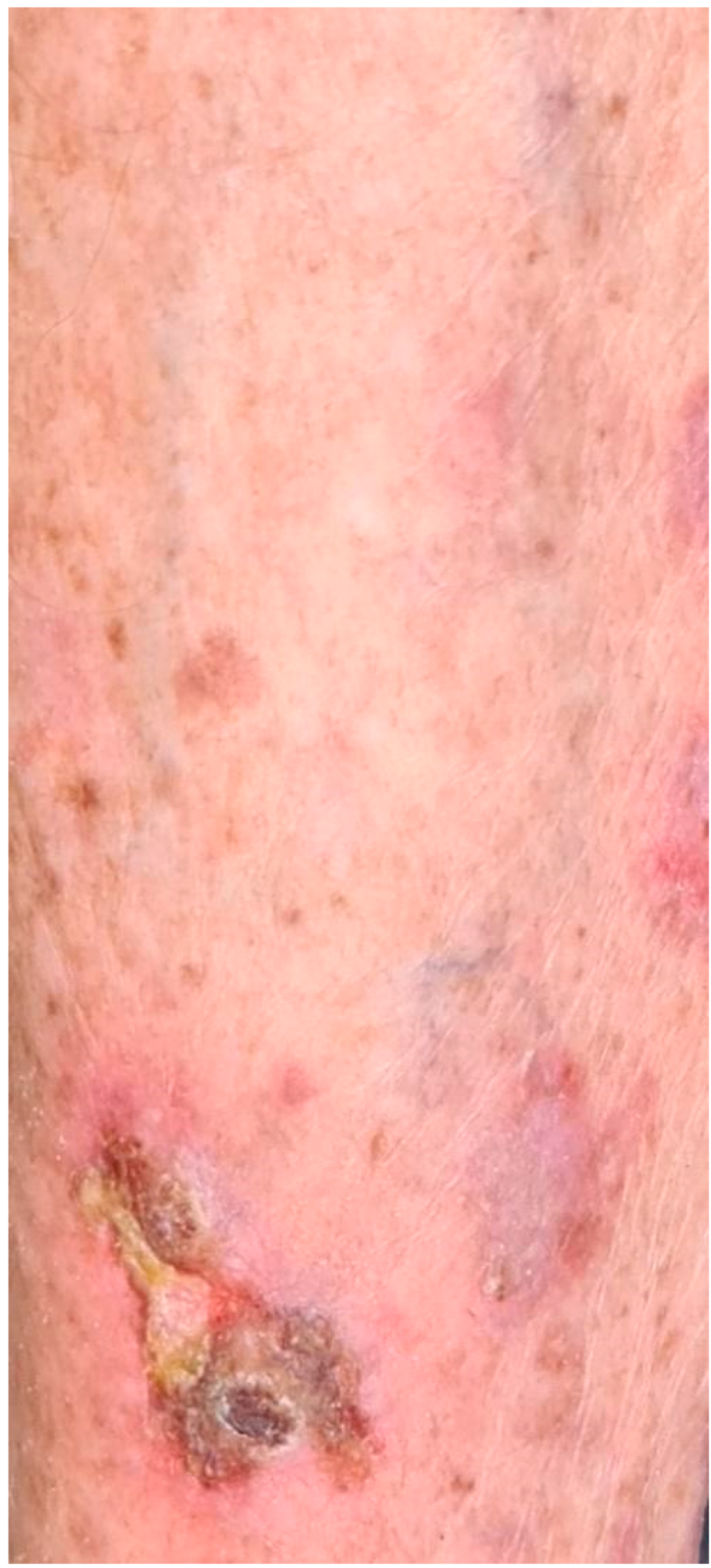
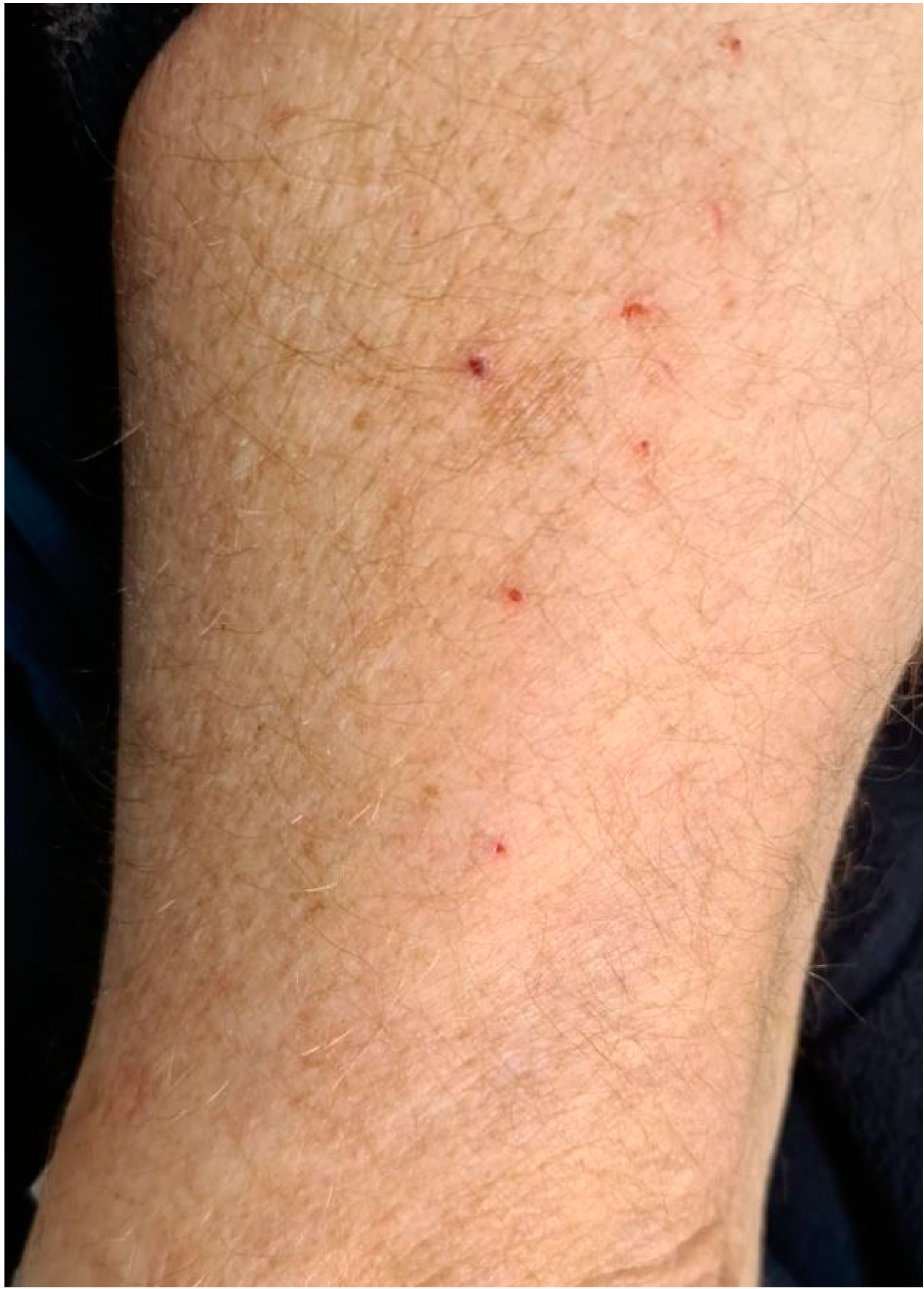
In the skin, the only adverse event reported was skin rash erythema (Figure 7 and Figure 8) found in seven patients. The clinical manifestations included widespread redness, small blisters with the development of crusts as a result of maceration (Figure 9 and Figure 10), and dryness of the skin in the affected areas. Patients also complained of moderate-to-intense itching. The most frequently affected areas are the abdomen, upper and lower limbs, back, face, and chest. All patients, following dermatological consultation, were treated with topical steroids and reported clinical and symptomatic improvement.
In five patients, skin erythema developed following the third infusion; in only two cases, a more rapid onset was recorded, following the first and second infusions (Table 3).

4. Discussion

The multiple toxicities reported following treatment with ICI drugs require a multidisciplinary approach. Early interception, with resolution or improvement of the lesion, could avoid the interruption of the immunotherapy and the management of severe sequelae.
In terms of overall toxicity, PD-1/PDL-1 inhibitor therapy has a better safety profile than drugs directed against the CTLA-4 receptor. The difference in behavior would seem to depend on the different localization of the two receptors: while CTLA-4 is present in T cells and regulatory T cells (T-REG) and its activation inhibits the immune response at an early stage, the PD-1 receptor is expressed mainly in peripheral tissues and in the tumor environment. The PD-1 inhibitor appears to target T cells with greater specificity [8]. For these reasons, CTLA-4 inhibitors lead to a broader immune response, with a greater occurrence of adverse events than PD-1/PDL-1 inhibitors. Immune-related adverse events in the skin and oral mucous membranes seem to have a different trend: these manifestations are more associated with treatment with anti-PD-1 drugs than with anti-CTLA-4 drugs [9]. From the analysis of the literature and our clinical experience, the correlation between therapy with ICI drugs and the onset of irAEs appears certain. The duration of administration may influence the onset of irAEs, since we have seen that episodes often occur after the first administration of the drug. It should also be emphasized that the aforementioned therapy can, sometimes, be combined with other types of treatments (e.g., chemotherapy), which often lead to a greater appearance of adverse reactions in many areas. It should be expected that the adverse reactions associated with the combined use of ICI drugs and chemotherapy may be more difficult to cure than with just one of these causal factors.
The present study, in accordance with the scientific literature, reported lichenoid reactions among the most frequent oral alterations associated with PD-1-born therapy. Clinically, they appear as whitish and reticulated plaques (Wickham’s striae), located mainly at the level of the cheek mucosa, lingual dorsum, and palate. They are generally asymptomatic and occur unilaterally, unlike oral lichen planus, where the distribution is characteristically bilateral. The onset is between the third and fourth doses of the drug, while later onset is described in the literature [10]. In addition, several studies have described the presence of erosive and atrophic elements in the context of whitish plaques that can lead to painful symptoms [11].
In three patients, erythematous areas of the cheek mucosa were found to appear at an early stage of treatment, between the first and second infusion of the drug. In the literature, such manifestations have been described as mucositis and concomitant appearance of ulcerative lesions [12]. They generally present low severity [1,2], while cases of severe immune mucositis associated with esophagitis are rare [13]. Among the examined patients, two reported the appearance of ulcerative lesions in the context of erythematous areas on the palate. Patients reported no painful symptoms as a result of these alterations. Following the appearance of the ulcerative lesion, only one patient complained of mild burning.
While no cases of candidiasis associated with patients receiving anti-PD-1 drugs are reported in the literature, candida infections were recorded in two cases, between the third and fourth infusions of the drug. In the first case, the fungal infection was not associated with other alterations in the oral mucous membranes, while in the second case, it arose in an inflammatory context. In a patient with skin cancer, the candidiasis and xerostomia could also be due to the radiation regimen.
Xerostomia is reported in the literature as the second most frequent oral adverse event following treatment with ICI, described as having an incidence of 6–7.2% [14]. The present study found only one case of xerostomia associated with difficulty swallowing. Often, the dryness of the mucous membranes also causes dysgeusia. Other reported symptoms include thick or sticky saliva, dry throat with hoarseness, and sensitivity to spicy and acidic foods. Cases of dry mouth with a high degree of severity have also been reported, which required temporary or permanent discontinuation of the ICI drug [15].
The most frequent oral adverse events, following treatment with anti-PD-1 drugs, therefore include mucositis, xerostomia, dysgeusia, and lichenoid reactions. Only one study described a case of pemphigoid of mucous membranes following therapy with pembrolizumab (PD-1) [16].
In the present study, six of seven patients with cutaneous irAEs also developed oral alterations. Only in two patients was this correlation not found: in one case, the presence of erythema on the face and chest was found, without any oral manifestation, while one patient reported inflammation of the palatal mucosa in the absence of skin changes. Moreover, adverse reactions were found in four of the six patients with lung cancer and treatment with ICI drugs. It could be analyzed, with a larger number of patients, whether the type of cancer being treated could be related not only to the onset but also to the type and location of the lesions that have arisen.
All patients who underwent radiotherapy developed oral lesions. Skin lesions were found in two of the three patients who received chemotherapy. The lesions found in the latter patients were always of the erythematous type, while the lesions in patients with radiotherapy were of both the erythematous and hyperkeratotic type.
ICI drugs have enabled a revolution in the treatment of cancer diseases, particularly for advanced cancers that are not responsive to conventional treatments. By contrast, the introduction of such therapies has led to the appearance of a new toxicity profile. The possible correlation between the onset of irAEs and the efficacy of the ICI drug has long been studied: it has been hypothesized that patients who show a better anticancer response, following treatment with ICI, have at the same time a greater probability of autoimmune toxicity [17]. This would seem to be caused by a more competent and reactive immune system, which stops the evolution of tumor pathology but also leads to an increase in responses against self [18]. Although several studies seem to confirm this relationship, there are still several disputes: the pathogenesis of these manifestations is not entirely clear, nor how the site, severity, timing of onset, and management of irAEs can influence the effectiveness of ICI drugs. Unfortunately, the research on biomarkers of response or toxicities is still an open field in immuno-oncology and we still do not know why certain cancer patients did not develop irAEs, and thus future research could be focused on the identification of toxicity biomarkers (sputum, microbiota). The only weapon currently available to avoid the occurrence of serious events is early interception, which is only possible through the knowledge of these manifestations. Close surveillance of patients being treated with immunosuppressive drugs allows a clinician to make a timely diagnosis when the adverse event occurs in the initial phase, allowing early treatment and avoiding progression to more advanced stages, which could lead to temporary or permanent suspension of the anticancer drug itself.
Potentially, irAEs can affect any organ. Adverse events reported more frequently include cutaneous, gastrointestinal, hepatic, endocrine, pulmonary, renal, rheumatological, neurological, cardiovascular, pericardial, and ophthalmological toxicity. These manifestations, in addition to being widely described in the literature, are reported in the guidelines “Management of Immunotherapy Toxicity” (Aimo, Italian Association of Medical Oncology), in which the correct approach for each individual toxicity is examined on the basis of the severity of these events. With regard to adverse events affecting the oral mucous membranes, there are not many studies that analyze their characteristics and methods of onset. It is possible that these events are underestimated as they are often mild or moderate and no symptomatology is associated with them. To intercept them, dedicated and accurate oral investigation is therefore necessary. For cancer patients planning to initiate immunotherapy, oral healthcare providers should be aware of any existing immune-mediated conditions. ICI therapy may exacerbate pre-existing immune-mediated conditions, and it is important to be able to distinguish a de novo irAE from exacerbation of an underlying disease process [19].
In addition, the present study showed an important correlation between skin changes and oral manifestations. Since dermatologic toxicities are among those with the highest incidence [20], it is necessary to deepen the investigations regarding irAEs involving the oral mucosa and introduce these manifestations to the guidelines for the management of immunotherapy toxicities, thereby providing clinicians with an important aid for their interception and treatment and improving the quality of life of the patient being treated with ICI drugs [21].

5. Conclusions

From the analysis of the literature and our clinical experience, the correlation between therapy with ICI drugs and the onset of irAEs appears certain. This work aims to propose clinical–diagnostic recommendations for general dentists and oral pathologists who are faced with certain situations. In the context of the management of oral lesions, pharmacological treatments (topical steroid therapies, hyaluronic acid drugs, and nystatin in the case of candidiasis) aiming to reduce the inflammatory state and burning symptomatology are useful; moreover, laser photobiomodulation treatments to prevent the onset of lesions and burning symptoms are considered to be a valid therapeutic choice [22]. For whitish lesions, it is possible to exclude potentially malignant lesions using cold-blade or laser-assisted biopsy and consequent histological examinations [23,24,25], although today, no onset of oral carcinomas following immunological therapy has been reported in the literature. In general, patients with a history of an oral irAE must be closely followed by their oral healthcare provider for flare-ups, recurrence, and/or new oral toxicities. In our study, considering the small sample size and the low grade of toxicities reported, we cannot evaluate the association between oral toxicity and tumor response because the patients in the present paper are all on a follow-up regimen, and a definitive evaluation after such a short period of therapy is still impossible. Further research is needed to elucidate the mechanism of these oral toxicities as well as the role they may play in predicting the antitumor efficacy of ICIs [26].

Author Contributions

Conceptualization, U.R. and A.P.; methodology, D.P. and G.P.; software, R.F.; validation, G.T., F.R. and A.C.; formal analysis, D.P.; investigation, G.P.; resources, A.B.; data curation, R.F.; writing—original draft preparation, D.P.; writing—review and editing, G.P.; visualization, U.R.; supervision, A.P.; project administration, G.T. All authors have read and agreed to the published version of the manuscript.

Funding

This research received no external funding.

Institutional Review Board Statement

The study was conducted according to the guidelines of the Declaration of Helsinki and approved by the Institutional Review Board of the Department of Oral and Maxillofacial Sciences of Sapienza University of Rome and was registered with the protocol number 001393 on 22 December 2020.

Informed Consent Statement

Informed consent was obtained from all subjects involved in the study.

Data Availability Statement

Data can be obtained from the corresponding author.

Conflicts of Interest

The authors declare no conflict of interest.

References

  1. Yang, Y. Cancer immunotherapy: Harnessing the immune system to battle cancer. J. Clin. Investig. 2015, 125, 3335–3337. [Google Scholar] [CrossRef] [Green Version]
  2. Finn, O.J. Immuno-oncology: Understanding the function and dysfunction of the immune system in cancer. Ann. Oncol. 2012, 23 (Suppl. S8), viii6–viii9. [Google Scholar] [CrossRef] [PubMed]
  3. Roberto, M.; Botticelli, A.; Cecere, F.; Cognetti, F.; Giusti, R.; Gelibter, A.; Lugini, A.; Nelli, F.; Nuti, M.; Santini, D.; et al. L’immunoterapia nel tumore del polmone non a piccole cellule: Ritorno al futuro [Immunotherapy in non-small cell lung cancer patients: Back to the future]. Recenti Prog. Med. 2019, 110, 587–593. (In Italian) [Google Scholar] [CrossRef] [PubMed]
  4. Lipson, E.J.; Drake, C.G. Ipilimumab: An anti-CTLA-4 antibody for metastatic melanoma. Clin. Cancer Res. 2011, 17, 6958–6962. [Google Scholar] [CrossRef] [Green Version]
  5. Topalian, S.L.; Hodi, F.S.; Brahmer, J.R.; Gettinger, S.N.; Smith, D.C.; McDermott, D.F.; Powderly, J.D.; Carvajal, R.D.; Sosman, J.A.; Atkins, M.B.; et al. Safety, activity, and immune correlates of anti-PD-1 antibody in cancer. N. Engl. J. Med. 2012, 366, 2443–2454. [Google Scholar] [CrossRef] [PubMed] [Green Version]
  6. Postow, M.A.; Sidlow, R.; Hellmann, M.D. Immune-related adverse events associated with immune checkpoint blockade. N. Engl. J. Med. 2018, 378, 158–168. [Google Scholar] [CrossRef]
  7. Dolladille, C.; Ederhy, S.; Sassier, M.; Cautela, J.; Thuny, F.; Cohen, A.A.; Fedrizzi, S.; Chrétien, B.; Da-Silva, A.; Plane, A.F.; et al. Immune Checkpoint Inhibitor Rechallenge After Immune-Related Adverse Events in Patients with Cancer. JAMA Oncol. 2020, 6, 865–871. [Google Scholar] [CrossRef]
  8. Boutros, C.; Tarhini, A.; Routier, E.; Lambotte, O.; Ladurie, F.L.; Carbonnel, F.; Izzeddine, H.; Marabelle, A.; Champiat, S.; Berdelou, A.; et al. Safety profiles of anti-CTLA4 and anti-PD-1 antibodies alone and in combination. Nat. Rev. Clin. Oncol. 2016, 13, 473–486. [Google Scholar] [CrossRef]
  9. Yang, W.; Li, S.; Yang, Q. Risk of dermatologic and mucosal adverse events associated with PD-1/PD-L1 inhibitors in cancer patients: A meta-analysis of randomized controlled trials. Medicine 2019, 98, e15731. [Google Scholar] [CrossRef]
  10. Dika, E.; Lambertini, M.; Gouveia, B.; Mussi, M.; Marcelli, E.; Campione, E.; Gurioli, C.; Melotti, B.; Alessandrini, A.; Ribero, S. Oral Manifestations in Melanoma Patients Treated with Target or Immunomodulatory Therapies. J. Clin. Med. 2021, 10, 1283. [Google Scholar] [CrossRef]
  11. Sibaud, V.; Eid, C.; Belum, V.R.; Combemale, P.; Barres, B.; Lamant, L.; Mourey, L.; Gomez-Roca, C.; Estilo, C.L.; Motzer, R.; et al. Oral lichenoid reactions associated with anti-PD-1/PD-L1 therapies: Clinicopathological findings. J. Eur. Acad. Dermatol. Venereol. 2017, 31, e464–e469. [Google Scholar] [CrossRef] [PubMed]
  12. Fazer, C.; Price, K.A. Management of immune-related dermatitis and mucositis associated with pembrolizumab in metastatic human papillomavirus-associated squamous cell carcinoma of the oropharynx. JCO Oncol. Pract. 2020, 16 (Suppl. S2), 20s–24s. [Google Scholar] [CrossRef] [PubMed]
  13. Acero Brand, F.Z.; Suter, N.; Adam, J.P.; Faulques, B.; Maietta, A.; Soulières, D.; Blais, N. Severe immune mucositis and esophagitis in metastatic squamous carcinoma of the larynx associated with pembrolizumab. J. Immunother. Cancer 2018, 6, 22. [Google Scholar] [CrossRef] [Green Version]
  14. Lacouture, M.; Sibaud, V. Toxic Side Effects of Targeted Therapies and Immunotherapies Affecting the Skin, Oral Mucosa, Hair, and Nails. Am. J. Clin. Dermatol. 2018, 19 (Suppl. S1), 31–39. [Google Scholar] [CrossRef] [PubMed] [Green Version]
  15. Warner, B.M.; Baer, A.N.; Lipson, E.J.; Allen, C.; Hinrichs, C.; Rajan, A.; Pelayo, E.; Beach, M.; Gulley, J.L.; Madan, R.A.; et al. Sicca Syndrome Associated with Immune Checkpoint Inhibitor Therapy. Oncologist 2019, 24, 1259–1269. [Google Scholar] [CrossRef] [PubMed] [Green Version]
  16. Del Vecchio, A.; Palaia, G.; Grassotti, B.; Tenore, G.; Ciolfi, C.; Podda, G.; Impellizzeri, A.; Mohsen, A.; Galluccio, G.; Romeo, U. Effects of laser photobiomodulation in the management of oral lichen planus: A literature review. Clin. Ther. 2021, 172, 467–483. [Google Scholar] [CrossRef]
  17. Zumelzu, C.; Alexandre, M.; Le Roux, C.; Weber, P.; Guyot, A.; Levy, A.; Aucouturier, F.; Mignot-Grootenboer, S.; Caux, F.; Maubec, E.; et al. Mucous membrane pemphigoid, bullous pemphigoid, and anti-programmed death-1/programmed death-ligand 1: A case report of an elderly woman with mucous membrane pemphigoid developing after pembrolizumab therapy for metastatic melanoma and review of the literature. Front. Med. 2018, 5, 268. [Google Scholar]
  18. Dascorresponding, S.; Johnson, D.B. Immune-related adverse events and anti-tumor efficacy of immune checkpoint inhibitors. J. Immunother. Cancer 2019, 7, 306. [Google Scholar] [CrossRef]
  19. Klein, B.A.; Shazib, M.A.; Villa, A.; de Abreu Alves, F.; Vacharotayangul, P.; Sonis, S.; Fedele, S.; Treister, N.S. Immune checkpoint inhibitors in cancer therapy: Review of orofacial adverse events and role of the oral healthcare provider. Front. Oral Health 2022, 3, 968157. [Google Scholar] [CrossRef]
  20. Riudavets, M.; Barba, A.; Maroto, P.; Sullivan, I.G.; Anguera, G.; Paez, D.; del Carpio, L.; Callejo, A.; Gonzalez Blanco, C.; Garcia Planellas, E.; et al. Correlation between immune-related adverse events (irAEs) and efficacy in patients with solid tumors treated with immune-checkpoints inhibitors (ICIs). J. Clin. Oncol. 2018, 36 (Suppl. S15), 3064. [Google Scholar] [CrossRef]
  21. Tattersall, I.W.; Leventhal, J.S. Cutaneous Toxicities of Immune Checkpoint Inhibitors: The Role of the Dermatologist. Yale J. Biol. Med. 2020, 93, 123–132. [Google Scholar] [PubMed]
  22. Del Vecchio, A.; Tenore, G.; Luzi, M.C.; Palaia, G.; Mohsen, A.; Pergolini, D.; Romeo, U. Laser Photobiomodulation (PBM)-A Possible New Frontier for the Treatment of Oral Cancer: A Review of In Vitro and In Vivo Studies. Healthcare 2021, 9, 134. [Google Scholar] [CrossRef] [PubMed]
  23. Palaia, G.; D’Alessandro, L.; Pergolini, D.; Carletti, R.; Di Gioia, C.R.T.; Romeo, U. In vivo clinical and histological thermal effect of a 445 nm diode laser on oral soft tissues during a biopsy. J. Oral Sci. 2021, 63, 280–282. [Google Scholar] [CrossRef]
  24. Palaia, G.; Renzi, F.; Pergolini, D.; Del Vecchio, A.; Visca, P.; Tenore, G.; Romeo, U. Histological Ex Vivo Evaluation of the Suitability of a 976 nm Diode Laser in Oral Soft Tissue Biopsies. Int. J. Dent. 2021, 2021, 6658268. [Google Scholar] [CrossRef]
  25. Pergolini, D.; Del Vecchio, A.; Mohsen, M.; Tenore, G. Inter-incisor ossifying fibroma removal by diode laser. Dent. Cadmos 2020, 88, 266–268. [Google Scholar] [CrossRef]
  26. Klein, B.A.; Alves, F.A.; de Santana Rodrigues Velho, J.; Vacharotayangul, P.; Hanna, G.J.; LeBoeuf, N.R.; Shazib, M.A.; Villa, A.; Woo, S.B.; Sroussi, H.; et al. Oral manifestations of immune-related adverse events in cancer patients treated with immune checkpoint inhibitors. Oral Dis. 2022, 28, 9–22. [Google Scholar] [CrossRef]
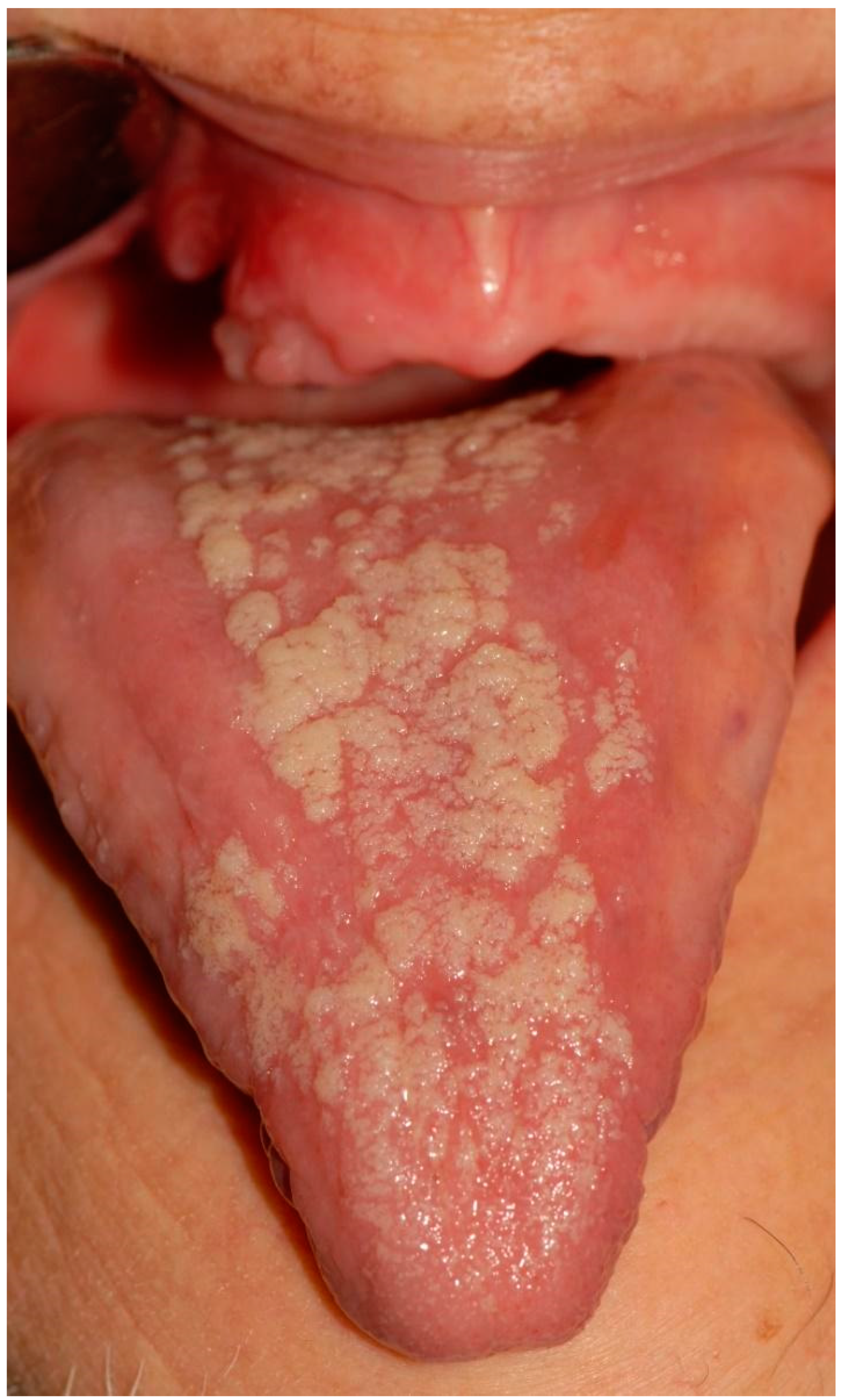
Figure 1. Patchy, erythematous depapillation of the dorsal tongue with regions of elongated filliform papillae. Histological examination showed a picture of atrophy as well as a hyperkeratosis without dysplasia (Patient no. 12, Table 2).
Applsci 12 12994 g001
Figure 2. Removable whitish plaques associated with pseudomembranous candidiasis (Patient no. 4, Table 2).
Applsci 12 12994 g002
Figure 3. Atrophic area of dorsal tongue associated with median rhomboid glossitis (Patient no. 4, Table 2).
Applsci 12 12994 g003
Figure 4. Mucosal erythema, palate (Patient no. 2, Table 2).
Applsci 12 12994 g004
Figure 5. Ulcerative lesion of the hard palate (Patient no. 13, Table 2).
Applsci 12 12994 g005
Figure 6. Ulcerative area of the palate (Patient no. 10, Table 2).
Applsci 12 12994 g006
Figure 7. Skin rash erythema (Patient no. 7, Table 3).
Applsci 12 12994 g007
Figure 8. Skin rash erythema (Patient no. 7, Table 3).
Applsci 12 12994 g008
Figure 9. Leg erythema (Patient no. 6, Table 3).
Applsci 12 12994 g009
Figure 10. Arm erythema (Patient no. 10, Table 3).
Applsci 12 12994 g010
Table 1. Patient data.
PatientCounty’SexCancer DiagnosisTherapy
Anti PD-1
DosageSmokeRadiotherapyChemotherapyPathological History
164FK urothelialpembrolizumab200 mg flat dose q21YesNoNoSystemic lupus erythematosus
267MK parotidpembrolizumab200 mg flat dose q21Former smoker10 sessionsNoHypertension
376MPulmonary ADKpembrolizumab200 mg flat dose q21NoNoNoRenal failure, aortic stenosis
468MK squamous skincemiplimab350 mg dropped dose q 21No2 sessionsNoRheumatoid arthritis
536MMelanomanivolumab240 mg q 14NoNoNoNothing to detect
681MK squamous cell oral cavitypembrolizumab200 mg flat dose q21Former smokerNoNoHypertension; diabetes II; dyslipidemia
768FK squamous cell oral cavitypembrolizumab200 mg flat dose q21NoNoNoDiabetes II; hypertension; chronic HBV; diverticular pathology
870MK urothelialpembrolizumab200 mg flat dose q21Yes No Cisplatin-gemcitabineNothing to detect
963MLung adenocarcinoma pembrolizumab 200 mg flat dose q21NoNoNoAtrial fibrillation
1069MPulmonary adenocarcinomanivolumab240 mg q 14Former smokerNoCisplatin alimtaHypertension; dyslipidemia; partial thyroidectomy; coronary stent
1166FLung adenocarcinoma pembrolizumab200 mg flat dose q21No No Cisplatin pemetrexedNothing to detect
1275MPulmonary adenocarcinomapembrolizumab200 mg flat dose q21NoNoNoNothing to detect
1371MPulmonary adenocarcinomapembrolizumab200 mg flat dose q21No10 sessionsNoHypertension
Table 2. Oral immune-related adverse events.
PatientClinical DescriptionType of LesionAnatomical SiteTherapyAnti-PD-1 Dose for Oral irAEs
1Nothing to detectNothing to detectNothing to detectNothing to detect
2Erythematous areaEpithelial atrophy without dysplasiaPalateAminogam® mouthwash1st dose
3Nothing to detectNothing to detectNothing to detectNothing to detect
4Removable whitish plates
Erythematous area
Xerostomia
Candidiasis (pseudomembranous candidiasis, median rhomboid glossitis)Dorsal tongue
Oral mucous membranes
Nystatin
Mucosamin® mouthwash
3rd dose
9th dose
5Nothing to detectNothing to detectNothing to detectNothing to detect
6Non-removable whitish lesionsHyperkeratosis Palate
Cheek mucosa
Aminogam® mouthwash4th dose
7Non-removable white lesionsHyperkeratosis Dorsal tongue
Palate
Aminogam® mouthwash3rd dose
8Nothing to detectNothing to detectNothing to detectNothing to detectNothing to detect
9Nothing to detectNothing to detectNothing to detectNothing to detect
10Erythematous and ulcerative areasEpithelial atrophy without dysplasia
Ulcer
Palate 2nd dose
11Nothing to detectNothing to detectNothing to detectNothing to detectNothing to detect
12Non-removable whitish lesionsEpithelial atrophy and hyperkeratosis without dysplasiaCheek
Dorsal tongue
Aminogam® mouthwash4th dose
13Erythematous area
Ulcer
Removable whitish plates
Epithelial atrophy without dysplasia
Ulcer
Candidiasis
Palate
Tuberosity
Dorsal tongue
Alveolar process
Nystatin2nd dose
4th dose
Table 3. Skin immune-related adverse events.
Patient LesionBody AreaTreatment Dermatological Anti-PD-1 Dose
1Nothing to detectNothing to detectNothing to detectNothing to detect
2Nothing to detectNothing to detectNothing to detectNothing to detect
3Nothing to detectNothing to detectNothing to detectNothing to detect
4ERYTHEMABACK TOPICAL STEROID3rd dose
5Nothing to detectNothing to detectNothing to detectNothing to detect
6ERYTHEMALEGSTOPICAL STEROID3rd dose
7ERYTHEMAABDOMENTOPICAL STEROID3rd dose
8Nothing to detectNothing to detectNothing to detectNothing to detect
9Nothing to detectNothing to detectNothing to detectNothing to detect
10ERYTHEMAARMS, LEGS, ABDOMENTOPICAL STEROID3rd dose
11ERYTHEMAFACE, CHESTTOPICAL STEROID3rd dose
12ERYTHEMAABDOMENTOPICAL STEROID1st dose
13ERYTHEMAABDOMENTOPICAL STEROID2nd dose
Disclaimer/Publisher’s Note: The statements, opinions and data contained in all publications are solely those of the individual author(s) and contributor(s) and not of MDPI and/or the editor(s). MDPI and/or the editor(s) disclaim responsibility for any injury to people or property resulting from any ideas, methods, instructions or products referred to in the content.