Abstract
The fields of micro- and nanomechanics are strongly interconnected with the development of micro-electro-mechanical (MEMS) and nano-electro-mechanical (NEMS) devices, their fabrication and applications. This article highlights the biomimetic concept of designing new nanodevices for advanced materials and sensing applications.
1. Introduction
Nowadays, technology of any kind is faced with the constant need to minimalize gauges as an imperative for lowering the energy consumption and reduction in materials utilization. The most contributing aspect in this technological revolution is science. By emulating nature’s patterns, science seeks sustainable solutions for everyday human challenges. It is vital to harness biomimetic concepts to develop advanced bio-inspired devices for various applications. This article describes the potentials of biomimetic concepts for the design of future nanodevices, especially NEMS, which could have a plethora of potential applications. A practical representation of the order of magnitude of nano devices is given in Figure 1.
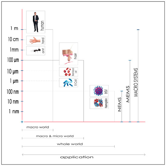
Figure 1.
Different scales in nature.
After a short introduction, we discuss the state-of-the-art bioinspired NEMS from a fundamental and design perspective, especially highlighting bio-oriented applications. We hope that this article will increase the awareness of the engineering community for biomimetics and highlight biomimetic-inspired concepts and solutions.
2. The Emergence of Bioinspired NEMS
Bioinspired devices are nanodevices with structure and functions designed to mimic examples from the natural world. Biomimetics is based on mimicking biological principals and patterns as a recipe for creating new materials and structures and integrating them into functional devices. The functionality of these devices also relies on the combined power of optics and nanoengineering. Figure 2 shows the synergy between nanoengineering, optics and biomimetics, which creates the new field of bioinspired NEMS.
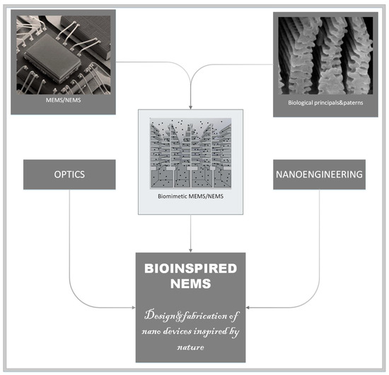
Figure 2.
NEMS–Synergy of different technical approaches.
Devices inspired by nature can be classified into two categories: (1) devices that incorporate natural biological structures within their systems and (2) biomimetic (bioinspired) devices that mimic structures and functions from the natural world.
The primary aim of this paper is to attract the interest of the broad material science community towards the bioinspired NEMS concept, which could be used to open new horizons in physical and material research. The authors of this article are confident that soon, in order to reach a cellular economy-driven society, we must develop technology that will be able to harness perfect engineering solutions designed by billions of years of evolution. For this approach to succeed, we must understand the complexity of the biological functions and patterns and their interconnections.
Biomimetics, to begin with [1,2,3], has a great potential for solving human problems by imitating the natural environment or learning from it. For example, an extensive review of bioinspired triboelectric nanogenerators includes a comparative analysis of structures and materials that draw inspiration from nature [4]. Additionally, a comprehensive review of nanomembranes and their application can be found in MDPI papers [5].
3. Fabrication of Bioinspired NEMS
Most of techniques for the fabrication of bioinspired NEMS use the lithographic method (Figure 3), which proved very suitable for processing nanoelements [6].
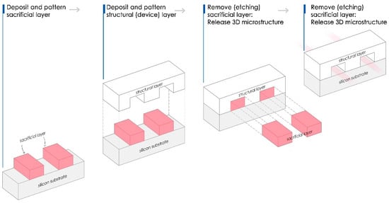
Figure 3.
Simplified scheme of a lithography process.
Fabrication of Bioinspired Nano-Structures
Recently, the additive engineering, i.e., 3D printing [7], is used for NEMS fabrication [8].
The future of bioNEMS technology is strongly connected with the development of new fabrication techniques (3D printing, self-assembly, etc.) and opens up many possibilities for potential applications in various fields, such as photonics, biomedicine, nanoelectronics, and sensing.
Micro molding is one of the most widely used techniques. The fabrication of replicas that perfectly match biostructure geometry is an enormous challenge for NEMS fabrication. The most straightforward technique used for replication consists of two-step process.
The development of a negative mold from a biopattern is the first step, and the second is making the positive replica. Kumar et al. [9] presents a precise micro-replication technique, as shown in Figure 4, to transfer surface microstructures of plant leaves onto a highly transparent, soft polymer material to design smooth surfaces with specific nano-corrugation. Structuring surfaces is beneficial for designing materials with controllable properties such as wetting, heat transfer, fluid flow, optical effects, etc.
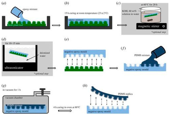
Figure 4.
Schematic sketch of the two-step replication process: (a) Fresh plant leaf glued on a plastic Petri dish, filled up with epoxy resin. (b) Curing of epoxy mixture for 15 h to produce negative epoxy mould. (c) Epoxy sample (which adhered with leaf surface) is kept for chemical treatment in potassium hydroxide solution on magnetic stirrer (at 60 ± 3 °C for 20 h). (d) Chemically treated sample washed in deionized water using an ultrasonicator. (e) Negative epoxy mould separated from the leaf surface. (f) Negative epoxy mould filled up with PDMS mixture. (g) Degassed in vacuum chamber to remove air trapped at the interface. (h) PDMS-positive replica peeled off from the epoxy mould. Reproduced with permission [9].
The unification of biostructures and nanosystems requires simple but reliable tools and techniques. The limited knowledge regarding materials leave us with just a few types of micro- and nanodevices. Modern technologies such as nanopatterning provide an excellent potential to obtain a new class of customized energetic materials for MEMS/NEMS application [10]. These materials allow advances in the processing of microscopic systems that are energy-demanding and may be even more important for bioinspired NEMS. An even more fascinating proposal is based on the utilization of so-called Pavlovian materials [11]. These materials are specially adapted to respond to certain stimuli, and they are proposed for application in integrated devices. As far as materials science is concerned, the inevitable dominance of bioinspired materials in technological applications will happen soon [12].
Recently, Shanker et al. designed melanin nanoparticles for new printable inks with a high boiling point to manage stable jetting and a high-printing efficiency [13]. The advantages of inkjet printing are mainly the reduction in processing times and materials requirement. An even more interesting example of nanofabrication is presented through the method of roll-to-plate (R2P) ultraviolet nanoimprint lithography (UV-NIL). This method uses the combined power of Nickel mold and transparent polycarbonate substrates [14]. The operation simplicity, high efficiency and low-cost fabrication make this antireflection technique promising. The imprint of nanostructures is also described elsewhere [15].
4. Bioinspired NEMS—The State of the Art
Here, we present a few exciting applications based on mimics of natural structures.
4.1. Hair-like Structures
Structures that resemble the hair are widespread in the living world. In biology, organisms often use these body parts to communicate with the environment [16]. Regarding the relatively simple shape of the hair, functional devices that mimic hair structures are constructed with the capability to mold mechanical responses as a function of the slightest changes in their environment. Inspired by the clavate hair-based sensory system used by crickets to sense gravitational acceleration and obtain information on their orientation, a one-axis-biomimetic accelerometer was developed and fabricated using different micro-machining processes [17,18]. Clavate hair is a receptor that is most sensitive to positional changes, while in practical terms, it can be seen as a pendulum subjected to external influences or accelerations from the environment. Staring at the halters of flies that serve to maintain balance based on Coriolis forces, a biomimetic gyroscope was made with the help of MEMS technology [19]. These are tiny organs by which flies sense the rotation of the body, practically opposing the orientation of wings during flight. The oblong extensions that are an integral part of most hair cells receive an external impulse and generate displacement to neurons as a mechanical response. The remarkable abilities of the fish and many sophisticated functions of their organs have led to the development of micro-biomimetic sensors made of artificial materials that resemble hair cells in shape and dimensions [20]. Practically, and in the physiological sense, sensory units are distributed throughout the body of the fish. These units receive flow rate information and convert it to electrical signals that pass through the innervated fibers directly to the brain for further processing. In the end, there are plenty of attractive models from nature to mimic, that could be used for the design of new functional devices.
4.2. BioMEMS: Medical Perspective
A significant subset of MEMS/NEMS devices is biomedical MEMS/NEMS or bioMEMS/bioNEMS. BioMEMS/BioNEMS refers to devices developed for biomedical and medical applications [21]. The potential for the applications of MEMS/NEMS in medicine is practically unlimited [22,23,24]. Scuor et al. [22] described a new appliance as MEMS-based in-plane biaxial cell stretcher used to study the influences of biaxial stresses on an individual living cell. By exploiting stretchable tools of the described device, the authors [22] recorded a displacement of several micrometers. Using this MEMS approach, it is possible at the same time to actuate and sense at a length scale comparable with single cells. In combination with biological microscopy techniques such as fluorescence, it is possible to visualize the effect of the stress. Parallel with Scuor research, Wang et al. [23] described the new micro-fabrication method for designing micro and nanosize artificial biocompatible capillaries.
Additionally, Tsuda et al. [24] observed a large multi-nuclear, single-cellular organism with no nervous system. The objective of his research was to make an integrated local sensor system that relies on intercellular information exchange. In search of an alternative to control and monitor the functioning of autonomous robots, Tsuda developed a system that works for an extended period without a power supply. This bio-hybrid creation is based on circuitry from amoeboid plasmodia of the slime mold, Physarum polycephalum. The circuits are connected to a hexapod robot that drives the system and exchanges information with the environment.
Soft robotics is an example of a field that has great potential for applications in medicine. Recently Kim et al. [25] developed an artificial micro-muscle fiber crafted from coiled shape–memory alloy (NiTi) springs.
4.3. The Last Decade—an Age of Great Promise
Ten years ago, an exciting moment occurred in materials science. The ability of all complex biological organisms to self-repair minor damages [26] was described, as well as the prospect of mimicking this feature in nanodevices.
“We continually learn things and borrow ideas from nature, but we design devices beyond nature” [27]. This beautiful, harmonious quote as well as the whole book by Di Zhang [27] represents a useful and exciting introduction to biomimetics. During this period, one gets the impression that thinking begins with a look at biomimetics; biomimetics finally gains its true meaning and becomes officially recognized.
Bioinspired NEMS can play a crucial role in curing or preventing disease. There are currently many NEMS applications in biology and biomedicine, and thus many articles in this field [28,29].
An example of such an application is a silicon micro-channel, used to provide an improved blood plasma separation from whole blood by acoustophoresis. Karthick et al. [30] harnessed the theory of acoustophoresis of dense suspension to understand the acoustic focusing of cells within blood capillaries. Such works are not only inspiring but also have promising applications.
A new lab-on-a-chip (LOC) technology is a kind of bioinspired nano system, recently described elsewhere [31,32,33]. It is a device that integrates one or several laboratory functions on a single integrated circuit. LOC can be defined as a subset of complex MEMS/NEMS devices. LOC is designed as a sensor capable of detecting different chemicals in bodily fluids and providing information about the current health condition. Currently, many MEMS are customized and programmed within LOC devices for various sensing applications. However, the ultimate goal is to detect any health issue without using expensive techniques and to stop the disease before it progresses. The potential uses of LOC in medicine and health application are unlimited. Recently, the New York Post highlighted LOC technology for non-specialists [33].
An exceptional example is a prototype of a 3D-printed artificial lung [34]. In brief, lungs are a mechanical sensor that reacts to the external effects, which is analogous to microsystems. Potkay [34] foresaw this product for short- and long-term respiratory support. In the beginning, it was used as a temporary measure while waiting for transplantation or another healing process, but it could become a permanent solution in the future. Although it does not look like an original organ overall, the structure is perfectly imitated. Thanks to the flexibility enabled by 3D printing, the variation in dimensions and shape is a huge advantage and a step towards new possibilities. Biomimicry is a constant inspiration to researchers and engineers, and now with new 3D technology, better production possibilities are affordable.
The successful replication of organ functions by natural materials, or biocompatible materials, represents the future of medical technology [35]. The possibility of replacing the lung is mind-blowing, with the potential to drastically change many patients’ lives.
Aside from that, You et al. [28] studied a self-organization of sessile bacteria within a controlled and closed environment. The ability of bacteria to sense the environment and its gradients and to adjust their movements to them, in combination with the hydrodynamic properties, has a significant impact on the bacterial colonization and management of nutrient resources [29] and could have a substantial impact on understanding complex ecological interactions.
Finally, the MEMS/NEMS applications reach the realm of pharmacy. The main challenge in pharmaceutical analytics is to find a fast and accurate test for sensing in the nano domain at a single-molecule or -particle level. Recently the PMTA method (Particle Mechanical Thermal Analyses) [36] was developed and uses a single particle as a resonator to determine changes in their mechanical properties. The PMTA opens the possibility of the characterization of materials at the single-particle level, which is crucial for developing future nano medicine devices. This achievement represents a remarkable advancement in pharmaceutical science. In recent years, various nanosystems targeting the drug delivery of different anticancer drugs were proposed and based on biomimetic approaches [37].
5. Biomimetics Meets Photonics and Nanomechanics
Recently, Pris et al. [38] and Zhang et al. [39] showed an interesting usage of biostructures. In their papers, the unique example of the management of thermal radiation by biophotonic structures of a Morpho butterfly is described. Additionally, the research described by Grujic et al. [40], pointed out the exciting mechanisms of thermophoresis/photophoresis within biophotonic structures, which could be harnessed for thermal radiation detection. Grujic et al. shows that, in the case of the Morpho butterfly, nature exploited natural photonic structures and their optical properties to develop IR detectors much more advanced than currently fabricated ones. Grujic et al.’s [40] experiment revealed a complex functional relationship between biological patterns and their functions. By imitating butterfly wing scales by polymer or composite materials, the thermophoretic effect can be further amplified for different sensing applications and the large-scale production of sensing devices. Polymers are particularly promising materials for infrared sensing applications because of their high IR absorption due to their organic bonds’ vibrational resonance modes and their high thermal expansion coefficient compared to metals and semiconductors [41]. Figure 5 shows a portion of a circular section of a sample, the microscopic image, and a holographic image of the investigated butterfly’s wing, accompanied with holographic reconstruction.
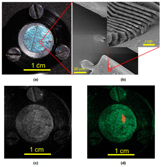
Figure 5.
Circular section of a butterfly’s wing: (a) A photograph, (b) a microscope image, (c) a hologram reconstruction and (d) a holographic image. Reproduced with permission [40].
In the end, to advance in nanoscience and biomimetics, it is imperative to understand the physics of the studied systems. When the microscale is reached, for example, when the distance between two structures or surfaces corresponds to the molecule-free path, thermal forces can occur [42]. Due to the temperature gradient, the surrounding gas causes the formation of a force that creates mechanical displacement at the microscale level. This phenomenon is called thermophoresis. These forces are mechanical forces that scale almost linearly with pressure and temperature. In their paper, Passian et al. [43] described the study of the pressure dependence of Knudsen forces by exploiting MEMS devices.
Moreover, when downscaling from the MEMS to NEMS level, forces between system elements cannot be described by using a classical physics framework. The quantum effect must be considered and controlled, which is one of the most challenging tasks for the development and applications of future NEMS. Figure 6 shows the cartoon image of “strangeness” of quantum mechanics [44].
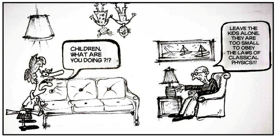
Figure 6.
Cartoon sketch of the limitations of classical physics and “strangeness” of quantum mechanics.
6. Conclusions
The possibility of harnessing biomimetics for the design of advanced NEMS devices is highlighted in this account. The topic discussed here is interesting from a fundamental perspective, with practically unlimited applications in the areas of photonics, sensing, and biomedicine. Even though, at the moment, bioinspired NEMS is still in its infant stage of development, the primary aim of this article is to attract the interest of the broad material and photonics science community for the biomimetic concept, which could open new horizons in material research.
Author Contributions
This study was conducted in partial fulfillment of the requirements for the PhD degree of Marina Simović Pavlović at the University of Belgrade, Faculty of Mechanical Engineering. Conceptualization: M.S.-P. and B.K.; Writing—original draft preparation: M.S.-P.; Writing—review and editing: M.S.-P., B.B., D.V. and B.K.; Visualization: B.B.; Supervision: B.K.; Administration: D.V. All authors have read and agreed to the published version of the manuscript.
Funding
This research was funded by NATO SPS (NATO Science for Peace and Security) 2019–2022, and by the Ministry of Science, Republic of Serbia grant number [Grant III 45016].
Institutional Review Board Statement
Not applicable.
Informed Consent Statement
Not applicable.
Acknowledgments
M.S.-P., D.V. and B.K. acknowledge support of the biological and bioinspired structures for multispectral surveillance, funded by NATO SPS (NATO Science for Peace and Security) 2019–2022. B.K., D.V. and B.B. acknowledge financial support of the Ministry of Science, Republic of Serbia (Grant III 45016). B.K. acknowledges support from FRS–FNRS. Authors warmly acknowledge the architects, Stevan Simovic and Tina Urosevic, for their assistance in creating the images for publication.
Conflicts of Interest
All authors confirmed that there is no conflict of interest among authors.
References
- Driessen-Mol, A. Biomimetics: A Molecular Perspective. Green Processing Synth. 2013, 2, 527. [Google Scholar] [CrossRef]
- Zheng, X.; Kamot, A.M.; Cao, M.; Kottapalli, A.G.P. Creating underwater vision through wavy whiskers: A review of the flow-sensing mechanisms and biomimetic potential of seal whiskers. J. R. Soc. Interface 2021, 18, 20210629. [Google Scholar] [CrossRef]
- Stenvinkel, P.; Painer, J.; Johnson, R.J.; Natterson-Horowitz, B. Biomimetics—Nature’s roadmap to insights and solutions for burden of lifestyle diseases. Rev. Simp. 2020, 287, 238–251. [Google Scholar] [CrossRef]
- Li, W.; Pei, Y.; Zhang, C.; Kottapalli, A.G.P. Bioinspired designs and biomimetic applications of triboelectric nanogenerators. Nano Energy 2021, 84, 105865. [Google Scholar] [CrossRef]
- Jakšić, Z.; Jakšić, O. Biomimetic nanomembranes: An overview. Biomimetics 2020, 5, 24. [Google Scholar] [CrossRef] [PubMed]
- Liu, L.; Sun, L.; Qi, L.; Guo, R.; Li, K.; Yin, Z.; Wu, D.; Zou, H. A low-cost fabrication method of nanostructures by ultraviolet proximity exposing lithography. AIP Adv. 2020, 10, 045221. [Google Scholar] [CrossRef]
- Duda, T.; Raghavan, L.V. 3D Metal Printing Technology. IFAC-PapersOnLine 2016, 49, 103–110. [Google Scholar] [CrossRef]
- Chen, Y.P.; Yang, M.D. Micro-Scale Manufacture of 3D Printing. Appl. Mech. Mater. 2014, 670, 936–941. [Google Scholar] [CrossRef]
- Kumar, C.; Le Houerou, V.; Speck, T.; Bohn, H.F. Straightforward and precise approach to replicate complex hierarchical structures from plant surfaces onto soft matter polymer. R. Soc. Open Sci. 2018, 5, 172132. [Google Scholar] [CrossRef]
- Rossi, C.; Zhang, K.; Esteve, D.; Alphonse, P.; Tailhades, P.; Vahlas, C. Nanoenergetic materials for MEMS: A review. J. Microelectromech. Syst. 2007, 16, 919–931. [Google Scholar] [CrossRef]
- Zhang, H.; Zeng, H.; Priimagi, A.; Ikkala, O. Pavlovian materials—Functional biomimetics inspired by classical conditioning. Adv. Mater. 2020, 32, 1906619. [Google Scholar] [CrossRef] [PubMed]
- Wang, Y.; Naleway, S.E.; Wang, B. Biological and bioinspired materials: Structure leading to functional and mechanical performance. Bioact. Mater. 2020, 5, 745–757. [Google Scholar] [CrossRef] [PubMed]
- Shanker, R.; Sardar, S.; Chen, S.; Gamage, S.; Rossi, S.; Jonsson, M.P. Noniridescent biomimetic photonic microdoms by inkjet printing. Nano Lett. 2020, 20, 7243–7250. [Google Scholar] [CrossRef]
- Sun, J.; Wang, X.; Wu, J.; Jiang, C.; Shen, J.; Cooper, M.A.; Wu, D. Biomimetic moth-eye nanofabrication: Enhanced antireflection with superior self-cleaning characteristic. Sci. Rep. 2018, 8, 1–10. [Google Scholar] [CrossRef]
- Chou, S.Y.; Krauss, P.R.; Renstrom, P.J. Imprint of sub-25 nm vias and trenches in polymers. Appl. Phys. Lett. 1995, 67, 3114–3116. [Google Scholar] [CrossRef]
- Seale, M.; Cummins, C.; Viola, I.M.; Mastropaolo, E.; Nakayama, N. Design principles of hair-like structures as biological machines. J. R. Soc. Interface 2018, 15, 20180206. [Google Scholar] [CrossRef]
- Droogendijk, H.; De Boer, M.J.; Sanders, R.G.P.; Krijnen, G.J.M. A biomimetic accelerometer inspired by the cricket’s calvate hair. J. R. Soc. Interface 2014, 11, 20140438. [Google Scholar] [CrossRef]
- Sakaguchi, D.S.; Murphy, R.K. The equilibrium detecting system of the cricket: Physiology and morphology of an identified interneuron. J. Comp. Physiol. 1983, 150, 141–152. [Google Scholar] [CrossRef]
- Droogendijk, H.; Brookhuis, R.A.; De Boer, M.J.; Sanders, R.G.P.; Krijnen, G.J.M. Towards a biomimetic gyroscope inspired by the fly’s haltere using microelectromechanical systems technology. J. R. Soc. Interface 2014, 11, 20140573. [Google Scholar] [CrossRef]
- Asadnia, M.; Kottapalli, A.G.P.; Miao, J.; Warkiani, M.E.; Triantafyllou, M.S. Artificial fish skin of self-powered micro-electromechanical systems hair cells for sensing hydrodynamic flow phenomena. J. R. Soc. Interface 2015, 12, 20150322. [Google Scholar] [CrossRef]
- Folch, A. Introduction to bioMEMS; CRC Press: Boca Raton, FL, USA, 2016. [Google Scholar]
- Scuor, N.; Gallina, P.; Panchawagh, H.V.; Mahajan, R.L.; Sbaizero, O.; Sergo, V. Design of a novel MEMS platform for the biaxial stimulation of living cells. Biomed. Microdevices 2006, 8, 239–246. [Google Scholar] [CrossRef]
- Wang, G.J.; Chen, C.L.; Hsu, S.H.; Chiang, Y.L. Bio-MEMS fabricated artificial capillaries for tissue engineering. Microsyst. Technol. 2005, 12, 120–127. [Google Scholar] [CrossRef]
- Tsuda, S.; Zauner, K.P.; Gunji, Y.P. Robot control with biological cells. Biosystems 2007, 87, 215–223. [Google Scholar] [CrossRef][Green Version]
- Kim, S.; Hawkes, E.; Choy, K.; Joldaz, M.; Foleyz, J.; Wood, R. Micro Artificial Muscle Fiber Using NiTi Spring for Soft Robotics. In Proceedings of the International Conference on Intelligent Robots and Systems, St. Louis, MO, USA, 10–15 October 2009; pp. 2228–2234. [Google Scholar]
- Nosonovsky, M.; Rohatgi, P.K. Biomimetics in Materials Science: Self-Healing, Self-Lubricating, and Self-Cleaning Materials; Springer Science & Business Media: Berlin/Heidelberg, Germany, 2011; Volume 152. [Google Scholar]
- Zhang, D. Morphology Genetic Materials Templated from Nature Species; Springer Science & Business Media: Berlin/Heidelberg, Germany, 2014. [Google Scholar]
- You, Z.; Pearce, D.J.G.; Sengupta, A.; Giomi, L. Geometry and mechanics of microdomains in growing bacterial colonies. Phys. Rev. X 2018, 8, 031065. [Google Scholar] [CrossRef]
- Desai, N.; Ardekani, A.M. Combined influence of hydrodynamics and chemotaxis in the distribution of microorganisms around spherical nutrient sources. Phys. Rev. E 2018, 98, 012419. [Google Scholar] [CrossRef] [PubMed]
- Karthick, S.; Sen, A.K. Improved understanding of acoustophoresis and development of an acoustofluidic device for blood plasma separation. Phys. Rev. Appl. 2018, 10, 034–037. [Google Scholar] [CrossRef]
- Zhang, J.M.; Ji, Q.; Liu, Y.; Huang, J.; Duan, H. An integrated micro-milli fluidic processing system. Lab Chip 2018, 18, 3393–3404. [Google Scholar] [CrossRef]
- Shuler, M.L. Advances in Organ-, body-, and Disease-on-a-chip Systems. Lab Chip 2019, 19, 9–10. [Google Scholar] [CrossRef] [PubMed]
- Tousignant, L. This Miracle Medical Chip Could One Day Heal almost Anything. New York Post, 8 August 2017. [Google Scholar]
- Richman, M. Breathing Easier; U.S. Department of Veterans Affairs: Washington, DC, USA, 2018. [Google Scholar]
- Tan, G.Z.; Zhou, Y. Electrospinning of biomimetic fibrous scaffolds for tissue engineering: A review. Int. J. Polym. Mater. Polym. Biomater. 2020, 69, 947–960. [Google Scholar] [CrossRef]
- Okeyo, P.O.; Larsen, P.E.; Kissi, E.O.; Ajalloueian, F.; Rades, T.; Rantanen, J.; Boisen, A. Single particles as resonators for thermomechanical analysis. Nat. Commun. 2020, 11, 1–11. [Google Scholar] [CrossRef]
- Li, A.; Zhao, Y.; Li, Y.; Jiang, L.; Gu, Y.; Liu, J. Cell-derived biomimetic nanocarriers for targeted cancer therapy: Cell membranes and extracellular vesicles. Drug Deliv. 2021, 28, 1237–1255. [Google Scholar] [CrossRef] [PubMed]
- Pris, A.D.; Utturkar, Y.; Surman, C.; Morris, W.G.; Vert, A.; Zalyubovskiy, S.J.; Deng, T.; Ghiradella, H.T.; Potyrailo, R.A. Towards high-speed imaging of infrared photons with bio-inspired nanoarchitecture. Nat. Photonics 2012, 6, 195–200. [Google Scholar] [CrossRef]
- Zhang, F.; Shen, Q.; Shi, X.; Li, S.; Wang, W.; Luo, Z.; He, G.; Zhang, P.; Tao, P.; Song, C.; et al. Infrared detection based on localized modification of Morpho butterfly wings. Adv. Matter. 2015, 27, 1077–1082. [Google Scholar] [CrossRef]
- Grujic, D.; Vasiljevic, D.; Pantelic, D.; Tomic, L.J.; Stamenkovic, Z.; Jelenkovic, B. Infrared camera on the butterfly’s wing. Opt. Express 2018, 26, 14143–14158. [Google Scholar] [CrossRef] [PubMed]
- Mueller, M.T. Biomimetic, Polymer-Based Microcantilever Infrared Sensors. Ph.D. Dissertation, University of California, Berkeley, CA, USA, 2007. [Google Scholar]
- Passian, A.; Wig, A.; Meriaudeau, F.; Ferrell, T.L.; Thundat, T. Knudsen forces on microcantilevers. J. Appl. Phys. 2002, 92, 6326–6333. [Google Scholar] [CrossRef]
- Passian, A.; Warmack, R.J.; Ferrel, T.L.; Thundat, T. Thermal transpiration at the microscale: A Crookes cantilever. Phys. Rev. Lett. 2003, 90, 124503. [Google Scholar] [CrossRef] [PubMed]
- Verstraete, C.; Mouchet, S.R.; Verbiest, T.; Kolarić, B. Linear and nonlinear optical effects in biophotonic structures using classical and nonclassical light. J. Biophotonics 2019, 12, e201800262. [Google Scholar] [CrossRef]
Publisher’s Note: MDPI stays neutral with regard to jurisdictional claims in published maps and institutional affiliations. |
© 2022 by the authors. Licensee MDPI, Basel, Switzerland. This article is an open access article distributed under the terms and conditions of the Creative Commons Attribution (CC BY) license (https://creativecommons.org/licenses/by/4.0/).