Article

Relationship between Hip Abductor Muscle Strength and Frontal Plane Kinematics: A Cross-Sectional Study in Elite Handball Athletes

by
Luis Ceballos-Laita
1,2,
Andoni Carrasco-Uribarren
3,*,
Sara Cabanillas-Barea
3,
Silvia Pérez-Guillén
3,
Ricardo Medrano-de-la-Fuente
1,2,
Ignacio Hernando-Garijo
1,2 and
Sandra Jiménez-del-Barrio
1,2
1
Department of Surgery, Ophthalmology, Otorhinolaryngology and Physiotherapy, University of Valladolid, c/Universidad s/n, 42004 Soria, Spain
2
Clinical Research in Health Sciences Group, University of Valladolid, c/Universidad s/n, 42004 Soria, Spain
3
Faculty of Medicine and Health Sciences, Universitat International de Catalunya, 08195 Sant Cugat del Vallés, Spain
*
Author to whom correspondence should be addressed.
Appl. Sci. 2022, 12(19), 10044; https://doi.org/10.3390/app121910044
Submission received: 7 August 2022 / Revised: 25 September 2022 / Accepted: 4 October 2022 / Published: 6 October 2022

Abstract

:
Frontal plane kinematics of the lower limb could be especially relevant in the risk of injuries in handball (HB) athletes. An association between lower limb frontal plane kinematics and hip abductor muscles strength has been investigated in different populations. However, the relationship between pelvis, hip, and knee frontal plane kinematics and the hip abductor strength in HB athletes has not been considered. Therefore, the objective of this study was to evaluate the relationship between hip abductor muscles strength and frontal plane kinematics (contralateral pelvic drop, femoral adduction, and knee valgus) in elite HB athletes using 2D analysis. Design: Cross-sectional and correlational study. Methods: Thirty-four male professional HB athletes were recruited. Athletes had to perform a deep single-leg squat. The frontal plane was recorded using the Camera app on iPhone (5SE). The clinical variables were hip abductor muscles strength assessed using a Lafayette hand-held dynamometer version 01165, and frontal plane kinematics measured with Kinovea 2D software version 0.9.4. Both variables were measured independently by two different examiners. Results: The correlation analysis showed a statistically significant negative correlation between the hip abductor muscles strength and the pelvic drop angle (p < 0.001; PCC: −0.873). A statistically significant positive correlation was found between the hip abductor muscles strength and the femoral adduction angle (p < 0.001; PCC: 0.767) and between the hip abductor muscles strength and the knee valgus angle (p < 0.001; PCC: 0.855). Conclusion: Hip abductor muscles strongly correlate with frontal plane kinematics in HB athletes.

1. Introduction

Handball (HB) is a sport with one of the highest rates of injury [1] because it involves physical contact, sudden acceleration, hard landing jumps, and rapid direction changes [2,3]. The speed of play, the increasing number of matches per season, as well as the physical and physiological demands on the musculoskeletal system, are some of the factors that have caused HB to present a higher risk of injury than other team and individual sports, with an average incidence of 2.7 injuries per 1000 player-hours. The incidence is higher in male athletes during competitions, with an incidence of 14.3 injuries per 1000 game hours [3]. The lower limbs present a higher injury prevalence than other body regions. The hip and knee joints are the most affected areas, which account for 54% of all injuries of the lower limbs [2]. Muscle strains in hamstrings and anterior cruciate ligament tears are among the most prevalent injuries in HB athletes [4].
Frontal plane kinematics of the lower limb seem to be one of the most important factors in the development of lower limb injuries. In the hip region, contralateral pelvic drop and femoral adduction have been analyzed during several single-leg tasks. The increase of contralateral pelvic drop and femoral adduction during a single-leg task have been related to different pathologies, such as iliotibial band syndrome, gluteal tendinopathy, or patellofemoral pain syndrome in runners [5,6,7,8] or basketball athletes [9]. In the knee region, the dynamic knee valgus has also been analyzed during jump landing or single-leg squat [10]. Increased dynamic knee valgus has been detected in runners, soccer, basketball, volleyball, and HB athletes with knee pain [7,11] and anterior cruciate ligament tears [12,13].
Several authors have suggested that pelvis, hip, and knee frontal plane kinematics are associated with hip abductor muscles strength (gluteus medius muscle, gluteus minimus muscle, and tensor fasciae latae muscle) [14,15,16,17]. Hip abductor muscles strengthening has shown a reduction in contralateral pelvic drop, femoral adduction, and dynamic knee valgus during single-leg tasks [18,19]. In addition, athletes with knee injuries have shown hip abductor muscles weakness [20]. However, these studies have been performed on runners, soccer, basketball, and volleyball athletes [14,15,19]. Even though HB has shown a higher risk of injury than runners, basketball, and volleyball athletes [3], to the best of our knowledge, no study has investigated whether there is a correlation between the frontal plane kinematics and the strength of the hip abductor muscles in HB athletes.
Currently, frontal plane kinematics is assessed using advanced three-dimensional (3D) motion capture systems. These systems are considered the gold standard for kinematic analysis [21]. However, these methodologies are used in biomechanical laboratories in controlled environments, require specific training, and only some clinicians can afford them because of their elevated costs.
In recent years, the introduction of smartphones in society has increased the demand for two-dimensional (2D) biomechanical analysis. The inclusion of new cameras and sensors, such as accelerometers, gyroscopes, and magnetometer in smartphones, has allowed clinicians to record patients’ movement patterns anywhere and at any time. There is only the need to choose the plane. Franko and Tirrel [22] reported ten years ago that most health practitioners own a smartphone. For this reason, 2D software is needed to analyze angles and/or spatio-temporal parameters. In his way, 2D analyses are a free alternative and have shown to be valid and reliable in measuring the frontal kinematic parameters of the lower limb during single-leg tasks [21,23].
Among the most used 2D programs, Kinovea has been used in several sports fields. Puig-Diví et al. [24] concluded that Kinovea was a valid instrument compared to AutoCAD, and that its reliability was excellent. Kinovea has been used to measure the lower limb kinematics in frontal [25] and sagittal planes [26], and in several populations such as cyclists, soccer players [27], and paralympic athletes [28], among others. In addition, reliable measurements can be expected with no previous experience [29]. According to this, Kinovea seems to be a free, portable, and easy-to-use software that may be used by clinicians and researchers
Clinicians and researchers have incorporated new objective assessments and feedback-driven interventions to evaluate movement quality. It is important to note that these new technologies try to improve primary and secondary injury risk. However, further economic and accessible technology development is required to address the clinical limitations in the kinematics assessment. In this sense, developing an economical and easy-to-use protocol and measurement tools could be essential in diagnosing, preventing, and controlling the risk factors of lower limb dysfunctions in HB athletes.
In this sense, frontal plane kinematics may be related to the increasing injury incidence and prevalence in the lower limbs of HB athletes. Lower limb kinematics seem to be associated to the hip abductor muscles strength. However, no clinical study has been found assessing the relationship between lower limb frontal plane kinematics and hip abductor muscles strength using 2D biomechanical analysis in elite HB athletes. Thus, this study aims to evaluate the relationship between hip abductor muscles strength and lower limb frontal plane kinematics (contralateral pelvic drop, femoral adduction, and knee valgus) in elite HB athletes using 2D analysis. Our hypothesis was that hip abductor muscles strength present a strong correlation with lower limb frontal plane kinematics measured with Kinovea software.

2. Materials and Methods

2.1. Study Design

A cross-sectional and correlational study was designed following the Strengthening the Reporting of Observational studies in Epidemiology (STROBE) guideline. The study was carried out between March 2022 and June 2022. The Clinical Research Ethics Committee of Valladolid Este provided ethical approval (CASVE-NM-21-504). All participants read the written consent and signed the informed consent before study enrollment.

2.2. Participants

The sample size was calculated using GRANMO v7.12 software (Institut Municipal d'Investigació Mèdica, Barcelona, Spain). The study of McCurdy et al. [30] was consulted to perform the calculation, estimating a two-side test with correlation coefficients of 0.7 at α = 0.05 and β = 0.20. A total of 10 participants fulfilling the inclusion criteria were included in a pilot study according to recommendations to determine the coefficient correlation [31]. A total of 14 participants were determined as a minimum to achieve the representation.
The recruitment was performed by contacting two professional HB clubs in Soria (Spain). Finally, 34 elite male participants agreed to participate and were recruited.
The inclusion criteria were: (1) male HB athletes, (2) aged between 18 and 30 years, (3) a minimum experience of 2 years practicing HB as an elite athlete, (4) and a minimum practice routine of 2 h/days and 3 days/week. The exclusion criteria were: (1) presence of low back pain or pain in any joint of the lower limbs, (2) previous fracture, dislocation, or surgery in the lumbar spine or any joint of the lower limbs, (3) other neurological or musculoskeletal disorders, (4) use of analgesics, muscle relaxants, or other pharmacological intervention, (5) previous physiotherapy treatment in the last month in the lumbar spine or any of the lower limbs.

2.3. Procedure

Sex, age, weight, height, body mass index (BMI), and years of practicing HB were registered as sociodemographic data. The dominant leg was determined by applying the clinical tests described by Van Melick et al. [32]. The six clinical tests used were: (1) kicking a ball, (2) picking marbles, (3) tracing shape, (4) stomping out the fire, (5) standing on one leg, and (6) jumping one leg. The participants were encouraged to kick a ball four meters away in the first test. In the second test, the participants were instructed to take five marbles and put them in a box using one foot while standing. The third test involved tracing the shape of a house using one foot. In the fourth test, the participants had to imagine a fire on a paper and were told to stomp it out with one foot while standing. The fifth test consisted of standing on one leg on an unstable surface with closed eyes. In the last test, the participants had to jump as far as possible with one leg. The first four tests were recommended by Schneiders et al. [33] to determine leg dominance, and the last two tests were added using Van Melick et al. [32]. Three trials of each test were performed. The dominant leg was considered the leg used in at least two of three trials.
The clinical variables were hip abductor muscles strength and frontal plane kinematics, which were measured in the dominant and non-dominant lower limb. The non-dominant lower limb was registered only to compare differences between both lower limbs. Hip abductor muscles strength and lower limb frontal plane kinematics were measured independently by two different examiners. The data from one examiner was hidden from the other in order to maintain the blinding.

2.3.1. Hip Abductor Muscles Strength

Hip abductor muscles strength was measured using a hand-held dynamometer (Lafayette 01165, Lafayette Instrument®, Lafayette, IN, USA,) according to the protocol described by Mentiplay et al. [34]. The participants were in a supine position, with the hips in a neutral position, and the knees extended. The hand-held dynamometer was placed immediately proximal to the lateral malleolus of the fibula. The test-retest reliability of this protocol has shown to be excellent (Intraclass Correlation Coefficient (ICC: 0.87–0.92)) [34]. The hand dynamometer was configured to record the maximal force of the hip abductor muscles in newtons. All participants performed two trials. Each trial lasted from 3 to 5 s, and a rest interval of 1 min was established between each trial. In each trial, the participants were encouraged to push as hard as possible. The value recorded in each trial was the maximum isometric force, and the mean of both tests was considered for statistical purposes. A rest interval of 1 min was established before the frontal plane kinematic assessments [35].
Before the strength test, a 5-min warm-up consisting of dynamic stretching and squatting exercises was performed.

2.3.2. Frontal Plane Kinematics

The frontal plane was recorded using the Camera app on iPhone (5SE). The smartphone was placed on a tripod perpendicular to the frontal plane. The tripod was set at the height of 1.05 and a distance of 2.0 m from the task-performing point. The analysis of the video record was performed using free 2D software (Kinovea version 0.9.4, available at http://www.kinovea.org (accessed on 29 September 2022)) [36].
Kinovea software is a free and open source (GPLv2) software (©Patreon, San Francisco, CA, USA), developed in France in 2009, that allows all types of researchers and clinicians, such as sports doctors, physiotherapists, or coaches, to perform movement analysis. Researchers and clinicians can manually measure angles, distances, and times, or semi-automatedly, in different frames. Kinovea software has been used to measure running gait analysis [37,38], gait analysis in patients with low back pain [26], knee valgus in single-leg tasks [39], and even facial movements [40]. This software has been shown to be valid and reliable in previous studies [37,41].
The measurements were performed in the same room after the training to ensure that environmental conditions remained stable. The participants were instructed to perform single-leg squats with the opposite knee flexed to approximately 90° [42]. Three practice attempts were allowed before starting recording. Three consecutive single squats were recorded. The deepest single-leg squat position was used in the frontal plane for statistical purposes. Hip and knee joints were considered the deepest position when no upward or downward movement was seen in the pelvis [41]. This position was determined by slowly advancing the video frame by frame.
Three angles were measured in the frontal plane: contralateral pelvic drop angle, femoral adduction angle, and dynamic knee valgus angle. The contralateral pelvic drop angle was recorded by drawing a first horizontal line from the stance leg’s anterior superior iliac spine [ASIS], and a second line joining the ASIS of the stance leg and the swing leg. The greater this angle, the greater the fall of the contralateral pelvis [36]. The femoral adduction angle was registered by drawing the first line joining the ASIS of the stance leg and the swing leg, and a second line connecting the ASIS of the stance leg with the knee joint center. Smaller femoral adduction angles represent more femoral adduction pelvis [36]. The knee valgus/varus frontal-plane angle was recorded by drawing a first line between the ASIS of the stance leg and the patella midpoint, and a second line between the patella midpoint and the anterior tibial tuberosity [39]. Minor knee valgus angles represent more knee valgus (Figure 1).

2.4. Reliability of the Measures

Test-retest reliability was assessed for all the variables before the study. Ten healthy HB athletes, different from the study participants with the same sociodemographic characteristics, were assessed on the same day. The sociodemographic characteristics of the participants were: mean age 23.50 (4.89) years, height 186.30 (7.08) cm, weight 86.30 (13.91) kg, BMI 24.90 (3.24) kg/m2, and years of practicing HB 12.46 (5.73). The examiners who carried out the measurements of the study performed the measurements. Hip muscle strength and frontal plane kinematics were assessed following the same protocols described above. These measurements were used to calculate the ICCs(2,1) (Table 1) [43].

2.5. Statistical Analysis

The statistical package for social studies (SPSS) version 20.0 for Windows (IBM SPSS, Chicago, IL, USA) was used for the statistical analysis. Means (M) and standard deviations (SD) were considered for sociodemographic and clinical variables (strength assessment and kinematics) for each participant for both lower limbs (the dominant and non-dominant). Each dependent variable’s normal or non-normal distribution was analyzed using the Shapiro-Wilk test. Within-group comparisons were performed using the paired t-test or the Wilcoxon test for normally distributed or non-normally distributed data. Only the dominant lower limb was considered for correlation analysis. The Pearson correlation coefficient (PCC) or Spearman rho was used to investigate the correlation between strength and kinematics variables. A p-value < than 0.05 was considered statistically significant. The Rank Correlation Coefficients were interpreted as weak (rho or PCC = 0–0.3), moderate (rho or PCC = 0.3–0.5), strong (rho or PCC = 0.5–0.7), or very strong (rho or PCC = 0.7–1) [44].

3. Results

Forty-five elite HB athletes were considered for the study. Thirty-four male HB athletes met all the inclusion criteria and were finally included in the study. The sociodemographic characteristics of the participants were: mean age 22.41 (4.25) years, height 186.94 (7.33) cm, weight 85.82 (11.21) kg, BMI 24.59 (2.78) kg/m2, and years of practicing HB 10.58 (5.09). Thirty participants were right-legged (88.23%), and four were left-legged (11.74%).
Both lower limbs showed similar results with no statistically significant differences for hip abductor strength and lower limb frontal plane kinematics (p > 0.05). The descriptive data of hip abductor strength, contralateral pelvic drop angle, femoral adduction angle, and knee values angle of the dominant and non-dominant limbs, and the significance between sides, are described in detail in Table 2.
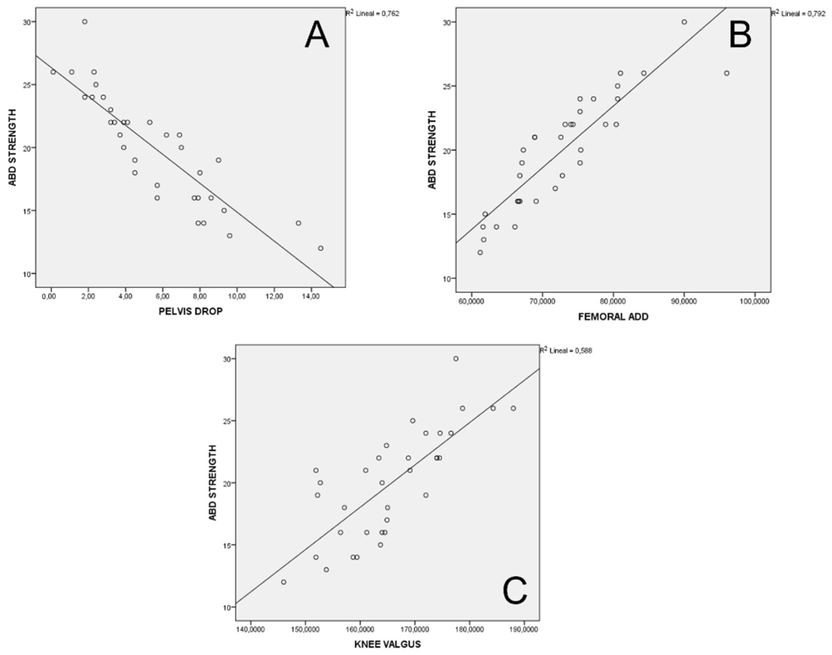
The correlation analysis showed a statistically significant negative correlation between the hip abductor muscles strength and the contralateral pelvic drop angle (p < 0.001). Figure 2a illustrates the negative correlation graphically, showing that the greater the hip abductor strength, the less contralateral pelvic drop angle. A statistically significant positive correlation was found between the hip abductor muscles strength and the femoral adduction angle (p < 0.001). Figure 2b shows the positive correlation indicating that the greater hip abductor muscle strength, the more femoral adduction angle. Additionally, a significant positive correlation was achieved between the hip abductor muscle strength and the knee valgus angle (p < 0.001). Figure 2c demonstrates the positive correlation showing that the greater the hip abductor muscle strength, the more knee valgus angle. Correlation analysis data is reported in Table 3.

4. Discussion

This is the first study investigating the relationship between hip abductor muscles strength and frontal plane kinematics in HB athletes. The results of this study confirmed the hypothesis raised and reported a very strong relationship between the hip abductor muscles strength and the pelvic drop, femoral adduction, and knee valgus in male HB athletes.
A negative relationship was found between hip abductor muscles strength and pelvic drop angle, while a positive relationship was identified with the femoral adduction angle. Thus, higher strength levels were related to less pelvic drop and femoral adduction in HB athletes. The results achieved in this study agree with those found by previous authors [18,45,46]. Selistre et al. [45] concluded that hip abductor strength explained 17% of contralateral pelvic drop and 21% of femoral adduction angle during walking in patients with knee osteoarthritis. Park et al. [46] reported that hip abductor muscles, knee flexor muscles, and ankle inversion muscles strength explained 34.4% of contralateral pelvic drop during walking in patients with knee osteoarthritis. Willy et al. [18] found a statistically significant reduction in contralateral pelvic drop and femoral adduction during a single-leg squat after a hip strengthening program in runners. Other studies showed no associations between hip muscle abductors strength and pelvis and hip kinematics. However, the task requested was not a single-leg task, which could lead to conflicting results [47,48]. Nevertheless, this is the first study showing a very strong correlation among HB athletes.
The results of this study also showed a positive relationship between hip abduction strength and dynamic knee valgus angle in HB athletes; greater strength was associated with less dynamic valgus. Previous studies support the hypothesis concerning the relationship between hip muscles and dynamic knee valgus in athletes using a 2D motion capture [19,49]. Willson et al. [49] showed a significant positive correlation between external rotation hip strength and the dynamic knee valgus in soccer, basketball, and volleyball athletes during single-leg squat. Ferri-Caruana et al. [19] reported a decrease in dynamic knee valgus at dynamic landing after applying a hip muscle strengthening protocol in soccer athletes. Suzuki et al. [50] reported similar results using a 3D motion capture system, showing a moderate correlation between hip muscles and knee kinematics in basketball athletes during single-legged medial drop. These findings suggest that using 2D software Kinovea may present similar results compared to a 3D system. Previous studies have analyzed the association between 2D software Kinovea and a 3D system (©VICON Motion System, Oxford, UK) for measuring hip, knee, and ankle angles during walking, and found a reasonable association with errors between 2º and 5º between both systems [51]. Thus, 2D systems may elicit similar results to 3D systems, and could be used in future studies and training programs to analyze injury risk.
Lower limb kinematics in the frontal plane seems to play a role in an athlete’s development of lower limb injuries [5,6,7,8,11,12,13]. This study evidences its relationship with the strength of the hip abductor muscles, and several authors suggest that these variables are modifiable with a physical therapy intervention based on strengthening [19,49,50]. Thus, its evaluation and management in clinical practice should be considered. In this sense, 2D motion capture systems for kinematic assessment appear to be reliable and accessible to clinicians. In addition, previous studies included runners [18,44,45], soccer [19,49], basketball [49,50], and volleyball [49] athletes, but this is the first study focused on HB athletes. Since HB showed a higher risk of injury than these sports [1], it is necessary to understand the relationship between biomechanical risk factors to develop injury prevention or rehabilitation strategies.
The results of the present study suggested a very strong relationship between the hip abductor muscles strength and the dynamic balance of the lower limb. Therefore, as a preventive measure, it should be considered to assess and improve the strength of the hip or knee muscles of HB athletes. Using 2D software Kinovea could facilitate the control of hip and knee kinematics in the clinical practice and sports environment.
This study presents some limitations. First, the study design is correlational, and the relationships observed do not imply causality. Second, the reliability measures described in this study may not be a valid determinant because they were calculated on the same day. Third, it used 2D motion capture systems, while 3D systems have been considered the gold standard. Fourth, kinematic analysis performed in a controlled environment may not accurately reproduce the athletic environment. Fifth, it was suggested that ipsilateral trunk tilt could act as a compensatory mechanism preventing the pelvic drop, but trunk kinematics were not assessed. Sixth, the test-retest reliability was measured on the same day, which could have favored the high ICC values. Finally, the sample was made up exclusively of men, so these results cannot be extrapolated to HB female athletes.
Future research should contrast our findings in HB athletes. Studies should include the frontal plane’s kinematic analysis of trunk tilt and its relationship to pelvic drop. Furthermore, future research should involve a prospective analysis, evaluating the effects on the number of injuries, strength, and function in HB athletes with different musculoskeletal conditions and kinematics of lower limbs. In addition, clinical trials should be carried out in order to analyze the effects of prevention programs and rehabilitation in HB athletes with lower limb dysfunctions on muscle strength and lower limb kinematics.

5. Conclusions

Hip abductor muscle strength strongly correlates with the pelvic drop, femoral adduction, and dynamic knee valgus in HB athletes. These results suggest that the hip abductor muscles strength plays an important role in the frontal plane lower limb kinematics in HB athletes.
This study highlights the importance of assessing lower limb kinematics and muscle function in HB athletes. Future studies should focus on prospective analysis of the kinematics of the lower limb, and on analyzing the effects of prevention and rehabilitation programs in HB athletes with lower limb dysfunction on muscle strength and lower limb kinematics.

Author Contributions

Conceptualization, L.C.-L. and S.J.-d.-B.; Methodology, A.C.-U., S.C.-B. and S.P.-G.; Software, R.M.-d.-l.-F. and I.H.-G..; Validation, A.C.-U., S.C.-B. and S.P.-G.; Formal Analysis, R.M.-d.-l.-F. and I.H.-G.; Investigation, A.C.-U., S.C.-B., S.P.-G., R.M.-d.-l.-F. and I.H.-G.; Resources, A.C.-U., S.C.-B., S.P.-G., R.M.-d.-l.-F. and I.H.-G.; Data Curation, R.M.-d.-l.-F. and I.H.-G.; Writing—Original Draft Preparation, L.C.-L., S.J.-d.-B., A.C.-U., S.C.-B., S.P.-G., R.M.-d.-l.-F. and I.H.-G.; Writing—Review and Editing, L.C.-L., S.J.-d.-B., A.C.-U., S.C.-B., S.P.-G., R.M.-d.-l.-F. and I.H.-G.; Supervision, L.C.-L., S.J.-d.-B., A.C.-U., S.C.-B., S.P.-G., R.M.-d.-l.-F. and I.H.-G.; Project Administration, L.C.-L. and S.J.-d.-B. All authors have read and agreed to the published version of the manuscript.

Funding

This research received no external funding.

Institutional Review Board Statement

This study design, protocol and consent forms were performed in accordance with the Helsinki Declaration of 1964 (revised in Fortaleza, 2013). The ethical approval for this study was obtained from the Clinical Research Ethics Committee of Valladolid Este (CASVE-NM-21-504).

Informed Consent Statement

Any research article describing a study involving humans should contain this statement. Please add “Informed consent was obtained from all subjects involved in the study.” OR “Patient consent was waived due to REASON (please provide a detailed justification).” OR “Not applicable.” for studies not involving humans. You might also choose to exclude this statement if the study did not involve humans. Written informed consent for publication must be obtained from participating patients who can be identified (including by the patients themselves). Please state “Written informed consent has been obtained from the patient(s) to publish this paper” if applicable.

Data Availability Statement

The data analyzed in this study are included in this published article. The dataset are available from the corresponding author on reasonable request.

Acknowledgments

Thanks to all the patients who participated in this study.

Conflicts of Interest

The authors declare no conflict of interest.

References

  1. Soligard, T.; Steffen, K.; Palmer, D.; Alonso, J.M.; Bahr, R.; Lopes, A.; Dvorak, J.; Grant, M.-E.; Meeuwisse, W.; Mountjoy, M.; et al. Sports injury and illness incidence in the Rio de Janeiro 2016 Olympic Summer Games: A prospective study of 11274 athletes from 207 countries. Br. J. Sports Med. 2017, 51, 1265–1271. [Google Scholar] [CrossRef] [PubMed] [Green Version]
  2. Bere, T.; Alonso, J.-M.; Wangensteen, A.; Bakken, A.; Eirale, C.; Dijkstra, H.P.; Ahmed, H.; Bahr, R.; Popovic, N. Injury and illness surveillance during the 24th Men's Handball World Championship 2015 in Qatar. Br. J. Sports Med. 2015, 49, 1151–1156. [Google Scholar] [CrossRef] [PubMed]
  3. Langevoort, G.; Myklebust, G.; Dvorak, J.; Junge, A. Handball injuries during major international tournaments. Scand. J. Med. Sci. Sports 2006, 17, 400–407. [Google Scholar] [CrossRef]
  4. Martín-Guzón, I.; Muñoz, A.; Lorenzo-Calvo, J.; Muriarte, D.; Marquina, M.; de la Rubia, A. Injury Prevalence of the Lower Limbs in Handball Players: A Systematic Review. Int. J. Environ. Res. Public Health 2021, 19, 332. [Google Scholar] [CrossRef] [PubMed]
  5. Barton, C.; Bonanno, D.; Carr, J.; Neal, B.; Malliaras, P.; Franklyn-Miller, A.; Menz, H. Running retraining to treat lower limb injuries: A mixed-methods study of current evidence synthesised with expert opinion. Br. J. Sports Med. 2016, 50, 513–526. [Google Scholar] [CrossRef] [Green Version]
  6. Ferber, R.; Noehren, B.; Hamill, J.; Davis, I. Competitive Female Runners with a History of Iliotibial Band Syndrome Demonstrate Atypical Hip and Knee Kinematics. J. Orthop. Sports Phys. Ther. 2010, 40, 52–58. [Google Scholar] [CrossRef] [PubMed] [Green Version]
  7. McCarthy, C.; Fleming, N.; Donne, B.; Blanksby, B. Barefoot running and hip kinematics: Good news for the knee? Med. Sci. Sports Exerc. 2015, 47, 1009–1016. [Google Scholar] [CrossRef] [PubMed]
  8. Neal, B.S.; Barton, C.J.; Gallie, R.; O’Halloran, P.; Morrissey, D. Runners with patellofemoral pain have altered biomechanics which targeted interventions can modify: A systematic review and meta-analysis. Gait Posture 2015, 45, 69–82. [Google Scholar] [CrossRef] [PubMed]
  9. Tsai, Y.-J.; Huang, Y.-C.; Chen, Y.-L.; Hsu, Y.-W.; Kuo, Y.-L. A Pilot Study of Hip Corrective Taping Using Kinesio Tape for Pain and Lower Extremity Joint Kinematics in Basketball Players with Patellofemoral Pain. J. Pain Res. 2020, 13, 1497–1503. [Google Scholar] [CrossRef]
  10. Larwa, J.; Stoy, C.; Chafetz, R.; Boniello, M.; Franklin, C. Stiff Landings, Core Stability, and Dynamic Knee Valgus: A Systematic Review on Documented Anterior Cruciate Ligament Ruptures in Male and Female Athletes. Int. J. Environ. Res. Public Health 2021, 18, 3826. [Google Scholar] [CrossRef]
  11. Petersen, W.; Rembitzki, I.; Liebau, C. Patellofemoral pain in athletes. Open Access J. Sports Med. 2017, 8, 143–154. [Google Scholar] [CrossRef] [PubMed] [Green Version]
  12. Koga, H.; Nakamae, A.; Shima, Y.; Iwasa, J.; Myklebust, G.; Engebretsen, L.; Bahr, R.; Krosshaug, T. Mechanisms for noncontact anterior cruciate ligament injuries: Knee joint kinematics in 10 injury situations from female team handball and basketball. Am. J. Sports Med. 2010, 38, 2218–2225. [Google Scholar] [CrossRef]
  13. Hewett, T.E.; Myer, G.D.; Ford, K.R.; Heidt, R.S., Jr.; Colosimo, A.J.; McLean, S.G.; Van Den Bogert, A.J.; Paterno, M.V.; Succop, P. Biomechanical Measures of Neuromuscular Control and Valgus Loading of the Knee Predict Anterior Cruciate Ligament Injury Risk in Female Athletes: A Prospective Study. Am. J. Sports Med. 2005, 33, 492–501. [Google Scholar] [CrossRef] [PubMed] [Green Version]
  14. Cashman, G.E. The Effect of Weak Hip Abductors or External Rotators on Knee Valgus Kinematics in Healthy Subjects: A Systematic Review. J. Sport Rehabil. 2012, 21, 273–284. [Google Scholar] [CrossRef] [PubMed] [Green Version]
  15. Wilczyński, B.; Zorena, K.; Ślęzak, D. Dynamic Knee Valgus in Single-Leg Movement Tasks. Potentially Modifiable Factors and Exercise Training Options. A Literature Review. Int. J. Environ. Res. Public Health 2020, 17, 8208. [Google Scholar] [CrossRef]
  16. Prins, M.R.; van der Wurff, P. Females with patellofemoral pain syndrome have weak hip muscles: A systematic review. Aust. J. Physiother. 2009, 55, 9–15. [Google Scholar] [CrossRef] [Green Version]
  17. Padua, D.A.; Marshall, S.W.; Beutler, A.I.; DeMaio, M.; Boden, B.P.; Yu, B.; Garrett, W.E. Predictors of Knee Valgus Angle During a Jump-landing Task. Med Sci Sport Exerc. 2005, 37, S398. [Google Scholar] [CrossRef]
  18. Willy, R.W.; Davis, I.S. The Effect of a Hip-Strengthening Program on Mechanics During Running and During a Single-Leg Squat. J. Orthop. Sports Phys. Ther. 2011, 41, 625–632. [Google Scholar] [CrossRef] [Green Version]
  19. Ferri-Caruana, A.; Prades Insa, B.; Serra Añó, P. Effects of pelvic and core strength training on biomechanical risk factors for anterior cruciate ligament injuries. J Sports Med Phys Fitness. 2020, 60, 1128–1136. [Google Scholar] [CrossRef]
  20. Bolgla, L.A.; Malone, T.R.; Umberger, B.R.; Uhl, T.L. Hip strength and hip and knee kinematics during stair descent in females with and without patellofemoral pain syndrome. J. Orthop. Sports Phys. Ther. 2008, 38, 12–18. [Google Scholar] [CrossRef]
  21. Schurr, S.A.; Marshall, A.N.; Resch, J.E.; Saliba, S.A. Two-Dimensional Video Analysis is Comparable To 3d Motion Capture in Lower Extremity Movement Assessment. Int. J. Sports Phys. Ther. 2017, 12, 163–172. [Google Scholar] [PubMed]
  22. Franko, O.I.; Tirrell, T.F. Smartphone App Use Among Medical Providers in ACGME Training Programs. J. Med Syst. 2011, 36, 3135–3139. [Google Scholar] [CrossRef]
  23. Herrington, L.; Alenezi, F.; Alzhrani, M.; Alrayani, H.; Jones, R. The reliability and criterion validity of 2D video assessment of single leg squat and hop landing. J. Electromyogr. Kinesiol. 2017, 34, 80–85. [Google Scholar] [CrossRef] [PubMed] [Green Version]
  24. Puig-Diví, A.; Escalona-Marfil, C.; Padullés-Riu, J.M.; Busquets, A.; Padullés-Chando, X.; Marcos-Ruiz, D. Validity and reliability of the Kinovea program in obtaining angles and distances using coordinates in 4 perspectives. PLoS ONE 2019, 14, e0216448. [Google Scholar] [CrossRef]
  25. Sañudo, B.; Rueda, D.; Pozo-Cruz, B.D.; de Hoyo, M.; Carrasco, L. Validation of a video analysis software package for quantifying movement velocity in resistance exercises. J strenght Cond Res. 2016, 30, 2934–2941. [Google Scholar] [CrossRef] [PubMed]
  26. Jiménez-del-Barrio, S.; Mingo-Gómez, M.T.; Estébanez-de-Miguel, E.; Saiz-Cantero, E.; del-Salvador-Miguélez, A.I.; Ceballos-Laita, L. Adaptations in pelvis, hip and knee kinematics during gait and muscle extensibility in low back pain patients: A cross-sectional study. J. Back Musculoskelet. Rehabil. 2020, 33, 49–56. [Google Scholar] [CrossRef]
  27. Ismail, S.I.; Adnan, R.; Sulaiman, N. Moderate Effort Instep Kick in Futsal. Procedia Eng. 2014, 72, 186–191. [Google Scholar] [CrossRef] [Green Version]
  28. Torralba, J.; Padullés, R.; Losada, L.; López, D.A. Ecological alternative in measuring long jump of paralimpics athletes. Cuad. De Psicol. Del Deporte 2016, 16, 69–76. [Google Scholar]
  29. Balsalobre-Fernandez, C.; Tejero-Gonzales, C.; Campo-Vecino, J.; Bavareso, N. The concurrent validity and reliability of a low-cost, high-speed camera-based method for measuring the flight time of vertical jumps. J. Strength Cond. Res. 2015, 28, 528–533. [Google Scholar] [CrossRef]
  30. McCurdy, K.; Walker, J.; Armstrong, R.; Langford, G. Relationship Between Selected Measures of Strength and Hip and Knee Excursion During Unilateral and Bilateral Landings in Women. J. Strength Cond. Res. 2014, 28, 2429–2436. [Google Scholar] [CrossRef]
  31. Hertzog, M.A. Considerations in determining sample size for pilot studies. Res. Nurs. Health 2008, 31, 180–191. [Google Scholar] [CrossRef] [PubMed]
  32. van Melick, N.; Meddeler, B.M.; Hoogeboom, T.J.; Nijhuis-van der Sanden, M.W.G.; van Cingel, R.E.H. How to determine leg dominance: The agreement between self-reported and observed performance in healthy adults. PLoS ONE 2017, 12, e0189876. [Google Scholar] [CrossRef] [Green Version]
  33. Schneiders, A.G.; Sullivan, S.J.; O'Malley, K.J.; Clarke, S.V.; Knappstein, S.A.; Taylor, L.J. A Valid and Reliable Clinical Determination of Footedness. PM&R 2010, 2, 835–841. [Google Scholar] [CrossRef]
  34. Mentiplay, B.F.; Perraton, L.G.; Bower, K.J.; Adair, B.; Pua, Y.-H.; Williams, G.P.; McGaw, R.; Clark, R.A. Assessment of Lower Limb Muscle Strength and Power Using Hand-Held and Fixed Dynamometry: A Reliability and Validity Study. PLoS ONE 2015, 10, e0140822. [Google Scholar] [CrossRef] [PubMed] [Green Version]
  35. Parcell, A.C.; Sawyer, R.D.; Tricoli, V.; Chinevere, T.D. Minimum rest period for strength recovery during a common isokinetic testing protocol. Med. Sci. Sports Exerc. 2002, 34, 1018–1022. [Google Scholar] [CrossRef]
  36. Dingenen, B.; Malliaras, P.; Janssen, T.; Ceyssens, L.; Vanelderen, R.; Barton, C.J. Two-dimensional video analysis can discriminate differences in running kinematics between recreational runners with and without running-related knee injury. Phys. Ther. Sport 2019, 38, 184–191. [Google Scholar] [CrossRef]
  37. Damsted, C.; Nielsen, R.O.; Larsen, L.H. Reliability of video-based quantification of the knee- and hip angle at foot strike during running. Int. J. Sports Phys. Ther. 2015, 10, 147–154. [Google Scholar]
  38. Dingenen, B.; Staes, F.F.; Santermans, L.; Steurs, L.; Eerdekens, M.; Geentjens, J.; Peers, K.H.; Thysen, M.; Deschamps, K. Are two-dimensional measured frontal plane angles related to three-dimensional measured kinematic profiles during running? Phys. Ther. Sport. 2018, 29, 84–92. [Google Scholar] [CrossRef]
  39. Llurda-Almuzara, L.; Pérez-Bellmunt, A.; López-De-Celis, C.; Aiguadé, R.; Seijas, R.; Casasayas-Cos, O.; Labata-Lezaun, N.; Alvarez, P. Normative data and correlation between dynamic knee valgus and neuromuscular response among healthy active males: A cross-sectional study. Sci. Rep. 2020, 10, 17206. [Google Scholar] [CrossRef]
  40. Baude, M.; Hutin, E.; Gracies, J.-M. A Bidimensional System of Facial Movement Analysis Conception and Reliability in Adults. BioMed Res. Int. 2015, 2015, 812961. [Google Scholar] [CrossRef]
  41. Dingenen, B.; Barton, C.; Janssen, T.; Benoit, A.; Malliaras, P. Test-retest reliability of two-dimensional video analysis during running. Phys Ther Sport. 2018, 33, 40–47. [Google Scholar] [CrossRef] [PubMed]
  42. Weeks, B.K.; Carty, C.P.; Horan, S.A. Kinematic predictors of single-leg squat performance: A comparison of experienced physiotherapists and student physiotherapists. BMC Musculoskelet Disord. 2012, 13, 207. [Google Scholar] [CrossRef] [PubMed]
  43. Koo, T.K.; Li, M.Y. A Guideline of Selecting and Reporting Intraclass Correlation Coefficients for Reliability Research. J. Chiropr. Med. 2016, 15, 155–163. [Google Scholar] [CrossRef] [PubMed] [Green Version]
  44. Hopkins, W.G.; Marshall, S.W.; Batterham, A.M.; Hanin, J. Progressive Statistics for Studies in Sports Medicine and Exercise Science. Med. Sci. Sports Exerc. 2009, 41, 3–13. [Google Scholar] [CrossRef] [Green Version]
  45. Selistre, L.F.A.; Gonçalves, G.H.; Nakagawa, T.H.; Petrella, M.; Jones, R.K.; Mattiello, S. The role of hip abductor strength on the frontal plane of gait in subjects with medial knee osteoarthritis. Physiother. Res. Int. 2019, 24, e1779. [Google Scholar] [CrossRef]
  46. Park, S.-K.; Kobsar, D.; Ferber, R. Relationship between lower limb muscle strength, self-reported pain and function, and frontal plane gait kinematics in knee osteoarthritis. Clin. Biomech. 2016, 38, 68–74. [Google Scholar] [CrossRef]
  47. Zeitoune, G.; Leporace, G.; Batista, L.A.; Metsavaht, L.; Lucareli, P.R.G.; Nadal, J. Do hip strength, flexibility and running biomechanics predict dynamic valgus in female recreational runners? Gait Posture. 2020, 79, 217–223. [Google Scholar] [CrossRef]
  48. Brindle, R.A.; Ebaugh, D.D.; Willson, J.D.; Finley, M.A.; Shewokis, P.A.; Milner, C.E. Relationships of hip abductor strength, neuromuscular control, and hip width to femoral length ratio with peak hip adduction angle in healthy female runners. J. Sports Sci. 2020, 38, 2291–2297. [Google Scholar] [CrossRef]
  49. Willson, J.D.; Ireland, M.L.; Davis, I. Core strength and lower extremity alignment during single leg squats. Med Sci Sports Exerc. 2006, 38, 945–952. [Google Scholar] [CrossRef] [Green Version]
  50. Suzuki, H.; Omori, G.; Uematsu, D.; Nishino, K.; Endo, N. The influence of hip strength on knee kinematics during a single-legged medial drop landing among competitive collegiate basketball players. Int. J. Sports Phys. Ther. 2015, 10, 592–601. [Google Scholar]
  51. Fernández-González, P.; Koutsou, A.; Cuesta-Gómez, A.; Carratalá-Tejada, M.; Miangolarra-Page, J.C.; Molina-Rueda, F. Reliability of Kinovea® Software and Agreement with a Three-Dimensional Motion System for Gait Analysis in Healthy Subjects. Sensors 2020, 20, 3154. [Google Scholar] [CrossRef] [PubMed]
Figure 1. (A) Kinovea analysis of the contralateral pelvic drop angle; (B) Kinovea analysis of the femoral adduction angle; (C) Kinovea analysis of the knee valgus angle.
Applsci 12 10044 g001
Figure 2. (A) Relationship between hip strength and contralateral pelvic drop. (B) Relationship between hip strength and femoral adduction. (C) Relationship between hip strength and knee valgus.
Applsci 12 10044 g002
Table 1. Test-retest reliability of the study variables.
ICC(2,1) (95% CI)
Hip abductor strength (NW)0.97 (0.95, 0.98)
Contralateral pelvic drop (°)0.92 (0.9, 0.94)
Femoral adduction (°)0.95 (0.9, 0.97)
Knee valgus (°)0.97 (0.96, 0.98)
Abbreviations: ICC: intraclass correlation coefficient; CI: confidence interval.
Table 2. Mean and standard deviations of hip strength and frontal plane kinematics.
Dominant LimbNon-Dominant LimbSignificance
Hip abductor strength (NW)204.38 (34.67)186.43 (50.05)0.165 b
Contralateral pelvic drop (°)5.42 (3.53)5.72 (3.36)0.671 a
Femoral adduction (°)72.24 (8.08)73.22 (8.42)0.648 a
Knee valgus (°)164.43 (10.58)166.44 (9.46)0.522 a
a: Paired sample t-test; b: Wilcoxon test. Abbreviations: SD: standard deviation; NW: newtons; °: Degrees.
Table 3. Correlation analysis.
Pearson CorrelationPelvic Drop (°)Femoral Adduction (°)Knee Valgus (°)
Hip abductor strength (NW)R = −0.873r = 0.767r = 0.855
Significance<0.001<0.001<0.001
R = Pearson correlation test. Abbreviations: NW: newtons; °: degrees.
Publisher’s Note: MDPI stays neutral with regard to jurisdictional claims in published maps and institutional affiliations.