Article

ATP-Binding Cassette (ABC) Transporters in Fusarium Specific Mycoparasite Sphaerodes mycoparasitica during Biotrophic Mycoparasitism

Department of Food and Bioproduct Sciences, University of Saskatchewan, Saskatoon, SK S7N 5A8, Canada
*
Author to whom correspondence should be addressed.
Present address: Plant Healthcare Research Institute, JAN153 Biotech Incorporated, Gwangju 61186, Korea.
Appl. Sci. 2022, 12(15), 7641; https://doi.org/10.3390/app12157641
Submission received: 29 May 2022 / Revised: 25 July 2022 / Accepted: 25 July 2022 / Published: 29 July 2022
(This article belongs to the Special Issue Plant Microbiome Responses to Environmental Changes)

Abstract

:
Recent transcriptomic profiling has revealed importance membrane transporters such as ATP-binding cassette (ABC) transporters in fungal necrotrophic mycoparasites. In this study, RNA-Seq allowed rapid detection of ABC transcripts involved in biotrophic mycoparasitism of Sphaerodes mycoparasitica against the phytopathogenic and mycotoxigenic Fusarium graminearum host, the causal agent of Fusarium head blight (FHB). Transcriptomic analyses of highly expressed S. mycoparasitica genes, and their phylogenetic relationships with other eukaryotic fungi, portrayed the ABC transporters’ evolutionary paths towards biotrophic mycoparasitism. Prior to the in silico phylogenetic analyses, transmission electron microscopy (TEM) was used to confirm the formation of appressorium/haustorium infection structures in S. mycoparasitica during early (1.5 d and 3.5 d) stages of mycoparasitism. Transcripts encoding biotrophy-associated secreted proteins did uncover the enrolment of ABC transporter genes in this specific biocontrol mode of action, while tandem ABC and BUB2 (non-ABC) transcripts seemed to be proper for appressorium development. The next-generation HiSeq transcriptomic profiling of the mycoparasitic hypha samples, revealed 81 transcripts annotated to ABC transporters consisting of a variety of ABC-B (14%), ABC-C (22%), and ABC-G (23%), and to ABC-A, ABC-F, aliphatic sulfonates importer (TC 3.A.1.17.2), BtuF, ribose importer (TC 3.A.1.2.1), and unknown families. The most abundant transcripts belonged to the multidrug resistance exporter (TC 3.A.1.201) subfamily of the ABC-B family, the conjugate transporter (TC 3.A.1.208) subfamily of the ABC-C family, and the pleiotropic drug resistance (PDR) (TC 3.A.1.205) subfamily of the ABC-G family. These findings highlight the significance of ABC transporter genes that control cellular detoxification against toxic substances (e.g., chemical pesticides and mycotoxins) in sustaining a virulence of S. mycoparasitica for effective biotrophic mycoparasitism on the F. graminearum host. The findings of this study provide clues to better understand the biotrophic mycoparasitism of S. mycoparasitica interacting with the Fusarium host, which implies that the ABC transporter group of key proteins is involved in the mycoparasite’s virulence and multidrug resistance to toxic substances including cellular detoxification.

1. Introduction

Mycoparasitism is a parasitic interaction between two fungi as a direct mechanism of biologically controlling plant pathogenic fungi where one organism, called a parasite, is benefited, whereas another organism, called a host or prey, is harmed. According to Naranjo-Ortiz and Gabaldón (2019), the best-studied groups of mycoparasites are within the Pezizomycotina [1], with representatives within the Sordariomycetes [2,3] and particularly in the Hypocreales [4,5,6]. The range from necrotrophic to biotrophic mycoparasitism depends on the parasite’s mode of action and its effect on a host as well as its host compatibility and specificity. Necrotrophic mycoparasitism is an aggressive mode of action where a mycoparasite kills or destroys a fungal prey and acquires or absorbs nutrients from the killed host cells, while biotrophic mycoparasitism is a more sophisticated mode of action combining a destructive and balanced strategy in which a mycoparasite lets nutrients of host cells flow out through intimate relationships between a parasite and a host. Necrotrophic mycoparasites are also known as generalists and include several Trichoderma species such as T. harzianum, T. viride, T. virens, T. atroviride, T. koningii, T. gamsii, and Clonostachys rosea, whereas biotrophic mycoparasites are known as specialists and include Ampelomyces quisqualis and Sphaerodes species such as S. mycoparasitica, S. quadrangularis, and S. retispora var. retispora [7].
Recently, biocontrol science has shifted from these non-specific to specific biocontrol fungal agents such as mycoparasitic Ampelomyces against powdery mildew (Erysiphales), Sphaerodes against Fusaria pathogens, and mycotoxins in cereal grains. Sphaerodes mycoparasitica is a biotrophic polyphagous mycoparasite of plant pathogenic and mycotoxigenic Fusarium species [2]. S. mycoparasitica produces hook-shaped contact structures [8] that eventually penetrate the host hyphae of the FHB Fusarium species through presumptive appressorium/haustorium-like structures during mycoparasitism. It is assumed that hyphal mechanical pressure combined with enzymatic hydrolysis is responsible for the invasion process in which S. mycoparasitica parasitizes the host. These assumptions related to several mycoparasitism-related genes and transcripts have not yet been investigated in S. mycoparasitica, which led to our attention.
Previous studies in our group have demonstrated that S. mycoparasitica parasitizes different Fusarium species through diphasic interactions, while its adaptability to the Fusarium hosts has been demonstrated using in vitro compatibility assays [2]. Moreover, the biodegradation or biotransformation ability of S. mycoparasitica to mycotoxins such as zearalenone, deoxynivalenol, and its derivatives (3-acetyl-deoxynivalenol and 15-acetyl-deoxynivalenol) has been uncovered by means of biodegradation experiments [9].
In general, mycoparasites produce cell wall degrading enzymes (CWDEs) such as glucanases and chitinases involved in the destruction of the phytopathogenic host cell wall [10,11]. The genes encoding CWDEs are often highly upregulated during the early stages of mycoparasitism [12,13]. Genes encoding the components of a mitogen-activated protein kinase (MAPK) [14,15], NADPH oxidase, oxalate decarboxylase, transcription factors [16,17], and the peroxisome and heat shock [18,19] are vital regulatory factors in mycoparasitism. In other instances, expression of mycoparasitism- and secondary metabolism-related genes are often differentially regulated in the presence or absence of a phytopathogenic host [20] which unveils the importance of in vivo interaction studies to discover different regulation pathways in mycoparasites.
When the mycoparasite interacts with a host in a particular environment, the cell wall (rigid and stable structure, also a storage site of regulatory molecules that sense the presence of pathogenic microbes) and the cell membrane (more flexible and responding to the dynamic changes) are the first barriers to recognize abiotic and biotic factors. The cell membrane commonly including membrane-bound or integrated proteins is expected to play a crucial role associated with mycoparasitism. A recent finding by Wu and Cox (2021) supported this insight. Five different lifestyle-specific genes, representing endophytism, entomopathogenism, plantpathogenism, mycoparasitism, and symbiosis [21], all encoded abundant proteins with multiple functional signatures. The genes were implicated in transmembrane transport and oxidation/reduction processes as integral components of membranes in fungi across their lifestyles and evolutionary functions.
In the well-known biocontrol agent Trichoderma, which is an extensively studied general mycoparasite, different ABC transporter genes have been reported to be upregulated during the tripartite mycoparasite-fungal pathogen–plant system [22]. Indeed, ABC transporter genes are involved in several Trichoderma’s mycoparasite activities by sustaining an efficient biocontrol. The pleiotropic drug resistance (PDR) family of ABC transporters such as the Taabc2 gene in Trichoderma atroviride, regulates fungal antagonism and the colonization process on phytopathogens under fluctuating environmental conditions [23]. Another PDR family of genes within the ABC-G subfamily members, including Pdr5p in the model yeast Saccharomyces cerevisiae, has been described as active efflux pumps conferring resistance to structurally and functionally unrelated drugs and xenobiotics: antibiotics, fungicides, detergents, ionophores, steroid hormones, and anticancer drugs [24,25,26]. In mycoparasite Clonostachys rosea, abcG5, belonging to the PDR family, seems to be an essential ABC transporter gene responsible for antagonistic and biocontrol activity [27]. Interestingly, abcG5 gene expression was induced by mycotoxin zearalenone [28], while its deletion in C. rosea resulted in lowering fungal tolerance to zearalenone produced by plant pathogenic Fusarium species [27].
Recent research on C. rosea during interactions with its hosts (Botrytis cinerea and F. graminearum) showed that transcriptional responses of C. rosea could predict more than half of the transcripts encoding membrane transporters (61%), followed by biosynthesis of secondary metabolites (12%) and carbohydrate-active enzymes (7%) [29]. Especially, the expressions of the two ABC transporter genes, i.e., abcC8 and abcG18 of C. rosea, were induced during the interaction with respective hosts B. cinerea and F. graminearum [29]. Moreover, the transcriptional responses of C. rosea genes such as abcC8 and abcG18 were induced by fungicides (boscalid and mefenoxam) and the mycotoxin zearalenone, respectively [29].
Despite significant research advancements in fungal functional genes and transcriptomics, the knowledge of transcriptomics in mycoparasitic specialists continues to lag far behind that of the mycoparasitic generalists. In addition, the distinction of Sphaerodes RNA-Seq profile from Trichoderma and Clonostachys transcriptomics remains unknown. This study on Sphaerodes focuses on membrane-bound or integral membrane proteins with diverse ABC transporter functions involved in the detoxification of toxic compounds/mycotoxins within the interactive mycoparasite–Fusarium zone. Moreover, a refined identification, classification, and evolutionary phylogeny of ABC transporters based on the transcripts may serve as predictive indications for differences in ecology and biocontrol efficiencies across different mycoparasitic lifestyles. An improved specific biocontrol against Fusarium graminearum in cereals is still waiting for genomic and transcriptomic solutions [30]. The need is further accentuated since the world production of cereals encountered a steady increase in F. graminearum incidence and experienced devastating losses due to Fusarium head blight (FHB) outbreaks and associated mycotoxins over the last decades [31]. In this study, we aimed to better understand the transcriptomic bases of mycoparasitism in S. mycoparasitica interacting with the Fusarium host, with particular emphasis on the ABC families of transporters possibly involved in the mechanisms of biocontrol, virulence, multiresistance, and cell detoxification. The results could further help to better explain the differences between distinct microparasitic lifestyles as well as the evolutionary advantages of the biotrophic-specific mycoparasitism as compared with well-known necrotrophic mycoparasitism.

2. Materials and Methods

In this study, we explore the transcriptome of Sphaerodes mycoparasitica Vujan. during biotrophic mycoparasitism with mycotoxigenic Fusarium graminearum Schwabe with an emphasis on the analysis of the ATP-binding cassette (ABC) transporters.

2.1. Co-Cultures

The S. mycoparasitica SMCD 2220-01 (mycopathogen) and its phytopathogenic host, F. graminearum SMCD 2243 aggressive 3-ADON chemotype were grown in co-culture. For general transcriptomic profiling of the mycoparasite during biotrophic mycoparasitism or to induce mycoparasitism-related gene expression, the mycoparasite was co-cultured on top of the host with a monofilament fabric, nylon mesh with 30 μm openings (SEFAR NITEX 03–48/31, Sefar Inc., Depew, NY, USA) placed between the mycoparasite and the host. The growth medium was potato glucose agar (PGA, Sigma-Aldrich, St. Louis, MO, USA) and co-cultures were incubated at 23 °C in darkness. The mycelia of the interactive mycoparasite used for RNA extraction were collected under sterile conditions using a Carl Zeiss Axioskop2 microscope at two different incubation time, 1.5 and 3.5 days. The method used in this study [32] allowed us to obtain a mycelium of the mycoparasite from co-cultures during an active mycoparasite–host interaction. The control samples consisted of the mycoparasite’s hyphae grown on fabric tissue without the host.

2.2. Transmission Electron Microscopy Analysis

To investigate morphological changes of the mycoparasite and the phytopathogenic host, two fungal isolates from the Saskatchewan Microbial Collection and Database (SMCD), a biocontrol fungus SMCD 2220-01 S. mycoparasitica Vujan. (2009) as a Fusarium specific mycoparasite [33,34], and SMCD 2243 F. graminearum Schwabe 3-ADON chemotype as a virulent plant pathogenic strain [2,8] were used as hosts for the mycoparasite. The fungi were grown on potato glucose agar (PGA, Sigma-Aldrich) medium at 23 °C in darkness for 7 days and fresh cultures were used. The fresh plugs of each culture (the approximate radius of the plugs was 0.8 cm and the distance between two plugs was about 2 cm) were inoculated on a slide culture system made of a thin layer of PGA (about 2 ml prior to solidification) on a sterilized slide glass and co-incubated for 8 days to be used for TEM. For a plate-based culture system, the mycoparasite was 3 days pre-inoculated on a PGA plate and co-cultured with the Fusarium host for 3 days.
The TEM analysis was conducted at the Western College of Veterinary Medicine Imaging Centre (WCVM), University of Saskatchewan, using a Hitachi HT7700 transmission electron microscope. Prior to the TEM analysis, the samples were stained with toluidine blue solution, which allowed us to screen the area of interest (the sample thickness was about 500 nm), and then ultrathin sections (approximate thickness 50 nm) were applied for the TEM analysis at 80.0 kV.

2.3. Transciptomic Analyses

The collected fungal samples containing fresh culture cells were used for further RNA extraction using an AurumTM Total RNA Mini Kit (Bio-Rad, Hercules, CA, USA), according to the manufacturer’s instructions. Thereafter, the extracted total RNAs were employed to perform the RNA evaluation followed by poly(A)+ RNA purification and cDNA library synthesis using a TrueSeq stranded mRNA Sample Prep kit (Illumina). Then, libraries were multiplexed with Illumina barcodes and sequenced per lane using an Illumina HiSeqTM 4000 system (PE 100 bp sequencing lane) based on the massively parallel sequencing protocol [35] at McGill University and Génome Québec Innovation Center (Montréal, QC, Canada).
The obtained raw reads were managed in Trimmomatic [36] for the trimming and clipping process, and then the clean reads were utilized for transcriptome de novo assembly which was accomplished by applying a general pipeline previously described [37,38] through the Trinity assembly software suite consisting of Inchworm, Chrysalis, and Butterfly at the Canadian Centre for Computational Genomics. The normalization counts, practically transcripts per million (TPM), were used as information on relative expression to compare different conditions. Further, the transcriptome BLAST search against GeneBank (NCBI) was performed with the BlastX program to identify protein-coding genes in cDNAs experimentally obtained and further predict potential functionality of the genes. Computational alignments were carried out by placing transcripts against the UniProtKB/Swiss-Prot (Swissprot) protein database (uniprot_sprot.trinotate_v2.0.pep) providing the highest level of annotation with a minimal level of redundancy along with NCBI non-redundant protein database and UniRef90. E-values less than 10–5 and sequence similarity greater than 60% were regarded as high homology. Further functional annotation was achieved through the Trinotate pipeline including Hmmer v.3.1b1 and PFAM for protein domain identification, SignalP v.4.1 and Tmhmm v.2.0c for protein signal peptide and transmembrane domain prediction, and EMBL Uniprot eggnog/GO pathways to search for orthologous groups.

2.4. Phylogenetic Analyses

S. mycoparasitica transcripts (i.e., Sm68082_c0_g1_i1 for biotrophy-associated secreted protein; Sm105692_c0_g1_i1, Sm72487_c0_g1_i2, Sm24426_c0_g1_i1, Sm52544_c0_g2_i1, Sm60015_c0_g1_i1, Sm107436_c0_g1_i1, and Sm72276_c0_g1_i2 for ABC-B; Sm78934_c5_g2_i8, Sm78625_c4_g2_i1, Sm78625_c4_g2_i3, Sm78625_c4_g2_i5, Sm75748_c0_g1_i1, Sm80145_c2_g9_i1, Sm75748_c0_g1_i3, Sm75748_c0_g1_i2, Sm76779_c0_g2_i1, and Sm79181_c0_g2_i4 for ABC-C; Sm80002_c1_g4_i2, Sm76040_c0_g1_i1 for ABC-F; Sm79490_c2_g2_i2 for ABC-G) of this Fusarium-specific mycoparasite were used to compare a biotrophy-associated protein and ABC-B, -C, -F, and -G transporters. Different fungi representing biotrophic, chemibiotrophic, necrotrophic, endophytic, and hemiendophytic lifestyles were selected for comparisons. Transcriptome (RNA sequencing) data of S. mycoparasitica are deposited in NCBI with currently available accession PRJNA757970.
The phylogenetic ABC transcript analyses of S. mycoparasitica as well as evolutionary relationships of this biotrophic mycoparasite with different fungal and other eukaryotes were investigated. The evolutionary history was inferred using the minimum evolution method [39]. The optimal phylogenetic trees with the sum of branch length equal to 2.46085065, 11.74499404, 53.69656248, 2.19433433, and 1.13037743 were shown. The trees were drawn to scale, with branch lengths in the same units as those of the evolutionary distances used to infer the phylogenetic trees. The evolutionary distances were computed using the maximum composite likelihood method [40] and were in the units of the number of base substitutions per site. The ME trees were searched using the close-neighbor-interchange (CNI) algorithm [41] at a search level of 1. The neighbor-joining algorithm [42] was used to generate the initial trees. All ambiguous positions were removed for each sequence pair (pairwise deletion option). There was a total of 1002, 7567, 458, 4870, and 3364 positions in the final datasets of the presented phylogenetic trees The evolutionary analyses were conducted in MEGA X [43].

3. Results

3.1. Investigation of Morphological Changes during Biotrophic Mycoparasitism

During the biotrophic mycoparasitism, there are some degrees of morphological alteration in Fusarium host cells affected by S. mycoparasitica. The results of the TEM analysis (Figure 1) depicted interesting dynamic changes in the host cell structure. In fact, S. mycoparasitica (1) formed the appressorium (Ap) to adhere tightly to the host cell surface and produced infective-constricted hypha to penetrate cell wall. Subsequently (Figure 1A), the mycoparasite (2) colonized the host cell and formed haustorium (H) to expend the cytoplasm, and (3) degraded host cell organelles and compartments into irregular shapes of the remains and cytoplasmic droplets. In Figure 1B, (4) a nearly empty host cell which has a host nucleus with folding of the cell and loss of integrity and rigidity of the cell wall can be observed. In Figure 1C, (5) an advanced degradation of the host cell wall appeared on the cellular side in proximity to a mucosa-like substance produced by the mycoparasite (highlighted in the yellow arrow), whereas the cell wall opposite to the mycoparasite was still undamaged.
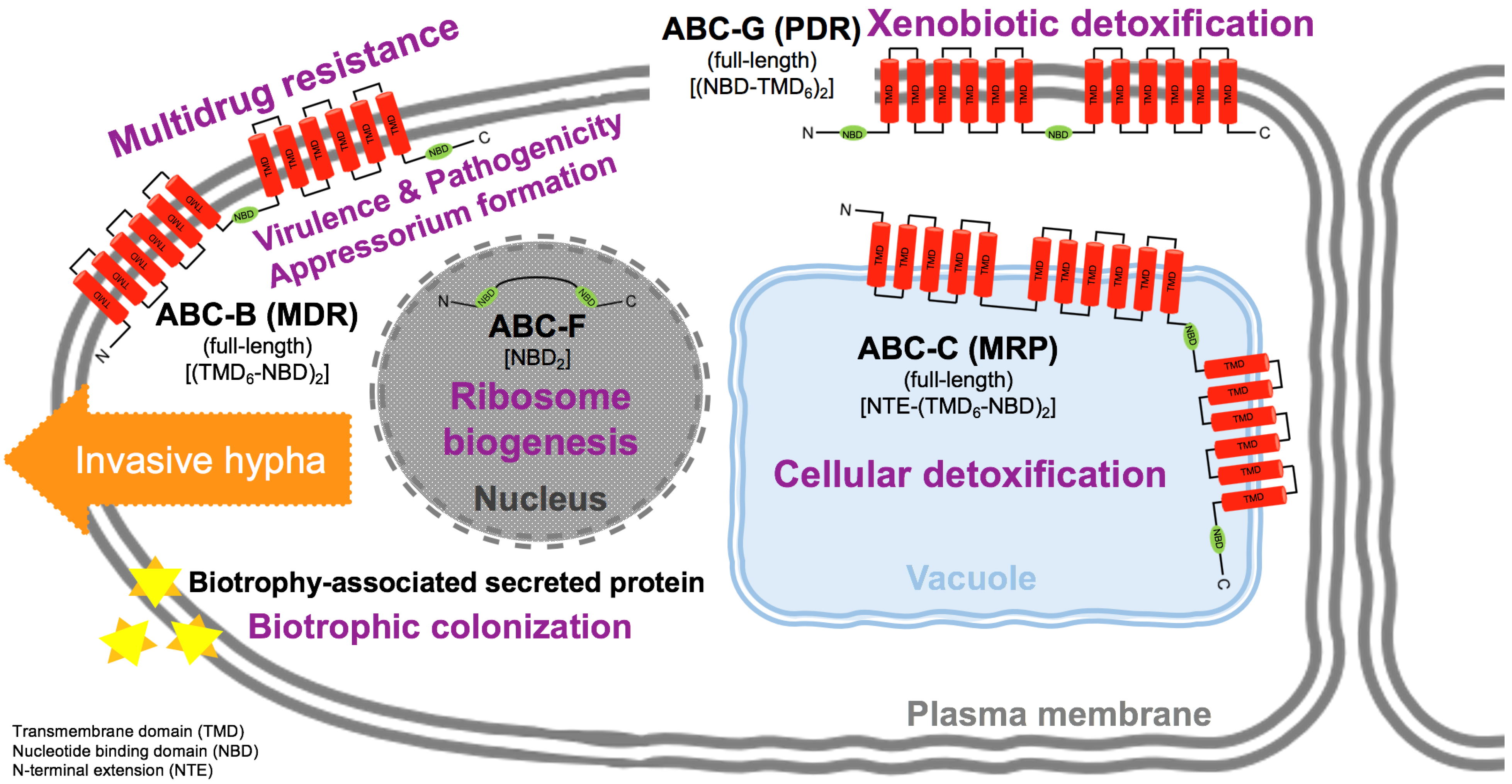
In addition to the host cell morpho-structural alteration, two S. mycoparasitica transcripts were associated with the early stages of biotrophic mycoparasitism. These two transcripts encoding biotrophy-associated secreted (BAS) proteins, Sm68082_c0_g1_i1 and Sm68082_c0_g1_i2, were upregulated on the interactive S. mycoparasitica at 1.5 days incubation time; the respective transcripts per million (TPM) values were 0.89 and 1.93 for the non-interactive transcriptome and the interactive transcriptome, respectively. Each transcript for BAS proteins showed the following identities: 47.1% (E-value 3.8 × 10⁻¹⁸ and Score 202) and 47.4% (E-value 1.6 × 10⁻¹⁷ and Score 190). This was different from the closest match to the biotrophy-associated secreted protein 2 in Magnaporthe oryzae; it is accumulated in the biotrophic interfacial complex (BIC), into and around the host cells. Further, a total of 81 transcripts of S. mycoparasitica during biotrophic mycoparasitism were annotated to ABC transporters located in the plasma or organelle membrane. Figure 2 schematically represents the predicted localization of the ABC transporters and BAS proteins in S. mycoparasitica whose functional transcripts were blasted against the UniProt Knowledgebase (UniProtKB, which is available at https://www.uniprot.org/).

3.2. Phylogenetic Evolutionary Analyses Based on the Interactive Transcriptome

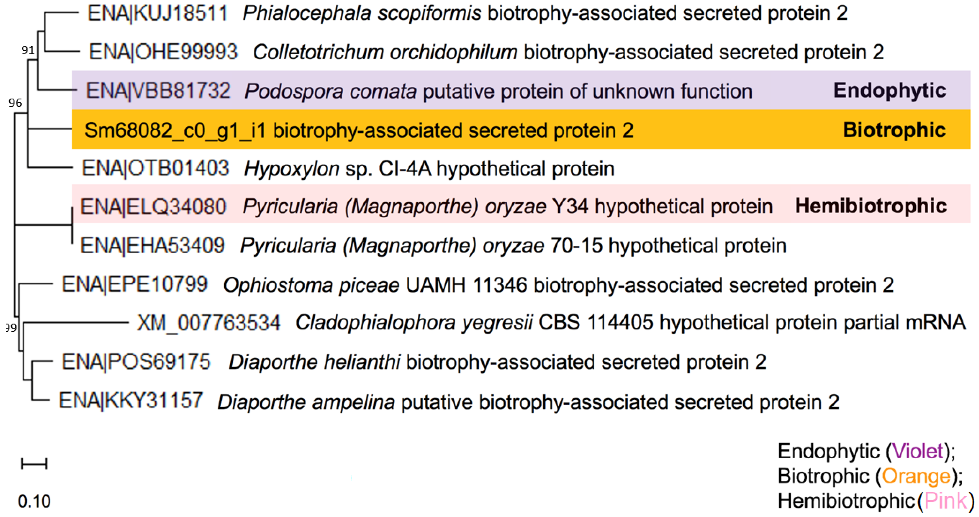
In the interactive transcriptome of S. mycoparasitica, the transcript, Sm68082_c0_g1_i1, annotated to a biotrophy-associated secreted protein 2 was the most closely related to the plant endophytic Podospora comata (Sordariomycetes, Sordariomycetidae, Sordariales, Chaetomiaceae, and Podospora), also a saprophytic fungus on herbivore dung [44], Colletotrichum orchidophilum (Glomerellales and Ascomycota), and Phialocephala scopiformis (Helotiales and Ascomycota), as shown in Figure 3.
In melanosporaceous S. mycoparasitica, the total number of (predicted) transcripts annotated to ABC transporters was 81. Among them, families of ABC-B (11 transcripts, 14%), ABC-C (18 transcripts, 22%), and ABC-G (19 transcripts, 23%) covered higher proportions than the other families belonging to ABC-A, ABC-F (2 transcripts), Aliphatic sulfonates importer (TC 3.A.1.17.2), BtuF, Ribose importer (TC 3.A.1.2.1), and unknown families.
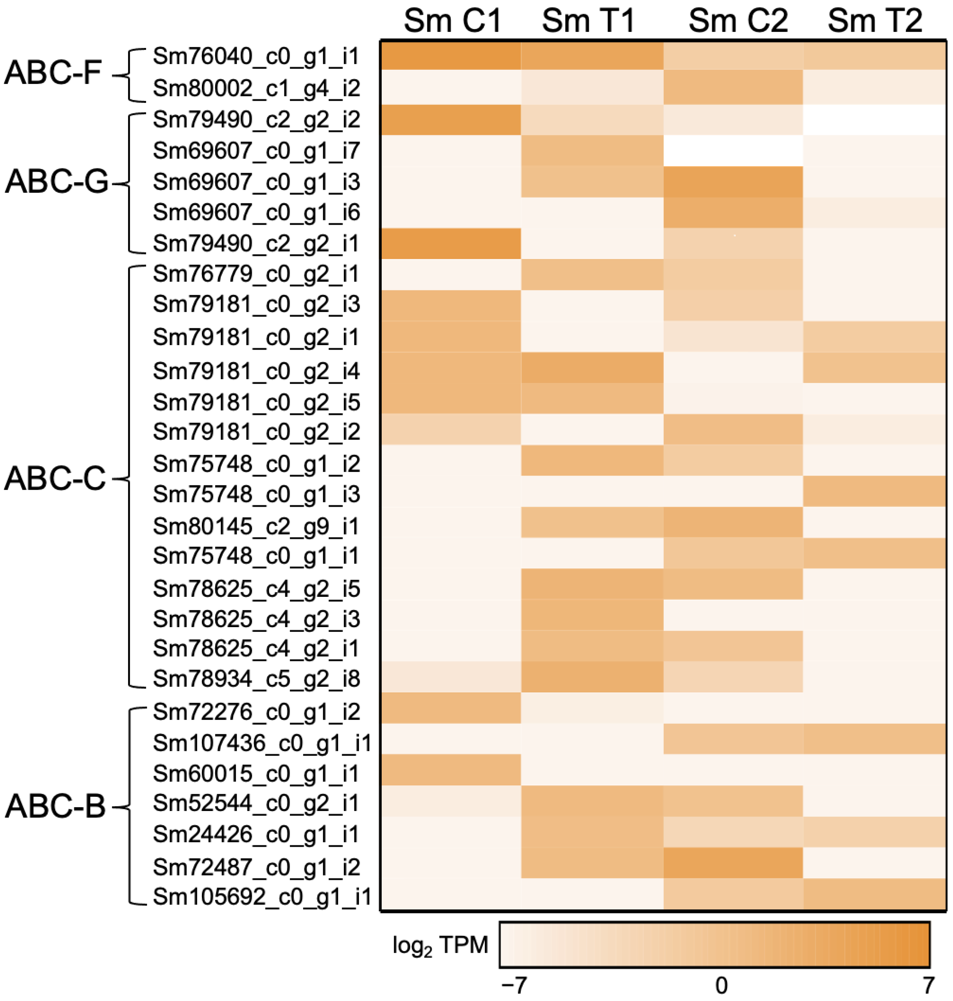
In fact, 11 transcripts for the multidrug resistance exporter (TC 3.A.1.201) subfamily of the ABC-B family were found along with 18 transcripts for the conjugate transporter (TC 3.A.1.208) subfamily of the ABC-C family, as well as 19 transcripts for the pleiotropic drug resistance (PDR) (TC 3.A.1.205) subfamily of the ABC-G family. Out of the total 81 transcripts, selected transcripts based on a relatively higher frequency and/or identity are presented in Figure 4, showing transcripts per million (TPM) values for 7, 14, 5, and 2 transcripts belonging to ABC-B, -C, -G, and -F families, respectively, and the detailed description follows.
In the transcriptome of S. mycoparasitica, the transcript, Sm105692_c0_g1_i1, annotated to ABC transporter 1 (alternative name, siderophore biosynthesis cluster protein ABC1 and gene name, ABC1) showed high identity (E-value 1.5 × 10⁻³¹, Score 293, and 72.7% identity) to the Ajellomyces capsulatus (an intracellular budding yeast) ABC transporter 1 (UniProt identifier: ABC1_AJECA) belonging to the multidrug resistance exporter (TC 3.A.1.201) subfamily, ABC-B family. The TPM value of the transcript, Sm105692_c0_g1_i1, annotated to the ABC transporter 1 was higher in the host-interactive sample (2.193904 TPM) than in the control (0.4872588 TPM) at 3.5 days; The control and the host-interactive samples at 1.5 days both had no expression of the transcript, as shown in Figure 4 (ABC-B).
In relation to virulence, the transcriptome of S. mycoparasitica depicted five transcripts which may be associated with virulence. One transcript (Sm78934_c5_g2_i8) was annotated to the multidrug resistance protein fer6 (alternative names, ATP-binding cassette subfamily C member fer6 and Fe-regulated protein 6, gene name fer6) belonging to the conjugate transporter (TC 3.A.1.208) subfamily, ABC-C family (UniProt identifier: FER6_USTMA). This protein is a part of the gene cluster that mediates the biosynthesis of siderophore ferrichrome A contributing to cell virulence [45,46]. The four remaining (Sm78625_c4_g2_i1, Sm78625_c4_g2_i3, Sm78625_c4_g2_i5, and Sm75748_c0_g1_i1) were annotated to the multiple drug resistance-associated protein-like transporter 1 (short name, MRP-like transporter 1; alternative name, vacuolar multidrug resistance ABC transporter MTL1) (UniProt identifier: MLT1_CANAL). The transcript, Sm78625_c4_g2_i1, had high similarity (E-value 9.5 ×10−37, Score 339, and 64.2% identity) showing a higher TPM value during the interaction with the host F. graminearum at 1.5 days (2.180728), as shown in Figure 4 (ABC-C).

3.2.1. Multidrug Resistance (MDR) Exporter Subfamily (ABC-B Family)

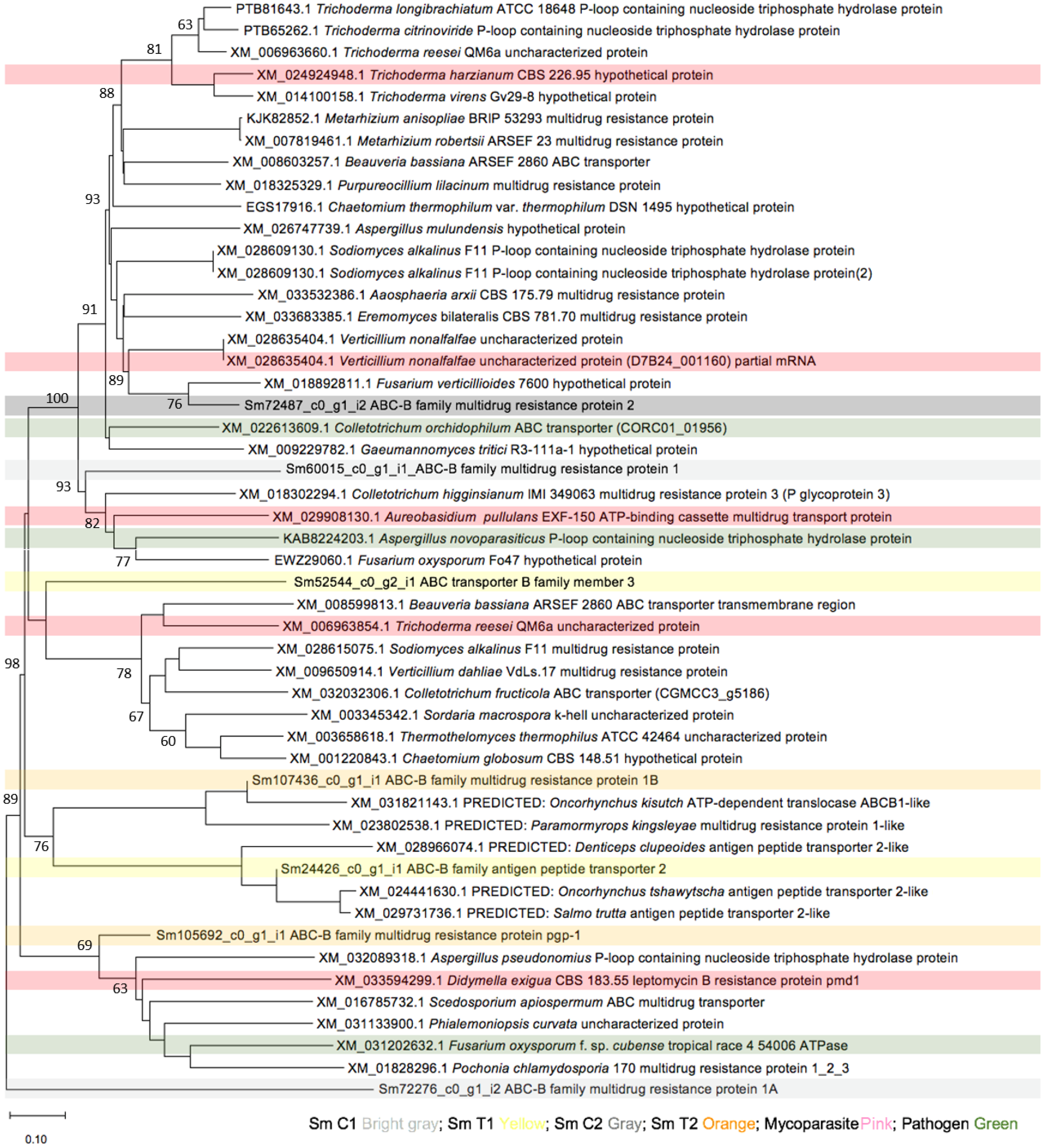
In the ABC-B family, the multidrug resistance exporter was the sole subfamily. Indeed, the ABC multidrug transporter atrC (pleiotropic ABC efflux transporter) was relatively more abundant than other ABC transporter proteins present in S.m transcriptome, where the transcript, Sm72487_c0_g1_i2, showed the highest identity of 69.5% (E-value 0.0 and Score 4398) to ABC multidrug transporter atrC (UniProt identifier: ATRC_EMENI) in Aspergillus nidulans (teleomorph Emericella nidulans). The transcript, Sm72487_c0_g1_i2, was upregulated at 3.5 days without interaction with the host, followed by, at 1.5 days, interaction with the host (Figure 4) and phylogenetically closely related to the Colletotrichum orchidophilum (Figure 5). At 1.5 days without interaction with the host, the transcript, Sm60015_c0_g1_i1, was upregulated, while the two transcripts, Sm24426_c0_g1_i1 and Sm52544_c0_g2_i1, were upregulated at 1.5 days with interaction with the host (Figure 4). The transcript, Sm107436_c0_g1_i1, annotated to ABC multidrug transporter MDR2 (alternative name, multidrug resistance protein 2, UniProt identifier: MDR2_TRIT1) in Trichophyton tonsurans (strain CBS 112818) (Scalp ringworm fungus) with 65.0% identity (E-value 2.4 × 10⁻²⁹ and Score: 281) showed upregulation on the interactive transcriptome at 3.5 days (Figure 4). The last transcript (Sm72276_c0_g1_i2) was annotated to ABC transporter BEA3 (alternative name, Beauvericin biosynthesis cluster protein 3 and UniProt identifier: BEA3_GIBF5) with 56.9% identity (E-value 0.0 and Score 3722).

3.2.2. Conjugate Transporter Subfamily (ABC-C Family)

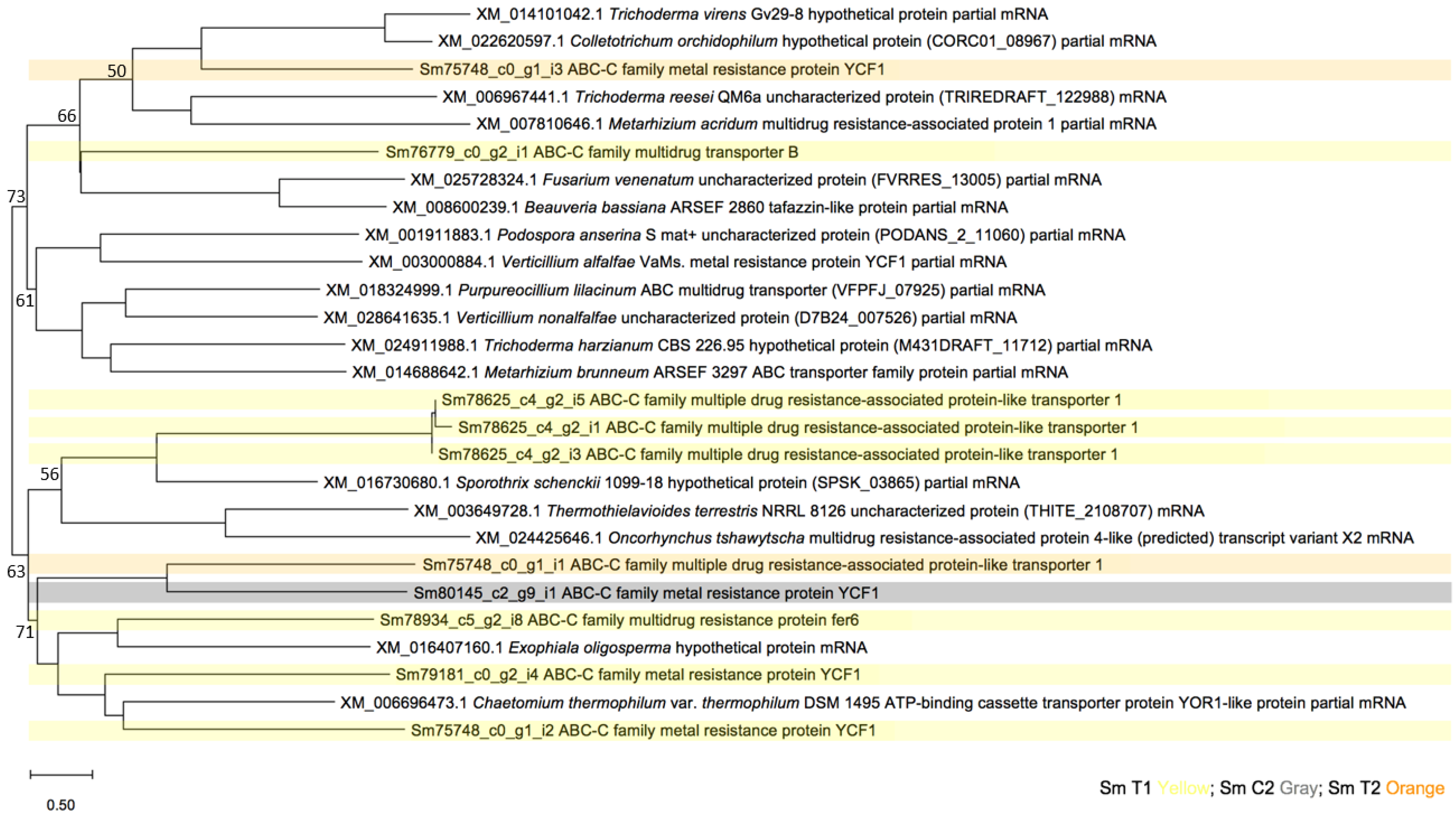
Importantly, eight transcripts (Sm80145_c2_g9_i1, Sm75748_c0_g1_i3, Sm75748_c0_g1_i2, Sm79181_c0_g2_i2, Sm79181_c0_g2_i5, Sm79181_c0_g2_i4, Sm79181_c0_g2_i1, and Sm79181_c0_g2_i3) were annotated to the metal resistance protein YCF1 (Saccharomyces cerevisiae (Baker’s yeast), UniProt identifier: YCFI_YEAST) which is also known as the ABC-type glutathione-S-conjugate transporter (conjugate transporter (TC 3.A.1.208) subfamily, ABC-C family) [47] involved in the ATP-dependent vacuolar transport of bilirubin and glutathione conjugates [48] contributing to cellular detoxification processes. The transcript, Sm80145_c2_g9_i1, showed the highest identity of 63.6% (E-value 3.3 × 10−38 and Score 349) and higher expression at 3.5 days without interaction with the host, whereas the transcript, Sm79181_c0_g2_i4, had the highest gene expression at 1.5 days interaction with the host (Figure 4). They seemed to phylogenetically be related to saprophytic fungus Chaetomium thermophilum, nematophagous fungus Purpureocillium lilacinum, and entomoparasite Metarhizium brunneum (Figure 6).
In addition to the metal resistance protein YCF1, the transcript, Sm76779_c0_g2_i1, was annotated to the ABC multidrug transporter B in Aspergillus fumigatus (UniProt identifier: ABCB_ASPFU, gene name abcB, 46.1% identity, E-value 1.6 × 10−98, and Score 537) which is known as a pleiotropic ABC efflux transporter that may be involved in A. fumigatus adaptation to azoles such as voriconazole. The transcript showed the highest expression at 1.5 days with interaction with the host (Figure 4) and phylogenetically closely related to entomopathogen Metarhizium acridum (Figure 6).

3.2.3. EF3 Subfamily (ABC-F Family)

The two transcripts, Sm80002_c1_g4_i2 and Sm76040_c0_g1_i1, were annotated to ABC transporter ATP-binding protein ARB1 (gene name ARB1, UniProt identifier: ARB1_YEAST) in Saccharomyces cerevisiae (Baker’s yeast) belonging to the EF3 subfamily of ABC-F family showed 69.3% identity (E-value 0.0 and Score 2194) and 68.2% identity (E-value 0.0 and Score 2170), respectively. The relative gene expression of the transcripts was higher when the mycoparasite was incubated without the host (Figure 4). The phylogenetic analyses for these transcripts indicated S. mycoparasitica was phylogenetically closely related to fungi with different lifestyles including Trichoderma gamsii (endophytic and parasitic lifestyle) and Metarhizium acridum (entomopathogenic lifestyle), as shown in Figure 7.

3.2.4. Pleiotropic Drug Resistance (PDR) Subfamily (ABC-G Family)

In this study, four transcripts (i.e., Sm79490_c2_g2_i1, Sm69607_c0_g1_i6, Sm69607_c0_g1_i3, and Sm69607_c0_g1_i7) were annotated to the ABC multidrug transporter C (pleiotropic ABC efflux transporter) in Aspergillus fumigatus (teleomorph Neosartorya fumigata) belonging to the PDR (TC 3.A.1.205) subfamily, ABC-G family (gene name abcC; synonyms abcB, abcG1, atrE, and cdr1B; UniProt identifier: ABCC_ASPFU) which confers resistance to structurally and functionally unrelated compounds. The transcript, Sm79490_c2_g2_i1, showed the highest identity of 65.0% with E-value 1.8 × 10−117 and Score 955.
The transcript, Sm79490_c2_g2_i2, was annotated to the ABC transporter CDR4 (gene name CDR4 and UniProt identifier: CDR4_CANAX) in Candida albicans (yeast) showing identity of 51.8% (E-value 0.0 and Score: 3716). The ABC transporter CDR4, involved in xenobiotic detoxification by transmembrane export across plasma membrane, belongs to the PDR (TC 3.A.1.205) subfamily of the ABC-G family. The relative expression of the transcript (Sm79490_c2_g2_i2) was extraordinarily high (Figure 4). Further phylogenetic analysis for the transcript Sm79490_c2_g2_i2, shown in Figure 8, indicated the transcript was closely related to the Phialemoniopsis mycoparasite (Phialemoniopsis curvata uncharacterized protein (E0L32_002615), partial mRNA (Blastn result 73.91% identity, E-value 0.0, and Accession number: XM_031136824.1)) and hemibiotrophic Colletotrichum parasite (Colletotrichum graminicola M1.001 ABC-2 type transporter partial mRNA (Blastn result 73.45% identity, E-value 0.0, and Accession number: XM_008100177.1)). The above blastn results were obtained based on the program discontiguous megaBLAST for more dissimilar sequences.

3.2.5. Mitotic Checkpoint Protein (Bub2) Interacting with ABC in Appressoria Formation

To initiate host cell infection, mycoparasitic Sm fungus develops an infection structure called the appressorium to penetrate underlying host cells. The transcript, Sm29848_c0_g1_i1, was annotated to a typical mitotic checkpoint protein Smbub2 interacting with ABC-C (Sm79181_c0_g2_i4) during an appressorium formation. The Smbub2 appressorium related transcript (Figure 9) also occurs in yeasts (UniProtKB, BUB2 P26448,) to monitor spindle integrity and prevent premature exit from mitosis.
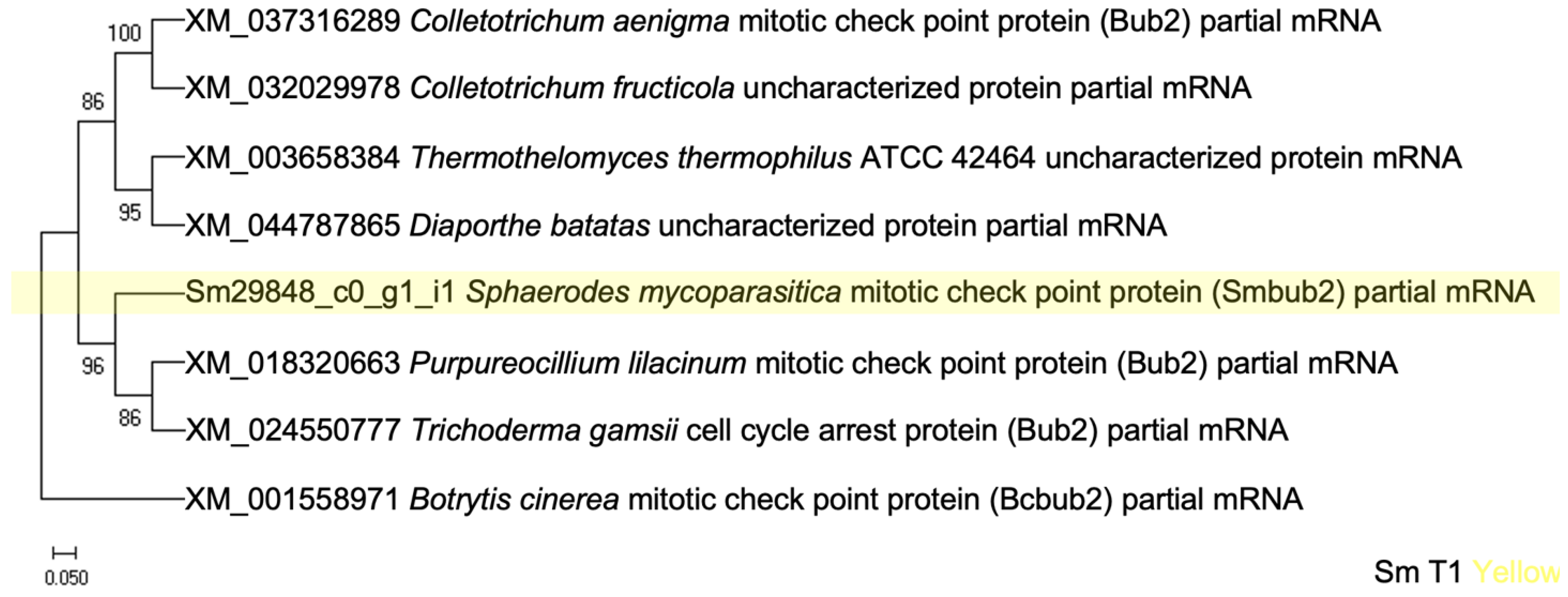
The highest Smbub2 gene expression at 1.5 days during the interaction with the host (Figure 9) coincides with ABC-type glutathione-S-conjugate transporter, ABC-C family (Figure 6) with close phylogenetic relatedness to nematophagous fungus Purpureocillium lilacinum (GenBank acc. no. XM_018320663) (Figure 10).

4. Discussion

Generally, ABC transporters are a major category of membrane-associated microbial protein structures involved in the transport of a wide range of substrates and biological processes. In eukaryotic fungi, the ABC transporters can be categorized into nine families (from A to I) and contain both uptake and efflux transport systems for fundamental cellular processes, pathogenesis, and tolerance against various toxic or xenobiotic compounds. In addition to the well-known function as an active transporter, ABCs act as ion channels and receptors and are also involved in mRNA translation and ribosome biogenesis. In this study, we performed a genome-wide identification of genes and transcripts encoding ABC transporter proteins on the interface of a suppressive cell-to-cell interactive zone between mycoparasitic S. mycoparasitica and mycotoxigenic F. graminearum.
Based on phylogenetic analyses and domain organization, the multixenobiotic resistance (MXR) proteins within ABC families are particularly seen as a competitive stress response in prokaryotes and eukaryotes across environments. However, the MXR-associated transcripts or proteins from the mycoparasite–phytopathogenic host interaction were not previously characterized to better explain the interface changes in an interactive, resilient and detoxifying hyphal tissue zone. MXR is a phenomenon in which ABC family proteins transfer harmful compounds out of cells, which was a prerequisite for early life on Earth. The MXR efflux activity in the eukaryotic cell immune system is mediated by ABC transporters of ABC-B and ABC-C as principally inducible under stress to prevent cytotoxicity. Taken together, it seems that ABC transporters form an active, physiological barrier at the tissue–environment interface to perform protection. In addition, ABC-G proteins are high-affinity ABC transporters to the biosynthesis of hormones orchestrating cellular growth and development in response to endogenous cues and environmental changes. However, the role of ABC transporters on the mycoparasite–mycotoxigenic fungal host interface is still poorly known, particularly in biotrophic fungal mycoparasites. Thus, the aim of this research was to investigate the possible candidate ABC genes responsible for the biotrophic mycoparasitism of S. mycoparasitica on the fungus–fungus interface using transcriptomic analyses.

4.1. Investigation of Morphological Changes during Biotrophic Mycoparasitism

Previously, monitoring shifts in Fourier transform infrared (FTIR) molecular signatures combined with scanning electron microscopy (SEM) and confocal laser scanning microscopy (CLSM) assessments at the fungus–fungus (Sphaerodes-Fusarium) interface has proven to be a useful tool for detecting early mycoparasitism-related interactome (proteins, lipids, and carbohydrates etc.) as a vital asset of the preventive biocontrol strategy against Fusarium pathogens [32]. Based on TEM analysis conducted in this study, it appears that S. mycoparasitica utilizes a specialized host-infecting structure, an appressorium/haustorium type structure. Thus, the lifestyles of S. mycoparasitica with Fusarium hosts belong to biotrophic parasitism and possess infection structures similar to biotrophic fungi (within ascomycetes) such as Blumeria graminis, Erysiphe pisi, Golovinomyces orontii, and Magnoportha oryzae, with mechanisms for the direct uptake of nutrients [49,50,51,52].
Another interesting feature during the biotrophism was also noticed. Indeed, Sm developed an infection structure called the appressorium to penetrate the host cells. The ABC-type glutathione-S-conjugate and the SmBub2 (non-ABC) genes were upregulated in tandem at 1.5 days which coincided with the appressorium formation on the host cell surface. The mitotic checkpoint protein Bub2 gene is a regulator of appressoria formed by germ tubes and appressorium-like structures developed by hyphal tips as observed in Magnaporthe oryzae [53,54]. In addition, the observed mucosa-like substance on cell surfaces, such as mucins, may act as barriers and sensors of the extracellular environment including the changes in pH, ionic composition, or physical interactions [55]. Mucins and mucin-like molecules are present in the cell surfaces of eukaryotes in the form of highly O-glycosylated proteins [56]. On the basis of the structure, function, and cellular localization of mucin proteins, mucins are classified into two categories, i.e., secreted mucins and membrane (cell surface-associated) mucins [57]. In M. oryzae, Msb2 was reported to be a cell surface (signaling) mucin gene and the Momsb2 mutant showed a significant reduction in appressorium formation on hydrophobic surfaces [58]. In the transcriptome of S. mycoparasitica, Msb2 was also found (data not shown). Importantly, the early stage of the interactive transcriptome of S. mycoparasitica showed upregulated transcripts annotated to MUC 5AC, one of the secreted gel-forming mucins (MUC 2, MUC 5AC, 5B, 6, and 19) [57].
In addition, biotrophy-associated secreted (BAS) proteins of Magnaporthe oryzae are distinctive plant pathogenic features [59]. Studying M. oryzae as a model chemibiotrophic system, Mosquera et al. (2009) demonstrated the localization of fluorescently labeled BAS proteins [60]. In fact, the accumulation of BAS1 and BAS2 was preferentially into biotrophic interfacial complexes along with known avirulence effectors. The localization of BAS3 was situated near cell wall crossing points, while the formation of a line of BAS4 was uniformly on growing invasive hyphae [60]. In this study, S.m. showed two different transcripts annotated to biotrophy-associated secreted (BAS) proteins. The phylogenetic analysis of the transcript Sm68082_c0_g1_i1 annotated to BAS2 showed relatively close relationships with saprophytic and endophytic fungus P. comata followed by hemibiotrophic fungus C. orchidophilum phylogenetically. The function of the transcript seemed to be focused on biotrophy due to the abundant appearance of BAS2. Within the same genus Colletotrichum, it has been demonstrated that BAS2, identified in C. gloeosporioides, a hemibiotrophic fungus, was involved in the penetration step into the host tissue [61]. Another S.m. transcript was phylogenetically related to the hypothetical protein close to the BAS cluster of hemibiotrophic Magnaporthe oryzae (Pyricularia oryzae, Sordariomycetes, Sordariomycetidae, Magnaporthales, Pyriculariaceae, and Pyricularia). Although several S. mycoparasitica transcripts discovered are related to its endophytism and hemibiotrophism, the functionality of BAS proteins in S.m. (e.g., BCA’s involvement in modulation of the host cell signaling and suppression of immune responses) may suggest a transcriptomic link to the biotrophic mycoparasitism.

4.2. Phylogenetic Evolutionary Analysis Based on the Interactive Transcriptome

Based on the interactive and comparative transcriptomic study between Clonostachys and Trichoderma, Karlsson et al. (2015) found the ABC profile to be an informative feature to distinguish hypocreaceous fungal mycoparasites. Among ABC transporters, especially ABC-B and ABC-G families were involved in fungal self-protection and/or tolerance toward toxic metabolites derived from the mycotoxigenic fungal prey or pesticides [62].
In S. mycoparasitica, the cluster of ABC-B transporters is represented with ABC transporter 1 (Sm105692_c0_g1_i1) belonging to the multidrug resistance exporter (TC 3.A.1.201) subfamily. In Ajellomyces capsulatus, ABC1 mediates the biosynthesis of hydroxamate-containing siderophores that play a critical role in virulence via intracellular iron acquisition during macrophage infection [63,64]. Probably the gene product is involved in the excretion of the extracellular siderophores [61]. Several classes of hydroxamate siderophores have, so far, been isolated such as ferrichromes, fusarinines, coprogens, and rhodotorulic acid [65]. In Magnaporthe grisea, from ABC1 to ABC4 were reported to regulate cytotoxicity and mediate multidrug resistance as well as, oxidative stress for successful infection [66]. Especially, MgABC1 and MgABC4 [65] were known to be responsible for pathogenicity as the M. grisea mutant for the ABC1 gene showed a strong reduction in its virulence. The mutant for the ABC4 gene was impaired in appressorium formation [67] and the transcription of MgABC1 was activated under oxidative condition [68], while MgABC2 [69] and MgABC3 [70] were required for multidrug resistance rather than pathogenicity. Sun et al. (2006) reported that M. oryzae mutated for deletion of the abc3 gene showed increased sensitivity to valinomycin and actinomycin D and showed high sensitivity to oxidative stress.
The analysis of vacuolar ABC transporters in S. mycoparasitica revealed multiple drug resistance-associated protein (MRP), possibly involved in virulence. These transcripts (including Sm78625_c4_g2_i1) annotated to MRP-like transporter 1 may be implicated in the transport of bilirubin and glutathione conjugates with an important role in yeasts’ virulence [71,72]. Further, the S.m. transcript (Sm72487_c0_g1_i2) annotated to the ABC multidrug transporter atrC (pleiotropic ABC efflux transporter) was also abundant. Previously, atrC was characterized as the ABC transporter-encoding gene from A. nidulans, known to be involved in the protection of the cells against a wide range of toxic compounds [73]. Andrade et al. (2000) demonstrated that the transcript levels of atrC with atrD were increased in fungal germlings under natural toxins and xenobiotics, where the expression of the atrC was highly constitutive as compared with atrD (atrD may also be involved in the antibiotic production, e.g., penicillin); the constitutive level of atrC expression implies its involvement in a metabolic stress controlling function.
Eight different S. mycoparasitica transcripts were annotated to the metal resistance protein YCF1 which is also known as the ABC-type glutathione-S-conjugate transporter subfamily. These transcripts of the ABC-C family [47] are expected to be involved in the ATP-dependent vacuolar transport of bilirubin and glutathione conjugates [48], contributing to cellular detoxification processes. The protein Ycf1p was originally characterized as a Cd2+ transporter. Then later, its function was discovered to enhance the recruitment of the soluble SNARE (soluble N-Ethylmaleimide-sensitive factor attachment protein receptor) Vam7p to vacuoles for efficient membrane fusion [74], resulting in cellular homeostasis. This unknown recruitment mechanism dependent on the ABC transporter Ycf1p may help to understand membrane trafficking and fusion [75]. More detailed information of different ABC-C transporters including Ycf1p for S. cerevisiae vacuole fusion has been reported by Sasser et al. (2014) [76].
In S. mycoparasitica, eleven PDR subfamily members (TC 3.A.1.205) of ABC transporters were identified. In Aspergillus fumigatus, it was demonstrated that two ABC transporters (ABC-A and ABC-B) shared the highest sequence similarity with Pdr5 in S. cerevisiae yeast [77]. The Pdr5 protein is a plasma membrane-localized pleiotropic ABC efflux transporter of multiple drugs. In addition, the ABC-B role was linked to azole resistance and ABC-B was required for normal pathogenesis of A. fumigatus tested in a Galleria mellonella (greater wax moth) infection model system [77]. The transcript, Sm80002_c1_g4_i2, was annotated to ABC transporter ATP-binding protein ARB1 of S. cerevisiae. The ARB1 protein in S. cerevisiae is essential for interacting with ribosomes and functioning in protein synthesis or ribosome biogenesis (stimulation of 40S and 60S ribosome biogenesis) [78]. In S. mycoparasitica, the two ABC-F/EF3 subfamily transcripts have close phylogenetic relatedness to mycoparasitic Trichoderma and Sporothrix generalists. It coincides with the ubiquitous nature of ABC-F proteins known as widespread translation factors within prokaryotes and eukaryotes. Interestingly, it was suggested that bacterial ABC-F proteins could mediate increased antibiotic resistance. Moreover, the anti-apoptotic function of ABCF2 has also been proposed during the infection of enteropathogenic E. coli [79].
The multidrug transporters ABC-G are involved in multiple functions or mechanisms leading to mycoparasitism such as fungal cell resistance to xenobiotic compounds and steroid transports. This ABC family may provide pesticide resistance, since this transporter is known as conferring resistance to structurally and functionally unrelated compounds including azoles such as itraconazole [80,81], posaconazole [82], and voriconazole [83]. This class of transporters is conferring resistance to cycloheximide, azoles, and a mycotoxin sporidesmin [84,85,86]. Among fungi, ABC-G1–5, ABC-G6, and ABC-G7 have been discovered as the major clusters within ascomycetes and basidiomycetes [87]. In this study, the ABC transporter-CDR4 found in biotrophic mycoparasite S. mycoparasitica belongs to the ABC-G/PDR cluster (TC 3.A.1.205) and might be involved in xenobiotic detoxification by transmembrane export across plasma membrane. Dubey et al. (2014) reported that deletion of abcG5 in necrotrophic mycoparasite C. rosea resulted in reduced ZEN mycotoxin tolerance and kept its partial ability to protect the barley plant against F. graminearum [27]. The ABCG5 gene provides instructions for making sterolin-1, which makes up half of a protein called sterolin. Sterolin is a transporter protein, which is a type of protein that moves substances across cell membranes.
Although we were unable to perform a comparison of genome assembly size in our study due to the absence of the genome assembly of S. mycoparasitica, genome size, gene number and gene turnover were discussed. Wu and Cox (2021) reported significant changes in gene number within the order Hypocreales including mycoparasitic and root symbiotic Trichoderma and plant pathogenic Fusarium species, as well as two other genera having endophytic and entomopathogenic lifestyles [21]. The mycoparasitic Trichoderma tended to have a lower gene number than the plant pathogenic Fusarium (genome size had the same decreasing pattern). In the same manner, the proportion of genus-specific orthogroups was reduced in the Trichoderma as compared with the Fusarium genus. With that, S. mycoparasitica might have the same pattern of reduction in genome size, gene number, and genus-specific orthogroups as inferred from the mycoparasitic lifestyle requiring a fewer number of core genes consisting of smaller genome size, lower gene number, and genus-specific orthogroups due to complementary means (gene turnover and positive selection). Consistent with phylogenetic classification belonging to Hypocreales, mycoparasitic generalists Trichoderma and Clonostachys and plant pathogenic Fusaria upregulated genes are more likely to be genus specific [21]. A subset of these genus-specific genes is shared by fungi with the same lifestyle in quite different evolutionary orders, thus, supporting the view that some genus-specific genes are necessary for specific lifestyles. While the mechanisms are highly variable, fungi with the same lifestyle often display gene expansions in certain strategic protein families [1] such as the ABC protein families. Further, the expressions of genus-specific genes are likely regulated in response to the host and environment as an evolutionary mechanism of fungal transition within this kingdom. In that regard, Trichoderma spp. seems to share genus-specific orthologous genes with common ancestors, mainly belonging to necrotrophic mycoparasites, then entomopathogenic (Metarhizium) and plant pathogenic (Fusarium) fungi, followed by some symbiotic (Epichloë) and root-associated fungi [21]. Similarly, this study’s fundings indicate that the transition of S. mycoparasitica (Melanosporales) into a specific mycoparasite may involve ABC genes aligned with fungal taxa having biotrophic, hemibiotrophic, and endophytic lifestyles.

5. Conclusions

This study is novel because it explores the transcriptome of S. mycoparasitica during biotrophic mycoparasitism with mycotoxigenic Fusarium graminearum, with an emphasis on the analysis of the ATP-binding cassette (ABC) transporters. The findings, through phylogenetic analyses and specific functional annotations, provide valuable clues to better understand the biotrophic mycoparasitism of S. mycoparasitica interacting with the Fusarium host, which implies that the ABC transporters group of key proteins are involved in S. mycoparasitica virulence and multidrug resistance to toxic substances, including cellular detoxification. Further, diversified ABC family transcripts (-B, -C, -F, and -G) might contribute to enhanced S. mycoparasitica adaptability to the hostile Fusarium host environment or stress factors in an active way of a defense system. It appears that genus-specific selection also contributes to the fungal lifestyle and multiple host transitions. The comparison of hypocreaceous Trichoderma and Clonostachys mycoparasites being generalists with melanosporaceus Sphaerodes specialists being adapted to control Fusarium pathogens and associated mycotoxins, suggests that fungal adaptation often requires a selected number of core genes to regulate a particular mycoparasitic lifestyle. A comparative analysis of ABC family genes reveals critical differences between various microparasitic lifestyles, while also pinpointing distinctive evolutionary advantages of S. mycoparasitica-specific mycoparasitism, thus, opening new directions for future research on biocontrol of mycotoxigenic Fusarium pathogens.

Author Contributions

S.H.K. and V.V. equally contributed to conceptualization, methodology, investigation, data analysis and interpretation, writing—original draft preparation, writing—review and editing; V.V. conceived project idea, acquired funding, and supervised. All authors have read and agreed to the published version of the manuscript.

Funding

This research was funded by a National Sciences and Engineering Research Council of Canada (NSERC) Discovery grant, grant number RGPIN-2017-05286 and the Government of Saskatchewan Agriculture Development Fund (ID 20160226), and the Saskatchewan Wheat Development Commission (171025-65) external to V. Vujanovic.

Institutional Review Board Statement

Not applicable.

Informed Consent Statement

Not applicable.

Data Availability Statement

The RNA sequence data are available in NCBI with accession PRJNA757970.

Acknowledgments

The authors would like to thank anonymous reviewers of this paper and Rhonda Sobchishin (WCVM) for technical TEM assistance.

Conflicts of Interest

The authors declare no conflict of interest. The funders had no role in the design of the study; in the collection, analyses, or interpretation of data; in the writing of the manuscript, or in the decision to publish the results.

References

  1. Naranjo-Ortiz, M.A.; Gabaldón, T. Fungal evolution: Major ecological adaptations and evolutionary transitions. Biol. Rev. Camb. Philos. Soc. 2019, 94, 1443–1476. [Google Scholar] [CrossRef] [PubMed] [Green Version]
  2. Vujanovic, V.; Kim, S.H. Adaptability of mitosporic stage in Sphaerodes mycoparasitica towards its mycoparasitic-polyphagous lifestyle. Mycologia 2018, 109, 701–709. [Google Scholar] [CrossRef]
  3. Goh, Y.K.; Vujanovic, V. Biotrophic mycoparasitic interactions between Sphaerodes mycoparasitica and phytopathogenic Fusariums species. Biocontrol. Sci. Technol. 2010, 20, 891–902. [Google Scholar] [CrossRef]
  4. Inglis, G.D.; Kawchuk, L.M. Comparative degradation of oomycete, ascomycete, and basidiomycete cell walls by mycoparasitic and biocontrol fungi. Can. J. Microbiol. 2002, 48, 60–70. [Google Scholar] [CrossRef]
  5. Atanasova, L.; Crom, S.L.; Gruber, S.; Coulpier, F.; Seidl-Seiboth, V.; Kubicek, C.P.; Druzhinina, I.S. Comparative transcriptomics reveals different strategies of Trichoderma mycoparasitism. BMC Genom. 2013, 14, 121. [Google Scholar] [CrossRef] [Green Version]
  6. Quandt, C.A.; Bushley, K.E.; Spatafora, J.W. The genome of the truffle-parasite Tolypocladium ophioglossoides and the evolution of antifungal peptaibiotics. BMC Genom. 2015, 16, 553. [Google Scholar] [CrossRef] [PubMed] [Green Version]
  7. Kim, S.H.; Vujanovic, V. Relationship between mycoparasites lifestyles and biocontrol behaviors against Fusarium spp. and mycotoxins production. Appl. Microbiol. Biotechnol. 2016, 100, 5257–5272. [Google Scholar] [CrossRef]
  8. Vujanovic, V.; Goh, Y.K. Sphaerodes mycoparasitica biotrophic mycoparasite of 3-acetyldeoxynivalenol- and 15-acetyldeoxynivalenol-producing toxigenic Fusarium graminearum chemotypes. FEMS Microbiol. Lett. 2011, 316, 136–143. [Google Scholar] [CrossRef] [PubMed]
  9. Kim, S.H.; Vujanovic, V. Biodegradation and biodetoxification of Fusarium mycotoxins by Sphaerodes mycoparasitica. AMB Express 2017, 7, 145. [Google Scholar] [CrossRef]
  10. Qualhato, T.F.; Lopes, F.A.C.; Steindorff, A.S.; Brandão, R.S.; Jesuino, R.S.A.; Ulhoa, C.J. Mycoparasitism studies of Trichoderma species against three phytopathogenic fungi: Evaluation of antagonism and hydrolytic enzyme production. Biotechnol. Lett. 2013, 35, 1461–1468. [Google Scholar] [CrossRef] [PubMed]
  11. Chatterton, S.; Punja, Z.K. Chitinase and β-1,3-glucanase enzyme production by the mycoparasite Clonostachys rosea f. catenulata against fungal plant pathogens. Can. J. Microbiol. 2009, 55, 356–367. [Google Scholar] [CrossRef] [PubMed]
  12. Elamathi, E.; Malathi, P.; Viswanathan, R.; Ramesh Sundar, A. Expression analysis on mycoparasitism related genes during antagonism of Trichoderma with Colletotrichum falcatum causing red rot in sugarcane. J. Plant Biochem. Biotechnol. 2018, 27, 351–361. [Google Scholar] [CrossRef]
  13. Steindorff, A.S.; Ramada, M.H.S.; Coelho, A.S.G.; Miller, R.N.G.; Pappas, G.J.; Ulhoa, C.J.; Noronha, E.F. Identification of mycoparasitism-related genes against the phytopathogen Sclerotinia sclerotiorum through transcriptome and expression profile analysis in Trichoderma harzianum. BMC Genom. 2014, 15, 204. [Google Scholar] [CrossRef] [Green Version]
  14. Reithner, B.; Schuhmacher, R.; Stoppacher, N.; Pucher, M.; Brunner, K.; Zeilinger, S. Signaling via the Trichoderma atroviride mitogen-activated protein kinase Tmk 1 differentially affects mycoparasitism and plant protection. Fungal Genet. Biol. 2007, 44, 1123–1133. [Google Scholar] [CrossRef] [PubMed] [Green Version]
  15. Sun, Z.-B.; Wang, Q.; Sun, M.-H.; Li, S.-D. The mitogen-activated protein kinase gene Crmapk is involved in Clonostachys chloroleuca mycoparasitism. Mol. Plant Microbe Interact. 2020, 33, 902–910. [Google Scholar] [CrossRef]
  16. Gruber, S.; Zeilinger, S. The transcription factor Ste12 mediates the regulatory role of the Tmk1 MAP kinase in mycoparasitism and vegetative hyphal fusion in the filamentous fungus Trichoderma atroviride. PLoS ONE 2014, 9, e111636. [Google Scholar] [CrossRef] [Green Version]
  17. Sun, Z.B.; Wang, Q.; Zhang, J.; Jiang, W.Z.; Wang, Q.; Li, S.D.; Ma, G.Z.; Sun, M.H. The transcription factor-encoding gene crtf is involved in Clonostachys chloroleuca mycoparasitism on Sclerotinia sclerotiorum. Microbiol. Res. 2018, 210, 6–11. [Google Scholar] [CrossRef]
  18. Seidl, V.; Song, L.; Lindquist, E.; Gruber, S.; Koptchinskiy, A.; Zeilinger, S.; Schmoll, M.; Martínez, P.; Sun, J.; Grigoriev, I.; et al. Transcriptomic response of the mycoparasitic fungus Trichoderma atroviride to the presence of a fungal prey. BMC Genom. 2009, 10, 567. [Google Scholar] [CrossRef] [PubMed] [Green Version]
  19. Sun, Z.-B.; Wang, Q.; Sun, M.-H.; Li, S.-D. The heat shock protein 70 gene is involved for colony morphology, sporulation and mycoparasitism of Clonostachys rosea. FEMS Microbiol. Lett. 2019, 366, fnz188. [Google Scholar] [CrossRef] [PubMed]
  20. Demissie, Z.A.; Foote, S.J.; Tan, Y.; Loewen, M.C. Profiling of the transcriptomic responses of Clonostachys rosea upon treatment with Fusarium graminearum secretome. Front. Microbiol. 2018, 9, 1061. [Google Scholar] [CrossRef] [Green Version]
  21. Wu, B.; Cox, M.P. Comparative genomics reveals a core gene toolbox for lifestyle transitions in Hypocreales fungi. Environ. Microbiol. 2021, 23, 3251–3264. [Google Scholar] [CrossRef] [PubMed]
  22. Marra, R.; Ambrosino, P.; Carbone, V.; Vinale, F.; Woo, S.L.; Ruocco, M.; Ciliento, R.; Lanzuise, S.; Ferraioli, S.; Soriente, I.; et al. Study of the three-way interaction between Trichoderma atroviride, plant and fungal pathogens by using a proteomic approach. Curr. Genet. 2006, 50, 307–321. [Google Scholar] [CrossRef]
  23. Ruocco, M.; Lanzuise, S.; Vinale, F.; Marra, R.; Turrà, D.; Woo, S.L.; Lorito, M. Identification of a new biocontrol gene in Trichoderma atroviride: The role of an ABC transporter membrane pump in the interaction with different plant-pathogenic Fungi. Mol. Plant Microbe Interact. 2009, 22, 291–301. [Google Scholar] [CrossRef] [Green Version]
  24. Piecuch, A.; Obłąk, E. Yeast ABC proteins involved in multidrug resistance. Cell. Mol. Biol. Lett. 2014, 19, 1–22. [Google Scholar] [CrossRef] [Green Version]
  25. Banerjee, D.; Lelandais, G.; Shukla, S.; Mukhopadhyay, G.; Jacq, C.; Devaux, F.; Prasad, R. Responses of pathogenic and nonpathogenic yeast species to steroids reveal the functioning and evolution of multidrug resistance transcriptional networks. Eukaryot. Cell 2008, 7, 68. [Google Scholar] [CrossRef] [Green Version]
  26. Mahé, Y.; Lemoine, Y.; Kuchler, K. The ATP binding cassette transporters Pdr5 and Snq2 of Saccharomyces cerevisiae can mediate transport of steroids in Vivo. J. Biol. Chem. 1996, 271, 25167–25172. [Google Scholar] [CrossRef] [Green Version]
  27. Dubey, M.K.; Jensen, D.F.; Karlsson, M. An ATP-binding cassette pleiotropic drug transporter protein is required for xenobiotic tolerance and antagonism in the fungal biocontrol agent Clonostachys rosea. Mol. Plant Microbe Interact. 2014, 27, 725–732. [Google Scholar] [CrossRef] [Green Version]
  28. Kosawang, C.; Karlsson, M.; Jensen, D.F.; Dilokpimol, A.; Collinge, D.B. Transcriptomic profiling to identify genes involved in Fusarium mycotoxin deoxynivalenol and zearalenone tolerance in the mycoparasitic fungus Clonostachys rosea. BMC Genom. 2014, 15, 55. [Google Scholar] [CrossRef] [Green Version]
  29. Nygren, K.; Dubey, M.; Zapparata, A.; Iqbal, M.; Tzelepis, G.D.; Durling, M.B.; Jensen, D.F.; Karlsson, M. The mycoparasitic fungus Clonostachys rosea responds with both common and specific gene expression during interspecific interactions with fungal prey. Evol. Appl. 2018, 11, 931–949. [Google Scholar] [CrossRef] [PubMed] [Green Version]
  30. Kim, S.H.; Lahlali, R.; Karunakaran, C.; Vujanovic, V. Specific mycoparasite-Fusarium graminearum molecular signatures in germinating seeds disabled fusarium head blight pathogen’s infection. Int. J. Mol. Sci. 2021, 22, 2461. [Google Scholar] [CrossRef]
  31. Powell, A.J.; Vujanovic, V. Evolution of fusarium head blight management in wheat: Scientific perspectives on biological control agents and crop genotypes protocooperation. Appl. Sci. 2021, 11, 8960. [Google Scholar] [CrossRef]
  32. Kim, S.H.; Vujanovic, V. Early transcriptomic response of the mycoparasite Sphaerodes mycoparasitica to the mycotoxigenic Fusarium graminearum 3-ADON, the cause of Fusarium head blight. Bioresour. Bioprocess. 2021, 8, 127. [Google Scholar] [CrossRef] [PubMed]
  33. Vujanovic, V.; Goh, Y.K. Sphaerodes mycoparasitica sp. nov., a new biotrophic mycoparasite on Fusarium avenaceum, F. graminearum and F. oxysporum. Mycol. Res. 2009, 113, 1172–1180. [Google Scholar] [CrossRef] [PubMed]
  34. Goh, Y.K.; Vujanovic, V. Ascospore germination patterns revealed ascomycetous biotrophic mycoparasite specificity to Fusarium hosts. Botany 2010, 88, 1033–1043. [Google Scholar] [CrossRef]
  35. Xiao, M.; Zhang, Y.; Chen, X.; Lee, E.J.; Barber, C.J.; Chakrabarty, R.; Desgagné-Penix, I.; Haslam, T.M.; Kim, Y.B.; Liu, E.; et al. Transcriptome analysis based on next-generation sequencing of non-model plants producing specialized metabolites of biotechnological interest. J. Biotechnol. 2013, 166, 122–134. [Google Scholar] [CrossRef] [PubMed]
  36. Bolger, A.M.; Lohse, M.; Usadel, B. Trimmomatic: A flexible trimmer for Illumina sequence data. Bioinformatics 2014, 30, 2114–2120. [Google Scholar] [CrossRef] [PubMed] [Green Version]
  37. Grabherr, M.G.; Haas, B.J.; Yassour, M.; Levin, J.Z.; Thompson, D.A.; Amit, I.; Adiconis, X.; Fan, L.; Raychowdhury, R.; Zeng, Q.; et al. Full-length transcriptome assembly from RNA-Seq data without a reference genome. Nat. Biotechnol. 2011, 29, 644–652. [Google Scholar] [CrossRef] [Green Version]
  38. Haas, B.J.; Papanicolaou, A.; Yassour, M.; Grabherr, M.; Blood, P.D.; Bowden, J.; Couger, M.B.; Eccles, D.; Li, B.; Lieber, M.; et al. De novo transcript sequence reconstruction from RNA-seq using the Trinity platform for reference generation and analysis. Nat. Protoc. 2013, 8, 1494–1512. [Google Scholar] [CrossRef]
  39. Rzhetsky, A.; Nei, M. A simple method for estimating and testing minimum-evolution trees. Mol. Biol. Evol. 1992, 9, 945–967. [Google Scholar]
  40. Tamura, K.; Nei, M.; Kumar, S. Prospects for inferring very large phylogenies by using the neighbor-joining method. Proc. Natl. Acad. Sci. USA 2004, 101, 11030–11035. [Google Scholar] [CrossRef] [Green Version]
  41. Nei, M.; Kumar, S. Molecular Evolution and Phylogenetics; Oxford University Press: Oxford, UK; New York, NY, USA, 2000. [Google Scholar]
  42. Saitou, N.; Nei, M. The neighbor-joining method: A new method for reconstructing phylogenetic trees. Mol. Biol. Evol. 1987, 4, 406–425. [Google Scholar] [PubMed]
  43. Kumar, S.; Stecher, G.; Li, M.; Knyaz, C.; Tamura, K. MEGA X: Molecular evolutionary genetics analysis across computing platforms. Mol. Biol. Evol. 2018, 35, 1547–1549. [Google Scholar] [CrossRef] [PubMed]
  44. Silar, P.; Dauget, J.-M.; Gautier, V.; Grognet, P.; Chablat, M.; Hermann-Le Denmat, S.; Couloux, A.; Wincker, P.; Debuchy, R. A gene graveyard in the genome of the fungus Podospora comata. Mol. Genet. Genom. 2019, 294, 177–190. [Google Scholar] [CrossRef]
  45. Eichhorn, H.; Lessing, F.; Winterberg, B.; Schirawski, J.; Kämper, J.; Müller, P.; Kahmann, R. A ferroxidation/permeation iron uptake system is required for virulence in Ustilago maydis. Plant Cell 2006, 18, 3332–3345. [Google Scholar] [CrossRef] [PubMed] [Green Version]
  46. Winterberg, B.; Uhlmann, S.; Linne, U.; Lessing, F.; Marahiel, M.A.; Eichhorn, H.; Kahmann, R.; Schirawski, J. Elucidation of the complete ferrichrome A biosynthetic pathway in Ustilago maydis. Mol. Microbiol. 2010, 75, 1260–1271. [Google Scholar] [CrossRef] [PubMed]
  47. Li, Z.-S.; Szczypka, M.; Lu, Y.-P.; Thiele, D.J.; Rea, P.A. The yeast cadmium factor protein (YCF1) is a vacuolar glutathione S-conjugate pump. J. Biol. Chem. 1996, 271, 6509–6517. [Google Scholar] [CrossRef] [PubMed] [Green Version]
  48. Petrovic, S.; Pascolo, L.; Gallo, R.; Cupelli, F.; Ostrow, J.D.; Goffeau, A.; Tiribelli, C.; Bruschi, C.V. The products of YCF1 and YLL015w (BPT1) cooperate for the ATP-dependent vacuolar transport of unconjugated bilirubin in Saccharomyces cerevisiae. Yeast 2000, 16, 561–571. [Google Scholar] [CrossRef]
  49. Martín, J.P.; Di Pietro, A. Morphogenesis and Pathogenicity in Fungi; Springer: Berlin/Heidelberg, Germany, 2012. [Google Scholar]
  50. Kuhn, H.; Kwaaitaal, M.; Kusch, S.; Acevedo-Garcia, J.; Wu, H.; Panstruga, R. Biotrophy at its best: Novel findings and unsolved mysteries of the Arabidopsis-powdery mildew pathosystem. Arabidopsis Book 2016, 14, e0184. [Google Scholar] [CrossRef] [PubMed] [Green Version]
  51. Micali, C.; Göllner, K.; Humphry, M.; Consonni, C.; Panstruga, R. The powdery mildew disease of Arabidopsis: A paradigm for the interaction between plants and biotrophic fungi. Arabidopsis Book 2008, 6, e0115. [Google Scholar] [CrossRef] [PubMed] [Green Version]
  52. Zhang, Z.; Henderson, C.; Perfect, E.; Carver, T.L.W.; Thomas, B.J.; Skamnioti, P.; Gurr, S.J. Of genes and genomes, needles and haystacks: Blumeria graminis and functionality. Mol. Plant Pathol. 2005, 6, 561–575. [Google Scholar] [CrossRef] [PubMed]
  53. Jayawardena, R.S.; Chen, Y.-J.; Konta, S.; Tibpromma, S.; Abeywickrama, P.D.; Gomdola, D.; Balasuriya, A.; Xu, J.; Lumyong, S. Diversity and Function of Appressoria. Pathogens 2021, 10, 746. [Google Scholar] [CrossRef]
  54. Kong, L.-A.; Li, G.-T.; Liu, Y.; Liu, M.G.; Zhang, S.-J.; Yang, J.; Zhou, X.-J.; Peng, Y.-L.; Xu, J.-R. Differences between appressoria formed by germ tubes and appressorium-like structures developed by hyphal tips in Magnaporthe oryzae. Fungal Genet. Biol. 2013, 56, 33–41. [Google Scholar] [CrossRef] [PubMed]
  55. Carraway, K.L.; Ramsauer, V.P.; Haq, B.; Carothers Carraway, C.A. Cell signaling through membrane mucins. BioEssays News Rev. Mol. Cell. Dev. Biol. 2003, 25, 66–71. [Google Scholar] [CrossRef] [PubMed]
  56. Pinzón Martín, S.; Seeberger, P.H.; Varón Silva, D. Mucins and pathogenic mucin-like molecules are immunomodulators during infection and targets for diagnostics and vaccines. Front. Chem. 2019, 7, 710. [Google Scholar] [CrossRef] [Green Version]
  57. Kasprzak, A.; Adamek, A. Mucins: The old, the new and the promising factors in hepatobiliary carcinogenesis. Int. J. Mol. Sci. 2019, 20, 1288. [Google Scholar] [CrossRef] [PubMed] [Green Version]
  58. Liu, W.; Zhou, X.; Li, G.; Li, L.; Kong, L.; Wang, C.; Zhang, H.; Xu, J.-R. Multiple plant surface signals are sensed by different mechanisms in the rice blast fungus for appressorium formation. PLoS Pathog. 2011, 7, e1001261. [Google Scholar] [CrossRef] [Green Version]
  59. Khang, C.H.; Berruyer, R.; Giraldo, M.C.; Kankanala, P.; Park, S.-Y.; Czymmek, K.; Kang, S.; Valent, B. Translocation of Magnaporthe oryzae effectors into rice cells and their subsequent cell-to-cell movement. Plant Cell 2010, 22, 1388–1403. [Google Scholar] [CrossRef] [PubMed] [Green Version]
  60. Mosquera, G.; Giraldo, M.C.; Khang, C.H.; Coughlan, S.; Valent, B. Interaction transcriptome analysis identifies Magnaporthe oryzae BAS1-4 as biotrophy-associated secreted proteins in rice blast disease. Plant Cell 2009, 21, 1273–1290. [Google Scholar] [CrossRef] [Green Version]
  61. An, B.; Wang, W.; Guo, Y.; Wang, Q.; Luo, H.; He, C. BAS2 is required for conidiation and pathogenicity of Colletotrichum gloeosporioides from Hevea brasiliensis. Int. J. Mol. Sci. 2018, 19, 1860. [Google Scholar] [CrossRef] [Green Version]
  62. Karlsson, M.; Durling, M.B.; Choi, J.; Kosawang, C.; Lackner, G.; Tzelepis, G.D.; Nygren, K.; Dubey, M.K.; Kamou, N.; Levasseur, A.; et al. Insights on the evolution of mycoparasitism from the genome of Clonostachys rosea. Genome Biol. Evol. 2015, 7, 465–480. [Google Scholar] [CrossRef] [PubMed]
  63. Hwang, L.H.; Mayfield, J.A.; Rine, J.; Sil, A. Histoplasma requires SID1, a member of an iron-regulated siderophore gene cluster, for host colonization. PLoS Pathog. 2008, 4, e1000044. [Google Scholar] [CrossRef] [Green Version]
  64. Silva-Bailão, M.G.; Bailão, E.F.; Lechner, B.E.; Gauthier, G.M.; Lindner, H.; Bailão, A.M.; Haas, H.; de Almeida Soares, C.M. Hydroxamate production as a high affinity iron acquisition mechanism in Paracoccidioides spp. PLoS ONE 2014, 9, e105805. [Google Scholar] [CrossRef] [PubMed] [Green Version]
  65. Winkelmann, G. Structures and functions of fungal siderophores containing hydroxamate and complexone type iron binding ligands. Mycol. Res. 1992, 96, 529–534. [Google Scholar] [CrossRef]
  66. Kim, Y.; Park, S.-Y.; Kim, D.; Choi, J.; Lee, Y.-H.; Lee, J.-H.; Choi, W. Genome-scale analysis of ABC transporter genes and characterization of the ABCC type transporter genes in Magnaporthe oryzae. Genomics 2013, 101, 354–361. [Google Scholar] [CrossRef] [PubMed] [Green Version]
  67. Gupta, A.; Chattoo, B.B. Functional analysis of a novel ABC transporter ABC4 from Magnaporthe grisea. FEMS Microbiol. Lett. 2008, 278, 22–28. [Google Scholar] [CrossRef] [PubMed] [Green Version]
  68. Urban, M.; Bhargava, T.; Hamer, J.E. An ATP-driven efflux pump is a novel pathogenicity factor in rice blast disease. EMBO J. 1999, 18, 512–521. [Google Scholar] [CrossRef] [PubMed] [Green Version]
  69. Lee, Y.-J.; Yamamoto, K.; Hamamoto, H.; Nakaune, R.; Hibi, T. A novel ABC transporter gene ABC2 involved in multidrug susceptibility but not pathogenicity in rice blast fungus, Magnaporthe grisea. Pestic. Biochem. Physiol. 2005, 81, 13–23. [Google Scholar] [CrossRef]
  70. Sun, C.B.; Suresh, A.; Deng, Y.Z.; Naqvi, N.I. A multidrug resistance transporter in Magnaporthe is required for host penetration and for survival during oxidative stress. Plant Cell 2006, 18, 3686. [Google Scholar] [CrossRef] [PubMed] [Green Version]
  71. Gaur, M.; Choudhury, D.; Prasad, R. Complete inventory of ABC proteins in human pathogenic yeast, Candida albicans. J. Mol. Microbiol. Biotechnol. 2005, 9, 3–15. [Google Scholar] [CrossRef] [PubMed]
  72. Theiss, S.; Kretschmar, M.; Nichterlein, T.; Hof, H.; Agabian, N.; Hacker, J.; Köhler, G.A. Functional analysis of a vacuolar ABC transporter in wild-type Candida albicans reveals its involvement in virulence. Mol. Microbiol. 2002, 43, 571–584. [Google Scholar] [CrossRef] [PubMed]
  73. Andrade, A.C.; Van Nistelrooy, J.G.M.; Peery, R.B.; Skatrud, P.L.; De Waard, M.A. The role of ABC transporters from Aspergillus nidulans in protection against cytotoxic agents and in antibiotic production. Mol. Gen. Genet. 2000, 263, 966–977. [Google Scholar] [CrossRef] [PubMed]
  74. Sasser, T.L.; Lawrence, G.; Karunakaran, S.; Brown, C.; Fratti, R.A. The yeast ATP-binding cassette (ABC) transporter Ycf1p enhances the recruitment of the soluble SNARE Vam7p to vacuoles for efficient membrane fusion. J. Biol. Chem. 2013, 288, 18300–18310. [Google Scholar] [CrossRef] [PubMed] [Green Version]
  75. Miner, G.E.; Starr, M.L.; Hurst, L.R.; Sparks, R.P.; Padolina, M.; Fratti, R.A. The central polybasic region of the soluble SNARE (soluble N-ethylmaleimide-sensitive factor attachment protein receptor) Vam7 affects binding to phosphatidylinositol 3-phosphate by the PX (phox homology) domain. J. Biol. Chem. 2016, 291, 17651–17663. [Google Scholar] [CrossRef] [PubMed] [Green Version]
  76. Sasser, T.L.; Fratti, R.A. Class C ABC transporters and Saccharomyces cerevisiae vacuole fusion. Cell. Logist. 2014, 4, e943588. [Google Scholar] [CrossRef] [Green Version]
  77. Paul, S.; Diekema, D.; Moye-Rowley, W.S. Contributions of Aspergillus fumigatus ATP-binding cassette transporter proteins to drug resistance and virulence. Eukaryot. Cell 2013, 12, 1619–1628. [Google Scholar] [CrossRef] [Green Version]
  78. Dong, J.; Lai, R.; Jennings, J.L.; Link, A.J.; Hinnebusch, A.G. The novel ATP-binding cassette protein ARB1 is a shuttling factor that stimulates 40S and 60S ribosome biogenesis. Mol. Cell Biol. 2005, 25, 9859–9873. [Google Scholar] [CrossRef] [Green Version]
  79. Fostier, C.R.; Monlezun, L.; Ousalem, F.; Singh, S.; Hunt, J.F.; Boël, G. ABC-F translation factors: From antibiotic resistance to immune response. FEBS Lett. 2021, 595, 675–706. [Google Scholar] [CrossRef] [PubMed]
  80. Bowyer, P.; Mosquera, J.; Anderson, M.; Birch, M.; Bromley, M.; Denning, D.W. Identification of novel genes conferring altered azole susceptibility in Aspergillus fumigatus. FEMS Microbiol. Lett. 2012, 332, 10–19. [Google Scholar] [CrossRef] [PubMed] [Green Version]
  81. Fraczek, M.G.; Bromley, M.; Buied, A.; Moore, C.B.; Rajendran, R.; Rautemaa, R.; Ramage, G.; Denning, D.W.; Bowyer, P. The cdr1B efflux transporter is associated with non-cyp51a-mediated itraconazole resistance in Aspergillus fumigatus. J. Antimicrob. Chemother. 2013, 68, 1486–1496. [Google Scholar] [CrossRef]
  82. Paul, S.; Diekema, D.; Moye-Rowley, W.S. Contributions of both ATP-binding cassette transporter and Cyp51A proteins are essential for azole resistance in Aspergillus fumigatus. Antimicrob. Agents Chemother. 2017, 61, e02748-16. [Google Scholar] [CrossRef] [Green Version]
  83. Ferreira, M.E.d.S.; Malavazi, I.; Savoldi, M.; Brakhage, A.A.; Goldman, M.H.S.; Kim, H.S.; Nierman, W.C.; Goldman, G.H. Transcriptome analysis of Aspergillus fumigatus exposed to voriconazole. Curr. Genet. 2006, 50, 32–44. [Google Scholar] [CrossRef] [PubMed]
  84. Balzi, E.; Wang, M.; Leterme, S.; Van Dyck, L.; Goffeau, A. PDR5, a novel yeast multidrug resistance conferring transporter controlled by the transcription regulator PDR1. J. Biol. Chem. 1994, 269, 2206–2214. [Google Scholar] [CrossRef]
  85. Bissinger, P.H.; Kuchler, K. Molecular cloning and expression of the Saccharomyces cerevisiae STS1 gene product. A yeast ABC transporter conferring mycotoxin resistance. J. Biol. Chem. 1994, 269, 4180–4186. [Google Scholar] [CrossRef]
  86. Hirata, D.; Yano, K.; Miyahara, K.; Miyakawa, T. Saccharomyces cerevisiae YDR1, which encodes a member of the ATP-binding cassette (ABC) superfamily, is required for multidrug resistance. Curr. Genet. 1994, 26, 285–294. [Google Scholar] [CrossRef] [PubMed]
  87. Kovalchuk, A.; Kochler, A.; Martin, F.; Asiegbu, F.O. Diversity and evolution of ABC proteins in mycorrhiza-forming fungi. BMC Evol. Biol. 2015, 15, 249. [Google Scholar] [CrossRef] [Green Version]
Figure 1. Morphogenesis of biotrophic mycoparasitism of S. mycoparasitica on a Fusarium host. The micrographs of TEM (magnification × 12.0k) were derived from the samples (co-culture of S. mycoparasitica (3 days pre-inoculated) with Fusarium isolate for 3 days on PGA plate (A); co-culture of S. mycoparasitica with Fusarium isolate for 8 days on PGA slide culture (B,C)). Ap, appressorium; CW, cell wall; Cy, cytoplasm; Ehy, empty hypha; ER, endoplasmic reticulum; H, haustorium; M, mitochondrion; N, nucleus; PM, cell membrane; V, vacuole.
Applsci 12 07641 g001
Figure 2. Schematic representation of predicted localization of the ABC transporters and the biotrophy-associated secreted (BAS) protein in S. mycoparasitica.
Applsci 12 07641 g002
Figure 3. Phylogenetic tree of the transcript related to the biotrophy-associated secreted protein in S. mycoparasitica. The branch support bootstrap values were obtained by 1000 replicates using the NJ method.
Applsci 12 07641 g003
Figure 4. RNA-Seq heat map revealing relative gene expression of selected transcripts for four families of ABC transporters (ABC-B, -C, -G, and -F) in S. mycoparasitica interacting with Fusarium graminearum 3-ADON. Sm C1 and Sm C2 represent S. mycoparasitica incubated for 1.5 and 3.5 days, respectively, without interaction with the host, whereas Sm T1 and Sm T2 represent S. mycoparasitica interacting with the host for respective incubation times (1.5 and 3.5 days). The log2 transformed transcripts per million (TPM) values were applied for comparisons of relative gene expression to depict the overexpressed genes.
Applsci 12 07641 g004
Figure 5. The unrooted phylogenetic tree for the ABC-B family transporters in S. mycoparasitica was built with MEGA-X by using the minimum evolution method [39]. The branch support bootstrap (>60%) values were obtained by 1000 replicates using the NJ method. Sm C1 (bright gray color) and Sm C2 (gray color) represent S. mycoparasitica incubated for 1.5 and 3.5 days, respectively, without interaction with the host, whereas Sm T1 (yellow color) and Sm T2 (orange color) represent S. mycoparasitica interacting with the host for respective incubation times (1.5 and 3.5 days). The mycoparasite and the pathogen were highlighted in red and green colors, respectively.
Applsci 12 07641 g005
Figure 6. The unrooted phylogenetic tree for the ABC-C family transporters in S. mycoparasitica was built with MEGA-X by using the minimum evolution method [39]. The branch support bootstrap (>50%) values were obtained by 1000 replicates using the NJ method. Sm C2 (gray color) represents S. mycoparasitica incubated for 3.5 days without interaction with the host. Sm T1 (yellow color) and Sm T2 (orange color) represent S. mycoparasitica interacting with the host for respective incubation times (1.5 and 3.5 days).
Applsci 12 07641 g006
Figure 7. The unrooted phylogenetic tree for the ABC-F family transporters in S. mycoparasitica was built with MEGA-X by using the minimum evolution method [39]. The branch support bootstrap (>50%) values were obtained by 1000 replicates using the NJ method. Sm C1 (bright gray color) and Sm C2 (gray color) represent S. mycoparasitica incubated for 1.5 and 3.5 days, respectively, without interaction with the host.
Applsci 12 07641 g007
Figure 8. The unrooted phylogenetic tree for the ABC-G family transporters in S. mycoparasitica was built with MEGA-X using the minimum evolution method [39]. The branch support bootstrap (>50%) values were obtained by 1000 replicates using the NJ method. Sm T2 (orange color) represents S. mycoparasitica interacting with the host for 3.5 days incubation time. The hemibiotrophic parasite and the mycoparasite were highlighted in bright red and red colors, respectively.
Applsci 12 07641 g008
Figure 9. Heat map revealing relative gene expression of SmBub2 (mitotic checkpoint protein in appressorium formation) in S. mycoparasitica interacting with Fusarium graminearum 3-ADON. Sm C1 and Sm C2 represent S. mycoparasitica incubated for 1.5 and 3.5 days, respectively, without interaction with the host, whereas Sm T1 and Sm T2 represent S. mycoparasitica interacting with the host for respective incubation times (1.5 and 3.5 days). Colors indicate the range of the transcripts per million (TPM) expressed as log2 TPM values for comparisons of relative gene expression to depict the overexpressed SmBub2 on SmT1.
Applsci 12 07641 g009
Figure 10. Phylogenetic tree for SmBub2 mitotic check point protein in S. mycoparasitica incubated for 1.5 days in interaction with Fusarium graminearum 3-ADON host. The branch support bootstrap values were obtained by 1000 replicates using the NJ method.
Applsci 12 07641 g010
Publisher’s Note: MDPI stays neutral with regard to jurisdictional claims in published maps and institutional affiliations.