Abstract
Molecular profiling has revolutionized the treatment of metastatic NSCLC. Uncommon mutations have been reported primarily in EGFR and BRAF genes and are frequently associated with atypical clinical presentations. Here, we present a rare case of a patient affected by BRAF exon 15 p.K601E-mutated lung cancer with synchronous peritoneal carcinomatosis. First line treatment with chemo-immunotherapy combinations provided a PFS of 8–9 months, whereas a second line treatment with BRAF and MEK inhibitors elicited a dissociated response. The latter clinical outcome suggests that these inhibitors have only partial activity against this rare mutation.
1. Introduction
Owing to the ever-growing number of potential molecular targets, comprehensive genomic profiling (CGP) and immunohistochemistry/immunocytochemistry (IHC/ICC) have become indispensable tools in the management of non-small cell lung cancer (NSCLC) patients. Indeed, over the last decade or so, survival expectations for patients harboring common oncogenic mutations, namely, EGFR and ALK, have improved considerably thanks to the long-term benefits of selective tyrosine kinase inhibitors (TKIs) [1]. On the other hand, the prognosis of patients affected by more rare mutations continues to be rather grim, mainly because of the scarce availability of targeted treatments. One such oncogenic driver is V-raf murine sarcoma viral oncogene homolog B1 (BRAF). Indeed, much effort is being dedicated to developing selective BRAF inhibitors capable of blocking the BRAF signalling pathway—A therapeutic strategy that has been shown to prolong the overall survival rates of some patients [2].
In detail, BRAF is involved in the activation of mitogen-activated protein kinases (MAPKs), including rat sarcoma (RAS); it is structurally composed of three conserved domains characteristic of the RAF kinase family: conserved region 1 (CR1), a Ras-GTP-binding self-regulatory domain; conserved region 2 (CR2), a serine-rich hinge region; and conserved region 3 (CR3), a catalytic protein kinase domain that phosphorylates a consensus sequence on protein substrates [2]. Subregions include the p-loop (residues 464–471), which stabilizes the non-transferable phosphate groups of ATP during enzyme ATP-binding, the nucleotide-binding pocket (NBP), the catalytic loop, the activation loop, and the DFG motif. All of these play a central role in regulating the MAP kinase/ERK signalling pathway, thereby affecting cell division, differentiation, and secretion [2]. In its active conformation, BRAF forms dimers via hydrogen-bonding and electrostatic interactions of its kinase domains [2]. When BRAF mutations occur, the activation of the RAS-RAF-MEK-ERK pathway is sustained, a phenomenon that eventually leads to uncontrolled cell growth and proliferation [3]. Found in 1% to 2% of lung adenocarcinoma, BRAF mutations typically occur in never-smokers, women, and aggressive micropapillary histological types; they also occur in non-Hodgkin lymphoma, colorectal cancer, malignant melanoma, papillary thyroid carcinoma, brain tumors, as well as in some inflammatory diseases such as Erdheim–Chester [4,5].
Alterations in BRAF are highly challenging to target because of their extreme molecular complexity. They are usually classified as p.V600 and p.non-V600 and divided into three classes based on the exon site. Approximately 50% of BRAF mutations in NSCLC patients are p.non-V600 [6]. Class I mutations, namely, mutant p.V600E/K/D/R, occur in the valine residue at amino acid position 600 of exon 15. These mutations lead to a persistent activation of BRAF kinases through the MAPK transduction pathway, thereby presenting high affinity for BRAF and MEK inhibitors [6]. Class II mutation, namely, p.K601, p.L597, p.G464, and p.G469, lie within either the activation segment or the p-loop and signal as RAS-independent dimers [7]. Therefore, such class increase intrinsic kinase activity, blocking the interactions with the P-loop and preserving the auto-inhibited kinase state [7]. Finally, class III mutations are typically found in the p-loop, catalytic loop, or DGF motif and negatively impact BRAF kinase activity. In fact, in contrast to class II, class III non-V600 BRAF mutants have less or no basal kinase activity than wild-type BRAF. Importantly, it has been preclinically demonstrated that these mutants signal as heterodimers with either CRAF or wild-type BRAF in a RAS-dependent dimerization process that leads to upstream activation of MAPK pathway [7]. Since all class II and III BRAF mutations are p.non-V600, they are generally less sensitive to current BRAF inhibitors [7].
Further complexity of BRAF mutations is given by deletions and translocations, being resistant to BRAF and MEK inhibitors. BRAF deletions enhance kinase activity by suppressing the αC helix in its active conformation, thereby functioning similarly to class I mutants [8]. Moreover, BRAF activating fusions, which are found in less than 1% of NSCLC cases, occur in truncation of the N-terminal CR1 auto-inhibitory domain, thereby functioning similarly to class II mutations. By the way, the absence of this domain leads to the constitutive activation of the BRAF pathway [4,9].
Drug insensitivity to BRAF exon 15 p.K601E-mutant NSCLC has also been reported. This mutation, which belongs to class II mutations, is slightly more frequent, occurring in about 5% of BRAF mutant lung adenocarcinomas. It consists of a single amino acid substitution of lysine with glutamic acid. The fact that its pathway is activated by a BRAF dimer diminishes the binding ability of specific inhibitors, including dabrafenib and vemurafenib, and increases the activity of MEK and ERK signals [6]. Poor drug response has also been documented in patients treated with trametinib. The clinical activity of this drug, which has a durable pharmacological effect on patients with BRAF exon 15 p.K601E-mutant melanoma, lasts instead only months in a BRAF exon 15 p.K601E-mutant lung adenocarcinoma analogue [10].
Here, we describe a rare case of a patient harboring BRAF exon 15 p.K601E mutant NSCLC featuring an unusual clinical presentation and report his response to first line chemo-immunotherapy and second-line combination treatment targeting BRAF and MEK pathways.
2. Case Report
A 68-year-old man with a history of heavy smoking was admitted to the emergency department of Sant’ Anna and San Sebastiano Hospital in Caserta, Italy, in April 2021. He presented with dyspnea and cough, which had worsened over the past few months. After being tested for SARS-CoV-2, he was referred for an urgent CT scan of the lungs. The scan revealed bilateral pleural effusions, a solid lesion of about 57 × 45 mm with spiculated margins in the left lower lobe with carcinomatous lymphangitis, and multiple parenchymal micronodulations in the right lung. In addition, it revealed pathological pericentimetric para-tracheal, pre- and sub-carinal homolateral, aortic–pulmonary, and Barety lymphadenopathies, measuring up to 25 mm in maximum diameter (Figure 1A–C). No lesions were reported in other body regions. He underwent evacuative palliative thoracentesis of about 1500 mL bloody pleural fluid and diagnostic bronchoscopy. The cytological picture depicted atypical epithelial cells with severe nuclear abnormalities organized in glandular structures; ICC analysis evidenced positivity for thyroid transcription factor 1 (TTF-1) and negativity for calretinin and Wilms tumor protein (WT1). Based on these clinical, radiological, morphological, and ICC features, he was diagnosed with NSCLC favor adenocarcinoma.
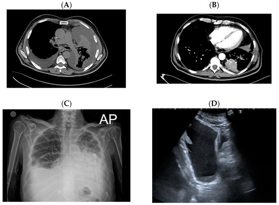
Figure 1.
April 2021. (A,B) Computer tomography showing bilateral pleural effusions, a solid lesion of about 57 × 45 mm2 with spiculated margins in the left lower lobe with carcinomatous lymphangitis. (C) Chest X-ray describing bilateral pleural effusion and synchronous carcinomatous lymphangitis. (D) Abdominal ultrasound highlighting pleural effusion.
The patient was thus referred to our clinical laboratory for molecular assessment of EGFR, ALK, ROS1, BRAF, and KRAS and evaluation of PD-L1 expression. The molecular test and the evaluation of PD-L1 expression were performed. No actionable mutations were identified, and PD-L1 was not expressed (tumor proportion score (TPS) < 1). Hospitalized in the Oncology Unit of Sant’ Anna and San Sebastiano Hospital in June 2021, he was begun on a first treatment cycle with carboplatin plus pemetrexed plus pembrolizumab based on the high burden of the disease and breathing difficulties. Moreover, a physical examination detected the presence of ascites, which were later confirmed by ultrasound (Figure 1D). Thus, evacuative paracentesis was immediately practiced and about 5 L of yellow citrine liquid was removed. Peritoneal cytological examinations identified TTF-1 positive malignant cells, suggesting peritoneal carcinomatosis (PCM) from lung cancer. Carcinoembryonic antigen (CEA) levels in the ascites were above 500 ng/mL. Cell-block analysis, which was performed in our clinical laboratory, revealed the atypical p.K601E of exon 15 of BRAF mutation and a PD-L1 expression level ≥ 50%. Owing to the limited data on targeted therapy in patients with BRAF exon 15 p.K601E-mutant lung adenocarcinoma, (Figure 2 and Figure 3) we preferred chemo-immunotherapy over targeted therapy to control the symptoms.
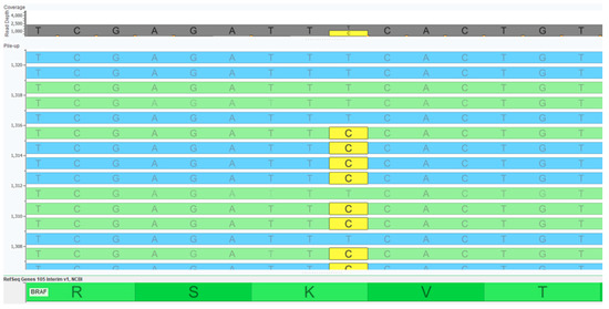
Figure 2.
Molecular evaluation. Next generation sequencing analysis revealed the presence of a BRAF exon 15 p.K601E point mutation.
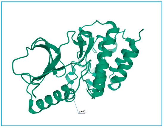
Figure 3.
3D representation of the BRAF protein. The blue arrow highlights the codon 601. This figure was created using Mol* PDB ID Mol* and Research Collaboratory for Structural Bioinformatics (RCSB) Protein Data Bank (PDB).
Remarkably, after the fourth cycle, ascites completely regressed. Concomitantly, breathlessness was significantly reduced. Indeed, home oxygen therapy was gradually reduced to 3 L/min for 15 min a day and used only on exertion. Overall, the patient’s performance status and appetite were markedly improved. Clinically, the size of the right lung nodules and of the primary heteroplastic lesion was reduced; carcinomatous lymphangitis was also decreased (Figure 4A,B). However, when seen at a follow-up outpatient visit in February 2022, his overall performance status seemed to have deteriorated. He complained of abdominal heaviness, swollen legs, and a worsening of dyspnea, thereby needing to increase oxygen therapy to 6 L/min for a couple of hours a day, even when at rest. Disease progression was also seen. Ascites had returned, as confirmed by ultrasound. Moreover, chest X-rays revealed a worsening of bilateral pleural effusion and carcinomatous lymphangitis. Therefore, an evacuative thoracentesis of about 2 L of bloody fluid was practiced. It was clear that the patient was experiencing cancer recurrence. Accordingly, he was begun on a second-line treatment with dabrafenib 300 mg/day plus trametinib 2 mg/day (Figure 5A,B). Stunningly, fifteen days later, ascites were no longer clinically appreciable and leg swelling was resolved. The treatment, however, had no effect on dyspnea. Indeed, since he required continuous oxygen therapy (6 L/min) throughout the day, he was subjected to evacuative thoracentesis. Despite this, in April 2022, the patient was admitted to a sub-intensive care unit (Figure 6) where a pleural drainage was placed. Palliative non-invasive ventilation was then administered until he died of cardiorespiratory failure.
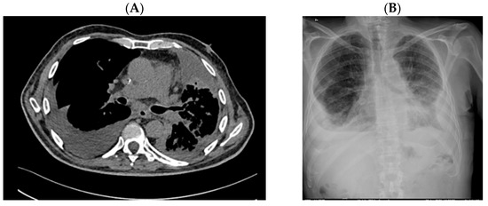
Figure 4.
September 2021. (A,B) Computer Tomography and X-ray highlighting simultaneous reduction of bilateral pleural effusion and carcinomatous lymphangitis.
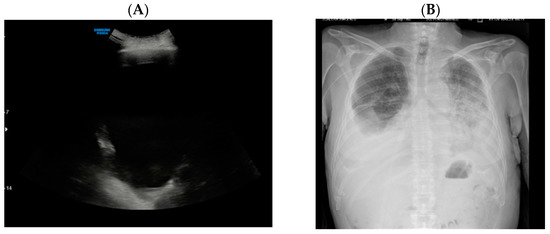
Figure 5.
February 2022. (A,B) Sharp worsening of pulmonary interstitial disease in chest X-ray and pleural effusion on abdominal ultrasound.
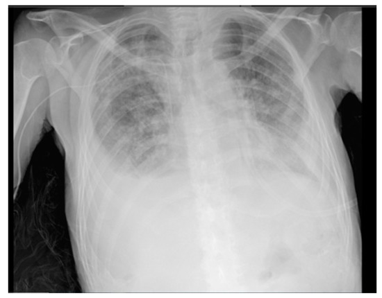
Figure 6.
April 2022. Diffuse pulmonary interstitial disease and lung progression during non-invasive ventilation.
3. Discussion
BRAF-p.V600 and non-p.V600 mutations in NSCLC are associated with different clinical and pathological features. The former has roughly the same incidence in both male and female smokers, whereas the latter has a higher incidence in male smokers [11,12]. Although the therapeutic efficacy of combination therapies against BRAF exon 15 p.V600E mutation in lung adenocarcinoma is well known, not much is known about the clinical benefit of these therapies in patients affected by class II and III BRAF mutations. The reason is that these mutations affect a much more heterogeneous and numerically inferior population than class I mutations, rendering them more difficult to analyze in phase II-III trials. Several insightful studies have attempted to shed some light on the aggressive behavior of these rare mutations in advanced NSCLC. For instance, a few years ago a study suggested that class I mutations may be less aggressive than non-p.V600 mutations, which are more likely to occur in brain metastases and RAS co-alterations. Indeed, the study reported that BRAF non-p.V600 patients had a shorter progression free survival (PFS) and overall survival (OS). It also suggested that the differences in survival rates might be driven by the fact that class I patients have fewer extra thoracic metastases and are more commonly treated with targeted therapies [13]. A more recent study evidenced that non-p.V600 mutations arise more frequently in smokers than in non-smokers and, intriguingly, that the smoking status is associated with a better response to immunotherapy [14]. Still, further research has recently suggested that since class II and III BRAF mutations coexist with KRAS mutations, which in turn are associated with higher PD-L1 expression levels, they are more sensitive to immune checkpoint inhibitors (ICIs) than class I mutations [15,16].
The possible molecular mechanism whereby the BRAF exon 15 p.K601E mutation evades immune checkpoint inhibitors has been described in several studies. BRAF exon 15 p.K601E mutation, which occurs in about 0.2% of NSCLC cases, affects the activation segment in the kinase domain, thus decreasing kinase auto-inhibition by hindering the bond with the phosphate-binding loop (p-loop) [6]. Apparently, this conformation increases in vitro kinase activity and phosphorylation of downstream MEK. Moreover p.K601E-mutated signals as a dimer, thus preventing BRAF inhibitors from binding to the monomeric kinase domain [7]. Indeed, recent research has reported that 12 cases harboring BRAF exon 15 p.K601E-driven tumors, namely, three NSCLC and nine melanoma patients, did not respond to treatment with single-agent selective BRAF inhibitors [17]. Still, another study has highlighted that whereas the MEK inhibitor trametinib provides durable clinical benefit in p.K601E-mutant melanoma [18], it has a very short-term clinical effect (four month) in an analogue of mutated lung adenocarcinoma [10]. On the other hand, studies show that combining BRAF and MEK inhibitors, such as dabrafenib and trametinib, may potently inhibit downstream signalling [19,20].
In a retrospective multicenter European BRAF cohort (EURAF), five out of six patients harboring non-p.V600E mutation and treated with specific targeted therapy exhibited a median overall survival of 11.8 months versus 25.3 months of class I mutated patients, most likely because they were resistant to BRAF-inhibitors [21].
The prevalence of BRAF mutations has been reported to be even lower in Chinese patients, affecting about 0.5–2% of NSCLC cases. In a retrospective multicenter study in Chinese patients with NSCLC harboring BRAF mutations [11], Mu et al. identified 11 patients with non-p.V600E mutations, including 4 patients affected by BRAF exon 15 p.K601E lung adenocarcinoma, and assessed the clinical outcome of several anticancer agents. Overall, patients’ response in terms of PFS was very discouraging. One patient, who had poorly responded to first-line treatment with platinum-based chemotherapy, developed progressive disease (PD) after receiving second line treatment with dabrafenib plus trametinib; two other patients experienced a PFS of 2.0 months and 4.0 months with first line anti-BRAF target therapy; and, finally, one patient achieved a PFS of 3.5 months after second line nivolumab plus chemotherapy [11]. Considering the differences between the genetic background of Caucasians and Asians, we propose that studying BRAF mutations in NSCLC Asian patients may be of great significance to study the molecular complexity of these mutations.
Although results from previous small-sample retrospective analyses have underlined an association between PD-L1 expression and BRAF-mutated NSCLC patients [22,23], the objective response rate to single anti-PD-(L)1 agents in BRAF-mutant lung adenocarcinoma is only 10–30%, with a median PFS of 2–4 months. Analogous results have been observed in studies of wild-type NSCLC treated with second-line immune-checkpoint inhibitor (ICI) monotherapy [14,24]. Specifically, no PFS differences were seen between V600E and non-V600E mutant NSCLC treated with a single-agent ICI [24]. Instead, a recent case report has shown that first-line ICI plus chemotherapy produces a durable response in a BRAF exon 15 p.V600E mutated lung cancer, with a PFS of about 20 months [25]. Based on these results, it would be interesting to assess the effects of chemo-immunotherapy combination in BRAF exon 15 p.K601E positive NSCLC.
Our report is interesting for several reasons. First, the clinical presentation of our case is quite atypical in that metastatic peritoneal carcinomatosis (MPC) is a rare NSCLC complication in clinical practice, often suggesting the presence of potentially targetable mutations [26]. As previously reported, the profile of MPC NSCLC is predominantly adenocarcinoma, often harboring EGFR mutations or ALK rearrangements. Notably, ascitic fluid cytology tests can successfully detect EGFR mutations in about 60% of patients whose tumor specimen harbor the mutation [27]. The presence or absence of a driver oncogene has been shown to determine an OS of 12.0 months versus 2.5 months, thereby drastically improving the prognosis [27,28]. As of today, though, data on MPC in BRAF-mutated NSCLC are still missing. A second interesting aspect of our report is that our patient carried the atypical BRAF exon 15 p.K601E mutation, for which there is no clinical experience describing the efficacy of first-line chemo-immunotherapy combination; moreover, there are only conflicting reports regarding the sensitivity of this rare mutation to targeted therapy. Specifically, after receiving first-line treatment with pembrolizumab plus carboplatin plus pemetrexed, our patient experienced a PFS of 8–9 months. Unfortunately, owing to cancer progression and subsequent death, we were unable to evaluate our patient’s PFS after receiving second-line targeted therapy. A final interesting aspect arising from this report is the hypothesis that our patient might have been affected by a biclonal disease given the absence of BRAF mutations in the pleural effusion. Such suspicion was clinically verified without resorting to invasive evacuative paracentesis but by rapidly controlling the peritoneal disease with dabrafenib plus trametinib treatment. However, the treatment failed to stop the rapid clinical lung and pleural progression. In this regard, we speculate that BRAF and MEK inhibitors might only partially act against BRAF class II exon 15 p.K601E-mutation.
4. Conclusions
BRAF mutations act as oncogenetic drivers and are grouped into three classes depending on their mechanism of action. Although dual MAPK pathway blockade with BRAF and MEK inhibitors is an efficient strategy for treating BRAF exon 15 p.V600E-mutated lung cancer, the potential role of this pharmaceutical strategy in atypical BRAF mutations is still unknown. Our case shows how BRAF exon 15 p.K601E mutation may underlie a biological phenotype of aggressive disease, with potential responsiveness to dabrafenib and trametinib combination. Moreover, it suggests that chemo-immunotherapy may be suitable as first line treatment especially for high-burden diseases to rapidly control symptoms.
Author Contributions
Conceptualization, M.D.F.; Methodology, all authors; Software, all authors; Validation, all authors; Formal Analysis, M.D.F., P.P., F.P., C.D.L., A.I., U.M., G.T., G.P.I.; Investigation, M.D.F., P.P., F.P., C.D.L., A.I., U.M., G.T., G.P.I.; Resources, M.D.F., P.P., F.P., C.D.L., A.I., U.M., G.T., G.P.I.; Data Curation, M.D.F., P.P., F.P., C.D.L., A.I., U.M., G.T., G.P.I.; Writing—Original Draft Preparation, M.D.F. and P.P.; Writing—Review and Editing, all authors; Visualization, M.D.F., P.P., F.P., C.D.L., A.I., U.M., G.T., G.P.I.; Supervision, U.M., G.T. and G.P.I.; Project Administration, U.M., G.T. and G.P.I. All authors have read and agreed to the published version of the manuscript.
Funding
The authors have not declared a specific grant for this review from any funding agency in the public, commercial or not-for-profit sectors.
Institutional Review Board Statement
Not applicable.
Informed Consent Statement
Written informed consent for publication has been obtained from participating patient to publish this paper.
Data Availability Statement
The data presented in this study are available on request from the corresponding author.
Acknowledgments
The authors thank Paola Merolla for editing the manuscript.
Conflicts of Interest
Pasquale Pisapia has received personal fees as speaker bureau from Novartis, unrelated to the current work. Umberto Malapelle has received personal fees (as consultant and/or speaker bureau) from Boehringer Ingelheim, Roche, MSD, Amgen, Thermo Fisher Scientifics, Eli Lilly, Diaceutics, GSK, Merck and AstraZeneca, Janssen, Diatech, Novartis, Hedera unrelated to the current work. Giancarlo Troncone reports personal fees (as speaker bureau or advisor) from Roche, MSD, Pfizer, Boehringer Ingelheim, Eli Lilly, BMS, GSK, Menarini, AstraZeneca, Amgen and Bayer, unrelated to the current work. The other authors have nothing to disclose.
References
- Huang, R.S.P.; Severson, E.; Haberberger, J.; Duncan, D.L.; Hemmerich, A.; Edgerly, C.; Ferguson, N.L.; Frampton, G.; Owens, C.; Williams, E.; et al. Landscape of Biomarkers in Non-small Cell Lung Cancer Using Comprehensive Genomic Profiling and PD-L1 Immunohistochemistry. Pathol. Oncol. Res. 2021, 27, 592997. [Google Scholar] [CrossRef]
- Leonetti, A.; Facchinetti, F.; Rossi, G.; Minari, R.; Conti, A.; Friboulet, L.; Tiseo, M.; Planchard, D. BRAF in Non-Small Cell Lung Cancer (NSCLC): Pickaxing Another Brick in the Wall. Cancer Treat. Rev. 2018, 66, 82–94. [Google Scholar] [CrossRef]
- Nguyen-Ngoc, T.; Bouchaab, H.; Adjei, A.A.; Peters, S. BRAF Alterations as Therapeutic Targets in Non-Small-Cell Lung Cancer. J. Thorac. Oncol. 2015, 10, 1396–1403. [Google Scholar] [CrossRef] [Green Version]
- Ross, J.S.; Wang, K.; Chmielecki, J.; Gay, L.; Johnson, A.; Chudnovsky, J.; Yelensky, R.; Lipson, D.; Ali, S.M.; Elvin, J.A.; et al. The Distribution of BRAF Gene Fusions in Solid Tumors and Response to Targeted Therapy. Int. J. Cancer. 2016, 138, 881–890. [Google Scholar] [CrossRef] [Green Version]
- Davies, H.; Bignell, G.R.; Cox, C.; Stephens, P.; Edkins, S.; Clegg, S.; Teague, J.; Woffendin, H.; Garnett, M.J.; Bottomley, W.; et al. Mutations of the BRAF Gene in Human Cancer. Nature 2002, 417, 949–954. [Google Scholar] [CrossRef]
- Dankner, M.; Rose, A.A.N.; Rajkumar, S.; Siegel, P.M.; Watson, I.R. Classifying BRAF Alterations in Cancer: New Rational Therapeutic Strategies for Actionable Mutations. Oncogene 2018, 37, 3183–3199. [Google Scholar] [CrossRef]
- Yao, Z.; Torres, N.M.; Tao, A.; Gao, Y.; Luo, L.; Li, Q.; de Stanchina, E.; Abdel-Wahab, O.; Solit, D.B.; Poulikakos, P.I.; et al. BRAF Mutants Evade ERK-Dependent Feedback by Different Mechanisms That Determine Their Sensitivity to Pharmacologic Inhibition. Cancer Cell 2015, 28, 370–383. [Google Scholar] [CrossRef] [Green Version]
- Foster, S.A.; Whalen, D.M.; Özen, A.; Wongchenko, M.J.; Yin, J.; Yen, I.; Schaefer, G.; Mayfield, J.D.; Chmielecki, J.; Stephens, P.J.; et al. Activation Mechanism of Oncogenic Deletion Mutations in BRAF, EGFR, and HER2. Cancer Cell 2016, 29, 477–493. [Google Scholar] [CrossRef] [Green Version]
- Zehir, A.; Benayed, R.; Shah, R.H.; Syed, A.; Middha, S.; Kim, H.R.; Srinivasan, P.; Gao, J.; Chakravarty, D.; Devlin, S.M.; et al. Mutational Landscape of Metastatic Cancer revealed from Prospective Clinical Sequencing of 10,000 Patients. Nat. Med. 2017, 23, 703–713. [Google Scholar] [CrossRef]
- Saalfeld, F.C.; Wenzel, C.; Aust, D.E.; Wermke, M. Targeted therapy in BRAF p.K601E–driven NSCLC: Case report and literature review. JCO Precis. Oncol. 2020, 4, 1163–1166. [Google Scholar]
- Mu, Y.; Yang, K.; Hao, X.; Wang, Y.; Wang, L.; Liu, Y.; Lin, L.; Li, J.; Xing, P. Clinical Characteristics and Treatment Outcomes of 65 Patients With BRAF-Mutated Non-small Cell Lung Cancer. Front. Oncol. 2020, 10, 603. [Google Scholar] [CrossRef]
- Marchetti, A.; Felicioni, L.; Malatesta, S.; Sciarrotta, M.G.; Guetti, L.; Chella, A.; Viola, P.; Pullara, C.; Mucilli, F.; Buttitta, F. Clinical features and outcome of patients with non-small-cell lung cancer harboring BRAF mutations. J. Clin. Oncol. 2011, 29, 3574–3579. [Google Scholar] [CrossRef]
- Litvak, A.M.; Paik, P.K.; Woo, K.M.; Sima, C.S.; Hellmann, M.D.; Arcila, M.E.; Ladanyi, M.; Rudin, C.M.; Kris, M.G.; Riely, G.J. Clinical characteristics and course of 63 patients with BRAF mutant lung cancers. J. Thorac. Oncol. 2014, 9, 1669–1674. [Google Scholar] [CrossRef] [Green Version]
- Mazieres, J.; Drilon, A.; Lusque, A.B.; Mhanna, L.; Cortot, A.; Mezquita, L.; Thai, A.A.; Mascaux, C.; Couraud, S.; Veillon, R.; et al. Immune checkpoint inhibitors for patients with advanced lung cancer and oncogenic driver alterations: Results from the immunotarget registry. Ann. Oncol. 2019, 30, 1321–1328. [Google Scholar] [CrossRef]
- Iaccarino, A.; Pisapia, P.; De Felice, M.; Pepe, F.; Gragnano, G.; De Luca, C.; Ianniello, G.; Malapelle, U. Concomitant Rare KRAS and BRAF Mutations in Lung Adenocarcinoma: A Case Report. J. Mol. Pathol. 2020, 1, 36–42. [Google Scholar] [CrossRef]
- Salimian, K.J.; Fazeli, R.; Zheng, G.; Ettinger, D.; Maleki, Z. V600E BRAF versus Non-V600E BRAF Mutated Lung Adenocarcinomas: Cytomorphology, Histology, Coexistence of Other Driver Mutations and Patient Characteristics. Acta Cytol. 2018, 62, 79–84. [Google Scholar]
- Kim, D.W.; Haydu, L.E.; Joon, A.Y.; Bassett, R.L., Jr.; Siroy, A.E.; Tetzlaff, M.T.; Routbort, M.J.; Amaria, R.N.; Wargo, J.A.; McQuade, J.L.; et al. Clinicopathological features and clinical outcomes associated with TP53 and BRAFNon-V600 mutations in cutaneous melanoma patients. Cancer 2017, 123, 1372–1381. [Google Scholar]
- Moiseyenko, F.V.; Egorenkov, V.V.; Kramchaninov, M.M.; Artemieva, E.V.; Aleksakhina, S.N.; Holmatov, M.M.; Moiseyenko, V.M.; Imyanitov, E.N. Lack of response to vemurafenib in melanoma carrying BRAF K601E mutation. Case Rep. Oncol. 2019, 12, 339–343. [Google Scholar]
- Su, P.L.; Lin, C.Y.; Chen, Y.L.; Chen, W.L.; Lin, C.-C.; Su, W.-C. Durable response to combined dabrafenib and trametinib in a patient with BRAF K601E mutation-positive lung adenocarcinoma: A case report. JTO Clin. Res. Rep. 2021, 2, 100202. [Google Scholar]
- Rogiers, A.; Thomas, D.; Vander Borght, S.; van den Oord, J.J.; Bechter, O.; Dewaele, M.; Rambow, F.; Marine, J.C.; Wolter, P. Dabrafenib plus trametinib in BRAF K601E-mutant melanoma. Br. J. Dermatol. 2019, 180, 421–422. [Google Scholar]
- Gautschi, O.; Bluthgen, M.V.; Smit, E.F.; Wolf, J.; Früh, M.; Peters, S.; Schuler, M.; Zalcman, G.; Milia, J.; Mazieres, J. Targeted therapy for patients with BRAF-mutant lung cancer: Results from the European EURAF cohort. J. Thorac. Oncol. 2015, 10, 1451–1457. [Google Scholar] [CrossRef]
- Yang, C.-Y.; Lin, M.-W.; Chang, Y.-L.; Wu, C.-T.; Yang, P.-C. Programmed Cell DeathLigand 1 Expression in Surgically Resected Stage I Pulmonary Adenocarcinoma and its Correlation with Driver Mutations and Clinical Outcomes. Eur. J. Cancer 2014, 50, 1361–1369. [Google Scholar] [CrossRef]
- Tseng, J.S.; Yang, T.Y.; Wu, C.Y.; Ku, W.H.; Chen, K.C.; Hsu, K.H.; Huang, Y.-H.; Su, K.-Y.; Yu, S.-L.; Cheng, G.-C. Characteristics and Predictive Value of PD-L1 Status in Real-World Non-Small Cell Lung Cancer Patients. J. Immunother. 2018, 41, 292–299. [Google Scholar] [CrossRef]
- Guisier, F.; Dubos-Arvis, C.; Viñas, F.; Doubre, H.; Ricordel, C.; Ropert, S.; Janicot, H.; Bernardi, M.; Fournel, P.; Lamy, R.; et al. Efficacy and Safety of Anti-PD-1 Immunotherapy in Patients with Advanced NSCLC With BRAF, HER2, or MET Mutations or RET Translocation: GFPC 01-2018. J. Thorac. Oncol. 2020, 15, 628–636. [Google Scholar] [CrossRef]
- Niu, X.; Sun, Y.; Planchard, D.; Chiu, L.; Bai, J.; Ai, X.; Lu, S. Durable Response to the Combination of Atezolizumab with Platinum-Based Chemotherapy in an Untreated Non-Smoking Lung Adenocarcinoma Patient With BRAF V600E Mutation: A Case Report. Front. Oncol. 2021, 11, 634920. [Google Scholar] [CrossRef]
- Hanane, K.; Salma, B.; Khadija, B.; Ibrahim, E.; Saber, B.; Hind, M.; Hassan, E. Peritoneal carcinomatosis, an unusual and only site of metastasis from lung adenocarcinoma. Pan Afr. Med. J. 2016, 23, 60. [Google Scholar] [CrossRef]
- Tani, T.; Nakachi, I.; Ikemura, S.; Nukaga, S.; Ohgino, K.; Kuroda, A.; Terai, H.; Masuzawa, K.; Shinozaki, T.; Ishioka, K.; et al. Clinical Characteristics and Therapeutic Outcomes of Metastatic Peritoneal Carcinomatosis in Non-Small-Cell Lung Cancer. Cancer Manag. Res. 2021, 13, 7497–7503. [Google Scholar] [CrossRef]
- Abbate, M.I.; Cortinovis, D.L.; Tiseo, M.; Vavalà, T.; Cerea, G.; Toschi, L.; Canova, S.; Colonese, F.; Bidoli, P. Peritoneal carcinomatosis in non-small-cell lung cancer: Retrospective multicentric analysis and literature review. Future Oncol. 2019, 15, 989–994. [Google Scholar] [CrossRef]
Publisher’s Note: MDPI stays neutral with regard to jurisdictional claims in published maps and institutional affiliations. |
© 2022 by the authors. Licensee MDPI, Basel, Switzerland. This article is an open access article distributed under the terms and conditions of the Creative Commons Attribution (CC BY) license (https://creativecommons.org/licenses/by/4.0/).