Article

Age, Sex, and Maxillary Position Are Associated with Successful Microimplant-Assisted Rapid Palatal Expansion in Adults

1
Graduate School of Medicine, Korea University, Seoul 05505, Korea
2
Department of Orthodontics, Asan Medical Center, University of Ulsan College of Medicine, 88 Olympic-ro 43-gil, Songpa-gu, Seoul 05505, Korea
*
Author to whom correspondence should be addressed.
These authors contributed equally to this work.
Appl. Sci. 2022, 12(14), 7324; https://doi.org/10.3390/app12147324
Submission received: 4 July 2022 / Revised: 19 July 2022 / Accepted: 20 July 2022 / Published: 21 July 2022

Abstract

:
The purpose of this study was to investigate the possible predictors of success of microimplant-assisted rapid palatal expansion (MARPE) in skeletally mature patients. Additionally, factors associated with the amount of maxillary expansion were analyzed. Factors associated with MARPE success were analyzed in 53 adult patients (27 males, 26 females, mean age 25.8 ± 8.9 years, and range 18.0 to 56.6 years) who had a maxillary transverse deficiency greater than 2 mm and a cervical vertebral maturation stage of 6. Age at pretreatment (T1), sex, sutural bone density at T1, type of appliance, mode of microimplant fixation, and lateral cephalometric variables at T1 were considered for inclusion as predictors for MARPE success. In patients who showed successful maxillary skeletal expansion, the linear distances of maxillary widths were measured on cone-beam-computed-tomography images at T1 and after MARPE (T2), and factors associated with the amount of expansion were analyzed. In total, 41 of the 53 patients showed successful maxillary expansion. Age (p = 0.019), sex (p = 0.002), and A-N perp (p = 0.015) were significantly associated with the success of MARPE. The factors associated with the amount of maxillary skeletal expansion were SN-MP and midpalatal-suture density at T1. In conclusion, there is a greater chance of failure in male patients who are older and have maxillary retrusion. A greater amount of maxillary expansion can be expected in patients with a higher mandibular-plane angle and with lower midpalatal-suture density.

1. Introduction

The prevalence of transverse discrepancy in the permanent dentition is approximately 12–14% [1,2]. The clinical implications of maxillary transverse deficiency include uni- or bilateral crossbite, narrow nasal cavity, and crowding of teeth [3,4]. Rapid maxillary expansion (RME) occurs as a result of the application of transverse forces that are greater than those required for tooth movement and the application of tensile forces on the circummaxillary sutures, leading to their separation. Bone apposition and remodeling take place after the sutures are separated.
RME using tooth-borne appliances may have undesirable effects, including alveolar bone bending and buccal tipping of the teeth along with maxillary expansion. This dentoalveolar effect occurs especially in postpubertal patients, whose circummaxillary sutures are mature, possibly leading to side effects such as marginal alveolar bone loss, a decrease in buccal bone thickness, and root resorption [5,6].
Microimplant-assisted rapid palatal expansion (MARPE) has been introduced to exert its forces through the microimplants anchored in the maxilla, thus minimizing the dentoalveolar side effects. Use of MARPE has been widespread among clinicians, especially in young adults, and many cases of successful maxillary skeletal expansion have been reported in nongrowing patients with severe maxillary transverse deficiency [7,8]. Choi et al. reported a success rate of 86.9% in adult patients who had undergone maxillary expansion using MARPE [9].
Despite the high success rate of MARPE, a possibility of failure still exists. If the success of MARPE can be predicted, it would help clinicians in treatment planning. Several indicators of skeletal maturation using occlusal radiographs, hand wrist radiographs, lateral cephalograms, and cone beam computed tomography (CBCT) have been proposed to predict the skeletal response to conventional RME [10,11,12,13,14]. Grünheid et al. suggested the midpalatal-suture density (MPSD) on a CBCT image as a useful clinical predictor of the skeletal response to RME [15]. Regarding the factors associated with the success of MARPE, older patients were more likely to experience failure [16,17]. Additionally, patients with thin and long palatal bone showed a higher chance of MARPE failure [17,18].
The purpose of this study was to investigate the possible predictors of the success of MARPE in skeletally mature patients. Among the factors affecting the success of MARPE, those associated with the amount of maxillary expansion in the circummaxillary sutures were also analyzed.

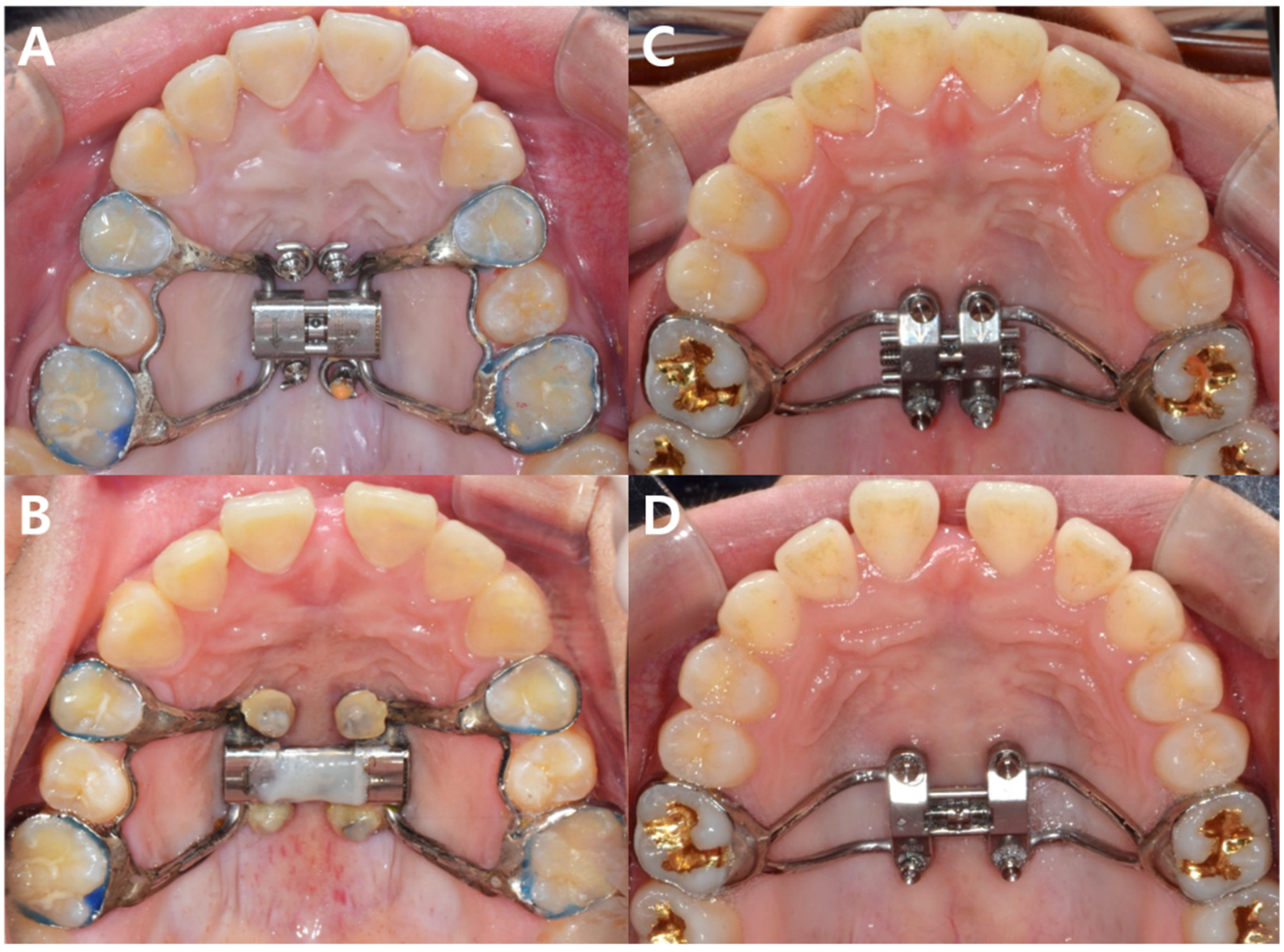
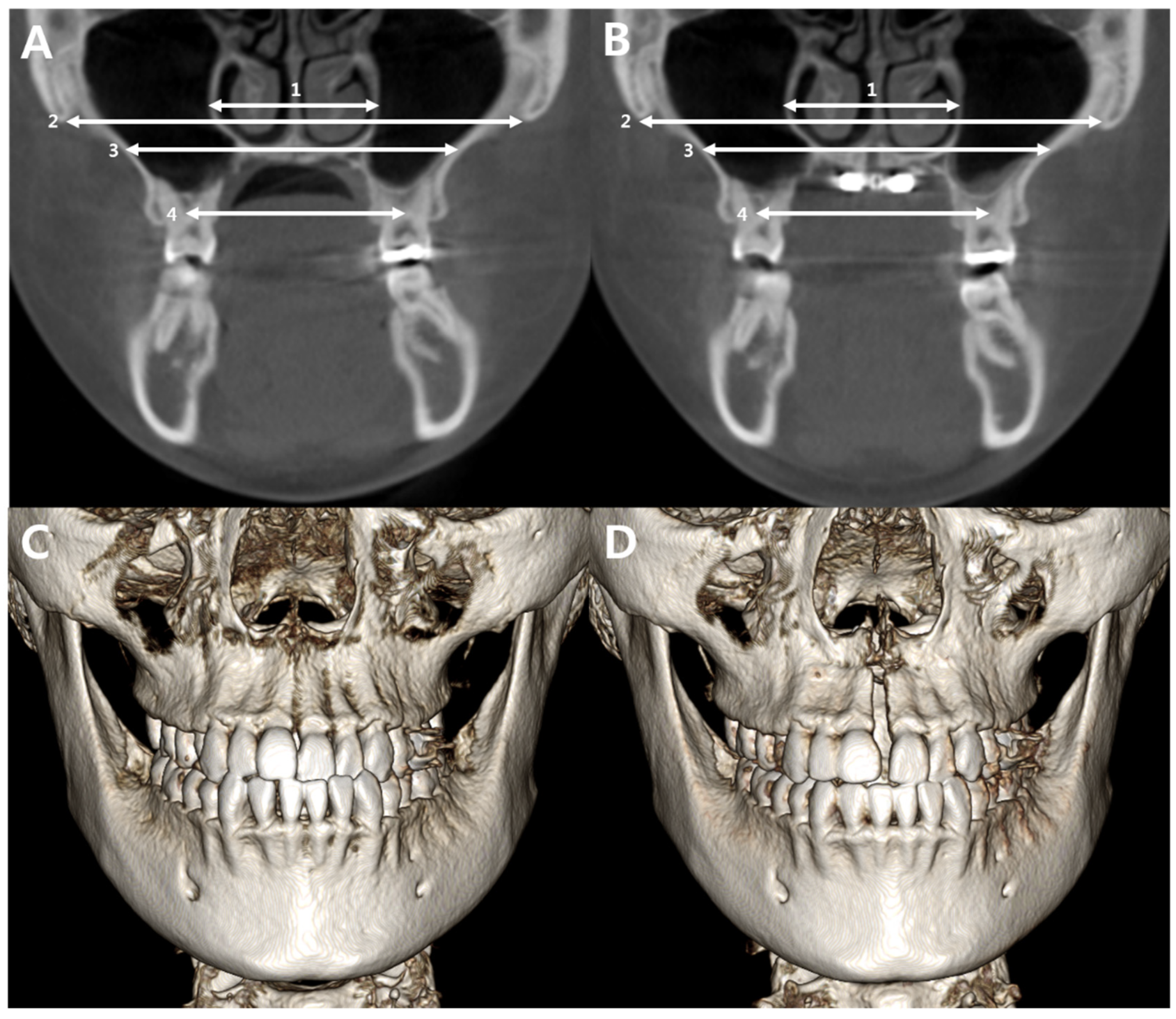
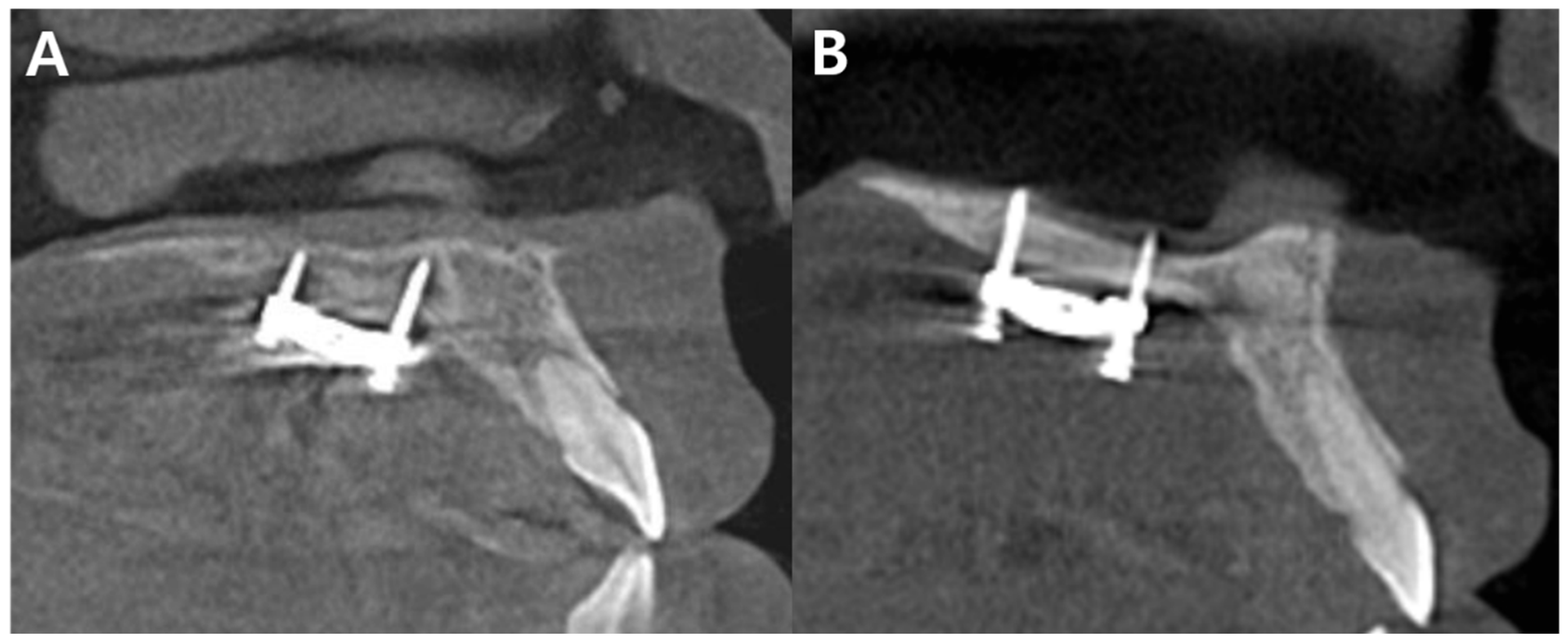
2. Materials and Methods

This retrospective study was approved by the Institutional Review Board of Korea University Anam Hospital (2019AN0288), and informed consent from patients for inclusion in the study was waived due to the retrospective nature of the study. Patients who were diagnosed with maxillary transverse deficiency and had undergone MARPE at the Department of Orthodontics at Korea University Anam Hospital, from September 2014 to March 2019, were reviewed for the study. The patients had agreed to undergo maxillary dental expansion in case of MARPE failure. The inclusion criteria were a maxillary transverse deficiency of >2 mm (according to the criteria suggested by Koo et al. [19]) an age of 18 years and older, cervical-vertebrae maturation index (CVMI) of 6 [20], and CBCT imaging performed before and after MARPE. Patients with cleft lip and palate were excluded.
The study included 53 patients (27 males, 26 females, mean age 25.8 ± 8.9 years, and range 18–56 years). Two types of bone-borne expanders for MARPE were used: (1) a modified Hyrax expander with soldered holes for microimplant placement on the palate and banded to the premolars and molars, and (2) a maxillary skeletal expander (MSE, Biomaterial Korea, Seoul, Korea) that had holes for microimplants incorporated in the expander and banded to the molars (Figure 1). A modified Hyrax expander was used in 2014–2016. The maxillary skeletal expander (MSE) was used in 2015–2019. After delivery of the appliances, microimplants were inserted under local anesthesia. Four microimplants (Jeil Medical Corporation, Seoul, Korea) with a diameter of 1.6 mm and lengths of 8 mm and 10 mm for anterior and posterior screws, respectively, were used for the modified Hyrax expanders; microimplants (Biomaterials Korea, Seoul, Korea) with a diameter of 1.5 mm and lengths of 11 mm for anterior and posterior screws were used for the MSEs.
Patients started maxillary expansion one week after the placement of microimplants at a rate of 0.4–0.5 mm per day. After two weeks of activation, patients who showed only dental tipping of the posterior teeth and no anterior diastema were considered as failure, and the expansion was discontinued. A CBCT scan was obtained to observe the midpalatal sutures. If a split in the midpalatal suture was not observed in the CBCT scan, the patient was classified in the failed group (F group) and dental expansion was performed using removable appliances to treat the posterior crossbite. Patients who showed an anterior diastema continued maxillary expansion as planned. At the end of maxillary expansion, the patients underwent CBCT, and separation of the midpalatal suture was confirmed. These patients that completed treatment were allocated to the success group (S group). Following expansion, the patients continued with the treatment of their malocclusions using a fixed orthodontic appliance. All procedures were performed by the same orthodontist (YK).
CBCT images were obtained using 3D eXam (KaVo Dental GmbH, Biberach, Germany) with the following parameters: field of view of 17 × 23 cm, at 120 kV, and 5 mAs, and pulsed scan time of 17.8 s. Scan data were reconstructed with a voxel size of 0.3 mm3.
In the patients belonging to the S group, the linear distances of maxillary widths were measured in the CBCT scans taken at pretreatment (T1) and after MARPE (T2) using Invivo5 software (Anatomage, San Jose, CA, USA). In the coronal plane passing through the furcation of the upper first molars, the distance between the furcation of the maxillary first molar, maxillary width at the level of the deepest palatal arch, the most inferior border of the zygomaticomaxillary sutures, and maximum nasal-cavity width were measured (Figure 2).
The bone mineral density of the two main resistance areas of skeletal expansion, the zygomaticomaxillary suture (zygomaticomaxillary-suture density, ZMSD) and the midpalatal suture (MPSD), was measured using CBCT at T1. A modification of the method by Grünheid et al. was used. 17 average grey density values were measured at the defined areas of the sutures (GDs), the midlevel of the soft palate (GDsp), the most inferior area of the zygomaticomaxillary sutures (GDzm), and the cortical-bone area of the mandibular symphysis (GDsy). The following equation was used to calculate the MPSD and ZMSD:
MPSD ratio = (GDs − GDsp)/(GDsy − GDsp)
ZMSD ratio = (GDzm − GDsp)/(GDsy − GDsp)
Furthermore, cephalometric analysis was performed using the CBCT-generated lateral cephalograms at T1 and T2 to analyze the skeletal changes following MARPE and investigate the possible associations between the patients’ skeletal patterns and success of MARPE. All linear measurements, bone density measurements, and cephalometric analyses were performed by the same investigator (JC).
Four weeks after the first measurement, 15 patients were randomly selected, and measurements were retaken by the same examiner (JC). An intraclass correlation analysis was performed; the intraclass correlation ranged from 0.875 to 0.980, showing that the measurement was highly reliable.

Statistical Analysis

After descriptive statistics of the patient demographics and lateral cephalometric variables, Fisher’s exact test was used to analyze the distribution of the patients in the F group and the S group according to sex, type of appliance, and presence of mono- vs. bicortical engagement of the microimplants (Figure 3). A logistic regression analysis was used to determine the factors associated with the success of MARPE. Age at T1, lateral cephalometric variables at T1, appliance type, mono- vs. bicortical engagement, and bone density at T1 (ZMSD and MPSD) were considered as possible predictor variables. Among the patients in group S, significant factors associated with the amount of expansion in the palate, zygomaticomaxillary sutures, and nasal cavity were analyzed using Pearson’s correlation analysis.
The statistical analyses were performed using SPSS software (version 22.0; IBM, Armonk, NY, USA). p values < 0.05 were considered statistically significant.

3. Results

The patients’ descriptive statistics, such as age and lateral cephalometric analyses in males and females, are presented in Table 1. The mean age of the patients was 25.8 ± 8.9 years (males, 24.6 ± 6.1 years; females, 27.0 ± 11.1 years). There were no significant differences in the age and cephalometric variables between male and female patients.
Of the 53 patients, successful maxillary skeletal expansion was performed in 41 (77.4%) patients. As a result of MARPE, SNA (p < 0.001), ANB (p = 0.001), A-Nperp (p < 0.001), SN-MP (p = 0.001), and Rickett’s LFH (p = 0.002) showed significant increases. Overbite showed a significant decrease following MARPE (p = 0.009, Table 2).
The proportion of females was higher in the S group than in the F group, while that of males was higher in the F group. This difference in the distribution of males and females in the two groups was significant (p = 0.002, Table 3), but there were no significant associations with the type of appliance and the mode of microimplant fixation (monocortical vs. bicortical) between the S and F groups (Table 3).
Based on a logistic regression model, significant factors associated with the success of MARPE and the odds ratios for each factor are displayed in Table 4. Age, sex, and A-N perp showed a significant association with the success of MARPE (Table 4). Other cephalometric variables and suture densities were not significant as predictors for MARPE success; Rickett’s lower facial height showed borderline nonsignificance. A yearly increase by age was likely to show MARPE failure with an odds ratio of 0.869 (p = 0.019). MARPE showed more tendency to fail in males than in females, with an odds ratio of 0.022 (p = 0.002). A greater A-N perp at T1 indicated a greater chance of success, with an odds ratio of 1.591 (p = 0.015).
With the use of MARPE, the mean amounts of expansion in the nasal cavity, zygomaticomaxillary suture, maxillary bone at the level of the roof of the palate, and bone between the furcation of the upper molars were 2.0 ± 0.9 mm, 2.2 ± 1.0 mm, 2.6 ± 1.6 mm, and 2.9 ± 1.2 mm, respectively. The factors showing significant association with the amount of maxillary skeletal expansion were MPSD at T1 and SN-MP at T1 (Table 5). The correlation coefficients indicated that higher SN-MP and lower MPSD at pretreatment were associated with a greater amount of skeletal expansion following MARPE.

4. Discussion

Despite the high success rate of MARPE, its failure can be a great burden on patients and clinicians. Therefore, alternative treatment options, such as dental expansion or surgically assisted RPE, should be considered before the start of the treatment. Our aim was to investigate the potential predictors of MARPE success that could be helpful for clinicians in predicting the prognosis of patients with maxillary transverse deficiencies, and thus help in the process of treatment planning. We found that younger patients, females, and those with a forward position of the maxilla showed a greater chance of MARPE success.
The mean age of patients in the S group was 25.1 ± 8.1 years as opposed to 27.7 ± 10.8 years in patients in the F group. Although the mean age was significantly greater in the F group, we were still able to expand the maxilla in some middle-aged patients. Out of the five patients aged over 40 years, three patients were successfully treated, and the oldest patient in the S group was 52 years old. All subjects were older than 18 years and had a CVMI of 6; thus, they were considered as skeletally mature, but there was an increased tendency of failure in older patients. However, Korbmacher reported that MPSD is the major source of resistance to maxillary expansion [21], rather than age. Our patients showed a weak positive correlation between age and MPSD, but it lacked statistical significance. There may be additional factors associated with older age, other than the sutural bone density, such as the increased interlocking pattern of the suture interdigitation that restricts maxillary expansion in older patients [13,22].
MPSD was associated with the amount of skeletal expansion, rather than the success itself. The zygomatic buttress area is one of the major areas of resistance to maxillary skeletal expansion, and significant lateral displacements in the zygomatic and maxillary bones are observed following MARPE [23]. However, the ZMSD was not associated with the success or amount of maxillary expansion. The association of MPSD with maxillary expansion following MARPE was similar to that of a previous study by Grünheid [15], conducted on growing patients. They reported greater maxillary expansion in patients with lower MPSD. In contrast, Knaup et al. conducted their study on adult patients and showed that the rate of ossified tissues in the midpalatal suture was low in all patients aged 18–63 years and that suture ossification did not restrict RPE in adults [24].
Patients with maxillary retrusion showed a lower chance of MARPE success, which is not a favorable result because patients with maxillary hypoplasia often show concurrent maxillary transverse deficiency and require maxillary skeletal expansion. Patients in the F group had a mean SNA of 78.2 ± 4.5 degrees, which is lower than normal. Clinicians should be aware that in patients with severe maxillary retrusion, there is a higher chance of MARPE failure. The Rickett’s LFH showed borderline nonsignificance as a predictor for success (p = 0.084). There was a tendency for success and greater amount of expansion in those with an increased vertical dimension. This may be associated with lower masticatory muscle forces, warranting further studies with a greater sample size to investigate the effect of vertical skeletal pattern in the success of MARPE.
Males showed a significantly greater failure rate than females did. Sex as a predictor of MARPE success showed a high statistical significance and post hoc power. However, based on our results, we could not fully explain the role of the patient’s sex on MARPE success. There was no significant difference in MPSD and ZMSD between males and females. Moreover, Jimenez-Valdivia et al. reported in their CBCT study that males showed less ossification in the midpalatal-suture area [25]. Further study is warranted to investigate the factors associated with MARPE with respect to sex-related differences.
We used two types of appliances for MARPE, one with four bands in the premolar and molars and the other with only molar bands. Although the pattern of force distribution of MARPE differs from the conventional RME [26] and among the different types of MARPE appliances [27,28], the two types of appliances used in this study showed no significant difference in the success rate of MARPE. This result coincided with that of a previous study, which showed successful maxillary expansion using different types of MARPE appliances. However, the amount of maxillary expansion was greater in the tooth-bone-anchored expander than the bone-anchored maxillary expander [29].
A limitation of this study was that the sample size was relatively small. A further, multicenter study with a larger sample size would provide a better understanding of the factors associated with MARPE success. Another limitation was that this study used CBCT for bone density measurements. Although ratios of grey values according to reference anatomic regions were used, there could still be errors due to different CBCT devices, image noise, exposure parameters, and patient positioning [30].

5. Conclusions

MARPE was performed in skeletally mature patients and a high success rate was observed. However, there was a greater chance of failure in patients who are male, older, and with maxillary retrusion. A greater amount of maxillary expansion can be expected in patients with a higher mandibular-plane angle and with lower MPSD at pretreatment.

Author Contributions

Conceptualization, Y.-J.K. and D.-Y.L.; methodology, B.G.G.; software, B.G.G. and J.-H.C.; formal analysis, J.-H.C. and Y.-J.K.; writing—original draft preparation, J.-H.C.; writing—review and editing, Y.-J.K.; visualization, B.G.G.; supervision, D.-Y.L. All authors have read and agreed to the published version of the manuscript.

Funding

This research received no external funding.

Institutional Review Board Statement

The study was conducted in accordance with the Declaration of Helsinki, and approved by the Institutional Review Board of Korea University Anam Hospital (2019AN0288).

Informed Consent Statement

Patient consent was waived due to the retrospective nature of the study.

Data Availability Statement

Not applicable.

Acknowledgments

This research did not receive any specific grant from funding agencies in the public, commercial, or not-for-profit sectors.

Conflicts of Interest

None.

References

  1. Perillo, L.; Masucci, C.; Ferro, F.; Apicella, D.; Baccetti, T. Prevalence of orthodontic treatment need in southern Italian schoolchildren. Eur. J. Orthod. 2010, 32, 49–53. [Google Scholar] [CrossRef] [PubMed]
  2. Jonsson, T.; Arnlaugsson, S.; Karlsson, K.O.; Ragnarsson, B.; Arnarson, E.O.; Magnusson, T.E. Orthodontic treatment ex-perience and prevalence of malocclusion traits in an Icelandic adult population. Am. J. Orthod. Dentofac. Orthop. 2007, 131, e11–e18. [Google Scholar] [CrossRef] [PubMed]
  3. McNamara, J.A. Maxillary transverse deficiency. Am. J. Orthod. Dentofac. Orthop. 2000, 117, 567–570. [Google Scholar] [CrossRef]
  4. Ramires, T.; Maia, R.A.; Barone, J.R. Nasal cavity changes and the respiratory standard after maxillary expansion. Braz. J. Otorhinolaryngol. 2008, 74, 763–769. [Google Scholar] [CrossRef] [Green Version]
  5. Seif-Eldin, N.F.; Elkordy, S.A.; Fayed, M.S.; Elbeialy, A.R.; Eid, F.H. Transverse skeletal effects of rapid maxillary expansion in pre and post pubertal subjects: A systematic review. Open Access Maced. J. Med. Sci. 2019, 7, 467–477. [Google Scholar] [CrossRef] [PubMed] [Green Version]
  6. Lo Giudice, A.; Barbato, E.; Cosentino, L.; Ferraro, C.M.; Leonardi, R. Alveolar bone changes after rapid maxillary expansion with tooth-born appliances: A systematic review. Eur. J. Orthod. 2018, 40, 296–303. [Google Scholar] [CrossRef] [PubMed]
  7. Lim, H.M.; Park, Y.C.; Lee, K.J.; Kim, K.H.; Choi, Y.J. Stability of dental, alveolar, and skeletal changes after minis-crew-assisted rapid palatal expansion. Korean J. Orthod. 2017, 47, 313–322. [Google Scholar] [CrossRef] [Green Version]
  8. Carlson, C.; Sung, J.; McComb, R.W.; Machado, A.W.; Moon, W. Microimplant-assisted rapid palatal expansion appliance to orthopedically correct transverse maxillary deficiency in an adult. Am. J. Orthod. Dentofac. Orthop. 2016, 149, 716–728. [Google Scholar] [CrossRef]
  9. Choi, S.H.; Shi, K.K.; Cha, J.Y.; Park, Y.C.; Lee, K.J. Nonsurgical miniscrew-assisted rapid maxillary expansion results in acceptable stability in young adults. Angle Orthod. 2016, 86, 713–720. [Google Scholar] [CrossRef] [Green Version]
  10. Wehrbein, H.; Yildizhan, F. The mid-palatal suture in young adults. A radiological-histological investigation. Eur. J. Orthod. 2001, 23, 105–114. [Google Scholar] [CrossRef] [Green Version]
  11. Revelo, B.; Fishman, L.S. Maturational evaluation of ossification of the midpalatal suture. Am. J. Orthod. Dentofac. Orthop. 1994, 105, 288–292. [Google Scholar]
  12. Franchi, L.; Baccetti, T.; McNamara, J.A. Postpubertal assessment of treatment timing for maxillary expansion and protrac-tion therapy followed by fixed appliances. Am. J. Orthod. Dentofac. Orthop. 2004, 126, 555–568. [Google Scholar]
  13. Baccetti, T.; Franchi, L.; Cameron, C.G.; McNamara, J.A. Treatment timing for rapid maxillary expansion. Angle Orthod. 2001, 71, 343–350. [Google Scholar] [PubMed]
  14. Angelieri, F.; Cevidanes, L.H.; Franchi, L.; Goncalves, J.R.; Benavides, E.; McNamara, J.A. Midpalatal suture maturation: Classification method for individual assessment before rapid maxillary expansion. Am. J. Orthod. Dentofac. Orthop. 2013, 144, 759–769. [Google Scholar]
  15. Grunheid, T.; Larson, C.E.; Larson, B.E. Midpalatal suture density ratio: A novel predictor of skeletal response to rapid maxillary expansion. Am. J. Orthod. Dentofac. Orthop. 2017, 151, 267–276. [Google Scholar]
  16. Oliveira, C.B.; Ayub, P.; Angelieri, F.; Murata, W.H.; Suzuki, S.S.; Ravelli, D.B.; Santos-Pinto, A. Evaluation of factors related to the success of miniscrew-assisted rapid palatal expansion. Angle Orthod. 2021, 91, 187–194. [Google Scholar]
  17. de Jesus, A.S.; de Oliveira, C.B.; Murata, W.H.; Suzuki, S.S.; dos Santos-Pinto, A. Would midpalatal suture characteristics help to predict the success rate of miniscrew-assisted rapid palatal expansion? Am. J. Orthod. Dentofac. Orthop. 2021, 160, 363–373. [Google Scholar]
  18. Shin, H.; Hwang, C.J.; Lee, K.J.; Choi, Y.J.; Han, S.S.; Yu, H.S. Predictors of midpalatal suture expansion by minis-crew-assisted rapid palatal expansion in young adults: A preliminary study. Korean J. Orthod. 2019, 49, 360–371. [Google Scholar]
  19. Koo, Y.J.; Choi, S.H.; Keum, B.T.; Yu, H.S.; Hwang, C.J.; Melsen, B.; Lee, K.J. Maxillomandibular arch width differences at estimated centers of resistance: Comparison between normal occlusion and skeletal Class III malocclusion. Korean J. Orthod. 2017, 47, 167–175. [Google Scholar]
  20. Franchi, L.; Baccetti, T.; McNamara, J.A. Mandibular growth as related to cervical vertebral maturation and body height. Am. J. Orthod. Dentofac. Orthop. 2000, 118, 335–340. [Google Scholar]
  21. Korbmacher, H.; Schilling, A.; Puschel, K.; Amling, M.; Kahl-Nieke, B. Age-dependent three-dimensional microcomputed tomography analysis of the human midpalatal suture. J. Orofac. Orthop. 2007, 68, 364–376. [Google Scholar] [PubMed]
  22. Melsen, B. Palatal growth studied on human autopsy material. A histologic microradiographic study. Am. J. Orthod. 1975, 68, 42–54. [Google Scholar] [PubMed]
  23. Cantarella, D.; Dominguez-Mompell, R.; Moschik, C.; Sfogliano, L.; Elkenawy, I.; Pan, H.C.; Mallya, S.M.; Moon, W. Zy-gomaticomaxillary modifications in the horizontal plane induced by micro-implant-supported skeletal expander, analyzed with CBCT images. Prog. Orthod. 2018, 19, 41. [Google Scholar]
  24. Knaup, B.; Yildizhan, F.; Wehrbein, H. Age-related changes in the midpalatal suture. A histomorphometric study. J. Orofac. Orthop. 2004, 65, 467–474. [Google Scholar] [PubMed]
  25. Jimenez-Valdivia, L.M.; Malpartida-Carrillo, V.; Rodriguez-Cardenas, Y.A.; Dias-Da Silveira, H.L.; Arriola-Guillen, L.E. Midpalatal suture maturation stage assessment in adolescents and young adults using cone-beam computed tomography. Prog. Orthod. 2019, 20, 38. [Google Scholar] [PubMed]
  26. Hartono, N.; Soegiharto, B.M.; Widayati, R. The difference of stress distribution of maxillary expansion using rapid maxillary expander (RME) and maxillary skeletal expander (MSE)—A finite element analysis. Prog. Orthod. 2018, 19, 33. [Google Scholar]
  27. Lee, H.K.; Bayome, M.; Ahn, C.S.; Kim, S.H.; Kim, K.B.; Mo, S.S.; Kook, Y.A. Stress distribution and displacement by different bone-borne palatal expanders with micro-implants: A three-dimensional finite-element analysis. Eur. J. Orthod. 2014, 36, 531–540. [Google Scholar]
  28. Yoon, S.; Lee, D.Y.; Jung, S.K. Influence of changing various parameters in miniscrew-assisted rapid palatal expansion: A three-dimensional finite element analysis. Korean J. Orthod. 2019, 49, 150–160. [Google Scholar]
  29. Oh, H.; Park, J.; Lagravere-Vich, M.O. Comparison of traditional RPE with two types of micro-implant assisted RPE: CBCT study. Semin. Orthod. 2019, 25, 60–68. [Google Scholar]
  30. Pauwels, R.; Jacobs, R.; Singer, S.R.; Mupparapu, M. CBCT-based bone quality assessment: Are Hounsfield units applicable? Dentomaxillofac. Radiol. 2015, 44, 20140238. [Google Scholar]
Figure 1. Two types of the MARPE appliance used in this study. A modified Hyrax expander with soldered holes for microimplant placement on the palate and banded to the premolars and molars, before (A) and after (B) maxillary expansion. MSE expander with holes for microimplants incorporated in the expander and banded to molars, before (C) and after (D) maxillary expansion.
Applsci 12 07324 g001
Figure 2. Linear measurements of the maxilla before and after microimplant-assisted rapid palatal expansion (MARPE). (A,B): (1) maximum nasal-cavity width; (2) distance between most inferior border of zygomaticomaxillary sutures; (3) maxillary width at the level of the deepest palatal arch; (4) distance between the furcation of the upper first molars. (C,D): 3D-rendered images of the cone beam computed tomography before and after MARPE.
Applsci 12 07324 g002
Figure 3. Monocortical versus bicortical fixation of the microimplant-assisted rapid palatal expander. (A) Monocortical fixation refers to the microimplants engaged only in the palatal cortical bone, and (B) bicortical fixation indicates microimplants engaged in the palatal and nasal layers of the cortical bone.
Applsci 12 07324 g003
Table 1. Patient characteristics.
VariablesTotalSex Differences
Male (n = 27)Female (n = 26)p-Value
MEAN ± SDMEAN ± SDMEAN ± SD
Age (year)25.7 ± 8.724.6 ± 6.127 ± 11.10.343
SNA (°)80.5 ± 4.080.3 ± 4.080.5 ± 4.20.798
SNB (°)79.8 ± 4.179.6 ± 4.879.8 ± 3.60.893
ANB (°)0.7 ± 3.30.6 ± 3.80.8 ± 3.00.870
A-N perp (mm)−0.2 ± 3.6−0.5 ± 4.0−0.2 ± 3.20.815
SN-MP (°)34 ± 5.632.8 ± 5.935.4 ± 50.095
Overbite (mm)0.6 ± 21.1 ± 20.2 ± 1.90.092
Overjet (mm)2.1 ± 2.51.9 ± 2.72.4 ± 2.30.458
Rickett’s LFH (mm)48.2 ± 4.448.5 ± 4.748.1 ± 4.30.778
SNA: Sella–nasion point A, SNB: Sella–nasion point B. ANB: Point A–nasion point B. A-N perp: Distance from point A to nasion perpendicular. SN-MP: Mandibular-plane angle defined as the angle between the sella–nasion plane and the mandibular plane. Rickett’s LFH: Rickett’s lower facial height.
Table 2. Comparison of the cephalometric variables before (T1) and after (T2) microimplant-assisted rapid palatal expansion.
VariablesT1T2p-Value
SNA (°)81.2 ± 3.882.5 ± 3.6<0.001
SNB (°)80.9 ± 3.680.8 ± 3.70.725
ANB (°)0.3 ± 3.21.6 ± 2.80.001
A-N perp (mm)0.6 ± 3.41.1 ± 3.2<0.001
SN-MP (°)34.3 ± 4.735.6 ± 4.70.001
Overbite (mm)0.3 ± 2.0−0.5 ± 1.80.009
Overjet (mm)1.6 ± 2.32.1 ± 2.40.091
Rickett’s LFH (mm)48.4 ± 4.249.6 ± 4.20.002
SNA (°)81.2 ± 3.882.5 ± 3.6<0.001
SNA: Sella–nasion point A, SNB: Sella–nasion point B. ANB: Point A–nasion point B. A-N perp: Distance from point A to nasion perpendicular. SN-MP: Mandibular-plane angle defined as the angle between the sella–nasion plane and the mandibular plane. Rickett’s LFH: Rickett’s lower facial height.
Table 3. Patient distribution of the success (S) and failed (F) groups according to sex, CVMI, type of appliance, and mode of fixation.
F GroupS Groupp-Value
(n = 15)(n = 41)
SexMale13160.002
Female225
Appliance typeHyrax370.999
MSE1234
FixationMonocortical6100.483
Bicortical620
CVMI: Cervical-vertebrae maturation index. Hyrax: A modified Hyrax expander with soldered holes for microimplant placement on the palate and banded to the premolars and molars. MSE: Maxillary skeletal expander (MSE, Biomaterial Korea, Seoul, Korea) that has holes for microimplants incorporated in the expander and banded to the molars.
Table 4. Factors associated with success of microimplant-assisted rapid palatal expansion. (Dependent variable: success [1] or failure [0]).
Independent VariableBOdds Ratiop-Value
Age−0.1400.8690.019
Sex *−3.8040.0220.002
A-N perp0.4641.5910.015
Rickett’s LFH0.2341.2640.084
* Odds ratio of males compared to females. A-N perp: distance from point A to nasion perpendicular line. Rickett’s LFH: Rickett’s lower facial height.
Table 5. Correlation coefficients of the factors associated with the amount of skeletal expansion.
MPSD_T1ZMSD_T1SN-MPANBA-NperpAge
Nasal_change−0.322−0.1920.382 *0.006−0.011−0.161
Zygo_change−0.371 *−0.192−0.002−0.1390.004−0.211
Palatal_change−0.027−0.0380.350 *−0.085−0.2170.002
Nasal_change: amount of expansion as a result of miniscrew-assisted rapid palatal expansion (MARPE) in the nasal cavity, Zygo_change: amount of expansion as a result of MARPE in the zygomaticomaxillary suture. Palatal_change: amount of expansion as a result of MARPE in the maxillary width at the level of the uppermost area of the palate. MPSD_T1: midpalatal-suture density before microimplant-assisted rapid palatal expansion at pretreatment. ZMSD_T1: zygomaticomaxillary-suture density at pretreatment. SN-MP_T1: sella–nasion line to mandibular-plane angle at pretreatment. * Statistically significant.
Publisher’s Note: MDPI stays neutral with regard to jurisdictional claims in published maps and institutional affiliations.

