Article

In Vitro Evaluation of the Strength of Dentin Replacement in Complex Posterior Tooth Restoration

1
Department of Conservative Dentistry, Faculty of Dentistry, Hasanuddin University, Makassar 90245, Indonesia
2
Faculty of Dentistry, Universitas Muslim Indonesia, Makassar 90231, Indonesia
3
Department of Oral Maxillofacial Surgery, Faculty of Dentistry, Hasanuddin University, Makassar 90245, Indonesia
4
Division of Clinical Cariology and Endodontology, Department of Oral Rehabilitation, School of Dentistry, Health Sciences University of Hokkaido, Tobetsu 061-0293, Japan
5
3D Global Biotech Inc. (Spin-Off Company from Taipei Medical University), New Taipei City 221, Taiwan
*
Author to whom correspondence should be addressed.
Appl. Sci. 2022, 12(14), 6877; https://doi.org/10.3390/app12146877
Submission received: 13 June 2022 / Revised: 4 July 2022 / Accepted: 5 July 2022 / Published: 7 July 2022
(This article belongs to the Collection Dental Composites and Adhesives in Dentistry)

Abstract

:
Due to the increasing interest in direct restoration, there is a need to address the shortcomings of these restorations, mainly by increasing the longevity of complex direct restorations. The present study aimed to evaluate the tensile strength differences in the complex restoration of posterior teeth with dentin replacement constructed by fiber and non-fiber materials. The samples were extracted from the mandibular permanent-molar and prepared using a complex cavity. The cavity was subsequently restored with the centripetal incremental technique using a nano-fill composite and different base materials, namely fiber dentin replacement, non-fiber dentin replacement, and flowable composite. The universal testing machine was used to consider the tensile strength and the fracture patterns were assessed using stereomicroscopy, followed by Scanning Electron Microscopy (SEM) examination. The data were statistically analyzed using the one-way ANOVA test. No significant differences were noted in the tensile strength of the three base materials. By using stereomicroscopy and SEM, the adhesive fracture patterns were observed more clearly in the cavities with fiber-based dentin replacement, whereas mixed fracture patterns were evident in cavities with non-fiber dentin replacement and flowable composite bases. The results indicated that the addition of fiber in dentin replacement did not affect the tensile strength in the complex restoration. Therefore, dentin replacement of both fiber and non-fiber materials is applicable as a base material for complex restoration of the posterior tooth.

1. Introduction

Extensive loss of tooth structure requires repeated replacement restoration to function again and ensure a long-lasting effect on the oral cavity [1,2,3,4]. Dental restoration can be classified as direct or indirect. Currently, direct restoration is more desirable due to its shorter procedures, minimal invasion, aesthetics, biocompatibility, improvement of mechanical properties, and longevity, while indirect restoration requires additional tissue removal, multiple visits, and higher costs [3,5,6,7,8,9].
Direct composite resin restorations exhibit certain limitations, such as resistance to fracture and polymerization shrinkage [8,9,10,11,12,13,14]. However, the mechanical properties and longevity of composite resin restorations are affected by their composition and viscosity [7,8,15,16]. Composite resin restorations, notably in posterior teeth, must consider mechanical properties, such as hardness, compressive strength, flexural strength, wear resistance, and tensile strength, which resembles the ability of natural teeth to withstand large masticatory forces. Accordingly, they are resistant to fracture [15,17]. Moreover, the fracture resistance of composite resin restoration decreases mainly in the complex cavity. Therefore, direct posterior restoration with a complex cavity requires materials that can improve the mechanical properties of composite resins, one of which is dentin replacement [8,10,16,18,19].
Dentin replacement is a base material that can be applied to the dental cavity and is followed by the application of the composite resin. This material possesses excellent adhesive properties towards dentin and self-leveling, which optimize the marginal integrity and adaptation of restorative materials [20,21,22]. In addition, this material can also function as a stress breaker with its low modulus of elasticity [23,24]. Currently, dentin replacement is being developed with the addition of fiber to increase its resistance to fractures and strengthen restorations, notably in teeth with complex cavities [8,25,26,27].
The mechanical properties of the restorative materials can be determined by assessing their tensile strength. Tensile strength is used to evaluate the material’s ability to experience plastic deformity when a force is applied [15]. Therefore, the present study aimed to investigate the tensile strength differences in the complex restoration of the posterior tooth with dentin replacement made from fiber and non-fiber materials.

2. Materials and Methods

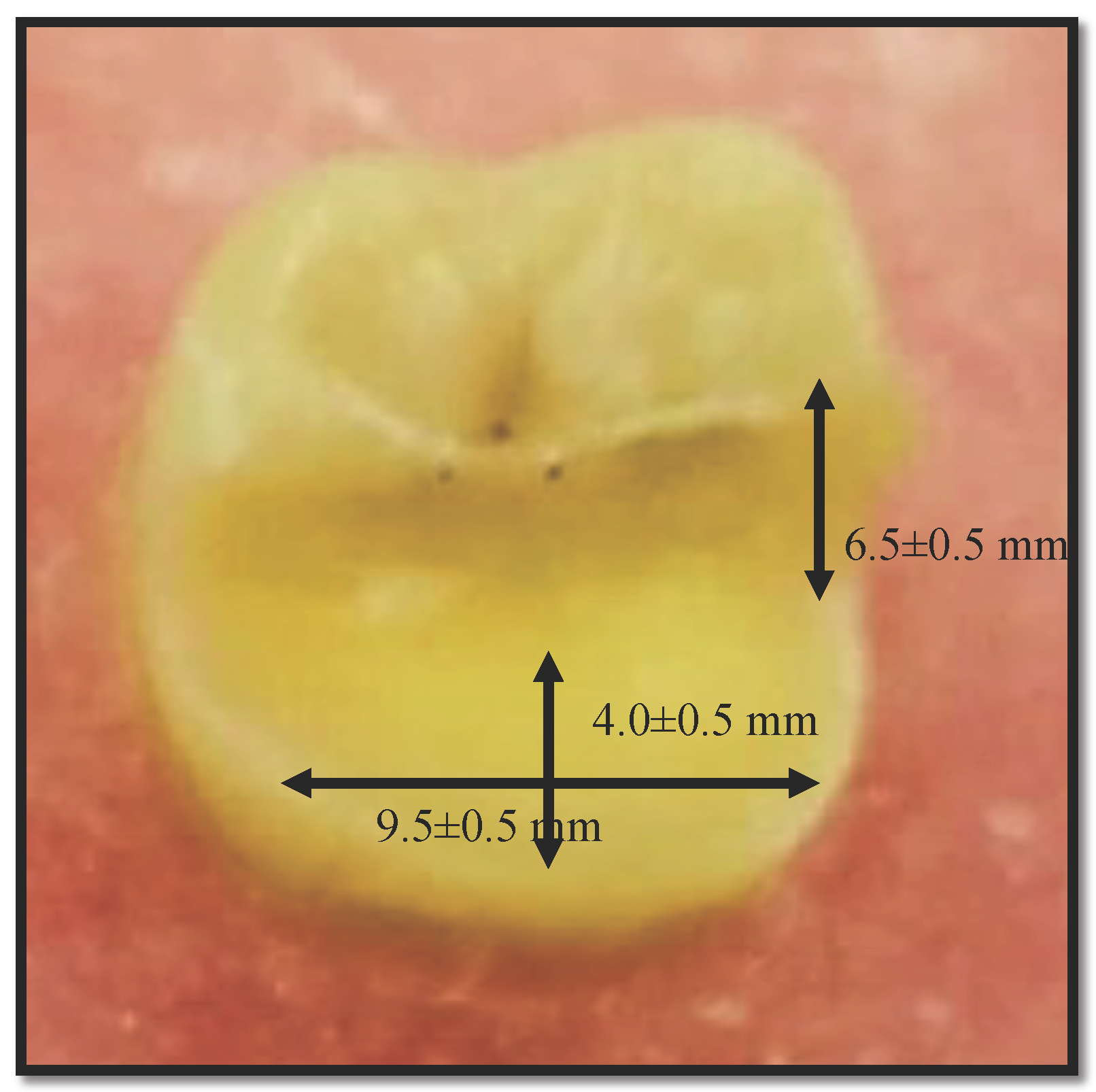
2.1. Teeth Preparation

A total of 36 extracted mandibular permanent-molar samples were used in this study, which fulfilled the following criteria: intact crown; no fractures; free of caries; sufficiently wide occlusal surface; and an extraction period not longer than six months. The extracted teeth were cleaned to remove the soft tissue and calculus, then were subsequently rinsed and stored in saline prior to treatment. The teeth were prepared using a high-speed handpiece (Panamax NSK, Tochigi, Japan) and diamond bur (Dentsply Maillefer, Ballaigues, Switzerland) with the following dimensions: 6.5 ± 0.5 mm in the cervico-occlusal; 9.5 ± 0.5 mm in the mesiodistal; and 4.0 ± 0.5 mm in the bucco-lingual (Figure 1).

2.2. Teeth Restoration

All samples were restored with a total etch adhesive system (Prime and Bond, Dentsply Detrey GmbH, Konstanz, Germany) according to the manufacturer’s instructions. Subsequently, the samples were divided into three complex restoration groups according to the type of restoration materials as follows: (I) complex restoration with fiber dentin replacement (EverX Posterior, GC CORPORATION, Tokyo, Japan) and packable nano-fill composites (Palfique LX 5, Tokuyama, Japan); (II) complex restoration with non-fiber dentin replacement (Surefil SDR, Dentsply, Milford, DE, USA) and packable nano-fill composites; (III) complex restoration with flowable composites (Filtek Z350XT, 3M ESPE, St. Paul, MN, USA) and packable nano-fill composites. Following the application of the adhesive, the nano-fill composite was applied using the centripetal technique to form the surface of the buccal cavity and form a marginal ridge, followed by the application of dentin replacement with a thickness of 4 mm. Finally, the application of 2 mm of composite resin (Palfique Bulk Flow, Tokuyama, Japan) was performed. All restored samples were immersed in artificial saliva for 24 h and incubated at 37 °C prior to tensile strength analysis.

2.3. Tensile Strength Analysis

The tensile strength of composite resin was measured using Universal Testing Machine (UTM; TISY Co., Ltd., Taichung, Taiwan). The maximum load used was 200 kgf with a pull speed of 1.0 mm/min.

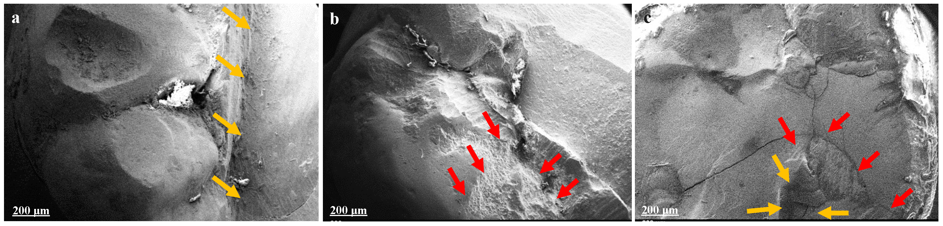
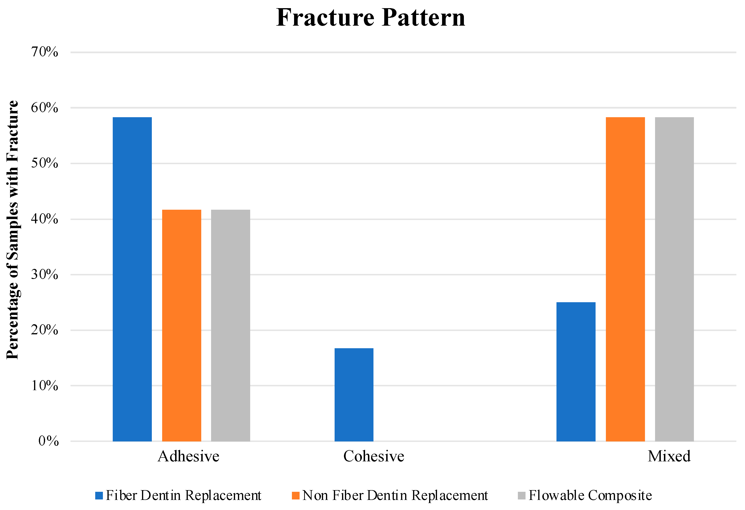
2.4. Fracture Pattern Evaluation

The fracture pattern of the sample was observed using a stereomicroscope (SFX 33 LED Portable, Inverness-shire, UK) and followed by Scanning Electron Microscopy (SEM; EVO MA 10, Zeiss, Cambridge, UK).

2.5. Statistical Analysis

The SPSS software with ANOVA test function (Version 17.0, SPSS Inc., Chicago, IL, USA) was adopted in the present study. The significance level was set at α = 0.05. All values reported indicate mean ± standard deviation.

3. Results

In this study, tensile strength and fracture pattern were the dependent variables, while fiber dentin replacement, non-fiber dentine replacement, and flowable composite were independent variables. Based on the test of homogeneity of variances obtained p-value (0.927) > 0.05, which indicates the data in this study is homogeneous. Furthermore, the normality test with Shapiro–Wilk obtained p-value (0.065) > 0.05, suggesting that the data were normally distributed.

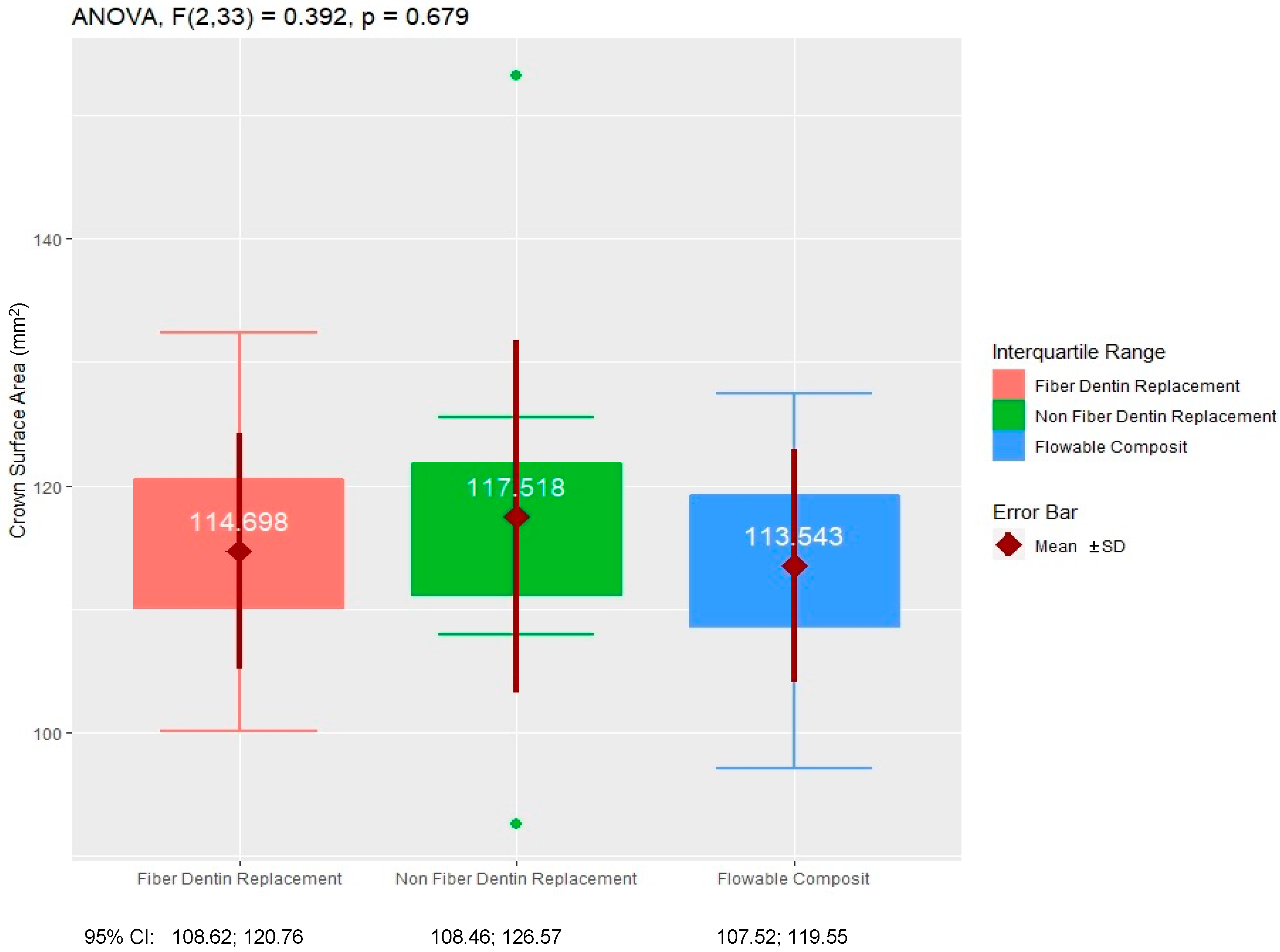
3.1. Sample Distribution

Figure 2 presents the distribution of the samples based on the crown surface area. Statistical analysis for the measurement of the extensive surface area indicated a p value of (0.679) > 0.05, suggesting no significant differences in the mean value of extensive surface area among the three groups.

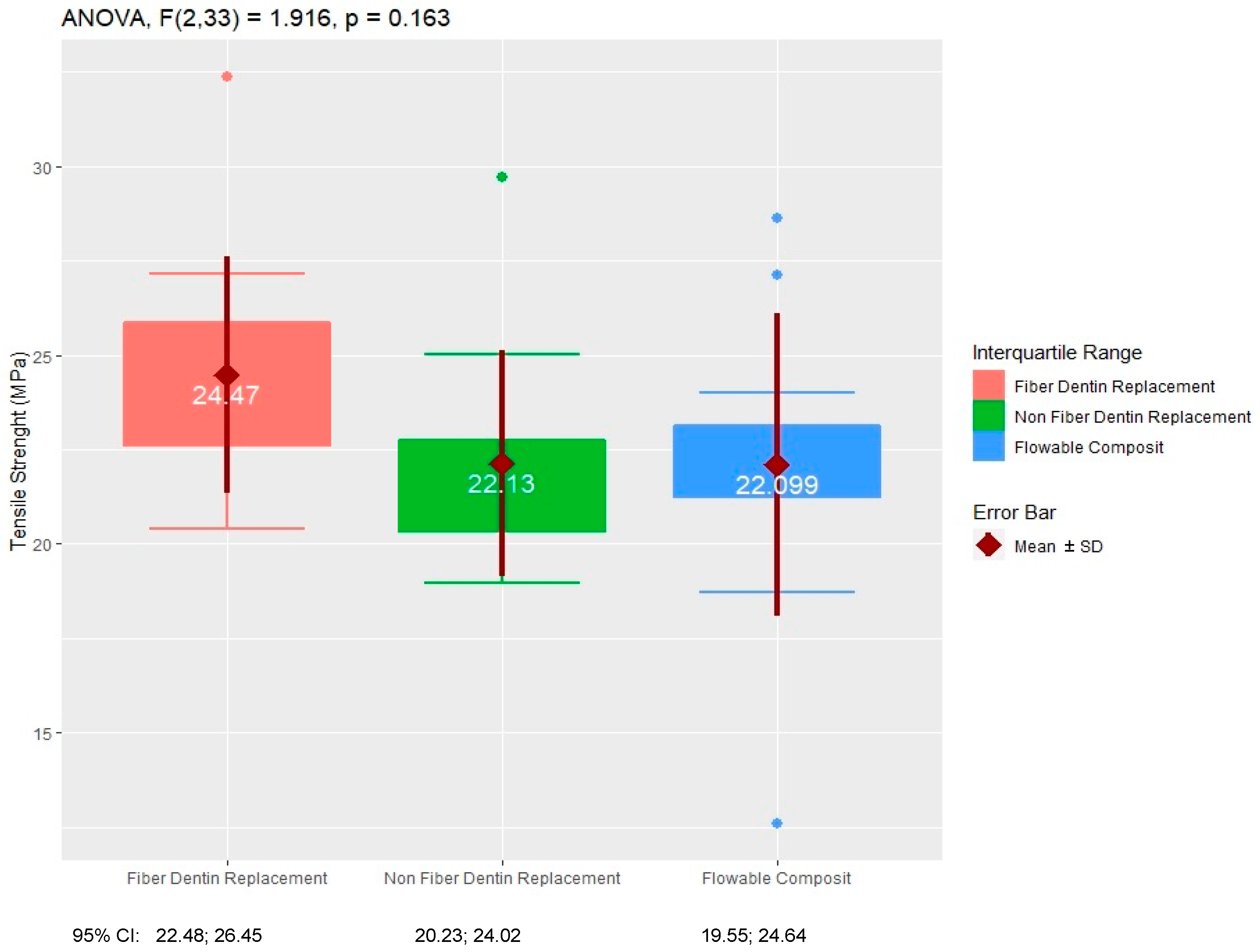
3.2. Tensile Strength Analysis

Table 1 expresses the descriptive statistics table of tensile strength in fiber dentin replacement, non-fiber dentin replacement, and flowable composite group, while the box plot is shown in Figure 3.
Statistical analysis of the tensile strength indicated a F (2,33) = 1.916, p (0.163) > 0.05, suggesting the absence of significant differences in the tensile strength among the three groups. However, the fiber dentin replacement group exhibited the highest average tensile strength than those noted for the non-fiber dentin replacement and flowable composite groups, with a value of 24.47 MPa.

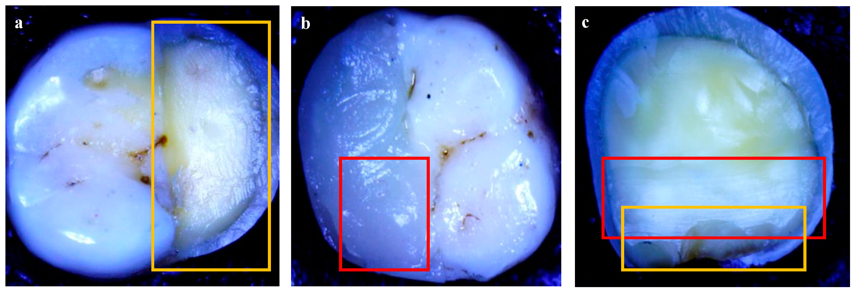
3.3. Fracture Pattern Evaluation

Figure 4 indicates various sample fracture patterns observed using stereomicroscopy. The adhesive fractures occur when the restoration separates from the tooth cavity and only one surface (in restoration or tooth) retains the bulk of the adhesive (Figure 4a); the adhesive, which is stuck on both surfaces is termed a cohesive fracture (Figure 4b); and the mixed fractures consist of adhesive and cohesive fractures (Figure 4c).
Figure 5 displays various sample fracture patterns detected using SEM. Figure 5a indicates the adhesive fracture with the restoration and the adhesive part detached from the tooth, while some of the adhesive material was still retained on the tooth surface. The other part was released with the restoration as shown in Figure 5b, and the combination of the two fracture patterns, (both adhesive and cohesive) is shown in Figure 5c.
Figure 6 illustrates various fracture patterns following quantification of the sample group. The adhesive fractures were the most common pattern in the fiber dentin replacement group, however, cohesive fractures only occurred in this group, while mixed fractures were more common in both the non-fiber replacement group and the flowable composite groups. The chi-square test designated a p value of (0.174) > 0.05, indicating the absence of significant relationship between restorations using fiber or non-fiber materials to fracture patterns of teeth.

4. Discussion

Composite resin is currently the first choice for tooth restoration due to its aesthetic appearance and reduced financial cost. However, the procedure consists of several stages and requires sensitive techniques [28,29,30]. Posterior teeth are synonymous with more extensive tooth decay. In these cases, the application of dentin replacement as a base is often recommended due to its optimal marginal adaptability. Dentin replacement has been developed with the addition of fiber to improve its mechanical properties [25,31,32,33].
The results of the present study indicated that the fiber dentin replacement group exhibited higher tensile strength compared with that of the non-fiber dentin replacement and flowable composite groups, although the difference was not significant. Fiber dentin replacement contains fibers, such as barium glass, glass fiber, bisphenol A-glycidyl methacrylate resin, triethyleneglycol dimethacrylate, silicon dioxide, polymethyl methacrylate (PMMA), and a photoinitiator. The content of glass fiber filler in dentin replacement can increase the material’s ability towards tensile loads. Following the application of a high tensile load to the tooth structure, the fiber undergoes a modification effect by absorbing and spreading the tensile load on the tooth surface [34,35]. A previous study reported relevant results to the findings of the current study, indicating that the fracture toughness and flexural strength of fiber dentin replacement were improved compared with those noted in the flowable composite resins [10,25]. This was due to the ability of the glass fiber to prevent cracks, while the PMMA could absorb and distribute the force from the polymer matrix to the glass fiber [10]. Polyethylene fiber ribbons found in dentin replacement aid the strengthening of tooth restoration by increasing the modulus of the elasticity and preventing fractures [34].
Non-fiber dentin replacement is a posterior bulk-fill composite resin containing a polymerization modulator, which is the main structure of SDR®. This polymerization modulator can reduce volumetric shrinkage by approximately 20% and polymerization force by 80%. Finally, it improves its mechanical properties [32,36,37].
Flowable composites are nanocomposite resins consisting of 82% nanoparticle fillers and nanofillers, in which the nanoparticles constituting the silica are categorized as non-agglomerated and non-aggregated, with 20 nm and 4–11 nm zirconia fillers. These particles enhance the mechanical and physical properties of the nanocomposite resin [32,34,37,38]. In contrast to the current study, a previous study that compared the flowable composites and the non-fiber dentin replacement in the mesio-occlusodistal (MOD) Class II cavities of molar teeth concluded that the tensile strength of the flowable composite resins was lower when compared with that of the non-fiber dentin replacement [39]. The differences in these results are probably associated with the adhesive system used in the study. The adhesive system plays a crucial role in maintaining the bond between the cavity wall and the restoration material. In the present study, all groups used an etch-and-rinse system that produced improved interlocking strength to increase the resistance of the fractures [15,32,40]. The research study conducted by Kumagai on molar teeth with class II MOD cavities indicated higher tensile strength in non-fiber dentin replacement compared with the flowable composites [41]. The results of that study may differ since conventional incremental applications were used, while in the present study, the application techniques used were centripetal-incremental. Centripetal-incremental approaches exhibit improved marginal adaptation and lower shrinkage polymerization, which can in turn enhance the mechanical properties of the restoration [37,42].
The difference in mechanical properties occurs due to the composite restoration material having different organic content. The small filler size and higher nanofiller volume can improve the mechanical properties of composite resins, such as tensile strength, compressive strength, and fracture resistance [34]. A previous study indicated that the tensile strength obtained in the non-fiber dentin replacement was higher than that noted in other composite resins due to differences in the composition of the polymer matrix and filler size [37].
Based on the fracture pattern observations noted in the present study, the most common dentin fiber replacement pattern was the adhesive pattern, which indicated optimal mechanical properties. High tensile strength indicated that the fiber dentin replacement ability was not deformed when a tensile force was applied. Bondable reinforcement fibers such as polyethylene fibers and glass fibers can increase the fracture resistance of the tooth structure. Therefore, when a tensile force is applied, a fracture occurs between the restoration and the tooth, which indicates that the fracture is more affected by the bonding ability of the adhesive material [37].
The use of fiber dentin replacement as a sub-structure under conventional composites has certain advantages, such as significantly increased strength of the restoration and the ability to prevent spread cracks and alter the fracture pattern [8]. In case a fracture is restored, the fracture direction will change and approach the coronal direction of the tooth [29,38].
The most common fracture in the non-fiber dentin replacement and flowable composites groups was a mixed pattern fracture since the material was less able to withstand cracks and plastic deformation under tensile pressure. An additional study demonstrated that the most frequent fracture pattern in the non-fiber dentin replacement was the adhesive fracture pattern in premolars with class II MOD cavities, which was not in line with the results reported in the current study and may be affected by differences in cavity extension [40]. The present study used non-fiber dentin replacement and flowable composites in complex cavities so that the tooth structure that received a load distribution was diminished. Non-fiber dentin replacement possesses a low elastic modulus so that when a force is applied, the load will be transferred on to the underlying structure [43].
The cohesive fracture pattern only occurs in the fiber dentin replacement group since, in the non-fiber dentin replacement and flowable composite groups, all the cohesive fracture patterns that occurred were accompanied by fractures on the tooth structure and were consequently grouped into mixed fracture patterns [43].
Thermocycling is a combination of hydraulic and thermal degradation that simulates extreme temperature changes in the oral cavity [44]. The thermocycling method is carried out to simulate the same conditions as those of the composite resin restoration, which is affected by the oral cavity [45]. In the present study, thermocycling was not performed. Therefore, the effects of the thermal stress on these three restoration groups were unidentified.
Based on the present study, the results indicated that the addition of fiber to dentin replacement did not show a significant increase in tensile strength compared with non-fiber dentin replacement. Accordingly, dentin replacement of both fiber and non-fiber material can be used as an alternative base material for the complex restoration of the posterior tooth. However, the limitations of the current study include: the actual condition of the oral cavity cannot be simulated; the tooth size of the sample is not all the same; and the absence of some tests such as compressive strength and flexural strength, which would also complement the mechanical properties of the material under study, and be able to simulate all the forces acting on the tooth. Furthermore, an additional study with a larger cohort is required that can be applied to various types of cavities in order to strengthen the present results.

5. Conclusions

The statistical test results for the measurement of the extensive surface area and tensile strength indicated no significant differences between fiber dentin replacement, non-fiber dentin replacement, and flowable composite. The samples were observed using stereomicroscopy and SEM, which indicated adhesive, cohesive, and mixed fracture patterns. Following quantification, the highest value of the fracture pattern corresponded to an adhesive pattern, while in the non-fiber dentin replacement flowable composite group the highest value of the fracture pattern corresponded to a mixed pattern. Therefore, dentin replacement, irrespective of its formation by a fiber material, is useful as the base material for the complex restoration of the posterior tooth.

Author Contributions

Conceptualization, N.N.; methodology, F.R.; validation, J.J.N. and C.A.R.; investigation, F.R.; data curation, S.S.; writing—original draft preparation, N.N.; writing—review and editing, N.N. and S.S.; visualization, M.R.; supervision, T.S. and K.-L.O. All authors have read and agreed to the published version of the manuscript.

Funding

This research received no external funding.

Institutional Review Board Statement

The current study was approved by the Ethics Committee Dentistry Faculty of Hasanuddin University (No.0020/PL.09/KEPK FKG-RSGM UNHAS/2018).

Informed Consent Statement

Informed consent was not applicable because the samples used in the present study were extracted teeth and were not specifically extracted for research purposes.

Data Availability Statement

Data are contained within the article.

Acknowledgments

The authors would like to thank to Nurlindah Hamrun for technical support.

Conflicts of Interest

The authors declare no conflict of interest.

References

  1. Alani, A.; Austin, R.; Djemal, S. Contemporary management of tooth replacement in the traumatized dentition. Dent. Traumatol. 2012, 28, 183–192. [Google Scholar] [CrossRef] [PubMed] [Green Version]
  2. Blum, I.R.; Lynch, C.D. Repair versus replacement of defective direct dental restorations in posterior teeth of adults. Prim. Dent. J. 2014, 3, 62–67. [Google Scholar] [CrossRef] [PubMed]
  3. Mackenzie, L.; Banerjee, A. Minimally invasive direct restorations: A practical guide. Br. Dent. J. 2017, 223, 163–171. [Google Scholar] [CrossRef]
  4. Zarow, M.; Ramırez-Sebastia, A.; Paolone, G.; de Ribot Porta, J.; Mora, J.; Espona, J.; Duran-Sindreu, F.; Roig, M. A new classification system for the restoration of root filled teeth. Int. Endod. J. 2018, 51, 17. [Google Scholar] [CrossRef] [PubMed] [Green Version]
  5. Pallesen, U.; van Dijken, J.W.; Halken, J.; Hallonsten, A.L.; Höigaard, R. Longevity of posterior resin composite restorations in permanent teeth in Public Dental Health Service: A prospective 8 years follow up. J. Dent. 2013, 41, 297–306. [Google Scholar] [CrossRef]
  6. da Veiga, A.M.; Cunha, A.C.; Ferreira, D.M.; da Silva Fidalgo, T.K.; Chianca, T.K.; Reis, K.R.; Maia, L.C. Longevity of direct and indirect resin composite restorations in permanent posterior teeth: A systematic review and meta-analysis. J. Dent. 2016, 54, 1–12. [Google Scholar] [CrossRef]
  7. Huang, N.C.; Bottino, M.C.; Levon, J.A.; Chu, T.G. The Effect of Polymerization Methods and Fiber Types on the Mechanical Behavior of Fiber-Reinforced Resin-Based Composites. J. Prosthodont. 2017, 26, 230–237. [Google Scholar] [CrossRef] [Green Version]
  8. Abouelleil, H.; Pradelle, N.; Villat, C.; Attik, N.; Colon, P.; Grosgogeat, B. Comparison of mechanical properties of a new fiber reinforced composite and bulk filling composites. Restor. Dent. Endod. 2015, 40, 262–270. [Google Scholar] [CrossRef]
  9. Angeletaki, F.; Gkogkos, A.; Papazoglou, E.; Kloukos, D. Direct versus indirect inlay/onlay composite restorations in posterior teeth. A systematic review and meta-analysis. J. Dent. 2016, 53, 12–21. [Google Scholar] [CrossRef]
  10. Garoushi, S.; Mangoush, E.; Vallittu, M.; Lassila, L. Short fiber reinforced composite: A new alternative for direct onlay restorations. Open Dent. J. 2013, 7, 181–185. [Google Scholar] [CrossRef] [Green Version]
  11. Barceleiro, M.; Soares, G.; Espindola, O.; Kahn, S.; Poiate, I.; Sampaio-filho, H. Low-shrinkage composites: An in vitro evaluation of sealing ability after occlusal loading. Gen. Dent. 2015, 63, e36–e40. [Google Scholar] [PubMed]
  12. Dionysopoulos, D.; Gerasimidou, O. Wear of contemporary dental composite resin restorations: A literature review. Restor. Dent. Endod. 2021, 46, e18. [Google Scholar] [CrossRef] [PubMed]
  13. Lotfy, M.; Mahmoud, N.A.; Riad, M.I. Effect of preheating on polymerization shrinkage strain of BIS-GMA free and containing resin composite restorative materials (in vitro study). Bull. Natl. Res. Cent. 2022, 46, 74. [Google Scholar] [CrossRef]
  14. Giachetti, L.; Scaminaci Russo, D.; Bambi, C.; Grandini, R. A Review of Polymerization Shrinkage Stress: Current Techniques for Posterior Direct Resin Restorations. J. Contemp. Dent. Pract. 2006, 7, 79–88. [Google Scholar] [CrossRef] [PubMed] [Green Version]
  15. Alkhudhairy, F.I. The effect of curing intensity on mechanical properties of different bulk-fill composite resins. Clin. Cosmet. Investig. Dent. 2017, 9, 1–6. [Google Scholar] [CrossRef] [Green Version]
  16. Cramer, N.B.; Stansbury, J.W.; Bowman, C.N. Recent advances and developments in composite dental restorative materials. J. Dent. Res. 2011, 90, 402–416. [Google Scholar] [CrossRef] [Green Version]
  17. Sakaguchi, R.L.; Powers, J.M. Craig’s Restorative Dental Materials, 13th ed.; Elsevier Mosby: Philadelphia, PA, USA, 2012; p. 21. [Google Scholar]
  18. Choudhury, W.R.; Nekkanti, S. Mechanical Properties of SDR™ and Biodentine™ as Dentin Replacement Materials: An In Vitro Study. J. Contemp. Dent. Pract. 2022, 23, 43–48. [Google Scholar] [CrossRef]
  19. Ausiello, P.; Dal Piva, A.M.d.O.; Borges, A.L.S.; Lanzotti, A.; Zamparini, F.; Epifania, E.; Mendes Tribst, J.P. Effect of Shrinking and No Shrinking Dentine and Enamel Replacing Materials in Posterior Restoration: A 3D-FEA Study. Appl. Sci. 2021, 11, 2215. [Google Scholar] [CrossRef]
  20. Orłowski, M.; Tarczydło, B.; Chałas, R. Evaluation of marginal integrity of four bulk-fill dental composite materials: In vitro study. Sci. World J. 2015, 2015, 701262. [Google Scholar] [CrossRef] [Green Version]
  21. Kapoor, N.; Bahuguna, N.; Anand, S. Influence of composite insertion technique on gap formation. J. Conserv. Dent. 2016, 19, 77–81. [Google Scholar] [CrossRef] [Green Version]
  22. Jaganath, B.; Krishnegowda, S.; Rudranaik, S.; Madanan, S.; Kurup, N.; Manjula, C.G. Influence of Composite Insertion Techniques (Bulk-fill and Incremental Nanofilled Composites) on Adaptability to the Pulpal Floor and Interfacial Gap Formation. Int. J. Prev. Clin. Dent. Res. 2017, 4, 225–231. [Google Scholar] [CrossRef]
  23. Arslan, S.; Demirbuga, S.; Ustun, Y.; Dincer, A.N.; Canakci, B.C.; Zorba, Y.O. The effect of a new-generation flowable composite resin on microleakage in Class V composite restorations as an intermediate layer. J. Conserv. Dent. 2013, 16, 189–193. [Google Scholar] [CrossRef] [PubMed] [Green Version]
  24. Lokhande, N.A.; Padmai, A.S.; Rathore, V.P.; Shingane, S.; Jayashankar, D.N.; Sharma, U. Effectiveness of flowable resin composite in reducing microleakage—An in vitro study. J. Int. Oral Health 2014, 6, 111–114. [Google Scholar]
  25. Garoushi, S.K.; Hatem, M.; Lassila, L.V.J.; Vallittu, P.K. The effect of short fiber composite base on microleakage and load-bearing capacity of posterior restorations. Acta Biomater. Odontol. Scand. 2015, 1, 6–12. [Google Scholar] [CrossRef] [Green Version]
  26. Tsujimoto, A.; Barkmeier, W.W.; Takamizawa, T.; Latta, M.A.; Miyazaki, M. Mechanical properties, volumetric shrinkage and depth of cure of short fiber-reinforced resin composite. Dent. Mater. J. 2016, 35, 418–424. [Google Scholar] [CrossRef] [PubMed] [Green Version]
  27. Lukarcanin, J.; Sadıkoğlu, İ.S.; Yaşa, B.; Türkün, L.Ş.; Türkün, M. Comparison of Different Restoration Techniques for Endodontically Treated Teeth. Int. J. Biomater. 2022, 2022, 6643825. [Google Scholar] [CrossRef] [PubMed]
  28. Didem, A.; Gözde, Y.; Nurhan, Ö. Comparative Mechanical Properties of Bulk-Fill Resins. Open J. Compos. Mater. 2014, 4, 117–121. [Google Scholar] [CrossRef] [Green Version]
  29. Atalay, C.; Yazici, A.R.; Horuztepe, A.; Nagas, E.; Ertan, A.; Ozgunaltay, G. Fracture Resistance of Endodontically Treated Teeth Restored With Bulk Fill, Bulk Fill Flowable, Fiber-reinforced, and Conventional Resin Composite. Oper Dent. 2016, 41, E131–E140. [Google Scholar] [CrossRef]
  30. Aminoroaya, A.; Neisiany, R.E.; Khorasani, S.N.; Panahi, P.; Das, O.; Madry, H.; Cucchiarini, M.; Ramakrishna, S. A review of dental composites: Challenges, chemistry aspects, filler influences, and future insights. Compos. Part B Eng. 2021, 216, 108852. [Google Scholar] [CrossRef]
  31. Ramdas, R.; Kumar, R.; Balan, P.; Rajeesh, M.G.; Palottil, A.S. Comparative Evaluation of Compressive And Flexural Strength of Newer Nanocomposite Materials with Conventional Hybrid Composites—An Invitro Study. IOSR-JDMS 2017, 16, 65–69. [Google Scholar]
  32. Braga, S.; Oliveira, L.; Rodrigues, R.B.; Bicalho, A.A.; Novais, V.R.; Armstrong, S.; Soares, C.J. The Effects of Cavity Preparation and Composite Resin on Bond Strength and Stress Distribution Using the Microtensile Bond Test. Oper. Dent. 2018, 43, 81–89. [Google Scholar] [CrossRef] [PubMed]
  33. Karatas, O. Effect of fiber reinforcement on color stability and degree of polymerization of different composite resins. J. Oral Res. Rev. 2021, 13, 25–30. [Google Scholar] [CrossRef]
  34. Garlapati, T.G.; Krithikadatta, J.; Natanasabapathy, V. Fracture resistance of endodontically treated teeth restored with short fiber composite used as a core material-An in vitro study. J. Prosthodont. Res. 2017, 61, 464–470. [Google Scholar] [CrossRef] [PubMed]
  35. Safwat, E.M.; Khater, A.G.A.; Abd-Elsatar, A.G.; Khater, G.A. Glass fiber-reinforced composites in dentistry. Bull. Natl. Res. Cent. 2021, 45, 190. [Google Scholar] [CrossRef]
  36. Chandrasekhar, V.; Rudrapati, L.; Badami, V.; Tummala, M. Incremental techniques in direct composite restoration. J. Conserv. Dent. 2017, 20, 386. [Google Scholar] [CrossRef]
  37. Alrahlah, A. Diametral Tensile Strength, Flexural Strength, and Surface Microhardness of Bioactive Bulk Fill Restorative. J. Contemp. Dent. Pract. 2018, 19, 13–19. [Google Scholar] [CrossRef]
  38. Vignesh, S.; Kandaswamy, D.; Lakshmi, B. Comparative evaluation of fracture resistance of endodontically treated teeth restored with bulk fill, packable and fiber reinforced composites. IJCR 2018, 10, 5. [Google Scholar]
  39. Rosatto, C.M.; Bicalho, A.A.; Veríssimo, C.; Bragança, G.F.; Rodrigues, M.P.; Tantbirojn, D.; Versluis, A.; Soares, C.J. Mechanical properties, shrinkage stress, cuspal strain and fracture resistance of molars restored with bulk-fill composites and incremental filling technique. J. Dent. 2015, 43, 1519–1528. [Google Scholar] [CrossRef]
  40. Atiyah, A.; Baban, L. Fracture resistance of endodontically treated premolars with extensive MOD cavities restored with different composite restorations (An In vitro study). J. Bagh. Coll. Dent. 2014, 26, 9. [Google Scholar] [CrossRef]
  41. Kumagai, R.Y.; Zeidan, L.C.; Rodrigues, J.A.; Reis, A.F.; Roulet, J.F. Bond Strength of a Flowable Bulk-fill Resin Composite in Class II MOD Cavities. J. Adhes. Dent. 2015, 17, 427–432. [Google Scholar] [CrossRef]
  42. Nadig, A.; Usha, G.; Karthik, J.; Rao, R.; Vedhavathi, B. Effect of Four Different Placement Techniques on Marginal Microleakage in Class II Composite Restorations: An in vitro Study. World J. Dent. 2011, 2, 111–116. [Google Scholar] [CrossRef]
  43. Ibrahim, B.; Al-Azzawi, H. Fracture Resistance of Endodontically Treated Premolar Teeth with Extensive MOD Cavities Restored with Different Bulk Fill Composite Restorations (An In vitro Study). J. Bagh. Coll. Dent. 2017, 29, 7. [Google Scholar] [CrossRef]
  44. Geerts, S.O.; Seidel, L.; Albert, A.I.; Gueders, A.M. Microleakage after Thermocycling of Three Self-Etch Adhesives under Resin-Modified Glass-Ionomer Cement Restorations. Int. J. Dent. 2010, 2010, 728453. [Google Scholar] [CrossRef] [PubMed] [Green Version]
  45. Souza, R.O.; Ozcan, M.; Michida, S.M.; de Melo, R.M.; Pavanelli, C.A.; Bottino, M.A.; Soares, L.E.; Martin, A.A. Conversion degree of indirect resin composites and effect of thermocycling on their physical properties. J. Prosthodont. 2010, 19, 218–225. [Google Scholar] [CrossRef] [Green Version]
Figure 1. Cavity preparation dimensions.
Applsci 12 06877 g001
Figure 2. Sample distribution based on the crown surface area.
Applsci 12 06877 g002
Figure 3. Box plot graphical representation of ANOVA result for tensile strength fiber dentin replacement, non-fiber dentin replacement, and flowable composite.
Applsci 12 06877 g003
Figure 4. Fracture patterns under stereomicroscopes: (a) Adhesive, indicated by yellow square; (b) Cohesive, indicated by red square; (c) Mixed.
Applsci 12 06877 g004
Figure 5. Fracture patterns under SEM: (a) Adhesive, with 31× magnifications, indicated by yellow arrow; (b) Cohesive, with 32× magnifications, indicated by red arrow; (c) Mixed, with 30× magnifications.
Applsci 12 06877 g005
Figure 6. Quantitative fracture pattern on fiber dentin replacement, non-fiber replacement and flowable composite group.
Applsci 12 06877 g006
Table 1. Descriptive statistics table of tensile strength in each group.
Descriptives
Tensile Strength (MPa)
NMeanStd. DeviationStd. Error95% Confidence Interval for MeanMinimumMaximum
Lower BoundUpper Bound
Fiber Dentin Replacement1224.473.120.9022.4826.4520.3932.36
Non-Fiber Dentin Replacement1222.132.980.8620.2324.0218.9629.70
Flowable Composite1222.104.001.1519.5524.6412.6028.62
Total3622.893.490.5821.7124.0812.6032.36
Publisher’s Note: MDPI stays neutral with regard to jurisdictional claims in published maps and institutional affiliations.

