Shear Wave Elastography Implementation on a Portable Research Ultrasound System: Initial Results
Abstract
1. Introduction
1.1. Shear Wave Elastography Principles
1.2. Background and Motivation
2. Materials and Methods
2.1. Ultrasound System Architecture
2.2. Push Beam Generation
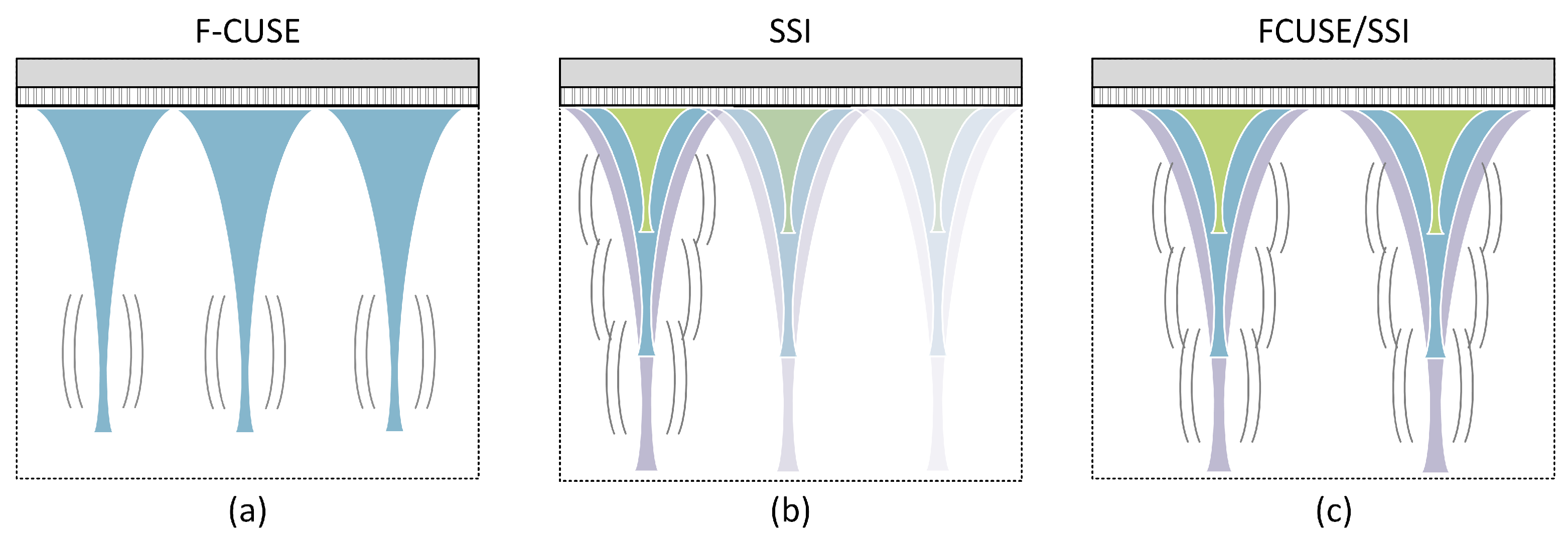
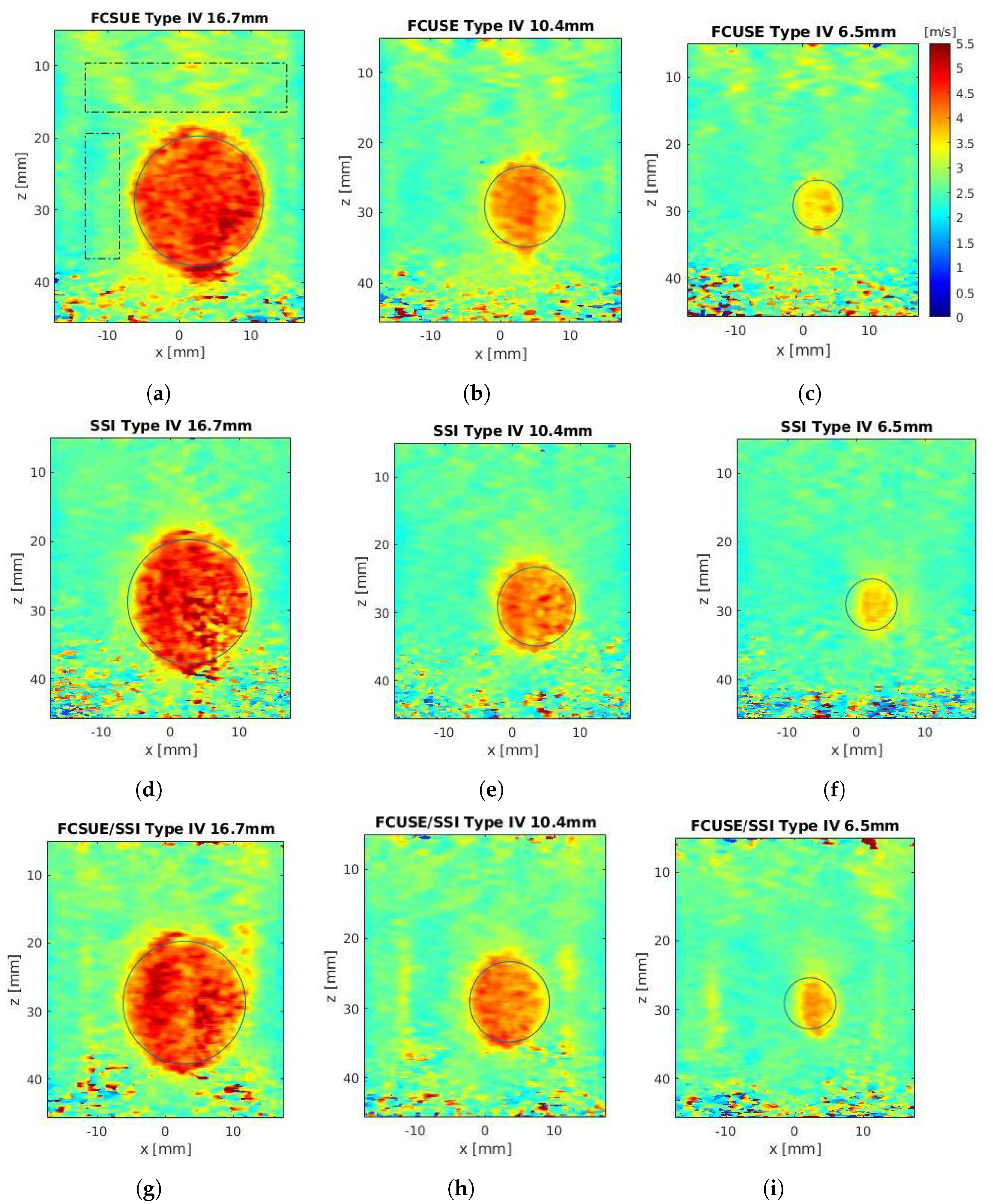
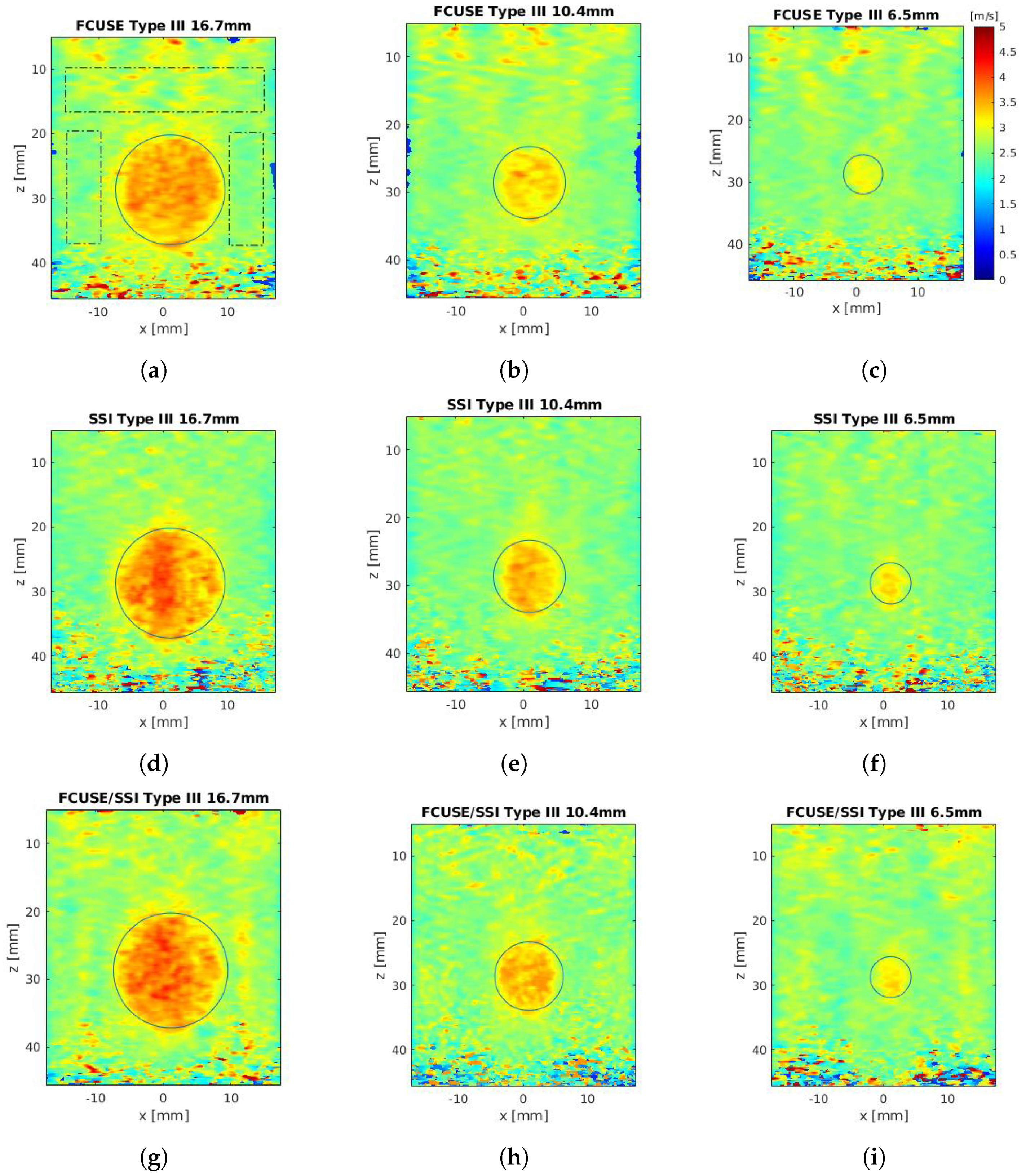
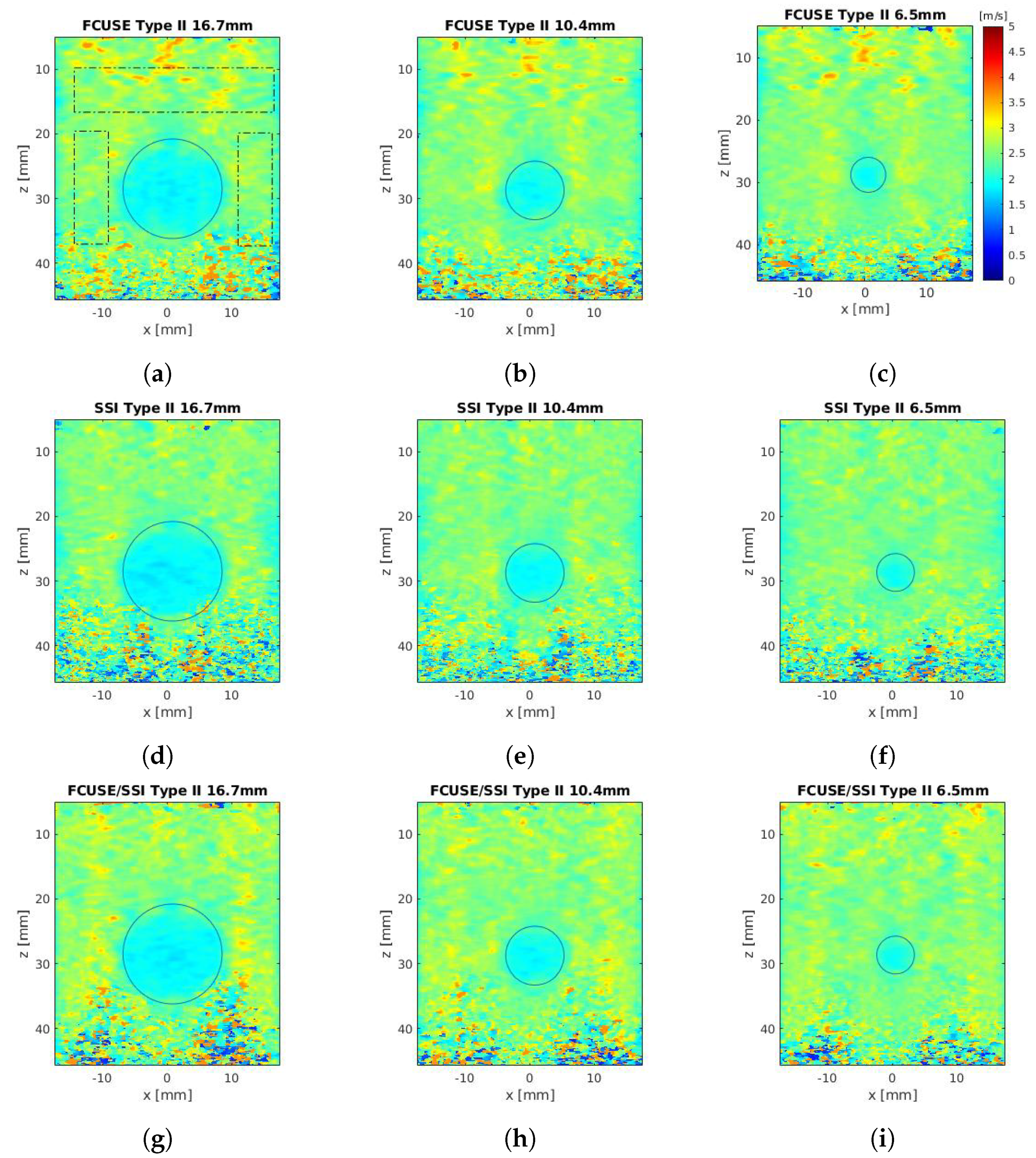
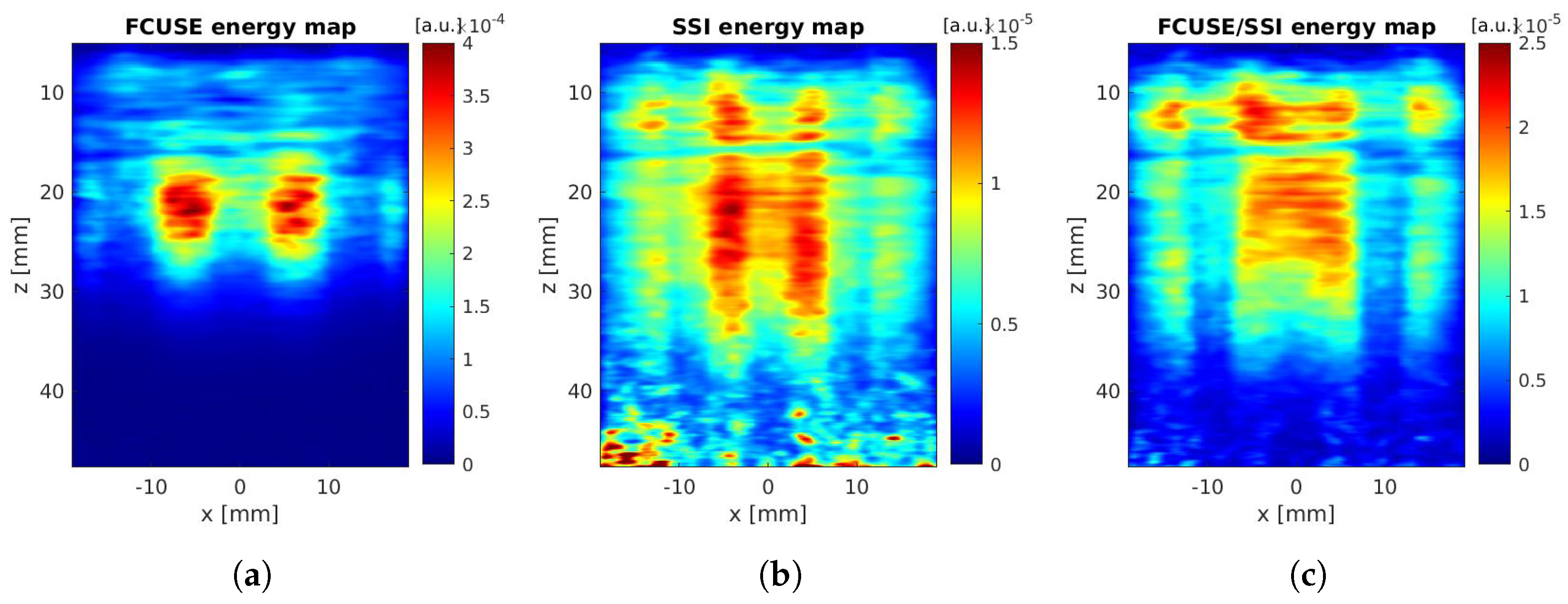
- FCUSE: The first method followed the principle that was proposed by Song et al. [21]. The transducer aperture was divided into three groups of 42 elements, each simultaneously producing a focused beam at a depth of 30 mm (). The push length was 800 s.
- SSI: The second method followed the principle that was proposed by Tanter et al. in [4]. Three acquisitions were used in total, each producing three laterally aligned beams that were focused at successive depths, which were generated sequentially by increasing the aperture size along with focusing the depth to maintain a constant f-number. The consecutive beams used 24, 50 and 66 elements and were focused at depths of 15 mm, 30 mm and 40 mm, respectively (constant ). The single push length was 80 s.
- FCUSE-SSI: In the third method, we proposed a combination of the two previous methods. Two focused beams were generated simultaneously, as in FCUSE, and then the beams were focused at consecutive depths and generated sequentially, as in SSI. It was equivalent to the generation of one FCUSE beam after another, increasing the focal point of the beam in each iteration. The consecutive beams used 24, 50 and 64 elements and were focused at depths of 15 mm, 30 mm and 40 mm and resulted in values of 2, 2 and 2.1, respectively. The single push length was 120 s.
2.3. Data Acquisition
2.4. Stiffness Map Reconstruction Algorithm
2.5. Phantom Experiments and Quality Metrics
3. Results
3.1. System Validation
3.2. Homogeneous Phantom Experiments
3.3. Heterogeneous Phantom Experiments
4. Discussion
4.1. System Validation
4.2. Phantom Experiments
4.3. Future Directions
5. Conclusions
Author Contributions
Funding
Institutional Review Board Statement
Informed Consent Statement
Data Availability Statement
Acknowledgments
Conflicts of Interest
Abbreviations
| ADC | Analog to digital converter |
| ARF | Acoustic radiation force |
| CPWI | Compounded plane wave imaging |
| DOF | Depth of field |
| FFT | Fast Fourier transform |
| FOV | Field of view |
| FPGA | Field-programmable gate array |
| fps | Frames per second |
| HV | High voltage |
| IC | Integrated circuit |
| PRF | Pulse repetition frequency |
| PRI | Pulse repetition interval |
| RAM | Random access memory |
| RF | Radio frequency |
| ROI | Region of interest |
| RX | Receive |
| SWE | Shear wave elastography |
| SWS | Shear wave speed |
| ToF | Time-of-flight |
| TX | Transmit |
| TXPB | Transmit push beamformer |
Appendix A
| Parameter | FCUSE | SSI | FCUSE-SSI |
|---|---|---|---|
| Inclusion Diameter = 16.7 mm | |||
| Inclusion SWS (m/s) | 4.48 ± 0.30 | 4.42 ± 0.42 | 4.39 ± 0.41 |
| Inclusion bias (%) | −7.9 | −9.0 | −9.6 |
| Inclusion SNR (dB) | 23.45 | 19.53 | 20.65 |
| Background SWS (m/s) | 2.71 ± 0.24 | 2.62 ± 0.19 | 2.76 ± 0.18 |
| Background bias (%) | +14.9 | +11.2 | +16.8 |
| Background SNR (dB) | 21.23 | 22.87 | 23.57 |
| CNR (dB) | 13.3 | 11.07 | 11.27 |
| Inclusion Diameter = 10.4 mm | |||
| Inclusion SWS (m/s) | 3.91 ± 0.30 | 4.02 ± 0.25 | 4.00 ± 0.31 |
| Inclusion bias (%) | −19.6 | −17.3 | −17.8 |
| Inclusion SNR (dB) | 22.30 | 24.02 | 22.32 |
| Background SWS (m/s) | 2.6 ± 0.19 | 2.55 ± 0.14 | 2.70 ± 0.19 |
| Background bias (%) | +10.2 | +7.9 | +14.5 |
| Background SNR (dB) | 22.51 | 25.26 | 22.99 |
| CNR (dB) | 11.28 | 14.15 | 11.09 |
| Inclusion Diameter = 6.5 mm | |||
| Inclusion SWS (m/s) | 3.41 ± 0.25 | 3.38 ± 0.31 | 3.51 ± 0.43 |
| Inclusion bias (%) | −29.8 | −30.4 | −27.7 |
| Inclusion SNR (dB) | 22.30 | 20.90 | 18.27 |
| Background SWS (m/s) | 2.60 ± 0.19 | 2.53 ± 0.13 | 2.68 ± 0.18 |
| Background bias (%) | +10.2 | +7.3 | +13.4 |
| Background SNR (dB) | 22.51 | 25.95 | 23.50 |
| CNR (dB) | 11.28 | 8.21 | 5.13 |
| Parameter | FCUSE | SSI | FCUSE-SSI |
|---|---|---|---|
| Inclusion Diameter = 16.7 mm | |||
| Inclusion SWS (m/s) | 3.43 ± 0.20 | 3.50 ± 0.27 | 3.52 ± 0.29 |
| Inclusion bias (%) | +1.8 | +4.0 | +4.4 |
| Inclusion SNR (dB) | 24.48 | 22.12 | 21.60 |
| Background SWS (m/s) | 2.64 ± 0.20 | 2.58 ± 0.18 | 2.70 ± 0.18 |
| Background bias (%) | +12.1 | +9.3 | +14.5 |
| Background SNR (dB) | 22.38 | 23.03 | 23.74 |
| CNR (dB) | 8.73 | 8.93 | 7.55 |
| Inclusion Diameter = 10.4 mm | |||
| Inclusion SWS (m/s) | 3.17 ± 0.17 | 3.28 ± 0.37 | 3.29 ± 0.22 |
| Inclusion bias (%) | −5.7 | −2.5 | −2.1 |
| Inclusion SNR (dB) | 25.39 | 19.06 | 23.51 |
| Background SWS (m/s) | 2.56 ± 0.24 | 2.85 ± 0.57 | 2.63 ± 0.17 |
| Background bias (%) | +8.7 | +20.80 | +11.4 |
| Background SNR (dB) | 20.59 | 13.92 | 23.63 |
| CNR (dB) | 6.32 | 3.94 | 7.52 |
| Inclusion Diameter = 6.5 mm | |||
| Inclusion SWS (m/s) | 2.95 ± 0.14 | 3.04 ± 0.19 | 3.04 ± 0.17 |
| Inclusion bias (%) | −5.7 | −9.6 | −9.6 |
| Inclusion SNR (dB) | 25.39 | 24.24 | 24.90 |
| Background SWS (m/s) | 2.56 ± 0.24 | 2.53 ± 0.14 | 2.66 ± 0.16 |
| Background bias (%) | +8.7 | +7.4 | +12.7 |
| Background SNR (dB) | 20.59 | 24.94 | 24.62 |
| CNR (dB) | 6.32 | 6.7 | 4.24 |
| Parameter | FCUSE | SSI | FCUSE-SSI |
|---|---|---|---|
| Inclusion Diameter = mm | |||
| Inclusion SWS (m/s) | 1.97 ± 0.15 | 1.94 ± 0.19 | 1.96 ± 0.16 |
| Inclusion bias (%) | +16.4 | +14.8 | +15.8 |
| Inclusion SNR (dB) | 22.41 | 20.19 | 21.51 |
| Background SWS (m/s) | 2.56 ± 0.25 | 2.50 ± 0.24 | 2.64 ± 0.23 |
| Background bias (%) | +8.3 | +6.2 | +12 |
| Background SNR (dB) | 20.31 | 20.39 | 21.19 |
| CNR (dB) | 6.15 | 5.28 | 7.63 |
| Inclusion Diameter = mm | |||
| Inclusion SWS (m/s) | 2.00 ± 0.12 | 1.97 ± 0.12 | 1.99 ± 0.13 |
| Inclusion bias (%) | +18.3 | +16.5 | +17.4 |
| Inclusion SNR (dB) | 24.31 | 24.26 | 23.89 |
| Background SWS (m/s) | 2.55 ± 0.21 | 2.50 ± 0.19 | 2.62 ± 0.20 |
| Background bias (%) | +8.1 | +6.1 | +11.2 |
| Background SNR (dB) | 21.78 | 22.62 | 22.37 |
| CNR (dB) | 7.13 | 7.61 | 8.59 |
| Inclusion Diameter = mm | |||
| Inclusion SWS (m/s) | 2.05 ± 0.13 | 2.08 ± 0.12 | 2.10 ± 0.14 |
| Inclusion bias (%) | +21.0 | +22.6 | +23.80 |
| Inclusion SNR (dB) | 23.95 | 24.50 | 23.72 |
| Background SWS (m/s) | 2.55 ± 0.20 | 2.49 ± 0.17 | 2.60 ± 0.17 |
| Background bias (%) | +7.9 | +5.5 | +10.2 |
| Background SNR (dB) | 21.92 | 23.45 | 23.94 |
| CNR (dB) | 6.25 | 5.96 | 7.44 |
References
- Ophir, J.; Cespedes, H.; Ponnekanti, Y.; Yazdi, Y.; Li, X. Elastography: A Quantitive Method for Imaging the Elasticity of Biological Tissues. Ultrason. Imaging 1991, 13, 111–134. [Google Scholar] [CrossRef] [PubMed]
- Sarvazyan, A.P.; Rudenko, O.V.; Swanson, S.D.; Fowlkes, J.B.; Emalianov, S.Y. Shear Wave Elasticity Imaging: A New Ultrasonic Technology of Medical Diagnostics. Ultrasound Med. Biol. 1998, 24, 1419–1435. [Google Scholar] [CrossRef]
- Pinton, G.F.; Dahl, J.J.; Trahey, G.E. Rapid Tracking of Small Displacements with Ultrasound. IEEE Trans. Ultrason. Ferroelectr. Freq. Control 2006, 53, 1103–1117. [Google Scholar] [CrossRef] [PubMed]
- Tanter, M.; Bercoff, J.; Athanasiou, A.; Deffieux, T.; Genisson, J.-L.; Mantaldo, G.; Muller, M.; Tardivon, A.; Fink, M. Quantitive Assessment of Breast Lesion Viscoelasticity: Initial Clinical Results Using Supersonic Shear Imaging. Ultrasound Med. Biol. 2008, 34, 1373–1386. [Google Scholar] [CrossRef] [PubMed]
- Palmeri, M.L.; Wang, M.H.; Dahl, J.J.; Frinkley, K.D.; Nightingale, K.R. Quantifying Hepatic Shear Modulus in vivo Using Acoustic Radiation Force. Ultrasound Med. Biol. 2008, 34, 546–558. [Google Scholar] [CrossRef]
- Rouze, N.C.; Wang, M.H.; Palmeri, M.L.; Nightingale, K.R. Parameters Affecting the Resolution and Accuracy of 2-D Quantitive Shear Wave Images. IEEE Trans. Ultrason. Ferroelectr. Freq. Control 2012, 59, 1729–1740. [Google Scholar] [CrossRef]
- Song, P.; Zhao, H.; Manduca, A.; Urban, M.W.; Greenleaf, J.F.; Chen, S. Comb-Push Ultrasound Shear Elastography (CUSE): A Novel Method for Two-Dimensional Shear Elasticity Imaging of Soft Tissues. IEEE Trans. Med. Imaging 2012, 31, 1821–1832. [Google Scholar] [CrossRef]
- Song, P.; Manduca, A.; Zhao, H.; Urban, M.W.; Greenleaf, J.F.; Chen, S. Fast Shear Compounding Using Robust 2-D Shear Wave Speed Calculation and Multi-Directional Filtering. Ultrasound Med. Biol. 2014, 40, 1343–1355. [Google Scholar] [CrossRef]
- Nabavizadeh, A.; Song, P.; Chen, S.; Greenleaf, J.F.; Urban, M.W. Multi-Source and Multi-Directional Shear Wave Generation with Intersecting Steered Ultrasound Push Beams. IEEE Trans. Ultrason. Ferroelectr. Freq. Control 2015, 62, 647–662. [Google Scholar] [CrossRef][Green Version]
- Rouze, N.C.; Wang, M.H.; Palmeri, M.L.; Nightingale, K.R. Robust Estimation of Time-of-Flight Shear Wave Speed Using a Radon Sum Transformation. IEEE Trans. Ultrason. Ferroelectr. Freq. Control 2010, 57, 2662–2670. [Google Scholar] [CrossRef]
- Li, Y.; Lv, Q.; Dai, J.; Tian, Y.; Guo, J. Shear Wave Velocity Estimation Using the Real-Time Curve Tracing Method in Ultrasound Elastography. Appl. Sci. 2021, 11, 2095. [Google Scholar] [CrossRef]
- Cosgrove, D.; Piscaglia, F.; Bamber, J.; Bojunga, J.; Correas, J.-M.; Gilja, O.H.; Klauser, A.S.; Sporea, I.; Calliada, F.; Cantisani, V.; et al. EFSUMB Guidelines and Recommendations on the Clinical Use of Ultrasound Elastography. Part 2: Clinical Applications. Ultraschall Med. 2013, 34, 238–253. [Google Scholar] [PubMed]
- Marais, L.; Pernot, M.; Khettab, H.; Tanter, M.; Messas, E.; Zidi, M.; Laurent, S.; Boutouyrie, P. Arterial Stiffness Assessment by Shear Wave Elastography and Ultrafast Pulse Wave Imaging: Comparison with Reference Techniques in Normatives and Hypertensives. Ultrasound Med. Biol. 2019, 45, 758–772. [Google Scholar] [CrossRef] [PubMed]
- Nightingale, K.; McAleavey, S.; Trahey, G. Shear-wave generation using acoustic radiation force: In vivo and ex vivo results. Ultrasound Med. Biol. 2003, 29, 1715–1723. [Google Scholar] [CrossRef]
- Bercoff, J.; Tanter, M.; Chen, S.; Fink, M. Supersonic shear imaging: A new technique for soft tissue elasticity mapping. IEEE Trans. Ultrason. Ferroelectr. Freq. Control 2004, 51, 396–409. [Google Scholar] [CrossRef]
- Tanter, M.; Bercoff, J.; Sandrin, L.; Fink, M. Ultrafast Compound Imaging for 2-D Motion Vector Estimation: Application to Transient Elastography. IEEE Trans. Ultrason. Ferroelectr. Freq. Control 2002, 49, 1363–1374. [Google Scholar] [CrossRef]
- Feng, F.; Goswami, S.; Khan, S.; McAleavey, S.A.W. Shear Wave Elasticity Imaging Using Nondiffractive Bessel Apodized Acoustic Radiation Force. IEEE Trans. Ultrason. Ferroelectr. Freq. Control 2021, 68, 3528–3539. [Google Scholar] [CrossRef]
- Boni, E.; Yu, A.C.H.; Freear, S.; Jensen, J.A.; Tortoli, P. Ultrasound Open Platforms for Next-Generation Imaging Technique Development. IEEE Trans. Ultrason. Ferroelectr. Freq. Control 2018, 65, 1078–1092. [Google Scholar] [CrossRef]
- Walczak, M.; Lewandowski, M.; Żołek, N. Optimization of real-time ultrasound PCIe data streaming and OpenCL processing for SAFT imaging. In Proceedings of the 2013 IEEE International Ultrasonics Symposium (IUS), Prague, Czech Republic, 21–25 July 2013; pp. 2064–2067. [Google Scholar]
- Witek, B.; Walczak, M.; Lewandowski, M. Characterization of the STHV748 integrated pulser for generating push sequences. In Proceedings of the 2015 IEEE International Ultrasonics Symposium (IUS), Taipei, Taiwan, 21–24 October 2015; pp. 1–4. [Google Scholar]
- Song, P.; Urban, M.W.; Manduca, A.; Zhao, H.; Greenleaf, J.F.; Chen, S. Comb-Push Ultrasound Shear Elastography (CUSE) with Various Ultrasound Push Beams. IEEE Trans. Med. Imaging 2013, 32, 1435–1447. [Google Scholar] [CrossRef]
- Deng, Y.; Palmeri, M.L.; Rouze, N.C.; Rosenzweig, S.J.; Abdelmalek, M.F.; Nightingale, K.R. Analyzing the Impact of Increasing Mechanical Index and Energy Deposition on Shear Wave Speed Reconstruction in Human Liver. Ultrasound Med. Biol. 2015, 41, 1948–1957. [Google Scholar] [CrossRef]
- Montaldo, G.; Tanter, M.; Bercoff, J.; Benech, N.; Fink, M. Coherent Plane-Wave Compounding for Very High Frame Rate Ultrasonography and Transient Elastography. IEEE Trans. Ultrason. Ferroelectr. Freq. Control 2019, 56, 489–506. [Google Scholar] [CrossRef] [PubMed]
- Lee, H.-K.; Greenleaf, J.F.; Urban, M.W. A New Plane Wave Compounding Scheme Using Phase Compensation for Motion Detection. IEEE Trans. Ultrason. Ferroelectr. Freq. Control 2022, 69, 702–710. [Google Scholar] [CrossRef] [PubMed]
- Kasai, C.; Namekawa, N.; Koyano, A.; Omoto, R. Real-time Two-Dimensional Blood Flow Imaging Using an Autocorrelation Technique. IEEE Trans. Ultrason. Ferroelectr. Freq. Control 1985, SU-32, 458–464. [Google Scholar]
- Manduca, A.; Lake, D.S.; Kruse, S.A.; Ehman, R.L. Spatio-temporal directional filtering for improved inversion of MR elastography images. Med. Image Anal. 2003, 7, 465–473. [Google Scholar] [CrossRef]
- Deffieux, T.; Gennison, J.-L.; Bercoff, J.; Tanter, M. On the Effects of Reflected Waves in Transient Shear Wave Elastography. IEEE Trans. Ultrason. Ferroelectr. Freq. Control 2011, 58, 2032–2035. [Google Scholar] [CrossRef]
- Racedo, J.; Urban, M.W. Evaluation of Reconstruction Parameters for 20D Comb-Push Ultrasound Shear Wave Elastopgrahy. IEEE Trans. Ultrason. Ferroelectr. Freq. Control 2018, 66, 254–263. [Google Scholar] [CrossRef]
- Zhao, H.; Song, P.; Urban, M.W.; Kinnick, R.R.; Yin, M.; Greenleaf, J.F.; Chen, S. Bias Observed in Time-of-Flight Shear Wave Speed Measurements Using Radiation Force of a Focused Ultrasound Beam. Ultrasound Med. Biol. 2011, 37, 1884–1892. [Google Scholar] [CrossRef]
- Deng, Y.; Rouze, N.C.; Palmeri, M.L.; Nightingale, K.R. On System-Dependent Sources of Uncertainty and Bias in Ultrasonic Quantitive Shear-Wave Imaging. IEEE Trans. Ultrason. Ferroelectr. Freq. Control 2016, 63, 381–393. [Google Scholar] [CrossRef]
- Harris, E.; Sinnatamby, R.; O’Flynn, E.; Kirby, A.; Bamber, J.C. Cross-Machine Comparison of Shear-Wave Speed Measurements Using 2D Shear-Wave Elastography in the Normal Female Breast. Appl. Sci. 2021, 11, 9391. [Google Scholar] [CrossRef]
- Edwards, C.; Cavanagh, E.; Kumar, S.; Clifton, V.; Fontanarosa, D. Intra-System Reliability Assessment of 2-Dimensional Shear Wave Elastography. Appl. Sci. 2021, 11, 2992. [Google Scholar] [CrossRef]





| Feature | Value |
|---|---|
| TX channels | 256 |
| HV transmit voltage | Configurable, up to 180 Vpp |
| Output current per channel | 2 A or 4 A |
| Transmit frequency range | 0.1– 50 MHz 1 |
| Transmit aperture | Arbitrary |
| Transmit pulse capability | Arbitrary 3-level square wave pattern for each channel |
| Transmit apodization | Yes 2 |
| Transmit delay | 0–327 s |
| Timing resolution | 5 ns |
| Theoretical maximum PRF | 100 kHz |
| Parameter | FCUSE | SSI | FCUSE-SSI |
|---|---|---|---|
| Average SWS (m/s) | 2.56 ± 0.13 | 2.54 ± 0.10 | 2.57 ± 0.13 |
| Average bias (m/s) | 0.21 (+9.0%) | 0.18 (+7.8%) | 0.22 (+9.2%) |
| SNR (dB) | 26.0 | 27.9 | 26.2 |
Publisher’s Note: MDPI stays neutral with regard to jurisdictional claims in published maps and institutional affiliations. |
© 2022 by the authors. Licensee MDPI, Basel, Switzerland. This article is an open access article distributed under the terms and conditions of the Creative Commons Attribution (CC BY) license (https://creativecommons.org/licenses/by/4.0/).
Share and Cite
Cacko, D.; Lewandowski, M. Shear Wave Elastography Implementation on a Portable Research Ultrasound System: Initial Results. Appl. Sci. 2022, 12, 6210. https://doi.org/10.3390/app12126210
Cacko D, Lewandowski M. Shear Wave Elastography Implementation on a Portable Research Ultrasound System: Initial Results. Applied Sciences. 2022; 12(12):6210. https://doi.org/10.3390/app12126210
Chicago/Turabian StyleCacko, Damian, and Marcin Lewandowski. 2022. "Shear Wave Elastography Implementation on a Portable Research Ultrasound System: Initial Results" Applied Sciences 12, no. 12: 6210. https://doi.org/10.3390/app12126210
APA StyleCacko, D., & Lewandowski, M. (2022). Shear Wave Elastography Implementation on a Portable Research Ultrasound System: Initial Results. Applied Sciences, 12(12), 6210. https://doi.org/10.3390/app12126210


