Review

Biomaterials for Ophthalmic Applications

by
Maria Pia Ferraz
1,2,3
1
Departamento de Engenharia Metalúrgica e de Materiais, Faculdade de Engenharia da Universidade do Porto, 4200-465 Porto, Portugal
2
i3S—Instituto de Investigação e Inovação em Saúde, Universidade do Porto, 4200-135 Porto, Portugal
3
INEB—Instituto de Engenharia Biomédica, Universidade do Porto, 4200-135 Porto, Portugal
Appl. Sci. 2022, 12(12), 5886; https://doi.org/10.3390/app12125886
Submission received: 20 May 2022 / Revised: 1 June 2022 / Accepted: 8 June 2022 / Published: 9 June 2022
(This article belongs to the Section Materials Science and Engineering)

Abstract

:
Ophthalmology is the branch of medicine that deals with diseases of the eye, the organ responsible for vision, and its attachments. Biomaterials can be made with different types of materials and can replace or improve a function or an organ, specifically the eye in the case of ophthalmic biomaterials. Biomaterials are substances that interact with biological systems for a medical purpose, either as a therapeutic (treat, augment, repair, or replace a tissue function of the body) or a diagnostic agent, and have continued to improve over the years, leading to the creation of new biomaterials. With the arrival of new generations, biomaterials have succeeded in reducing complications and toxicity and improving biocompatibilities associated with older generations. With the aging population, eye problems are becoming more prevalent, and biomaterials have helped in recent years to improve or restore vision, improving the quality of life of many patients. This review focuses on the most clinically used ophthalmic biomaterials, including contact lenses, intraocular lenses, artificial tears, inlays and vitreous replacements. Tissue engineering is presented as a new tool that is able to be treat several ophthalmologic disorders.

1. Introduction

The eye is an organ of great complexity and, compared to our other organs, is easier to observe and easier to access for surgery. It is also the first organ in which a foreign material was implanted to fulfil the function of what is now called a biomaterial. With the introduction of synthetic hydrogels, i.e., polymers capable of addressing and retaining water without dissolving in an aqueous medium, the range of ophthalmic biomaterials has grown considerably [1]. The use of biomaterials in general and ophthalmic biomaterials in particular is growing and integrates knowledge and ideas from multiple disciplines, such as medicine, biology, chemistry, physics, materials science and engineering [2].
The eye is the organ responsible for vision situated in a bony cavity, called the orbit, is connected to the brain via optic pathways and is surrounded by a nutrient membrane, the choroid, as well as a protective membrane, the sclera. The eye is a hollow, spherical structure, surrounded by attachments with a motor paper or protective paper. The anatomy and physiology of the eye are very complex and extensively documented [3,4,5].
The consequences of an increasingly ageing society on the health system have become a topic of great importance and concern. Concerning the maintenance of vision, adequate care is vitally important as it helps to maintain the independence of the elderly. All tissues and all aspects of the eye are affected by ageing to some degree. The most common vision disorders associated with increasing age are presbyopia, dry eyes, cataract or glaucoma [1].
In relation to ophthalmic biomaterials, contact lenses are the oldest and most well-known of these, and are presently composed of hydrogel and silicone hydrogel. Intraocular lenses can replace a clouded eye lens that is removed during cataract operation. They are usually composed of derivative acrylics or silicone copolymers [6,7]. Other biomaterials widely marketed are artificial tears, used mainly for dry eye treatment. They reduce irritation symptoms, reduce friction, increase lubrication, stabilize the tear film and protect against dehydration. They are dispensed in the form of eye drops, gel or ointment [8]. Inlays are used to correct presbyopia and work according to different approaches [9].
Locations for the possible ophthalmic application of biomaterials is provided by several authors [10,11]. Briefly, in the anterior segment, soft contact lenses and artificial tears may be used. Intraocular lenses and inlays can be used in the lens. On the posterior segment, biomaterial application mainly involves vitreous substitutes.
In pathological situations, such as retinal detachment, it is necessary to replace the biological vitreous [12]. The ideal vitreous substitute should have a variety of properties to perfectly mimic the necessary physicochemical properties [13,14].

2. Biomaterials in Ophthalmology
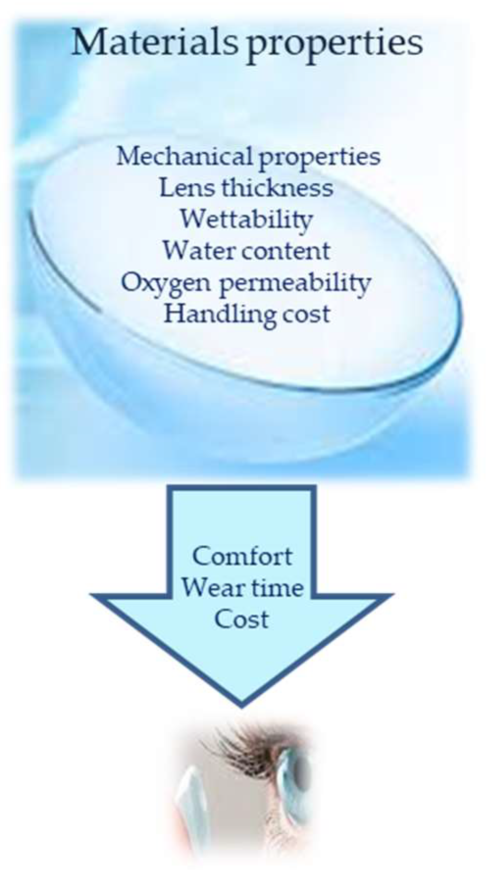
The history of ophthalmic biomaterials is relatively brief. Onofrio Abbate, in 1862, implanted a foreign material for a purpose similar to that of a biomaterial. It was a glass disc enclosed in a two-ring skirt forming an artificial cornea. This device was tested on the cornea of animals, but it was unable to stay in place for more than a week. Later, Dimmer attempted to construct an artificial cornea composed of celluloid (a mixture of nitrocellulose and camphor with stabilizers), implanting it in four human patients; however, it was rejected in the first few months. It was not until half a century later that a fully synthetic polymer was used as an implantable ophthalmic biomaterial composed of polyvinyl alcohol (PVA) gel. It was followed by the first artificial cornea made from polymethyl methacrylate (PMMA) [15]. A few years later, a synthetic polymer, poly(1-vinyl-2-pyrrolidone), was implanted in the vitreous cavity as a vitreous substitute. It was mainly thanks to the introduction of synthetic hydrogels that the range of biomaterials to treat ophthalmic lesions developed [1].
The main goal of developing successive generations of biomaterials is to bridge the gaps or defects of previous generations and to improve safety, efficiency and comfort. Innovations were made to improve quality standards, production or production efficiency to reduce costs. Market pressure to reduce costs exists in order to promote competitiveness and provide better accessibility. Ophthalmic biomaterials are now highly sophisticated devices and their usefulness has increased dramatically in recent years [1]. Many important requirements must be met by ophthalmic biomaterials, including the ability to deliver oxygen to tissues, refractive changes, tissue protection during surgery, tissue integration and healing modulation [16,17].
Contact lenses are especially important as an ophthalmic biomaterial, as they are in contact with ocular surface components, primarily the corneal epithelium. They also, economically speaking, represent the most important category and have undergone the most important and complex evolution [18].
Hydrogels have already been approved for several ophthalmic applications, but currently, several are under investigation. Hydrogels are successfully marketed as soft contact lenses [19], foldable intraocular lenses [20], or in situ gelling vehicles for ophthalmic drug delivery [21]. Efforts are being mounted to improve the sustained release of antibiotics, anti-inflammatory drugs, therapeutic proteins and nucleic acids. Furthermore, hydrogels are also being investigated as potential vitreous substitutes [13]. A wide range of natural, semisynthetic and synthetic polymers can be used as starting materials for hydrogels. Examples of natural origin polymers include alginate, collagen, and hyaluronic acid (HA). Poly(ethylene glycol) (PEG), poly(vinyl alcohol) (PVA), polymers based on acrylate monomers, and siloxanes are examples of synthetic gel-forming materials [10].
Tissue engineering helps to regenerate lost tissue, specifically in severe cases such as burns, ulcers, diabetes, bone defects and liver failure among others. It is based on stem cells, growth factors and biomaterials [22,23]. The general process of tissue engineering is based on the implantation of living cells in a scaffold specially designed to promote tissue replacement and regeneration [24,25]. It is therefore considered as the next step in the development of biomaterials. Regenerative medicine relies on interdisciplinary research and applications based on repair, replacement or the regeneration of cells, tissues or organs to restore impaired function. However, adult humans only have a limited capacity for spontaneous regeneration. Induced regeneration has been reported in conjunctiva and cornea. Tissue engineering in the eye has mainly been reported in the anterior segment (cornea and conjunctiva) and significant progress has been made in cell therapies to treat degenerative diseases of the retina. Developments in biomaterials are being made using new manufacturing techniques that allow for the production of personalized tissues through advances in stem cell programming, generation tissue imaging and computer-aided design [1,26].

2.1. Contact Lenses

Obtaining good visual acuity may require the use of an optical correction, which can be achieved by converging or diverging contact lenses, whose power is expressed in diopters. There are four common refractive disorders, namely myopia; hypermetropia; astigmatism and presbyopia. The first glass contact lenses appeared in the 19th century. In the 1880sm the first scleral glass lenses appeared with Adolf Fick. The process of making contact lenses was described in 1888 [2,27]. Rohm and Haas introduced PMMA, a material considered biologically inert, light, easy to manufacture and resistant to breakage, in 1936. Hard lenses apparently appeared due to an error during the process of manufacturing PMMA scleral lenses and led to the popularization of contact lenses [2,27].
Silicone elastomer lenses were introduced in the 1960s as a silicone soft contact lens that does not retain water, is very permeable to gases, and has a hydrophobic surface [27].
Hydrophilic lenses date back to 1972 with the introduction of the soft lens on the market, which was an immediate success thanks to its comfort and its superior biocompatibility; however, an improvement in their gas permeability was required, after which they were made thinner and had a higher aqueous content [28]. In 1974, the first rigid gas permeable lenses were invented. One of the first materials was cellulose acetate butyrate (CAB), which provided a gas permeability higher than that of PMMA, but was prone to deformation. Norman Gaylord successfully incorporated silicone into the basic structure of PMMA to introduce a new family of polymer contact lenses, silicone acrylate. In 1994, a technique drastically reduced the production costs and helped to conceive of the first daily contact lenses. More than one decade of intensive research and development was needed to develop silicone hydrogel contact lenses which improved hypoxia problems related to contact lenses [27,29,30].
Soft contact lenses are the most popular type of contact lens, accounting for 88% of worn lenses [28,31,32]. Hydrogel contact lenses are made with a component of stable polymer, which can absorb or bind water. Polymer pores allow the liquid to penetrate the material, making it hydrated. Due to the particular environment of the eye, the lens must be safe, inert, non-toxic, biocompatible, easy to produce, maintain a stable and continuous tear film, be permeable to oxygen, maintain normal corneal metabolism, be ion permeable to maintain the movement of eyes, be comfortable and provide a stable and clear view [27,28,31,33,34].
Nowadays, soft contact lenses are able to provide high oxygen transmission to the cornea, good tear film moisture for comfort and superior vision, good material strength and adequate water permeability to maintain lens movement [28]. Silicone hydrogel is the result of years research to combine silicone with conventional hydrogel monomers, leading to a superior capacity of oxygen transmission and lower likelihood of dehydration in the eyes [28,33,34].
There are several polymers and polymerization conditions suitable for contact lens manufacturing. The variety of materials explains why there is a wide range of contact lenses available. In Figure 1, the most important factors when considering materials for contact lenses are presented. This choice of material influences comfort, wear time and cost, and in turn influences patient/practitioner choice. In Table 1, the advantages and disadvantages of the most commonly used materials to produce contact lenses are shown.
PVA is a synthetic polymer that contains many hydroxy (–OH) groups, one in each repeating monomer unit. This leads to PVA’s excellent hydrophilic and biocompatible properties [35]. PVA contact lens hydrogels have low protein absorption rates. PVA has been used as a tool for producing more comfortable lenses [6] or to facilitate the loading of a colored pigment into the lens.
Silicone-based hydrogels include silicone, siloxanes, fluorosiloxanes, and derivative materials. Their extensive use is linked to the fact that these contact lenses have the highest oxygen permeability of all contact lens materials. Silicone contact lenses are often durable, attributable to their high Si–O bonding energy, often with a higher modulus than conventional polymer hydrogels. The high modulus is linked to irritation to the eye, such as the conjunctiva of the inner eyelid [27]. Discomfort and dryness of silicone-based lenses are two of the main reasons for users’ discontinuation [36].
HEMA and related hydrogels are high-water content, oxygen-permeable polymeric materials. These hydrogels can have a water content in the range of 20–80% depending on the comonomers, with a hydrogel composed of only HEMA containing about 38% water. The highly polar properties of HEMA make it attractive for applications where wettability is important, producing very comfortable contact lenses. The oxygen permeability of these gels is suitable for longer wear, but not to the same extent as silicone-based lenses. HEMA is commonly copolymerized with monomers to increase the water content of hydrogels. The mechanical properties can be improved by using a cross-linking molecule, such as ethylene glycol dimethacrylate (EGDMA). EGDMA has two functional groups allowing for the formation of covalent bonds between two individual polymer chains. This increases the mass of the polymer and improves its ability to form a gel network. However, the crosslinks reduce the polymer-chain motion, which can decrease swelling and oxygen transport. The water content and cross-linking affect the modulus and oxygen permeability of the hydrogels; therefore, a balance must be reached between these parameters when designing a contact lens for a particular application [37].
PMMA has little to no oxygen permeability due to the lack of mobility of polymer chains which prevents the flow of oxygen or internal water to mediate oxygen flow. This occurs due to intermolecular forces, such as dipole–dipole bonding and physical entanglement, which is prevalent between polymer chains. The dipoles are created by the negatively charged (electrochemical negative) oxygen compared with the adjacent positively charged (electrochemical positive) carbon and hydrogen atoms. As a result, neighbouring polymer chains can attract each other to provide thermodynamic stability to the polymer. These intermolecular forces lead to a low free volume in PMMA; therefore, chains do not rotate or move easily. Additionally, PMMA does not contain large pendant chains inhibiting the interaction of neighbouring chains. All these factors together prevent oxygen flow through the polymer. However, functionalization of the PMMA surface can improve hydrophilicity, which can be useful [38].
Soft contact lenses are the most common and most successful commercial application of hydrogels. The selected hydrogel materials used for the manufacture of soft contact lenses are: allyl methacrylate (AMA); ethylene glycol dimethacrylate (EGDMA); 2-hydroxyethyl methacrylate (HEMA); methacrylic acid (MAA); 4-(2-methacryloyloxyethyl)-2-(2H-benzotriazol-2-yl)phenol (MAEBTP); 2-methacryloxyethyl phosphorylcholine (MPC); N-vinylpyrrolidone (NVP); poly(ethylene propylene glycol) dimethacrylate (PEPGDMA); poly(vinyl alcohol) (PVA); 4-tert-butyl-2-hydroxycyclohexyl methacrylate (TBHMA); sodium methacrylate (SMA); 1,1,1-tris(hydroxymethyl)propane; and trimethacrylate (TRIM).
Recent studies aimed to improve the oxygen permeability and wearing comfort of soft contact lenses [39] and the development of new polymers with antifouling properties to reduce the adsorption of proteins and cells on the lens surface [40]. This may help to improve the biocompatibility of soft contact lenses, especially for long-term wear.
Poly(dimethyl siloxane) (PDMS)-based lens materials combine the extremely high oxygen permeability of PDMS and the wearing comfort of conventional pHEMA hydrogels. Besides their application as corrective lenses, soft contact lenses can provide a drug delivery system for the anterior segment of the eye [41,42].
The use of soft contact lenses as a drug delivery system is a challenging approach with no approved products on the market. The main challenge is to reach adequate levels of drug loading while ensuring controlled drug release at the same time. Although well-established polymers are used, the biocompatibility, transparency, and oxygen permeability of drug-loaded soft contact lenses have to be tested. Extensive in vitro and in vivo studies will be required to prove the safety and efficacy of the developed drug delivery systems.
Soft contact lenses are manufactured mainly by lathe-cutting, spin-casting, cast-molding or a combination of these three [27,32].
Lathe-cutting is the most expensive due to the number and variability of stages. It is usually reserved for the production of customized lenses with features that do not allow for mass production. The dry polymer is placed in a lathe where it rotates and a diamond tool generates the back surface of the lens. It is then mounted with adhesive wax, where the same process takes place to generate the front surface of the lens. It is then removed from the lathe and the edges are polished before inspection. The dry lens is hydrated in saline solution to obtain its final shape. It is inspected again before being placed in a sealed glass bottle, labeled and sterilized in an autoclave [27,32].
Spin-casting fabrication is performed using a stainless-steel tool allowing for the manufacture of hundreds of thousands of female molds, produced by the pressure of a male tool in cast polypropylene which, once cooled, hardens. The female mold is placed on an axis which rotates about the lens axis, with the concavity upwards, and liquid monomers are introduced into the rotating mold. The final shape of the lens is determined by a combination of the temperature, gravity, centrifugal force, tension surface, the amount of liquid in the mold and rotation speed. The monomers are then irradiated with UV light to initiate polymerization. The dry lens is removed from the mold, its edges polished and the mold removed. The final steps are the same as for lathe-cutting, except that they are placed in a bag containing saline solution. The blister is sealed and sterilized in an autoclave [27,32].
Cast-molding fabrication is the preferred way to make lenses. Both male and female casts are made of stainless steel, and the contour of the male tool head defines the shape of the front surface of the lens, while the contour of the head of the female tool defines the shape of the back surface of the lens. The mold’s tongue and groove are made by pressing the tools in molten polypropylene which, once cooled, harden. The female mold is assembled, the concavity is made upwards and the liquid monomers are introduced into it. The male mold is then placed in the female mold. Excess polymer is expelled to the outside. The monomers are irradiated with UV or thermal energy to initiate polymerization. The dry lens is removed from the mold, which is discarded, and is inspected. The end of production follows the same method as that of spin-casting [27,32].
Contact lenses must be cleaned, disinfected and stored until the next use (except for the single use ones). The need to clean them is due to the wide variety of debris that can adhere to the lens surface. Cleaning also improves the process of disinfection, reducing the number of microorganisms on contact lenses [34,43].
Chlorhexidine, a biocide, is probably the most widely used antiseptic [44]. Thiomersal, while less effective in general, is generally more effective against fungi. A combination of chlorhexidine and thimerosal was used before toxic reactions were reported and hypersensitivity due to the lens absorption of these agents [43,45]. Hydrogen peroxide has a broad spectrum effectiveness against bacteria, viruses and yeasts that produce radicals and can be chemically broken down into oxygen and water, making it eco-friendly. Although extremely effective in terms of antimicrobial action, it is toxic to the eyes and requires neutralization before use [46].
Multipurpose solutions (MPS) are the most used lens care systems today; they do not require other components in the lens care process once they combine cleaning, washing and disinfection in a single product [47]. MPS usually contain polyhexanide, being active against a wide range of bacteria, has a higher molecular weight than chlorhexidine, therefore it cannot penetrate the lens matrix decreasing the likelihood of potential toxic reactions or hypersensitivity. Usually, polyquaternium-1 is also used as a preservative. Nowadays, MPS products usually use two disinfectants instead of one, with the process being called double disinfection [27,45,48].
Contact lenses are nowadays also seen as a tool for ophthalmic treatments that require the delivery of a drug to the eye. Eye drops have a low bioavailability of common ophthalmic treatments; therefore, new lenses are being developed based on drug delivery materials [49,50]. Several methods are being used, namely drug-loading by nanoparticle or polymeric encapsulation [51,52,53,54], β-cyclodextrin delivery [55,56], molecular imprinting [57,58], and solution soaking [59,60,61]. These are ongoing research works; however, more studies are needed including clinical trials or tests that assess the feasibility of manufacturing.

2.2. Intraocular Lenses

Cataract is the clouding of the lens and is the leading cause of blindness in the world. It is due to changes in lens proteins, a natural phenomenon that occurs during aging, but can be accentuated by various factors, such as exposure to UV rays or smoking. It is treated by replacing the biological lens with a polymer-based substitute [1]. The most common procedure is phacoemulsification (FACO), creating a small incision in the cornea through which a probe is used to break the cloudy lens which is removed by suction. An intraocular lens artificial device (IOL) is then placed in the intact capsular bag [1,62].
The first IOL was in PMMA [63,64] a rigid, non-collapsible and hydrophobic polymer requiring a large incision. Thomas Mazzocco invented the first foldable silicone lens that can be implanted through a 3 mm incision [65].
An ideal IOL should provide the patient with good vision for a long period of time and the surgeon should be able to implant it easily, without causing complications [63]. The material and its design must allow for a low degree of postoperative inflammation and the production process must be relatively simple for the IOL to be accessible [66]. Currently, all IOLs include a chromophore in their composition to block UV light. Blue light is harmful and can cause damage to the retina due to increased oxidative stress [66].
IOLs can be divided into the following two groups: an acrylic/methacrylate polymer comprising hydrophobic acrylic, hydrophilic acrylic or hydrogel (rigid or flexible) and hydrophobic (flexible) silicone/silicone elastomers. Non-folding PMMA lenses from the group of acrylic polymers are almost non-existent in Europe and the USA due to the size of the incision needed for implantation [43,45,67,68].

2.2.1. Intraocular Acrylic Lenses

Hydrogel and acrylic are joined with silicone to make flexible lenses. Flexible acrylic lenses exist in a hydrophilic or hydrophobic form. They can be folded at room temperature and can regain their original shape and size when inserted into the eye. Compared to silicone IOLs, implantation is more slowly and easily controlled. However, improper handling during loading and implantation can leave permanent marks on the surface of the optics [65]. In general, hydrophobic acrylic lenses absorb very little water, less than 1%, while the hydrophilic ones absorb between 18 and 38% of water [69,70].

2.2.2. Intraocular Elastomer Lenses

Silicone lenses were the first flexible IOLs made available on the market and introduced in 1984 [70]. Currently, silicone lenses are extremely easy to bend and their intraocular implantation is very fast notwithstanding difficulties in controlling them [69,70].

2.3. Artificial Tears

Dry eye is defined as a multifactorial disease of the ocular surface characterized by a loss of tear film homeostasis and accompanied by ocular symptoms, including instability, tear film hyperosmolarity, inflammation, an ocular surface and sensorineural abnormalities [71]. It is a disorder of the ocular surface, affecting millions of people worldwide, with varying degrees in severity ranging from simple discomfort to pain or fluctuating vision [72]. Dry eye is associated with several names, including keratoconjunctivitis sicca (KCS); dry eye syndrome (DES); dry eye disease (DED) or dysfunctional tear syndrome (STD). KCS is the traditional name that involves the drying and inflammation of the ocular surface. DES or DED is the most widely recognized term [73,74].
Aetiologias of DED are often classified as environmental, aqueous tear deficient or evaporative [72,75]. A problem in lipid secretion, mucins or water or an increase in tear film evaporation can cause a DED, which often has a multifactorial origin [72,76]. DED is usually related to other pathologies and may be triggered by the environment, be a side effect of a drug and its prevalence increases with age. Dry eye is classified according to risk factors and pathophysiology characteristics to improve diagnosis and treatment. Dry eye diagnosis is complex, due to the lack of consistent results from current clinical trials, as well as individual variability and the subjective nature of symptoms [72]. DED can be episodic or chronic. Patients often complain of eye irritation and occasional blurred vision, but if patients with chronic DED are not treated, symptoms may persist and cause eye damage without impairing vision. Cornea or conjunctiva erosion are rare complications. The treatment of DED reduces symptoms and prevents eye damage; furthermore, artificial tears are the most used treatment for this condition, regardless of disease severity [75].

2.3.1. Lacrimal Film Composition

The tear film is a trilaminar structure composed of a lipid layer of about 0.1 µm, a 71 µm thick aqueous layer and finally the mucin layer which is 0.02–0.05 µm thick [8]. The lipid layer is secreted by the meibomian glands [8,72]. It delays the evaporation of the lower aqueous layer to reduce the tension surface of the tear film, thereby preventing tears from spreading on the cheeks [8,72,77]. Lipids play a critical role in tear film stabilization [78]. The watery layer is secreted by the lacrimal glands [8,72], provides access for atmospheric oxygen to the avascular corneal epithelium, antibacterial protection, a smooth surface for optimal vision and removes debris from the cornea and conjunctival sac [8,77]. The mucin layer is secreted by conjunctival goblet cells [8,72] and covers the ocular surface [77]. It allows the film tear acts as a lubricant for eyelid movement on the ocular surface [8], protects the cornea during blinking and reduces the hydrophobia of epithelial cells [72,77].

2.3.2. Formulation of Lacrimal Supplements and Tear Substitutes

The term artificial tears mainly refers to products sold without medical prescription, whose purpose is to replace and/or supplement the natural tear film; however, they do not allow the underlying pathophysiology of DED to be addressed [75,79]. Tear substitutes include a variety of products to target one or more layers of the tear film and have similar compositions. Artificial tears must have several short-term benefits, should not irritate the eye, should improve eye lubrication, decrease tear film evaporation, have a good retention time and should not change the eye optics. The ultimate goal is to prevent corneal damage and alleviate symptoms with few side effects. The aqueous base is the more abundant component. To improve its lubrication time and retention on the ocular surface, several viscosity improvers are usually incorporated [8,79]. Each artificial tear varies in terms of composition, viscosity, duration of action, presence or absence of preservative, osmolarity and pH [80], although they generally have a pH of 6.5 to 7.5, close to the pH of human tears [75].
Conventional ophthalmic dosage forms such as eye drops (formulated as solutions or suspensions), gels and ointments are preferred for administering drugs to the ocular surface. Its relative ease of use, non-invasiveness, low cost of production and its ease of manufacture offer undeniable advantages. However, the ocular aqueous solutions experience a very short contact time with the ocular surface, due to rapid nasolacrimal drainage, resulting in low ocular bioavailability [81]. Ointments are formulated with a specific blend of mineral oil and petroleum. Some contain lanolin, which can irritate the eyes and delay wound healing in the cornea or contain parabens as preservatives. In general, ointments do not promote bacterial growth and therefore do not require preservatives but are not well tolerated by patients with severe dry eye [82]. Ointments can be used for prolonged action, especially during eye surgery or during night applications. The change in the refractive index between the tear film and ointment causes blurred vision, which is one of the main disadvantages [75,83]. Gels containing cross-linked high-weight molecular acrylic-acid polymers, have longer retention times than artificial tear solutions, but have a lower visual staining effect than Vaseline ointments [82]. During its use, vision is initially blurred, but the phenomenon disappears quickly [75].
Viscosity-improving agents include carbomer 940, carboxymethyl cellulose (CMC), hyaluronic acid (HA), hydroxypropylmethylcellulose (HPMC) and polyvinylpyrrolidone (PVP), as well as mixtures thereof [75,79,80]. They can increase the time of permanence in the eye due to their mucoadhesive properties [74,83]. HPMC and PVA are widely used in artificial tears, although they have a short duration of action [83]. Viscosity agents make it possible to increase the tear film thickness, improve tear retention to the surface and protection of the ocular surface, decrease drying and help maintain physiological corneal thickness [79].
Osmoprotectors are a group of solutes capable of maintaining normal cellular metabolism, even under extreme osmotic stress, decreasing cellular apoptosis and inflammatory cytokines and increasing the number of goblet cells. Trehalose is a natural disaccharide with the dual property of bioprotection and osmoprotection that prevents desiccation, apoptosis of eye cells, protects against ultraviolet-induced oxidative damage, accelerated corneal healing and restores osmotic balance as well [79].
Quercetin, epigallocatechin gallate, n-propyl gallate and gallic acid showed good bioavailability, were effective in scavenging free radicals and can be effective in protecting the corneal epithelium from oxidative damage [79].
Benzalkonium chloride (BAC) is the most used preservative in eye drops. It causes eye irritation, is responsible for significant toxicity to the surface of the eye and cornea and aggravates dry eye in particular if artificial tears are used more than 4 times a day. Other preservatives may cause less irritation, such as sodium perborate, sodium chlorite, and polyquaternium-1 [75,79].
Due to the particular structure of eye tissues, the number of acceptable excipients is limited, consisting mainly of ionic and non-ionic isotonic agents. There are few studies on the effect of excipients on the ocular surface [84,85].
The lipid layer of the tear film plays an important role in preventing the evaporation of tears. A variety of oils have been incorporated into the formulations of eye lubricant to help restore the lipid layer of the tear film. Drops containing lipids are formulated as emulsions, but are not easily formed and a great deal of pressure is required to overcome the effects of surface tension. The types of lipids used include phospholipids, saturated and unsaturated fatty acids, and triglycerides. It also includes mineral oil in various concentrations, castor oil, olive oil, coconut oil, soy oil and lecithin, in combination with various emulsifying agents and surfactants. There are many types of phospholipids and, of these, two are commonly found in tears: phosphatidylcholine and phosphatidylethanolamine [79,84,85].
Table 2 presents the main components and functions of artificial tears.

2.4. Inlays

The history of ophthalmic inlays begins in the 1940s, when Barraquer introduced the idea of correcting corneal refraction through an implant to increase cornea curvature [87]. Barraquer proved that no existing material could be used due to their lack of biocompatibility (corneal inflammation, nutritional deprivation, lack of precision in refractive correction). The use of living corneal tissue did not prove to be a viable alternative due to difficult surgical procedures [87,88].
The discovery of hydrogels deepened the concept of the intracorneal inlay [9]. It was Dohlman, in the late 1960s, who first described the use of a hydrophilic hydrogel polymer to permit nutritional flow. Inlay migration, epithelial growth at the interface and crystal formation represent some of the most common complications. The shapes were limited by the materials and available equipment used to modify surfaces.
Inlays continued to be improved with the emergence of new biocompatible materials and were more tolerated. Femtosecond lasers developed at the end of 1990s made obtaining more accurate stromal pouches possible; therefore, a better centralization of the intracorneal inlay and a better estimation of the implementation depth were achieved [9].
Presbyopia is a progressive decrease in the amplitude of accommodation, related with age, which is responsible for a reduction in near visual acuity. Presbyopia is a growing problem in view of an aging global population and increasingly, patients desire spectacle-free solutions to address this condition. This loss in accommodation amplitude is due to the aging of the zonula, capsule, lens and ciliary bodies. The lens becomes thicker and stiffer, the capsule increases in thickness and decreases in elasticity, and the zonula becomes more fragile and rigid; therefore, the ciliary body remains in a contracted position because of elasticity loss in the lens. Non-invasive methods to correct presbyopia include corrective lenses and contact lenses, however, these do not restore the accommodation process. Surgical methods can reduce visual acuity and quality of vision. Deploying inlays is technically easy and incurs less risk than an intraocular procedure [9].

2.5. Vitreous Substitutes

The vitreous body, also called the vitreous or vitreous humor, fills the posterior space of the eye, between the lens and the retina. It occupies more than two thirds of the eye volume. It is a clear gel, highly transparent, inhomogeneous and consists of several parts with different densities and biochemical compositions [12,13,89]. The vitreous is composed of water, proteins (mainly collagen), GAG (hyaluronic acid, chondroitin sulfate and heparan), metabolites (glucose and lactic acid), ascorbic acid, amino acid, fatty acid, prostaglandin, cells (hyalocytes, fibrocytes/fibroblasts and macrophages) and enzymes [13,86,89,90].
The viscoelastic properties of the vitreous are due to collagen fibers, while the hyaluronic acid provides it with shock-absorbing properties. This protects the structures and tissue of the eye, maintaining the shape of the eyeball and keeping the lens and retina in place [12,13]. The vitreous has four main functions, which are as follows: a structural function, it supports the eye growth, volume and elasticity; an optical function while maintaining transparency and improving accommodation; a barrier function, forming a barrier to biochemical substances; and a nutritional role in providing nutrients and for metabolism [86].
At birth, the vitreous body is completely gelatinous, with age, the vitreous body gradually liquefies. It is this liquefaction of the vitreous that plays a role in posterior vitreous detachment (PVD), corresponding to vitreous cortex detachment of the retina [13,89].
An ideal vitreous substitute should be (i) non-toxic and biocompatible with eye tissues, (ii) clear and transparent with a refractive index and density similar to natural glass, (iii) must remain transparent without opacifying after the operation, (iv) an effective buffering agent, (v) allow for the transfer of metabolites, proteins and solutes, (vi) if possible non-absorbable and not biodegradable, (vii) hydrophilic and not soluble in water, (viii) injectable in a small needle, (ix) retain its properties after injection, (x) can be preserved and sterilized without the loss of properties mentioned above. The ideal vitreous substitute does not yet exist and remains a goal to be achieved [13,89].
The various vitreous substitutes available today make it possible to replace the mechanical role of the vitreous, but they are toxic in the long run. They can be classified into the following broad categories: gas (air, sulfur hexafluoride (SF6)), perfluoropropane (C3F8); liquid (physiological solution, perfluorocarbon fluids (PFCL), semi-fluorinated alkanes (SFA), natural and semi-synthetic polymers namely hyaluronic acid and chitosan, silicone oil; and experimental substitutes.
Currently, silicone oil (OS) is the only substance used for long-term vitreous replacement despite some clinical complications [89].
Regarding experimental substitutes, these correspond to the search for a substance with the same molecular structure than the vitreous, as well as the same chemical and physiological properties. These are mainly synthetic polymers, especially hydrogels that can be divided into hydrogels and smart hydrogels. They seem to be promising materials because they have excellent transparency, biocompatibility and can absorb viscoelastic shocks, thus mimicking the behavior of natural vitreous [13].
Hydrogels and smart hydrogels seem to be good candidates as long-term vitreous substitutes once they show excellent transparency and good biocompatibility. They can act as viscoelastic shock-absorbing materials, thereby closely mimicking the behaviour of the natural vitreous body. Hydrogels are networks of polymer chains that contain 99.9% water, they are hydrophilic and not flowable, they swell in aqueous solutions without being solved, can be injected in an aqueous form forming a gel in situ [91]. However, many issues, such as retinal toxicity, increased intraocular pressure, and the formation of opacities still need to be addressed. Fragmentation and changes in viscoelastic properties and resiliency after injection through a small-gauge needle have also been found in some types of hydrogels.
Smart hydrogels are a relatively new class of stimuli-sensitive hydrogels. They possess the common properties of conventional hydrogels, and they can respond to a variety of signals, including PH, temperature, light, pressure, electric fields, and chemicals [12,86,89,90]. These interactions lead to better gelation, drug diffusion, and gel expansion. However, there is still insufficient information about its toxicity or inflammatory action. These materials are still in the experimental phase, as certain complications are still not well understood. Generally, smart hydrogels appear promising, but research on their use is still at an early experimental stage, and their effects on long-term toxicity are unknown [5].
Recent research in vitreous substitutes mostly include cross-linked hydrogels; these materials show an enhanced retention time in the eye and are capable of acting as a tamponade agent. New developments in existing hydrogel-based vitreous substitutes have been reported. A study conducted by Leone et al. reported PVA hydrogels synthesis through cross-linking with non-toxic trisodium trimetaphosphate (STMP), resulting in rheological properties similar to the natural vitreous body. These properties were preserved after injection and in vitro cytotoxicity presented good results. In vivo studies to prove the long-term compatibility of this promising material have yet to be conducted [92]. Swindle-Reilly et al. copolymerized acrylamide with bisacryloylcystamine, obtaining good mechanical properties similar to the natural vitreous humor with in vivo animal testing showing good biocompatibility. However, long-term testing is still needed [93]. Tao et al. proposed a different cross-linking approach using two reactive PEG derivatives and tested it on rabbits. The obtained hydrogel was stable during the time of the in vivo rabbit study (9 months), the mechanical and optical properties were very similar to the natural vitreous body and no adverse reactions were found [94].
Natural polymers for vitreous replacement have also been investigated with hyaluronic acid (HA) the most promising material. Schramm et al. compared two different HA hydrogels regarding their suitability as a vitreous substitute with different cross-linking methods (adipic dihydrazide (ADH) as well as 1-ethyl-3-(3-dimethylaminopropyl)carbodiimide (EDC) and the photopolymerization of glycidyl methacrylate groups. During in vitro cell culture experiments, ADH/EDC cross-linked hydrogels induced mild cytotoxicity, whereas photopolymerized HA gels presented no toxicity. During 6 weeks of in vivo testing, the photopolymerized gels showed appropriate biocompatibility [95]. However, another study found no toxic effects with similar ADH cross-linked HA hydrogels not only in vitro but also in vivo [96].
Although significant improvements have been achieved over the past years, there are currently no artificial vitreous substitutes that reach the properties of the natural vitreous body available. Even in situ cross-linkable hydrogels, which are generally favored due to the absence of shear thinning, do not meet all of the requirements to act as long-term vitreous substitutes [89].
Another possibility is the use of tissue engineering and gene therapy to artificially synthesize the vitreous via the proliferation of hyalocytes. This research has been aided by the use of a reverse transcriptase polymerase chain reaction to analyze and compare the expression profiles of several genes involved in the synthesis of the vitreous [86,97].
Table 3 summarizes the main classes of ophthalmic applications involving biomaterials and related studies.

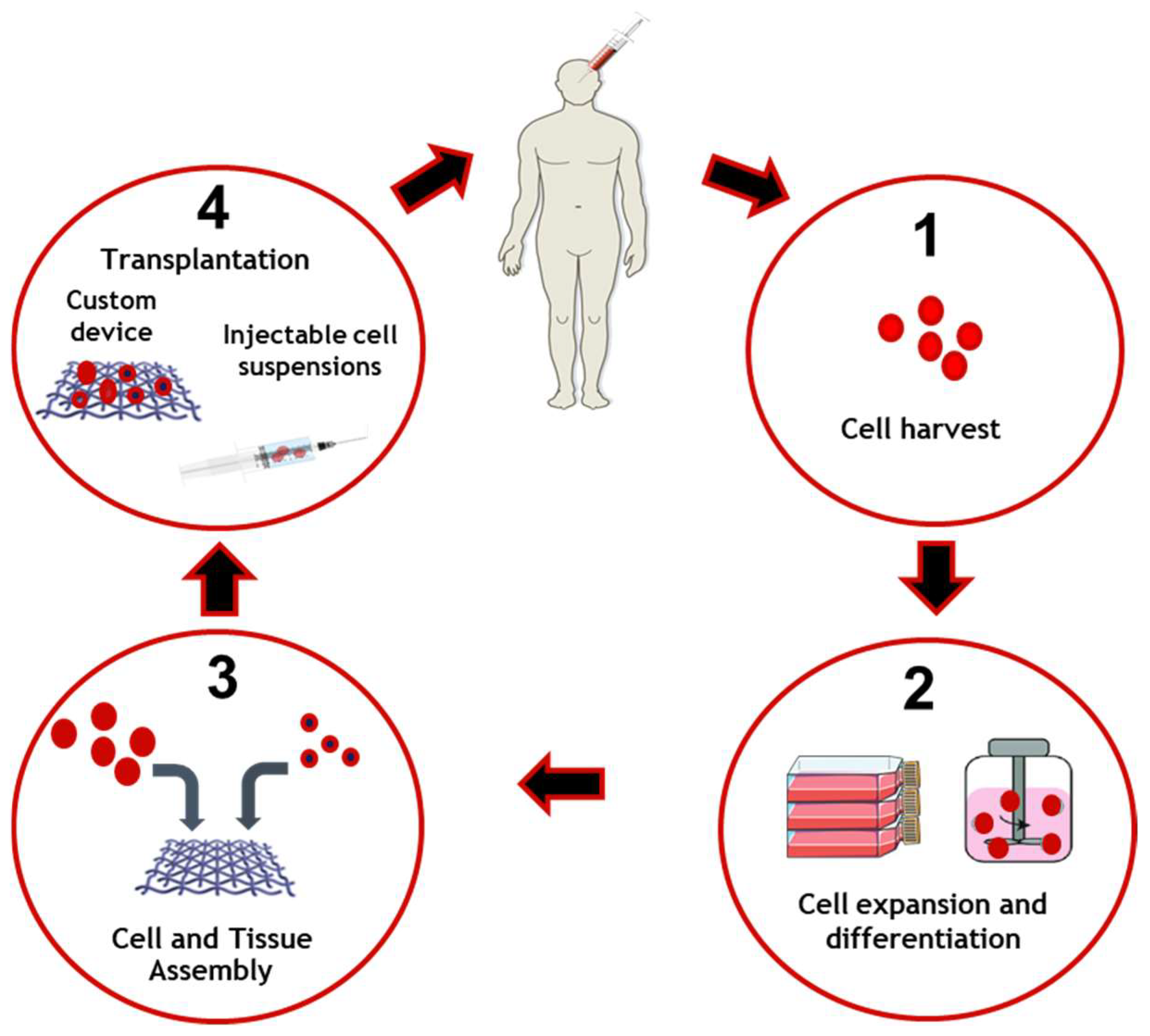
2.6. Tissue Engineering

Tissue engineering evolved from biomaterials development and is based on the combination of scaffolds, cells, and biologically active molecules to promote the development of functional tissues. The goal of tissue engineering is to assemble functional constructs that restore, maintain, or improve damaged tissues or whole organs. The approach was conceived to address the critical gap between the growing number of patients on the waiting list for organ transplantation and the limited number of donated organs available [25].
The general process of tissue engineering is based on the implantation of living cells in a scaffold specially designed to promote tissue replacement and regeneration [24,25] as is schematized in Figure 2. It is therefore considered as the next step in the development of biomaterials. Regenerative medicine relies on interdisciplinary research and applications based on repair, replacement or the regeneration of cells, tissues or organs to restore impaired functions. However, adult humans have only a limited capacity for spontaneous regeneration. Induced regeneration has been reported in conjunctiva and cornea.
Tissue engineering in the eye has been reported mainly in the anterior segment (cornea and conjunctiva) and significant progress has been made in cell therapies to treat degenerative diseases of the retina. Ocular regenerative therapies are a new tool to treat several blinding disorders, namely corneal disease, cataract, glaucoma, retinitis pigmentosa, and age-related macular degeneration. Several transplantable products, delivered as cell suspensions or as preformed 3D scaffolds combining cells and natural or artificial substrates, are being studied. Bioengineering approaches with advance cell product manufacturing are being developed, thereby enhancing stem cell-based medicine [138].

3. Concluding Remarks and Future Perspectives

The eye is a complex organ composed of a container with three membranes and different transparent elements. As we age, the eye, similarly to every other organ in the human body, suffers changes, leading to a deterioration of vision [1]. Biomaterials try to compensate for vision loss which may or may not be related to age. Contact lenses, composed of several polymers are produced in industrial quantities [27]. The introduction of multipurpose solutions allows for the cleaning and disinfection of lenses with a single solution, also allowing for better patient compliance [33]. Intraocular lenses are introduced into the eye to replace the clouded lens in cataract [1]. They can be composed of acrylic compounds or silicone elastomers and are kept inside the capsular bag with haptics [63]. Artificial tears are used for dry eyes [75], which can be used as eye drops, gel or ointment [81]. Artificial tears are composed of a thickening agent, osmoprotectant, antioxidant, preservative (except in single dose form), buffers, excipients, electrolytes and lipid supplements [8,79]. Inlays compensate for the loss in amplitude for the accommodation of presbyopia [9]. Vitreous body liquefies with age and can lead to pathological situations that require its replacement; however, the ideal substitute still does not exist and efforts should be taken to remedy this [13]. Different vitreous substitutes are used, in the form of gas or liquid [86], but the most promising for the future are those in hydrogels [12].
Today, contact lenses are increasingly seen as a tool for specific ophthalmic treatments that require the delivery of a drug to the eye. The low bioavailability of common ophthalmic treatments, such as eye drops, is a concern in delivering effective treatments. Therefore, practitioners rely on new lenses to be developed to improve these treatments. This is of scientific interest; however, more should be done to facilitate their practical application. This can include clinical trials or the practicality of manufacturing.
The intraocular application of hydrogels may lead to significant advances in improving their clinical application. Intravitreal drug delivery systems should be injectable and permit controlled drug release over several weeks or months. After complete drug release, the hydrogel should degrade without significant swelling. Similar requirements are imposed on hydrogel-based vitreous substitutes; however, these materials must be fully transparent and mechanically stable during the entire application period; therefore, research is needed to achieve such results.
Biomaterials have been demonstrated to be highly beneficial in their role in solving ophthalmic conditions in many studies. Advances have been made in recent years, particularly in terms of smart or stimuli-responsive hydrogels. However, many of these formulations are not commercially available, mainly because many of them have yet to undergo clinical trials. This would be a vital step in improving the quality of life of patients.
With an ageing population, eye diseases are becoming more widespread. Biomaterials have contributed to numerous medical devices for the restoration of eyesight, thereby improving many patients’ quality of life. Therefore, biomaterials, tissue engineering and regenerative medicine are becoming increasingly important to advances in ophthalmology and optometry. However, more research is needed to improve the treatment of severe, vision-threatening diseases.

Funding

This research received no external funding.

Institutional Review Board Statement

Not applicable.

Informed Consent Statement

Not applicable.

Data Availability Statement

Not applicable.

Conflicts of Interest

The author declares no conflict of interest.

References

  1. Chirila, T.; Harkin, D.; ScienceDirect. Biomaterials and Regenerative Medicine in Ophthalmology; Woodhead Publishing: Duxford, UK, 2016. [Google Scholar]
  2. Amato, S.; Amato, S.F.; Ezzell, R.M.; Amato, S.F. ProQuest. In Regulatory Affairs for Biomaterials and Medical Devices, 1st ed.; Woodhead Publishing: Cambridge, UK, 2015. [Google Scholar]
  3. Friedman, N.J.; Kaiser, P.K. Essentials of Ophthalmology, 1st ed.; Saunders Elsevier: Philadelphia, PA, USA, 2007; 294p. [Google Scholar]
  4. Basak, S.K. Essentials of Ophthalmology, 7th ed.; Jaypee Brothers Medical Publishers: New Delhi, India, 2019; 606p. [Google Scholar]
  5. Decker, S. Essentials of Ophthalmology; Foster Academics: New York, NY, USA, 2021; 432p. [Google Scholar]
  6. Winterton, L.C.; Lally, J.M.; Sentell, K.B.; Chapoy, L.L. The elution of poly (vinyl alcohol) from a contact lens: The realization of a time release moisturizing agent/artificial tear. J. Biomed. Mater. Res. Part B Appl. Biomater. 2007, 80, 424–432. [Google Scholar] [CrossRef] [PubMed]
  7. Powell, C.H.; Lally, J.M.; Hoong, L.D.; Huth, S.W. Lipophilic versus hydrodynamic modes of uptake and release by contact lenses of active entities used in multipurpose solutions. Contact Lens Anterior Eye 2010, 33, 9–18. [Google Scholar] [CrossRef] [PubMed]
  8. Hopkins, G.A.; Hopkins, G.; Pearson, R.M.; ScienceDirect. Ophthalmic Drugs: Diagnostic and Therapeutic Uses, 5th ed.; Butterworth-Heinemann Elsevier: Edinburgh, UK, 2007. [Google Scholar]
  9. Moarefi, M.A.; Bafna, S.; Wiley, W. A Review of Presbyopia Treatment with Corneal Inlays. Ophthalmol. Ther. 2017, 6, 55–65. [Google Scholar] [CrossRef] [PubMed] [Green Version]
  10. Kirchhof, S.; Goepferich, A.M.; Brandl, F.P. Hydrogels in ophthalmic applications. Eur. J. Pharm. Biopharm. 2015, 95, 227–238. [Google Scholar] [CrossRef]
  11. Lynch, C.R.; Kondiah, P.P.D.; Choonara, Y.E.; du Toit, L.C.; Ally, N.; Pillay, V. Hydrogel Biomaterials for Application in Ocular Drug Delivery. Front. Bioeng. Biotechnol. 2020, 8, 228. [Google Scholar] [CrossRef] [Green Version]
  12. Donati, S.; Caprani, S.M.; Airaghi, G.; Vinciguerra, R.; Bartalena, L.; Testa, F.; Mariotti, C.; Porta, G.; Simonelli, F.; Azzolini, C. Vitreous substitutes: The present and the future. Biomed. Res. Int. 2014, 2014, 351804. [Google Scholar] [CrossRef]
  13. Baino, F. Towards an ideal biomaterial for vitreous replacement: Historical overview and future trends. Acta Biomater. 2011, 7, 921–935. [Google Scholar] [CrossRef] [Green Version]
  14. Yadav, I.; Purohit, S.D.; Singh, H.; Bhushan, S.; Yadav, M.K.; Velpandian, T.; Chawla, R.; Hazra, S.; Mishra, N.C. Vitreous substitutes: An overview of the properties, importance, and development. J. Biomed. Mater. Res. B Appl. Biomater. 2021, 109, 1156–1176. [Google Scholar] [CrossRef]
  15. Santos, L.; Ferraz, M.P.; Shirosaki, Y.; Lopes, M.A.; Fernandes, M.H.; Osaka, A.; Santos, J.D. Degradation studies and biological behavior on an artificial cornea material. Investig. Ophthalmol. Vis. Sci. 2011, 52, 4274–4281. [Google Scholar] [CrossRef] [Green Version]
  16. Zhang, X.; Williams, D.F. Definitions of Biomaterials for the Twenty-First Century: Proceedings of a Consensus Conference Held in Chengdu, People’s Republic of China, June 11th and 12th 2018, Organized under the Auspices of the International Union of Societies for Biomaterials Science & Engineering; Hosted and Supported by Sichuan University, Chengdu and the Chinese Society for Biomaterials, China; Elsevier: Amsterdam, The Netherlands, 2019. [Google Scholar]
  17. Williams, D.F. Essential Biomaterials Science; Cambridge University Press: Cambridge, UK, 2014. [Google Scholar]
  18. Morgan, P.B.; Efron, N. Global contact lens prescribing 2000–2020. Clin. Exp. Optom. 2022, 105, 298–312. [Google Scholar] [CrossRef]
  19. Nicolson, P.C.; Vogt, J. Soft contact lens polymers: An evolution. Biomaterials 2001, 22, 3273–3283. [Google Scholar] [CrossRef]
  20. Findl, O.; Leydolt, C. Meta-analysis of accommodating intraocular lenses. J. Cataract Refract. Surg. 2007, 33, 522–527. [Google Scholar] [CrossRef]
  21. Agrawal, A.K.; Das, M.; Jain, S. In situ gel systems as ‘smart’ carriers for sustained ocular drug delivery. Expert Opin. Drug Deliv. 2012, 9, 383–402. [Google Scholar] [CrossRef]
  22. Teixeira, S.; Fernandes, M.H.; Ferraz, M.P.; Monteiro, F.J. Proliferation and mineralization of bone marrow cells cultured on macroporous hydroxyapatite scaffolds functionalized with collagen type I for bone tissue regeneration. J. Biomed. Mater. Res. A 2010, 95, 1–8. [Google Scholar] [CrossRef]
  23. Teixeira, S.; Yang, L.; Dijkstra, P.J.; Ferraz, M.P.; Monteiro, F.J. Heparinized hydroxyapatite/collagen three-dimensional scaffolds for tissue engineering. J. Mater. Sci. Mater. Med. 2010, 21, 2385–2392. [Google Scholar] [CrossRef]
  24. Mozafari, M.; Mozafari, M.; Sefat, F.; Atala, A.; ScienceDirect. Handbook of Tissue Engineering Scaffolds; Woodhead Publishing: Duxford, UK, 2019. [Google Scholar]
  25. Lanza, R.P.; Langer, R.; Vacanti, J.; Atala, A.; Science, D. Principles of Tissue Engineering, 5th ed.; Academic Press: London, UK, 2020. [Google Scholar]
  26. Sun, M.T.; O’Connor, A.J.; Wood, J.; Casson, R.; Selva, D. Tissue Engineering in Ophthalmology: Implications for Eyelid Reconstruction. Ophthalmic Plast. Reconstr. Surg. 2017, 33, 157–162. [Google Scholar] [CrossRef]
  27. Efron, N. Contact Lens Practice; Elsevier: Edinburgh, UK, 2018. [Google Scholar]
  28. Hom, M.M.; Bruce, A.S. Manual of Contact Lens Prescribing and Fitting with CD-ROM; Butterworth Heinemann: Oxford, UK, 2006. [Google Scholar]
  29. Guillon, M.; Dumbleton, K.; Patel, T.; Patel, K.; Gupta, R.; Maissa, C.A. Corrigendum to “Quantification of contact lens wettability after prolonged visual device use under low humidity conditions” [Cont. Lens Anterior Eye 42 (2019) 386–391]. Contact Lens Anterior Eye 2020, 43, 91. [Google Scholar] [CrossRef] [Green Version]
  30. Morgan, P.B.; McCullough, S.J.; Saunders, K.J. Corrigendum to “Estimation of ocular axial length from conventional optometric measures” [Cont. Lens Anterior Eye 43 (1) (2020) 18–20]. Contact Lens Anterior Eye 2020, 43, 413. [Google Scholar] [CrossRef]
  31. Musgrave, C.S.A.; Fang, F. Contact Lens Materials: A Materials Science Perspective. Materials 2019, 12, 261. [Google Scholar] [CrossRef] [Green Version]
  32. Pillay, R.; Hansraj, R.; Rampersad, N. Historical Development, Applications and Advances in Materials Used in Spectacle Lenses and Contact Lenses. Clin. Optom. 2020, 12, 201–202. [Google Scholar] [CrossRef]
  33. Millis, E.A.W. Medical Contact Lens Practice; Elsevier Butterwoth Heinemann: Edinburgh, UK; New York, NY, USA, 2005. [Google Scholar]
  34. Bennett, E.S.; Bennett, E.S.; Henry, V.A. Clinical Manual of Contact Lenses, 5th ed.; Wolters Kluwer Health/Lippincott Williams & Wilkins: Philadelphia, PA, USA, 2020. [Google Scholar]
  35. Baker, M.I.; Walsh, S.P.; Schwartz, Z.; Boyan, B.D. A review of polyvinyl alcohol and its uses in cartilage and orthopedic applications. J. Biomed. Mater. Res. B Appl. Biomater. 2012, 100, 1451–1457. [Google Scholar] [CrossRef]
  36. Stapleton, F.; Stretton, S.; Papas, E.; Skotnitsky, C.; Sweeney, D.F. Silicone hydrogel contact lenses and the ocular surface. Ocul. Surf. 2006, 4, 24–43. [Google Scholar] [CrossRef]
  37. Seo, E.; Kumar, S.; Lee, J.; Jang, J.; Park, J.H.; Chang, M.C.; Kwon, I.; Lee, J.S.; Huh, Y.I. Modified Hydrogels Based on Poly(2-hydroxyethyl methacrylate) (pHEMA) with Higher Surface Wettability and Mechanical Properties. Macromol. Res. 2017, 25, 704–711. [Google Scholar] [CrossRef]
  38. Ozgen, O.; Hasirci, N. Modification of Poly(methyl methacrylate) Surfaces with Oxygen, Nitrogen and Argon Plasma. J. Biomater. Tiss. Eng. 2014, 4, 479–487. [Google Scholar] [CrossRef]
  39. Peterson, R.C.; Wolffsohn, J.S.; Nick, J.; Winterton, L.; Lally, J. Clinical performance of daily disposable soft contact lenses using sustained release technology. Contact Lens Anterior Eye 2006, 29, 127–134. [Google Scholar] [CrossRef]
  40. Goda, T.; Ishihara, K. Soft contact lens biomaterials from bioinspired phospholipid polymers. Expert Rev. Med. Devices 2006, 3, 167–174. [Google Scholar] [CrossRef]
  41. Xinming, L.; Yingde, C.; Lloyd, A.W.; Mikhalovsky, S.V.; Sandeman, S.R.; Howel, C.A.; Liewen, L. Polymeric hydrogels for novel contact lens-based ophthalmic drug delivery systems: A review. Contact Lens Anterior Eye 2008, 31, 57–64. [Google Scholar] [CrossRef]
  42. Carvalho, I.M.; Marques, C.S.; Oliveira, R.S.; Coelho, P.B.; Costa, P.C.; Ferreira, D.C. Sustained drug release by contact lenses for glaucoma treatment-a review. J. Control. Release 2015, 202, 76–82. [Google Scholar] [CrossRef]
  43. Cardona, G.; Alonso, S.; Yela, S. Compliance versus Risk Awareness with Contact Lens Storage Case Hygiene and Replacement. Optom. Vis. Sci. 2022, 99, 449–454. [Google Scholar] [CrossRef]
  44. Barros, J.; Grenho, L.; Fontenente, S.; Manuel, C.M.; Nunes, O.C.; Melo, L.F.; Monteiro, F.J.; Ferraz, M.P. Staphylococcus aureus and Escherichia coli dual-species biofilms on nanohydroxyapatite loaded with CHX or ZnO nanoparticles. J. Biomed. Mater. Res. A 2017, 105, 491–497. [Google Scholar] [CrossRef]
  45. Gasson, A.; Morris, J.; Askews. The Contact Lens Manual: A Practical Guide to Fitting, 4th ed.; Butterworth/Heinemann: Edinburgh, UK, 2010. [Google Scholar]
  46. Nichols, J.J.; Chalmers, R.L.; Dumbleton, K.; Jones, L.; Lievens, C.W.; Merchea, M.M.; Szczotka-Flynn, L. The Case for Using Hydrogen Peroxide Contact Lens Care Solutions: A Review. Eye Contact Lens 2019, 45, 69–82. [Google Scholar] [CrossRef] [PubMed]
  47. Wu, Y.T.; Willcox, M.; Zhu, H.; Stapleton, F. Contact lens hygiene compliance and lens case contamination: A review. Contact Lens Anterior Eye 2015, 38, 307–316. [Google Scholar] [CrossRef] [PubMed]
  48. Bradley, C.S.; Sicks, L.A.; Pucker, A.D. Common Ophthalmic Preservatives in Soft Contact Lens Care Products: Benefits, Complications, and a Comparison to Non-Preserved Solutions. Clin. Optom. 2021, 13, 271–285. [Google Scholar] [CrossRef] [PubMed]
  49. Choi, S.W.; Kim, J. Therapeutic Contact Lenses with Polymeric Vehicles for Ocular Drug Delivery: A Review. Materials 2018, 11, 1125. [Google Scholar] [CrossRef] [Green Version]
  50. Xu, J.; Xue, Y.; Hu, G.; Lin, T.; Gou, J.; Yin, T.; He, H.; Zhang, Y.; Tang, X. A comprehensive review on contact lens for ophthalmic drug delivery. J. Control. Release 2018, 281, 97–118. [Google Scholar] [CrossRef]
  51. Cheng, R.; Meng, F.; Deng, C.; Klok, H.A.; Zhong, Z. Dual and multi-stimuli responsive polymeric nanoparticles for programmed site-specific drug delivery. Biomaterials 2013, 34, 3647–3657. [Google Scholar] [CrossRef]
  52. Behl, G.; Iqbal, J.; O’Reilly, N.J.; McLoughlin, P.; Fitzhenry, L. Synthesis and Characterization of Poly(2-hydroxyethylmethacrylate) Contact Lenses Containing Chitosan Nanoparticles as an Ocular Delivery System for Dexamethasone Sodium Phosphate. Pharm. Res. 2016, 33, 1638–1648. [Google Scholar] [CrossRef]
  53. ElShaer, A.; Mustafa, S.; Kasar, M.; Thapa, S.; Ghatora, B.; Alany, R.G. Nanoparticle-Laden Contact Lens for Controlled Ocular Delivery of Prednisolone: Formulation Optimization Using Statistical Experimental Design. Pharmaceutics 2016, 8, 14. [Google Scholar] [CrossRef] [Green Version]
  54. Maulvi, F.A.; Lakdawala, D.H.; Shaikh, A.A.; Desai, A.R.; Choksi, H.H.; Vaidya, R.J.; Ranch, K.M.; Koli, A.R.; Vyas, B.A.; Shah, D.O. In vitro and in vivo evaluation of novel implantation technology in hydrogel contact lenses for controlled drug delivery. J. Control. Release 2016, 226, 47–56. [Google Scholar] [CrossRef]
  55. dos Santos, J.F.; Alvarez-Lorenzo, C.; Silva, M.; Balsa, L.; Couceiro, J.; Torres-Labandeira, J.J.; Concheiro, A. Soft contact lenses functionalized with pendant cyclodextrins for controlled drug delivery. Biomaterials 2009, 30, 1348–1355. [Google Scholar] [CrossRef]
  56. Phan, C.M.; Subbaraman, L.N.; Jones, L. In vitro drug release of natamycin from beta-cyclodextrin and 2-hydroxypropyl-beta-cyclodextrin-functionalized contact lens materials. J. Biomater. Sci. Polym. Ed. 2014, 25, 1907–1919. [Google Scholar] [CrossRef]
  57. Alvarez-Lorenzo, C.; Yanez, F.; Barreiro-Iglesias, R.; Concheiro, A. Imprinted soft contact lenses as norfloxacin delivery systems. J. Control. Release 2006, 113, 236–244. [Google Scholar] [CrossRef]
  58. Malaekeh-Nikouei, B.; Vahabzadeh, S.A.; Mohajeri, S.A. Preparation of a molecularly imprinted soft contact lens as a new ocular drug delivery system for dorzolamide. Curr. Drug Deliv. 2013, 10, 279–285. [Google Scholar] [CrossRef]
  59. Lesher, G.A.; Gunderson, G.G. Continuous drug delivery through the use of disposable contact lenses. Optom. Vis. Sci. 1993, 70, 1012–1018. [Google Scholar] [CrossRef]
  60. Schrader, S.; Wedel, T.; Moll, R.; Geerling, G. Combination of serum eye drops with hydrogel bandage contact lenses in the treatment of persistent epithelial defects. Graefe’s Arch. Clin. Exp. Ophthalmol. 2006, 244, 1345–1349. [Google Scholar] [CrossRef]
  61. Lee, D.; Cho, S.; Park, H.S.; Kwon, I. Ocular Drug Delivery through pHEMA-Hydrogel Contact Lenses Co-Loaded with Lipophilic Vitamins. Sci. Rep. 2016, 6, 34194. [Google Scholar] [CrossRef]
  62. Khandelwal, S.S.; Jun, J.J.; Mak, S.; Booth, M.S.; Shekelle, P.G. Effectiveness of multifocal and monofocal intraocular lenses for cataract surgery and lens replacement: A systematic review and meta-analysis. Graefe’s Arch. Clin. Exp. Ophthalmol. 2019, 257, 863–875. [Google Scholar] [CrossRef] [Green Version]
  63. Güell, J.L. Cataract; Karger: Basel, Switzerland, 2013; 153p. [Google Scholar]
  64. Shekelle, P.G.; Khandelwal, S.; Jun, J. United StatesDepartment of Veterans AffairsHealth Services, RDevelopment, S.Quality Enhancement Research, I.West Los Angeles, V.A.M.C.E.-B.S.P.CComparative Effectiveness of Multifocal, Accommodative, and Monofocal Intraocular Lenses for Cataract Surgery and Lens Replacement; Department of Veterans Affairs, Veterans Health Administration, Quality Enhancement Research Initiative, Health Services Research & Development Service: Washington, DC, USA, 2018. [Google Scholar]
  65. Buratto, L.; Brint, S.F.; Boccuzzi, D.; Ebook Central Academic Complete. Cataract Surgery and Intraocular Lenses; Slack Incorporated: Thorofare, NJ, USA, 2014. [Google Scholar]
  66. Traxler, L.; Bayer, N.; Reutterer, B.; Drauschke, A. Improvement of Optics, Mechanics and the Usability of a Mechanical Eye Model for Vision Quality Evaluation of IOLs. IFAC-PapersOnLine 2015, 48, 1–18. [Google Scholar] [CrossRef]
  67. Leung, T.G.; Lindsley, K.; Kuo, I.C. Types of intraocular lenses for cataract surgery in eyes with uveitis. Cochrane Database Syst. Rev. 2014, 3, CD007284. [Google Scholar] [CrossRef] [Green Version]
  68. Wang, X. Intraocular Lens; IntechOpen: London, UK, 2020. [Google Scholar]
  69. Nguyen, J.; Werner, L. Intraocular Lenses for Cataract Surgery. In Webvision: The Organization of the Retina and Visual System; Kolb, H., Fernandez, E., Nelson, R., Eds.; University of Utah Health Sciences Center: Salt Lake City, UT, USA, 1995. [Google Scholar]
  70. Werner, L. Intraocular Lenses: Overview of Designs, Materials, and Pathophysiologic Features. Ophthalmology 2021, 128, e74–e93. [Google Scholar] [CrossRef]
  71. Craig, J.P.; Nichols, K.K.; Akpek, E.K.; Caffery, B.; Dua, H.S.; Joo, C.K.; Liu, Z.; Nelson, J.D.; Nichols, J.J.; Tsubota, K.; et al. TFOS DEWS II Definition and Classification Report. Ocul. Surf. 2017, 15, 276–283. [Google Scholar] [CrossRef]
  72. Clayton, J.A. Dry Eye. N. Engl. J. Med. 2018, 378, 2212–2223. [Google Scholar] [CrossRef]
  73. Yao, W.; Davidson, R.S.; Durairaj, V.D.; Gelston, C.D. Dry eye syndrome: An update in office management. Am. J. Med. 2011, 124, 1016–1018. [Google Scholar] [CrossRef]
  74. Ocular surface, d.; Holland, E.J.; Mannis, M.J.; Lee, W.B.; ScienceDirect. Ocular Surface Disease: Cornea, Conjunctiva and Tear Film; Elsevier/Saunders: London, UK; New York, NY, USA, 2013. [Google Scholar]
  75. Marshall, L.L.; Roach, J.M. Treatment of Dry Eye Disease. Consult. Pharm. 2016, 31, 96–106. [Google Scholar] [CrossRef]
  76. Bron, A.J.; Tomlinson, A.; Foulks, G.N.; Pepose, J.S.; Baudouin, C.; Geerling, G.; Nichols, K.K.; Lemp, M.A. Rethinking dry eye disease: A perspective on clinical implications. Ocul. Surf. 2014, 12, S1–S31. [Google Scholar] [CrossRef]
  77. Willcox, M.D.P.; Argueso, P.; Georgiev, G.A.; Holopainen, J.M.; Laurie, G.W.; Millar, T.J.; Papas, E.B.; Rolland, J.P.; Schmidt, T.A.; Stahl, U.; et al. TFOS DEWS II Tear Film Report. Ocul. Surf. 2017, 15, 366–403. [Google Scholar] [CrossRef] [Green Version]
  78. Sweeney, D.F.; Millar, T.J.; Raju, S.R. Tear film stability: A review. Exp. Eye Res. 2013, 117, 28–38. [Google Scholar] [CrossRef]
  79. Jones, L.; Downie, L.E.; Korb, D.; Benitez-Del-Castillo, J.M.; Dana, R.; Deng, S.X.; Dong, P.N.; Geerling, G.; Hida, R.Y.; Liu, Y.; et al. TFOS DEWS II Management and Therapy Report. Ocul. Surf. 2017, 15, 575–628. [Google Scholar] [CrossRef] [PubMed]
  80. Tong, L.; Petznick, A.; Lee, S.; Tan, J. Choice of artificial tear formulation for patients with dry eye: Where do we start? Cornea 2012, 31 (Suppl. 1), S32–S36. [Google Scholar] [CrossRef] [PubMed]
  81. Al-Kinani, A.A.; Zidan, G.; Elsaid, N.; Seyfoddin, A.; Alani, A.W.G.; Alany, R.G. Ophthalmic gels: Past, present and future. Adv. Drug Deliv. Rev. 2018, 126, 113–126. [Google Scholar] [CrossRef] [PubMed] [Green Version]
  82. Pflugfelder, S.C.; Geerling, G.; Kinoshita, S.; Lemp, M.A.; McCulley, J.P.; Nelson, D.; Novack, G.N.; Shimazaki, J.; Wilson, C. Management and therapy of dry eye disease: Report of the Management and Therapy Subcommittee of the International Dry Eye WorkShop (2007). Ocul. Surf. 2007, 5, 163–178. [Google Scholar] [CrossRef]
  83. Velpandian, T.; SpringerLink. Pharmacology of Ocular Therapeutics, 1st ed.; Springer International Publishing: Cham, Switzerland, 2016. [Google Scholar]
  84. Klenkler, B.; Sheardown, H.; Jones, L. Growth factors in the tear film: Role in tissue maintenance, wound healing, and ocular pathology. Ocul. Surf. 2007, 5, 228–239. [Google Scholar] [CrossRef]
  85. Lorentz, H.; Jones, L. Lipid deposition on hydrogel contact lenses: How history can help us today. Optom. Vis. Sci. 2007, 84, 286–295. [Google Scholar] [CrossRef]
  86. Alovisi, C.; Panico, C.; de Sanctis, U.; Eandi, C.M. Vitreous Substitutes: Old and New Materials in Vitreoretinal Surgery. J. Ophthalmol. 2017, 2017, 3172138. [Google Scholar] [CrossRef] [Green Version]
  87. Binder, P.S. Intracorneal Inlays for the Correction of Presbyopia. Eye Contact Lens 2017, 43, 267–275. [Google Scholar] [CrossRef]
  88. Fenner, B.J.; Moriyama, A.S.; Mehta, J.S. Inlays and the cornea. Exp. Eye Res. 2021, 205, 108474. [Google Scholar] [CrossRef]
  89. Kleinberg, T.T.; Tzekov, R.T.; Stein, L.; Ravi, N.; Kaushal, S. Vitreous substitutes: A comprehensive review. Surv. Ophthalmol. 2011, 56, 300–323. [Google Scholar] [CrossRef]
  90. Gao, Q.Y.; Fu, Y.; Hui, Y.N. Vitreous substitutes: Challenges and directions. Int. J. Ophthalmol. 2015, 8, 437–440. [Google Scholar] [CrossRef]
  91. Barros, J.A.R.; Melo, L.D.R.; Silva, R.; Ferraz, M.P.; Azeredo, J.; Pinheiro, V.M.C.; Colaco, B.J.A.; Fernandes, M.H.R.; Gomes, P.S.; Monteiro, F.J. Encapsulated bacteriophages in alginate-nanohydroxyapatite hydrogel as a novel delivery system to prevent orthopedic implant-associated infections. Nanomedicine 2020, 24, 102145. [Google Scholar] [CrossRef] [Green Version]
  92. Leone, G.; Consumi, M.; Aggravi, M.; Donati, A.; Lamponi, S.; Magnani, A. PVA/STMP based hydrogels as potential substitutes of human vitreous. J. Mater. Sci. Mater. Med. 2010, 21, 2491–2500. [Google Scholar] [CrossRef]
  93. Swindle-Reilly, K.E.; Shah, M.; Hamilton, P.D.; Eskin, T.A.; Kaushal, S.; Ravi, N. Rabbit study of an in situ forming hydrogel vitreous substitute. Investig. Ophthalmol. Vis. Sci. 2009, 50, 4840–4846. [Google Scholar] [CrossRef] [PubMed]
  94. Tao, Y.; Tong, X.; Zhang, Y.; Lai, J.; Huang, Y.; Jiang, Y.R.; Guo, B.H. Evaluation of an in situ chemically crosslinked hydrogel as a long-term vitreous substitute material. Acta Biomater. 2013, 9, 5022–5030. [Google Scholar] [CrossRef] [PubMed]
  95. Schramm, C.; Spitzer, M.S.; Henke-Fahle, S.; Steinmetz, G.; Januschowski, K.; Heiduschka, P.; Geis-Gerstorfer, J.; Biedermann, T.; Bartz-Schmidt, K.U.; Szurman, P. The cross-linked biopolymer hyaluronic acid as an artificial vitreous substitute. Investig. Ophthalmol. Vis. Sci. 2012, 53, 613–621. [Google Scholar] [CrossRef] [PubMed]
  96. Su, W.Y.; Chen, K.H.; Chen, Y.C.; Lee, Y.H.; Tseng, C.L.; Lin, F.H. An injectable oxidated hyaluronic acid/adipic acid dihydrazide hydrogel as a vitreous substitute. J. Biomater. Sci. Polym. Ed. 2011, 22, 1777–1797. [Google Scholar] [CrossRef]
  97. Kashiwagi, Y.; Nishitsuka, K.; Takamura, H.; Yamamoto, T.; Yamashita, H. Cloning and characterization of human vitreous tissue-derived cells. Acta Ophthalmol. 2011, 89, 538–543. [Google Scholar] [CrossRef]
  98. McMahon, T.T.; Zadnik, K. Twenty-five years of contact lenses: The impact on the cornea and ophthalmic practice. Cornea 2000, 19, 730–740. [Google Scholar] [CrossRef]
  99. Alio, J.L.; Belda, J.I.; Artola, A.; Garcia-Lledo, M.; Osman, A. Contact lens fitting to correct irregular astigmatism after corneal refractive surgery. J. Cataract Refract. Surg. 2002, 28, 1750–1757. [Google Scholar] [CrossRef]
  100. Wang, J.; Fonn, D.; Simpson, T.L. Topographical thickness of the epithelium and total cornea after hydrogel and PMMA contact lens wear with eye closure. Investig. Ophthalmol. Vis. Sci. 2003, 44, 1070–1074. [Google Scholar] [CrossRef] [Green Version]
  101. Thean, J.H.; McNab, A.A. Blepharoptosis in RGP and PMMA hard contact lens wearers. Clin. Exp. Optom. 2004, 87, 11–14. [Google Scholar] [CrossRef]
  102. Subbaraman, L.N.; Glasier, M.A.; Senchyna, M.; Sheardown, H.; Jones, L. Kinetics of in vitro lysozyme deposition on silicone hydrogel, PMMA, and FDA groups I, II, and IV contact lens materials. Curr. Eye Res. 2006, 31, 787–796. [Google Scholar] [CrossRef]
  103. Lee, M.J.; Park, S.Y.; Sung, A.Y. Poly (Ethylene Glycol) Methyl Ether Methacrylate-Based Hydrogel and Cerium(IV) Oxide Nanoparticles as Ophthalmic Lens Material. Micromachines 2021, 12, 1111. [Google Scholar] [CrossRef]
  104. Wang, Z.; Li, X.; Zhang, X.; Sheng, R.; Lin, Q.; Song, W.; Hao, L. Novel Contact Lenses Embedded with Drug-Loaded Zwitterionic Nanogels for Extended Ophthalmic Drug Delivery. Nanomaterials 2021, 11, 2328. [Google Scholar] [CrossRef]
  105. Zare, M.; Bigham, A.; Zare, M.; Luo, H.; Rezvani Ghomi, E.; Ramakrishna, S. pHEMA: An Overview for Biomedical Applications. Int. J. Mol. Sci. 2021, 22, 6376. [Google Scholar] [CrossRef]
  106. Maissa, C.; Guillon, M.; Cockshott, N.; Garofalo, R.J.; Lemp, J.M.; Boclair, J.W. Contact Lens Lipid Spoliation of Hydrogel and Silicone Hydrogel Lenses. Optom. Vis. Sci. 2014, 91, 1071–1083. [Google Scholar] [CrossRef]
  107. Dogru, M.; Ward, S.K.; Wakamatsu, T.; Ibrahim, O.; Schnider, C.; Kojima, T.; Matsumoto, Y.; Ogawa, J.; Shimazaki, J.; Tsubota, K. The effects of 2 week senofilcon-A silicone hydrogel contact lens daily wear on tear functions and ocular surface health status. Contact Lens Anterior Eye 2011, 34, 77–82. [Google Scholar] [CrossRef]
  108. Chalmers, R. Overview of factors that affect comfort with modern soft contact lenses. Contact Lens Anterior Eye 2014, 37, 65–76. [Google Scholar] [CrossRef]
  109. Sulley, A.; Young, G.; Hunt, C. Factors in the success of new contact lens wearers. Contact Lens Anterior Eye 2017, 40, 15–24. [Google Scholar] [CrossRef]
  110. Tasci, Y.Y.; Gurdal, C.; Sarac, O.; Onusever, A. Evaluation of the Tear Function Tests and the Ocular Surface in First-Time Users of Silicone Hydrogel Contact Lenses. Curr. Eye Res. 2017, 42, 976–981. [Google Scholar] [CrossRef]
  111. Hyon, S.H.; Cha, W.I.; Ikada, Y.; Kita, M.; Ogura, Y.; Honda, Y. Poly(vinyl alcohol) hydrogels as soft contact lens material. J. Biomater. Sci. Polym. Ed. 1994, 5, 397–406. [Google Scholar] [CrossRef]
  112. Phan, C.M.; Walther, H.; Riederer, D.; Lau, C.; Lorenz, K.O.; Subbaraman, L.N.; Jones, L. Analysis of polyvinyl alcohol release from commercially available daily disposable contact lenses using an in vitro eye model. J. Biomed. Mater. Res. B Appl. Biomater. 2019, 107, 1662–1668. [Google Scholar] [CrossRef] [Green Version]
  113. Daza, J.H.U.; Righetto, G.M.; Chaud, M.V.; da Conceicao Amaro Martins, V.; Lopes Baratella da Cunha Camargo, I.; Maria de Guzzi Plepis, A. PVA/anionic collagen membranes as drug carriers of ciprofloxacin hydrochloride with sustained antibacterial activity and potential use in the treatment of ulcerative keratitis. J. Biomater. Appl. 2020, 35, 301–312. [Google Scholar] [CrossRef] [PubMed]
  114. Zha, X.J.; Zhang, S.T.; Pu, J.H.; Zhao, X.; Ke, K.; Bao, R.Y.; Bai, L.; Liu, Z.Y.; Yang, M.B.; Yang, W. Nanofibrillar Poly(vinyl alcohol) Ionic Organohydrogels for Smart Contact Lens and Human-Interactive Sensing. ACS Appl. Mater. Interfaces 2020, 12, 23514–23522. [Google Scholar] [CrossRef] [PubMed]
  115. Jain, S.; Chauhan, A.; Rajshekar, K.; Vashist, P.; Gupta, P.; Mathur, U.; Gupta, N.; Gupta, V.; Dutta, P.; Gauba, V.K. Generic and vision related quality of life associated with different types of cataract surgeries and different types of intraocular lens implantation. PLoS ONE 2020, 15, e0240036. [Google Scholar] [CrossRef] [PubMed]
  116. Maedel, S.; Evans, J.R.; Harrer-Seely, A.; Findl, O. Intraocular lens optic edge design for the prevention of posterior capsule opacification after cataract surgery. Cochrane Database Syst. Rev. 2021, 8, CD012516. [Google Scholar] [CrossRef]
  117. Shiraki, A.; Sakimoto, S.; Oie, Y.; Soma, T.; Miki, A.; Usui, S.; Sato, S.; Matsushita, K.; Sakaguchi, H.; Nishida, K. Inferior Removal of Dislocated Polymethyl Methacrylate Intraocular Lens and Scleral Refixation in Glaucomatous Eyes. Ophthalmol. Ther. 2022, 11, 881–886. [Google Scholar] [CrossRef]
  118. Tetz, M.; Jorgensen, M.R. New Hydrophobic IOL Materials and Understanding the Science of Glistenings. Curr. Eye Res. 2015, 40, 969–981. [Google Scholar] [CrossRef]
  119. Huang, J.F.; Zhong, J.; Chen, G.P.; Lin, Z.T.; Deng, Y.; Liu, Y.L.; Cao, P.Y.; Wang, B.; Wei, Y.; Wu, T.; et al. A Hydrogel-Based Hybrid Theranostic Contact Lens for Fungal Keratitis. ACS Nano 2016, 10, 6464–6473. [Google Scholar] [CrossRef]
  120. Cooper, R.C.; Yang, H. Hydrogel-based ocular drug delivery systems: Emerging fabrication strategies, applications, and bench-to-bedside manufacturing considerations. J. Control. Release 2019, 306, 29–39. [Google Scholar] [CrossRef]
  121. Pimenta, A.F.R.; Serro, A.P.; Colaco, R.; Chauhan, A. Optimization of intraocular lens hydrogels for dual drug release: Experimentation and modelling. Eur. J. Pharm. Biopharm. 2019, 141, 51–57. [Google Scholar] [CrossRef]
  122. Jaitli, A.; Roy, J.; Chatila, A.; Liao, J.; Tang, L. Effect of time and temperature-dependent changes of IOL material properties on IOL: Lens capsule interactions. Exp. Eye Res. 2021, 211, 108726. [Google Scholar] [CrossRef]
  123. Chen, S.Y.; Xie, C.; Wang, Y.; Shen, Y. Full-vision maintenance in extra-high myopia from implantable collamer lens to trifocal intraocular lens implantation. Int. J. Ophthalmol. 2018, 11, 1239–1242. [Google Scholar] [CrossRef]
  124. Tychsen, L.; Reynolds, M.; Hoekel, J. Reply: Safety of phakic intraocular collamer lens implantation in 95 highly myopic special-needs children. J. Cataract Refract. Surg. 2021, 47, 1606–1607. [Google Scholar] [CrossRef]
  125. Pinto, C.; Monteiro, T.; Franqueira, N.; Faria-Correia, F.; Mendes, J.; Vaz, F. Posterior chamber collamer phakic intraocular lens implantation: Comparison of efficacy and safety for low and moderate-to-high myopia. Eur. J. Ophthalmol. 2022, 32, 894–901.e1451. [Google Scholar] [CrossRef]
  126. Mulet, M.E.; Alio, J.L.; Knorz, M.C. Hydrogel intracorneal inlays for the correction of hyperopia: Outcomes and complications after 5 years of follow-up. Ophthalmology 2009, 116, 1455–1460. [Google Scholar] [CrossRef]
  127. Casini, G.; Loiudice, P.; De Cilla, S.; Radice, P.; Nardi, M. Sulfur hexafluoride (SF6) versus perfluoropropane (C3F8) tamponade and short term face-down position for macular hole repair: A randomized prospective study. Int. J. Retin. Vitr. 2016, 2, 10. [Google Scholar] [CrossRef] [Green Version]
  128. Yu, Q.; Liu, K.; Su, L.; Xia, X.; Xu, X. Perfluorocarbon liquid: Its application in vitreoretinal surgery and related ocular inflammation. Biomed. Res. Int. 2014, 2014, 250323. [Google Scholar] [CrossRef] [Green Version]
  129. Thacker, M.; Tseng, C.L.; Lin, F.H. Substitutes and Colloidal System for Vitreous Replacement and Drug Delivery: Recent Progress and Future Prospective. Polymers 2020, 13, 121. [Google Scholar] [CrossRef]
  130. Pruett, R.C.; Schepens, C.L.; Swann, D.A. Hyaluronic-Acid Vitreous Substitute—6-Year Clinical-Evaluation. Arch. Ophthalmol. 1979, 97, 2325–2330. [Google Scholar] [CrossRef]
  131. Barth, H.; Crafoord, S.W.; Vinchon, C.; Andreasson, S.; Ghosh, F.K. A cross-linked hyaluronic acid hydrogel (Healaflow (R)) as a potential vitreous substitute. Investig. Ophthalmol. Vis. Sci. 2014, 55, 2338. [Google Scholar]
  132. Barth, H.; Crafoord, S.; Andreasson, S.; Ghosh, F. A cross-linked hyaluronic acid hydrogel (Healaflow((R))) as a novel vitreous substitute. Graefe’s Arch. Clin. Exp. Ophthalmol. 2016, 254, 697–703. [Google Scholar] [CrossRef]
  133. Schulz, A.; Rickmann, A.; Wahl, S.; Germann, A.; Stanzel, B.V.; Januschowski, K.; Szurman, P. Alginate- and Hyaluronic Acid-Based Hydrogels as Vitreous Substitutes: An In Vitro Evaluation. Transl. Vis. Sci. Technol. 2020, 9, 34. [Google Scholar] [CrossRef]
  134. Wang, S.; Chi, J.H.; Jiang, Z.W.; Hu, H.W.; Yang, C.Z.; Liu, W.S.; Han, B.Q. A self-healing and injectable hydrogel based on water-soluble chitosan and hyaluronic acid for vitreous substitute. Carbohydr. Polym. 2021, 256, 117519. [Google Scholar] [CrossRef]
  135. Yu, S.Q.; Wang, S.; Xia, L.X.; Hu, H.W.; Zou, M.Y.; Jiang, Z.W.; Chi, J.H.; Zhang, Y.J.; Li, H.J.; Yang, C.Z.; et al. Injectable self-crosslinking hydrogels based on hyaluronic acid as vitreous substitutes. Int. J. Biol. Macromol. 2022, 208, 159–171. [Google Scholar] [CrossRef]
  136. Greco, G.; Lamponi, S.; Leone, G.; Consumi, M.; Magnani, A. In Vitro Biocompatibility of New Hydrogels as Vitreous Body Substitutes. Eur. J. Ophthalmol. 2017, 27, E97. [Google Scholar]
  137. Lin, K.T.; Wang, A.; Nguyen, A.B.; Iyer, J.; Tran, S.D. Recent Advances in Hydrogels: Ophthalmic Applications in Cell Delivery, Vitreous Substitutes, and Ocular Adhesives. Biomedicines 2021, 9, 1203. [Google Scholar] [CrossRef]
  138. Stern, J.H.; Tian, Y.; Funderburgh, J.; Pellegrini, G.; Zhang, K.; Goldberg, J.L.; Ali, R.R.; Young, M.; Xie, Y.; Temple, S. Regenerating Eye Tissues to Preserve and Restore Vision. Cell Stem Cell 2018, 23, 453. [Google Scholar] [CrossRef] [PubMed]
Figure 1. From the material point of view, contact lenses depend on several materials properties. Final contact lenses require a consideration of wear time, comfort and cost. These characteristics depend on material properties, manufacturing processes and final treatment.
Applsci 12 05886 g001
Figure 2. Tissue engineering applied to cell and tissue manufacture and delivery. Cells are collected and harvested in order to perform expansion and differentiation. Cells and bioengineered scaffolds are then assembled to be able to be transplanted on the patient.
Applsci 12 05886 g002
Table 1. Advantages and disadvantages of the most commonly used contact lens biomaterials.
BiomaterialDisadvantagesAdvantages
Poly(vinyl alcohol) (PVA)Low permeability to oxygen; fixed water contactLow cost; biocompatible; easy manufacturing
Silicon hydrogelExpensive; abrasive behaviorHigh permeability to oxygen; high durability
Hydroxy ethyl methacrylate (HEMA) hydrogelLow permeability to oxygen; protein deposition problemsLow cost; biocompatible; several copolymer possibilities
Polymethyl methacrylate (PMMA)Impermeable to oxygen; not flexible in the eyes; abrasive behaviorLow cost; well-studied polymer
Table 2. Main components and functions of artificial tears.
ComponentMain FunctionReferences
Viscosity-improving agents (Carbomer 940, Carboxymethyl cellulose (CMC), Hyaluronic acid (HA), Hydroxypropylme-thylcellulose (HPMC), polyvinylpyrrolidone (PVP))Increase the time of permanence in the eye due to their mucoadhesive properties[81,86]
OsmoprotectorsMaintaining normal cellular metabolism, even under extreme osmotic stress[71,79]
TrehaloseBioprotection and osmoprotection[71,72,77,79]
Quercetin, epigallocatechin gallate, n-propyl gallate and gallic acidProtecting the corneal epithelium from oxidative damage[75,79]
Benzalkonium chloride (BAC), Sodium perborate, sodium chlorite, Polyquaternium-1Preservatives[75,79]
Table 3. Main classes of ophthalmic applications involving biomaterials.
Clinical ApplicationMaterialReferences
Contact lensesPMMA[98,99,100,101,102]
HEMA hydrogel[37,103,104,105]
Silicone hydrogel[106,107,108,109,110]
PVA[111,112,113,114]
Intraocular lensesPMMA[115,116,117]
Hydrophobic acrylate polymers[70,118]
Hydrophilic acrylate polymers[119,120,121]
Siloxanes[70,104,122]
Collamer[123,124,125]
InlaysHydrophilic hydrogels[126]
Vitreous substitutesGas (air, sulfur hexafluoride (SF6) and perfluoropropane (C3F8)[14,127]
Liquid (physiological solution, perfluorocarbon fluids (PFCL), semi-fluorinated alkanes (SFA)[128,129]
Natural and semi-synthetic polymers (hyaluronic acid and chitosan, silicone oil)[130,131,132,133,134,135]
Hydrogels[135,136,137]
Publisher’s Note: MDPI stays neutral with regard to jurisdictional claims in published maps and institutional affiliations.

