Article

Gastroprotective Effects of Fermented Gold Kiwi (Actinidia chinenesis L.) Extracts on HCl/EtOH-Induced Gastric Injury in Rats

1
Department of Laboratory Animal Medicine, College of Veterinary Medicine, Jeonbuk National University, Iksan-si 54596, Jeollabuk-do, Korea
2
R&D Team, Food & Supplement Helath Claims, Vitech, #602 Giyeon B/D 141 Anjeon-ro, Iseo-myeon, Wanju-gun 55365, Jeollabuk-do, Korea
*
Author to whom correspondence should be addressed.
These authors contributed equally to this work.
Appl. Sci. 2022, 12(10), 5271; https://doi.org/10.3390/app12105271
Submission received: 20 April 2022 / Revised: 14 May 2022 / Accepted: 21 May 2022 / Published: 23 May 2022
(This article belongs to the Special Issue Research and Development of Functional Foods)

Abstract

:
Gastritis and gastric ulcers caused by stressors such as drinking are common. The ability of functional foods to protect the stomach more effectively and reduce the risk of side effects is of interest. The fermentation process can increase the preservation and bioactive compound content of kiwi fruits. This study produced fermented kiwi powder using two lactic acids separated from gold kiwi fruits. Gold kiwi puree (Actinidia chinensis L.) was fermented using beneficial bacteria. Fermentation increased the content of bioactive compounds such as organic acids, flavonoids, and carotenoids. We investigated whether fermented gold kiwi (FGK) extract had antioxidant and gastric protective effects in an HCl/EtOH-induced gastritis animal model and pyloric ligation animal model. FGK increased radical scavenging activity in a dose-dependent manner. In the gastritis model, FGK inhibited inflammation-related factors such as iNOS, COX-2, IL-6, and TNF-α, while increasing the expression of the protective molecule PGE2. Furthermore, FGK administration improved gastric lesion site appearance, clinical symptoms, and mucosal thickness in rats. FGK also reduced gastric fluid volume, free acidity, total acidity, and pepsin activity in the pyloric ligation model. These results suggest that FGK can decrease the inflammatory response and protect the gastric mucosa. FGK therefore has the potential to prevent and treat gastritis and gastric ulcers.

1. Introduction

Gastritis and gastric ulcers are common diseases that afflict millions of people worldwide. Gastritis is an inflammatory disease of the gastric mucosa that occurs due to an imbalance between the gastric mucosa’s defense mechanisms and attack factors [1]. The stomach wall consists of four layers. Gastritis refers to damage of only the first gastric wall layer, the gastric mucosa, whereas gastric ulcers refer to damage extending to the second layer or deeper [2]. Endogenous factors such as gastric acid, histamine, pepsin, and gastrin secretion can damage the mucosa, in concert with exogenous factors such as Helicobacter pylori infection, excessive drinking, and NSAIDs [3].
Gastritis and gastric ulcers are treated with acid-suppressant drugs, such as histamine receptor antagonists (H2 receptor antagonists) and proton pump inhibitors [4,5]. Although these drugs have excellent therapeutic effects, they also have side effects, such as diarrhea, headache, rash, high blood pressure, and joint pain, when used long term [6,7,8]. Therefore, functional foods to protect the gastric mucosa are of great interest. These functional foods would ideally reduce the recurrence of gastritis and gastric ulcers and reduce the burden of long-term use of acid-suppressant drugs.
Kiwis are plants in the genus Actinidia whose fruits are edible; kiwis grow primarily in temperate zones, and there are 64 varieties worldwide. Kiwi fruits contain bioactive compounds with various physiologically active substances, such as organic acid, flavonoids, and carotenoids [9]. Kiwi fruits have bioactivity compound content 2–3 times higher than that of apples and grapes and contain organic acids such as quinic acid, malic acid, and citric acid [10]. Kiwi fruits have been shown to have antioxidant activity, anticancer activity, and to improve constipation [11,12,13,14].
Ethylene production results in softening of the flesh of kiwis, decomposition, synthesis of fragrance and pigment components, and reduction of organic acids. Therefore, kiwis have a short shelf life and rapidly lose commercial quality during storage [15]. Research has been conducted to determine how to preserve the biologically active compounds in kiwis. Although various fermented kiwi products have been studied, most of these products are processed foods and beverages with low utilization [15,16,17].
Recently, attempts have been made to improve the storage properties, nutritional quality, and physiological activity of fermented kiwi using various beneficial bacteria [18,19]. Our lab has screened the ability of several lactic acid bacteria as starter cultures to produce valuable substances when used to ferment Jeju gold kiwi fruit puree. In the current study, kiwi fruit puree was fermented with Lactococcus lactis VI-01 (KCTC 14351BP) and Lactobacillus paracasei VI-02 (KCTC 14352 BP), both of which were originally isolated from Jeju gold kiwi. We used this fermented gold kiwi (FGK) extract in an experimental gastric ulcer model and pyloric ligation model to investigate whether it exerted gastroprotective effects and could be used in the development of functional foods with therapeutic effects.

2. Materials and Methods

2.1. Sample Preparation

For FGK production, Jeju gold kiwi (Actinidia chinensis L.) puree with peels and seed removed was purchased from Namuang Foods. Vitech. Co., Ltd. Bacteria used for fermentation were Lactococcus lactis VI-01 (KTCT 14351 BP) and Lactobacillus paracase VI-02 (KTCT 14352 BP) isolated from gold kiwi, and each strain was cultured in MRS Broth at 37 °C. Collected seed-cultured bacteria were pre-cultured for 9 h at 37 °C in an industrial medium. The pre-culture solution was mixed with the prepared kiwi puree and cultured at 37 °C for 8 h to prepare fermented gold kiwi. The cultured fermented product was collected and prepared as a freeze-dried powder.

2.2. Chemicals and Drugs

Citric acid, quinic acid, 2,2-diphenyl-1-picrylhydrazyl (DPPH), 2,2′-azino-bis (3-ethylbenzothiazoline-6-sulfonic acid) (ABTS), potassium phosphate, ethanol, sodium hydroxide (NaOH), phenolphthalein solution, hemoglobin, and trichloroacetic acid solution were purchased from Sigma-Aldrich (Burlington, MO, USA). Malic acid, lutein, and quercetin dehydrate were purchased from Merck (Darmstadt, Germany). Sucralfate powder was purchased from Cayman Chemical (Ann Arbor, MI, USA). HCl was purchased from Wako (Osaka, Japan). Interleukin-6 (IL-6), tumor necrosis factor-alpha (TNF-α), and prostaglandin E2 (PGE2) were purchased from R&D Systems (Minneapolis, MN, USA). Inducible nitric oxide synthase (iNOS) antibody, cyclooxygenase-2 (COX-2) antibody, and beta-actin (β-actin) antibody were purchased from Cell Signaling Technology (Beverly, MA, USA). SuperSignal West Dura Extended Duration Substrate was purchased from Thermo Scientific (Waltham, MA, USA).

2.3. Determination of the Content of Bioactive Compounds in FGK

2.3.1. Determination of Organic Acid Content

To analyze the organic acid concent (citric acid, malic acid, and quinic acid), a citric acid sample (Cas No. 77-92-9 as a standard), malic acid sample (Cas No. 6915-15-7 as a standard), and quinic acid sample (Cas No. 77-95-2 as a standard) were dissolved in DW at 0.02 g/mL. The HITACHI chromaster (HITACHI EzchromElite, HITACHI, Tokyo, Japan) was used. Chromatographic separation was performed on an Aminex HPX-87H Ion Exclusion instrument (300 mm × 7.8 mm). The column temperature was set at 25 °C, and the flow rate was 0.6 mL/min. The solvent was 4 mM sulfuric acid, and the injection volume was 10 μL. A UV detector generated DAD chromatograms at a 355 nm wavelength, and results were expressed as mg of each standard/g.

2.3.2. Determination of Carotenoid Content

To analyze the β-carotene, samples (or β-carotene; Cas No. 7235-40-7 as a standard) were dissolved in a mixture solvent ratio of 9:1 (3% pyorogallol ethanol and 60% potassium hydroxide) at 8 mg/mL. The Agilent 1200 series gradient HPLC system (Agilent Technologies, Santa Clara, CA, USA) was used. Chromatographic separation was performed on an exsil phase C18 HPLC column (5 μm particle size, L × I.D 2.5 cm × 4.6 mm). The column temperature was set at 40 °C and the flow rate was 1.0 min/mL. Mobile phase A and B consisted of acetonitrile and methanol, 85:15, v/v, and dichloromethane, respectively. The gradient system was as follows: 0 min, 70% A; 12 min, 75% A; 15.5 min, 70% A; 27.5 min, 100% A; 28 min, 70% A. UV/Vis detection was performed at 450 nm wavelength [20], and results were expressed as mg of each standard/g.
To analyze the lutein, samples (or lutein; Cas No. 127-40-2 as a standard) were dissolved in DW at 13.3 mg/mL and we added 30 mL of ethyl acetate. The Agilent 1200 series gradient HPLC system (Agilent Technologies, Santa Clara, CA, USA) was used. Chromatographic separation was performed on a nucleosil C18 HPLC column (3 μm particle size, L × I.D 7.5 cm × 4.6 mm). The column temperature was set at 40 °C and the flow rate was 1.5 min/mL. Mobile phase A and B consisted of 70% hexane and 30% ethyl acetate, respectively. The gradient system was as follows: 0 min, 25% B; 1 min, 50% B; 20 min, 70% B, 25 min, 100% B, 30 min, 100% B, 35 min, 25% B. UV/Vis detection was performed at 4460 nm wavelength [20], and results were expressed as mg of each standard/g.

2.3.3. Determination of Total Flavonoid Content

The total flavonoid content of fermented gold kiwi fruit (FGK) and general gold kiwi (GK) was determined using a slight modification of the method of Wu et al. [15]. Each sample was mixed with 1.2 mL 1 mol/L NaOH methanol and 20.18 mL NaNO2 and reacted at room temperature for 6 min. An 8% Al(NO3)3 of 30.36 mL was added, reacted for 5 min, and then 1 mol/L NaOH was added to 1.2 mL. After 15 min, absorbance was measured at 510 nm. Results are expressed as mg of each standard/g.

2.3.4. Determination of Total Quercetin Derivative Content

The 200 mg amounts of total quercetin derivatives in FGK were determined according to the method of Dadakova et al. [21]. In the method, all glycoside forms of 1.2 M HCl and 50% methanol are hydrolyzed in the presence of ascorbic acid in the antioxidant. The released quercetin is isolated and preconcentrated to extract the solid phase (RP-18, Merck, Germany). The absorbed substance is eluted with methanol (1.4 mL). Results are expressed as mg of each standard/g.

2.4. DPPH Free Radical Scavenging Activity

The stable free radical 2,2-diphenyl-1-picryl-hydrazyl (DPPH) was used to evaluate the radical scavenging activity of FGK using a previously described method [22]. Ascorbic acid was used as a standard. FGK was dissolved in ethanol to concentrations of 1.25, 2.5, 5, and 10 mg/mL, and then 0.2 mM DPPH solution was added at a ratio of 1:9. After reaction for 10 min in the dark, absorbance was measured at 517 nm using a microplate reader (Synergy 2, BioTek Instruments, Winooski, VT, USA).

2.5. ABTS Free Radical Scavenging Activity

An assay of 2,2′-azino-bis (3-ethylbenzothiazoline)-6-sulfonic acid (ABTS) radical scavenging activity was used to evaluate the antioxidant activity of FGK [23]. Ascorbic acid was used as a standard. FGK was dissolved in ethanol to a concentration of 1, 5, 10, or 25 mg/mL. Then, 7.4 mM ABTS and 2.6 mM potassium persulfate were mixed and placed in the dark for 16 h. The solution was diluted with absolute ethanol to an absorbance value at 734 nm of 0.70 ± 0.02. Sample and diluted solution were reacted at a ratio of 1:9 in the dark for 1 min, and then absorbance was measured at 734 nm using a microplate reader (Synergy 2, BioTek Instruments, Winooski, VT, USA).

2.6. Experimental Animals

Six-week-old male Sprague-Dawley (SD) rats (Damul, Daejeon, Korea), weighing 150–200 g, were used. All animals were allowed free access to food and water. During the experimental period, animals were monitored once a day for health status by assessing body weight, food intake, and behavior changes. All animals were cared for in accordance with the guidelines of Jeonbuk National University Institutional Animal Care and Use Committee, which approved the study protocol (JBNU 2021-064).

2.7. HCl/EtOH-Induced Acute Gastric Lesions

The SD rats were randomly distributed into six groups: Normal (normal control), Control (negative control), SC (sucralfate 50 mg/kg, positive control), and FGK 50, 125, 250 (mg/kg, experimental groups). Each group had eight experimental animals who were fasted and allowed free access to water for 24 h before induction of acute gastritis. Sucralfate and FGK were orally administered using DW as a vehicle. One hour after administration, 2 mL of 150 mM HCl/60% EtOH solution was administered orally to induce the formation of gastric lesions. Thirty minutes after HCl/EtOH administration, clinical symptoms were observed by three individuals for 1 h. Then, rats were euthanized under anesthesia, and the stomach was removed from the abdomen. The removed stomach was incised along the greater curvature and fixed in 2% paraformaldehyde for 15 min at room temperature. Fixed stomachs were photographed using an optical digital camera (DSC-HX50V, Sony Japan), and the hemorrhagic lesion area was measured using Image J software (version 1.8.0., National Institutes of Health, Bethesda, MD, USA).
Gastric lesion area (%) = (gastric lesion/total area) × 100

2.8. Clinical Symptom Score after Gastritis Induction

Clinical symptom score was assessed based on a previously described method [24]. Points were assigned as follows: 1 point for normal walking and excited behavior, 5 points for slow movement compared to movement or normal movement, 10 points for slight movement when little movement is present or stimulated, and 15 points for breathlessness and deep breathing.

2.9. Macroscopic Damage Score after Gastritis Induction

After the fixed stomachs were photographed, the size and number of ulcers formed in the gastric mucosa were measured using Image J software version 1.8.0 (National Institutes of Health, Bethesda, MD, USA). The gastric ulcer index was calculated based on a previous study [25] and was determined as shown in Table 1.

2.10. Gastric Mucosa Thickness Measurement

The dissected gastric tissue was fixed at 4% paraformaldehyde for 24 h. All selected samples were fitted into the paraffin and cut into 5-micron sections using a microtome. The tissue sections were deparaffinized using a xylene and graduated alcohol series to water. The tissue sections were stained with hematoxylin and eosin (H&E) and observed under an optical microscope (Zeiss, Germany). The gastric mucosa thickness visualized the cross-section of the H&E-dyed specimen, and the area of the dyed site (in pixels) was quantified using the ImagePro analysis program.

2.11. Gastric Tissue Western Blotting

Protein in gastric tissues was subjected to 8–12% SDS-page gels and then transferred to PVDF membranes (Bio-Rad, Hercules, CA, USA). After blocking in 5% skim milk in PBS-T, membranes were incubated with primary antibodies for COX-2 (D5H5, Cell Signaling Technology, Beverly, MA, USA), iNOS (D6B6S, Cell Signaling Technology, USA), or β-actin (4967L, Cell Signaling Technology, USA) diluted 1:1000 in 1% skim milk in PBS-T overnight at 4 °C. The blots were then incubated with secondary antibody diluted 1:10,000 at room temperature for 1 h. Immunoreactions were detected using SuperSignal West Dura Extended Duration Substrate (Thermo Fisher Scientific, San Jose, CA, USA) and analyzed using the Chemi-Imager system (Alpha Innotech, San Leandro, CA, USA).

2.12. Analysis of Inflammatory Factors

Concentrations of TNF-α (RTA00, R&D Systems, Minneapolis, MN, USA), IL-6 (ER3IL6, Thermo scientific, Frederick, MD, USA), and PGE2 (KGE004B, R&D Systems) in gastric tissue were determined with ELISA kits. Concentrations of TNF-α, IL-6, and PGE2 were calculated from standard curves. Absorbance was measured at 450 nm using a microplate reader (Synergy 2, BioTek instrument).

2.13. Analysis of Antisecretory Activity in Pyloric Ligation

After 24 h of fasting, SD rats were randomly distributed into five groups: CON (control), SC (sucralfate 50 mg/kg, positive control), and FGK (50, 125, and 250 mg/kg) groups. Each group had eight experimental animals. Thirty minutes after sample administration, rats were anesthetized with isoflurane. The abdominal cavity was opened, and pyloric ligation was performed. Rats were moved to cages after suturing the surgical site. Rats were euthanized 6 h after pyloric ligation, and gastric juice was collected. Gastric juice supernatant was obtained by centrifugation (2000× g, 10 min). Volume of gastric juice and pH were measured using a pH meter (Hanna, Woonsocket, RI, USA). Acidity was measured by titration with 0.05 N NaOH, using phenolphthalein as an indicator. Total acidity was calculated according to the following formula [26]:
Total Acidity (mEq/6h) = Vol. of gastric juice (mL) × Vol. of NaOH (mL) × normality of 0.05 N NaOH

2.14. Pepsin Activity

The gastric juice supernatant obtained by centrifugation was used to evaluate pepsin activity. This assay is based on the stop-point assay of hemoglobin degradation developed by Anson [27]. The rate of hydrolysis of denatured hemoglobin was measured. Absorbance of the obtained supernatant was measured at 280 nm using a microplate reader (synergy 2, BioTek instrument). Pepsin activity was calculated as follows:
Units/mL = [(A280 Sample − A280 blank) × df]/t × v

2.15. Statistical Analysis

Data are summarized as mean ± SEM. The significance of differences between groups was determined by one-way ANOVA using GraphPad Prism version 5.0 (GraphPad Software, San Diego, CA, USA), and differences among groups were considered significant based on p < 0.05 as obtained by Tukey’s multiple range test.

3. Results

3.1. Characteristics of the FGK

Organic acid and total flavonoid changes as a result of the fermentation of gold kiwi using Lactococcus lactis VI-01 (KTCT14351BP) and Lactobacillus paracasei VI-02 (KTCT 14352BP) are shown in Table 2. Citric acid, malic acid, and quinic acid content increased by 8.4%, 75.3%, and 26.2% in kiwis that underwent fermentation, respectively. In addition, there was an increase in flavonoid content after fermentation. Among flavonoids, the content of quercetin increased the most, by 35.1%. These results suggest that the fermentation of kiwi with lactic acid bacteria promoted the formation of organic acids and various active compounds.

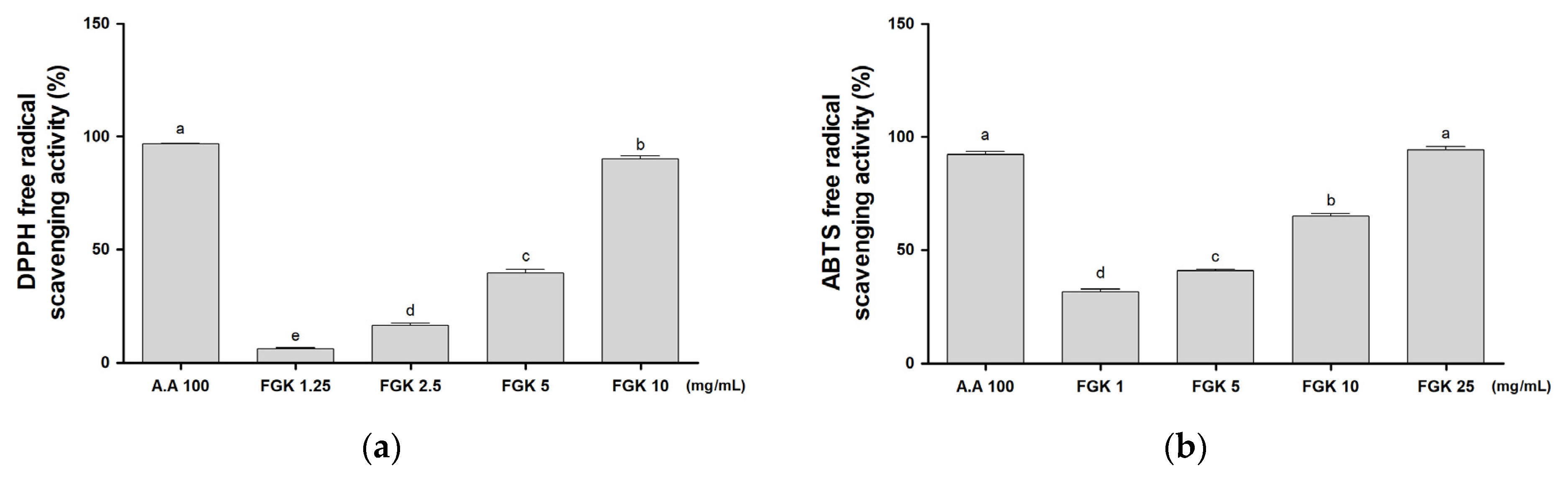
3.2. Activity of FGK on Antioxidant Effect

Among many disease conditions, the most critical oxygen-containing component is a highly reactive species in the cell membrane that damages biologically related molecules such as DNA, protein, carbohydrates, and lipids in the nucleus and cell membrane. Antioxidants delay or inhibit cellular damage, mainly through their free radical scavenging activity [28]. The DPPH/ABTS radical scavenging activities of FGK are shown in Figure 1; FGK showed dose-dependent free radical scavenging activity in both DPPH and ABTS assays, consistent with a previous study [16]. These results suggest that metabolites produced by fermentation, such as phenolics, contribute to radical scavenging activity.

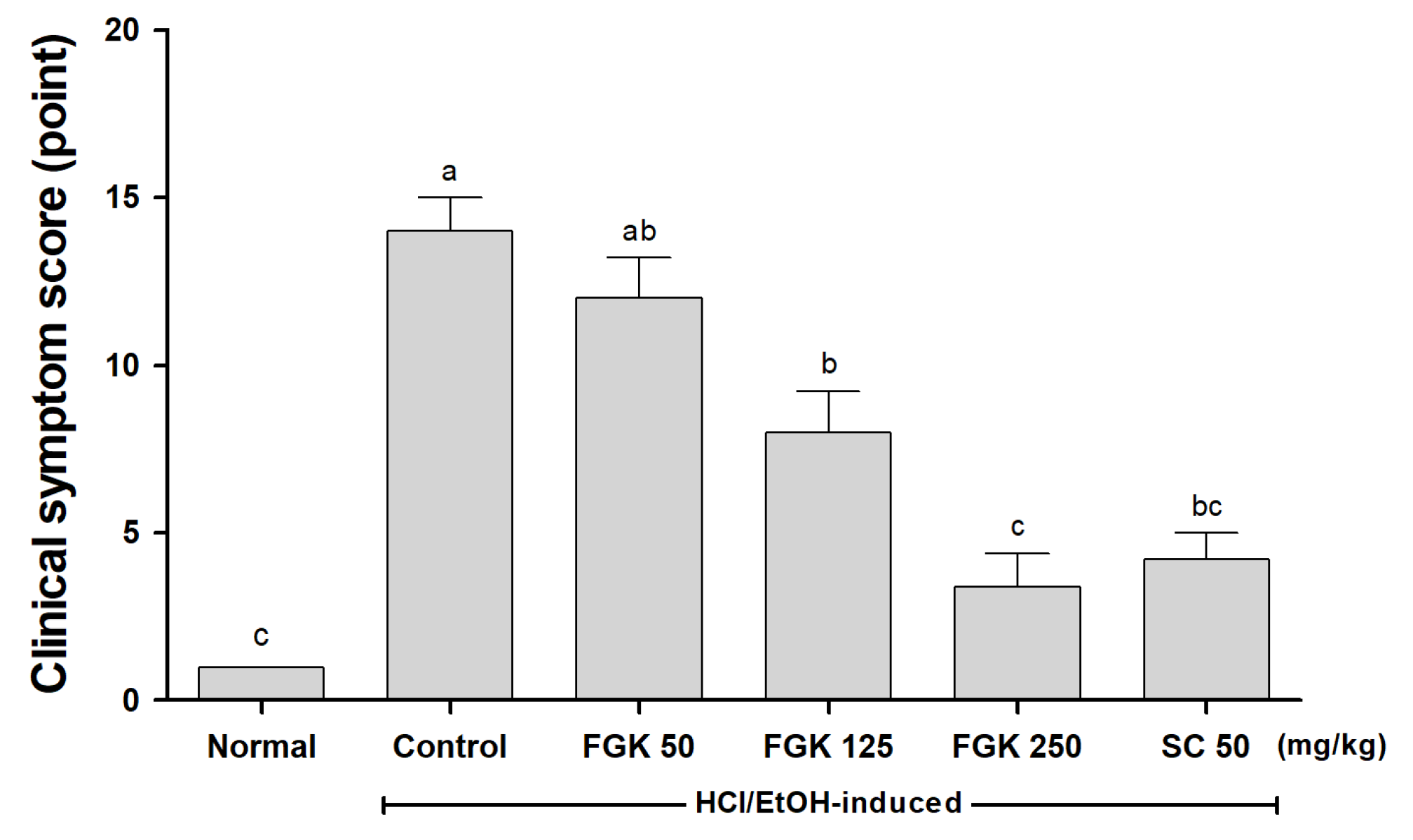
3.3. Effect of FGK on Clinical Symptoms after HCl/EtOH-Induced Gastritis Model

A gastritis animal model was obtained by oral administration of HCl/EtOH. The observed movement and clinical symptoms 30 min after administration of HCl/EtOH solution were evaluated (Figure 2). In the Normal group administered distilled water (DW) orally, rats displayed normal behaviors such as grooming and walking. In contrast, there was no movement in the Control group, which was treated with HCl/EtOH, and only slight activity in response to stimulation. Clinical symptoms decreased in a dose-dependent manner with the administration of FGK compared to the Control group.

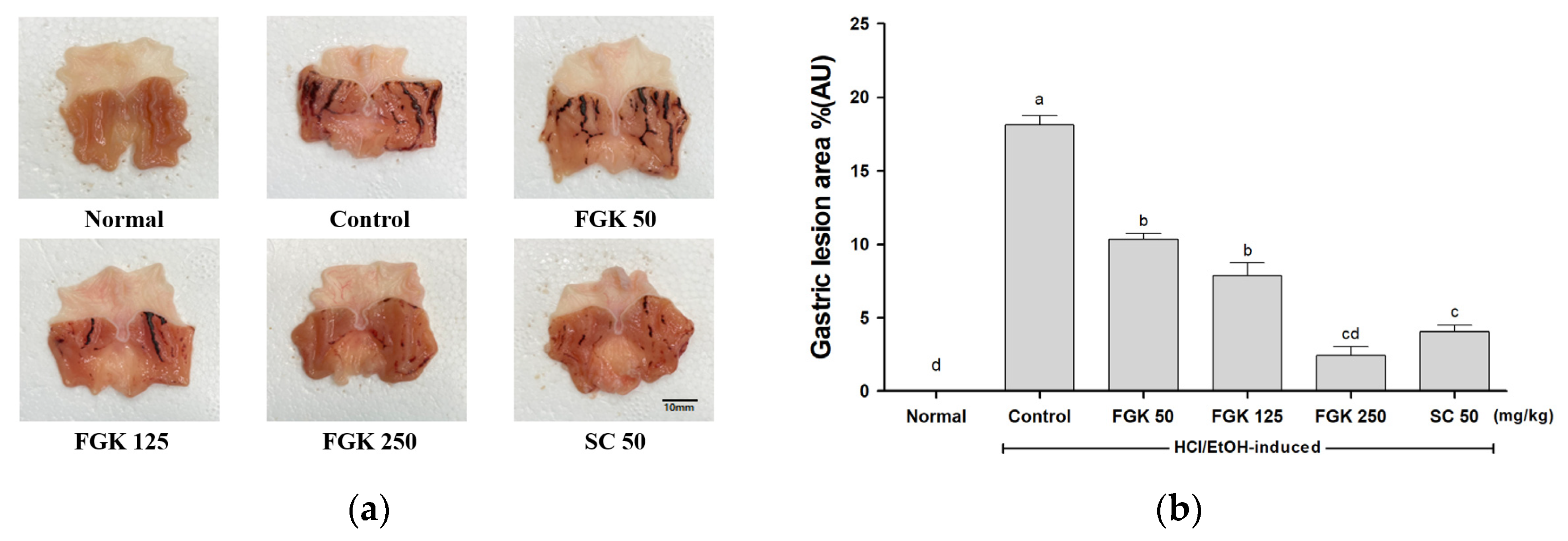
3.4. Effect of FGK on HCl/EtOH-Induced Gastric Lesions and Mucosa Damage

3.4.1. Gastric Lesions

The solution of HCl/EtOH damages the gastric mucosa and stimulates the stomach by increasing gastric motility [29]. Administration of HCl/EtOH produced extensive visible black hemorrhagic lesions and redness of the gastric mucosa. The ulcer-inhibiting effect of FGK in HCl/EtOH-induced gastric lesions is shown in Figure 3a. The area of gastric ulcer formation was significantly reduced in the group pretreated with sucralfate (SC) or FGK compared to the Control group. In the FGK group, the lesion area was controlled in a dose-dependent manner. The Control group had a significantly larger lesion area (18.14 ± 1.05 mm) than the Normal group. Meanwhile, administration of FGK at 50, 125, 250 mg/kg significantly reduced the lesion area by 10.35 ± 0.61 mm, 7.87 ± 1.52 mm, and 2.45 ± 1.00 mm, respectively, compared to the Control group. Treatment with 250 mg/kg FGK (2.45 ± 1.00 mm) had a greater gastric mucosal protective effect than treatment with SC (4.04 ± 0.75 mm) (Figure 3b).

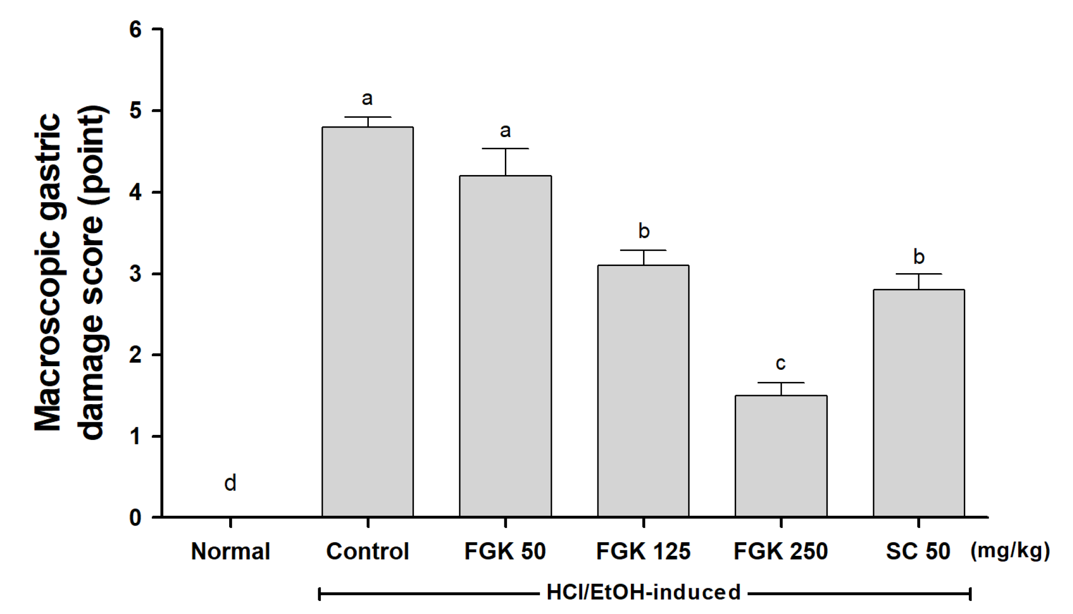
3.4.2. Gastric Mucosal Damage

In the HCl/EtOH-induced gastritis animal model, the sizes of the ulcers formed in the gastric mucosa were measured and converted into a macroscopic damage index score (Figure 4). Ulcer size was significantly higher in the Control group than the Normal group (4.8 ± 0.27 %AU vs. 0.00 ± 0.00 %AU), while ulcer size decreased significantly in the FGK groups in a dose-dependent manner. In addition, SC group rats had significantly smaller ulcers than Control group rats.

3.5. Effects of FGK on Mucosal Thickness

Histological observation of the HCl/EtOH-induced gastric lesion in the Control group revealed relatively wide-ranging gastric mucosa damage. Necrotic lesions, extensive edema, and leukocyte penetration into the submucosal layer were observed (Figure 5a). The FGK group showed relatively better gastric mucosal protection, as evidenced by the reduced ulcer area and reduced submucosal edema and leukocyte infiltration. The average gastric mucosal thickness of the Normal group was 1181.7 ± 279.3 μm. In contrast, the gastric mucosal thickness of the Control group was 491.4 ± 95.8 μm. The gastric mucosal thickness of rats that received 50, 125, or 250 mg/kg FGK was 660.30 ± 158.96 μm, 721.98 ± 143.42 μm, and 811.99 ± 145.41 μm, respectively, compared with the Control group (Figure 5b), indicating that FGK exerted cytoprotective effects in a dose-dependent manner.

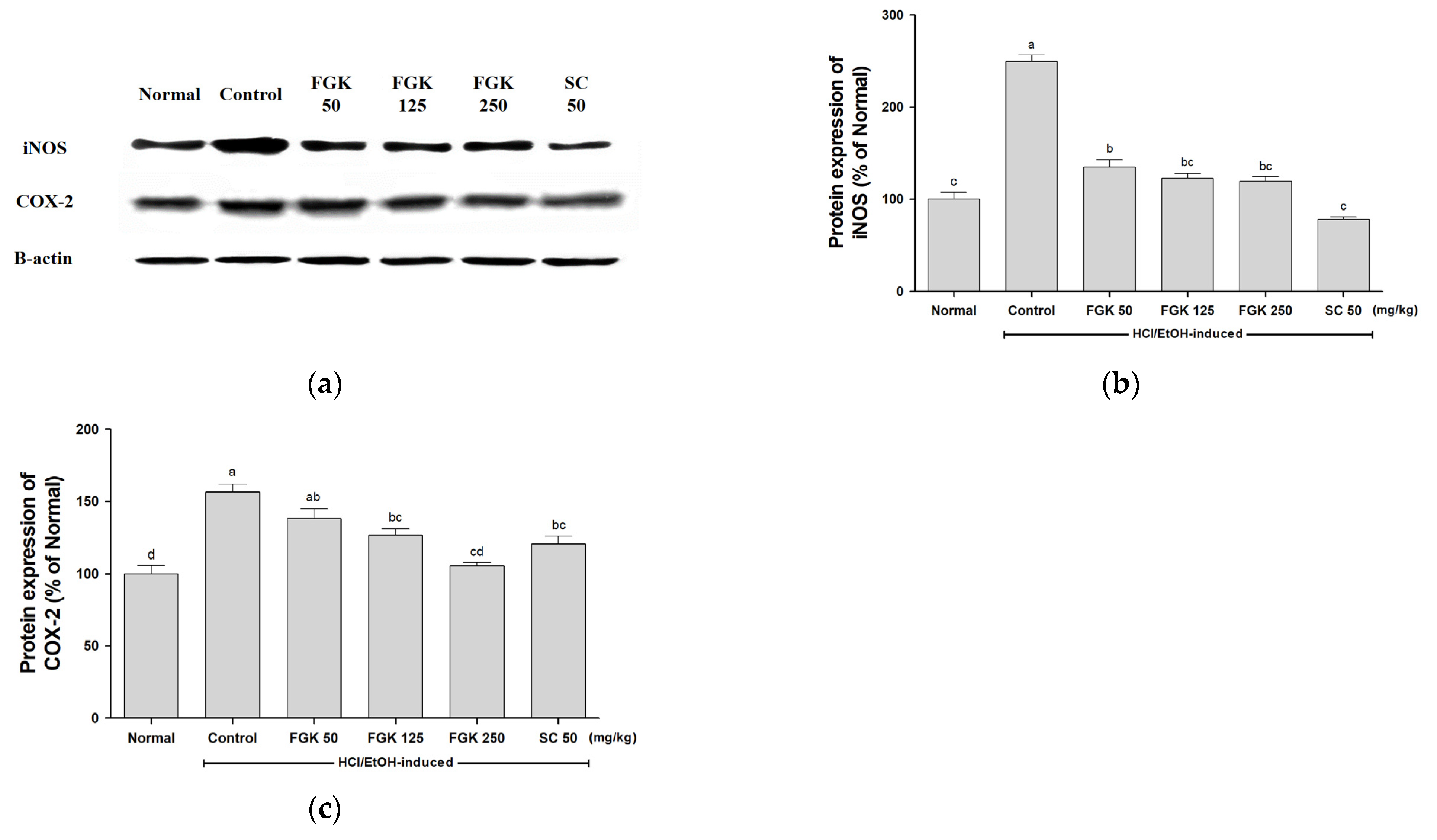
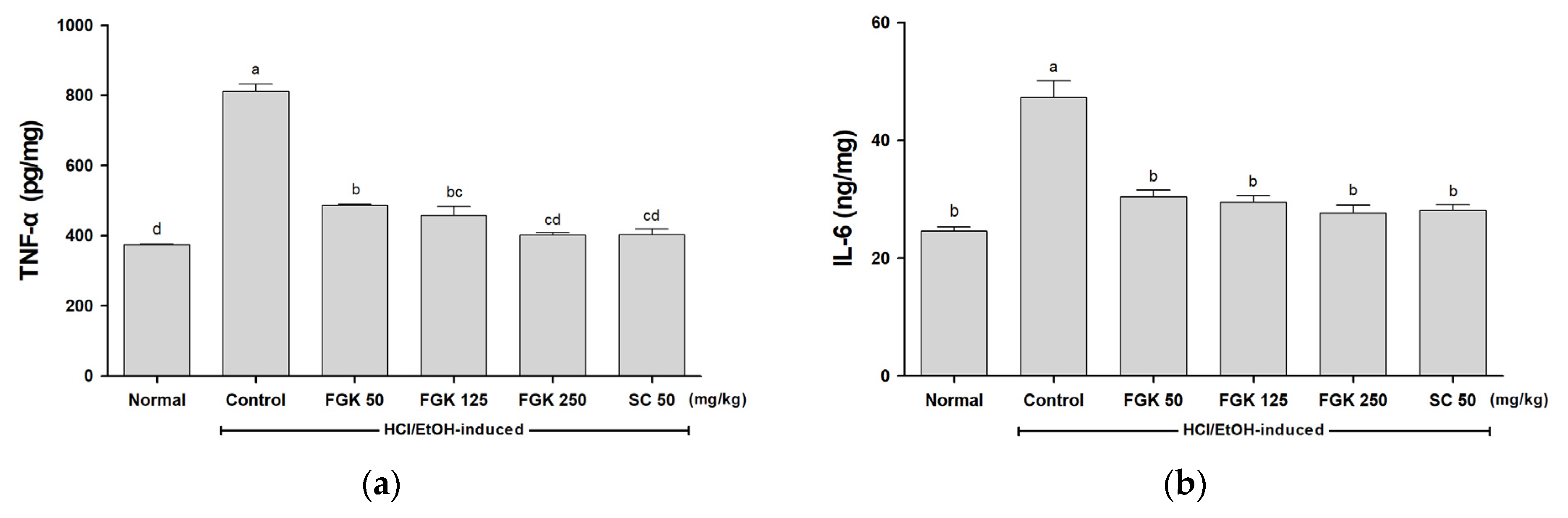
3.6. Effect of FGK on Inflammatory Factors

NF-κB activation in response to oxidative stress results in its transmigration into the nucleus and the activation of several inflammatory factors, such as iNOS and COX-2 [30]. In addition, iNOS-derived NO and PGE2 synthesized by COX-2 play a pivotal role in the pathogenesis of acute and chronic inflammation [31]. In this experiment, the expression levels of various signaling molecules involved in NF-κB activation were assessed to determine the effects of FGK.
In gastric tissue, inflammatory factors iNOS and COX-2 were measured by Western blotting, while the expression of the pro-inflammatory cytokines TNF-α and IL-6 was measured by ELISA. The results showed that the increased iNOS and COX-2 protein expression in the Control group decreased significantly in a dose-dependent manner following FGK pre-administration (Figure 6).
In addition, the concentrations of TNF-α and IL-6 increased in response to HCl/EtOH administration. TNF-α decreased significantly in a dose-dependent manner following FGK pre-administration (Figure 7a).
These results confirmed that the inhibition of inflammation by FGK was achieved through its effects on the NF-κB signaling pathway. FGK effectively reduced the expression of inflammatory factors (iNOS, COX-2) and inflammatory cytokines (TNF-α, IL-6), exerting an anti-inflammatory effect. The rats were part of a HCL-EtOH induced gastritis model, and the FGK 50 mg/kg levels could not significantly reduce acute gastritis or the expression level of COX-2. However, in the FGK 125 mg/kg intake group, a significant decrease and normalization of the inflammation value that was increased by the induction of gastritis was confirmed. In the case of FGK 250 mg/kg, a similar inflammatory relief trend to the SC50 group, which was a positive control group, was confirmed. As a result, the degree of inflammatory relief due to the concentration of FGK can be assured, suggesting that FGK administration is helpful for inflammatory relief in the acute gastritis rat model generated by HCl/EtOH administration.

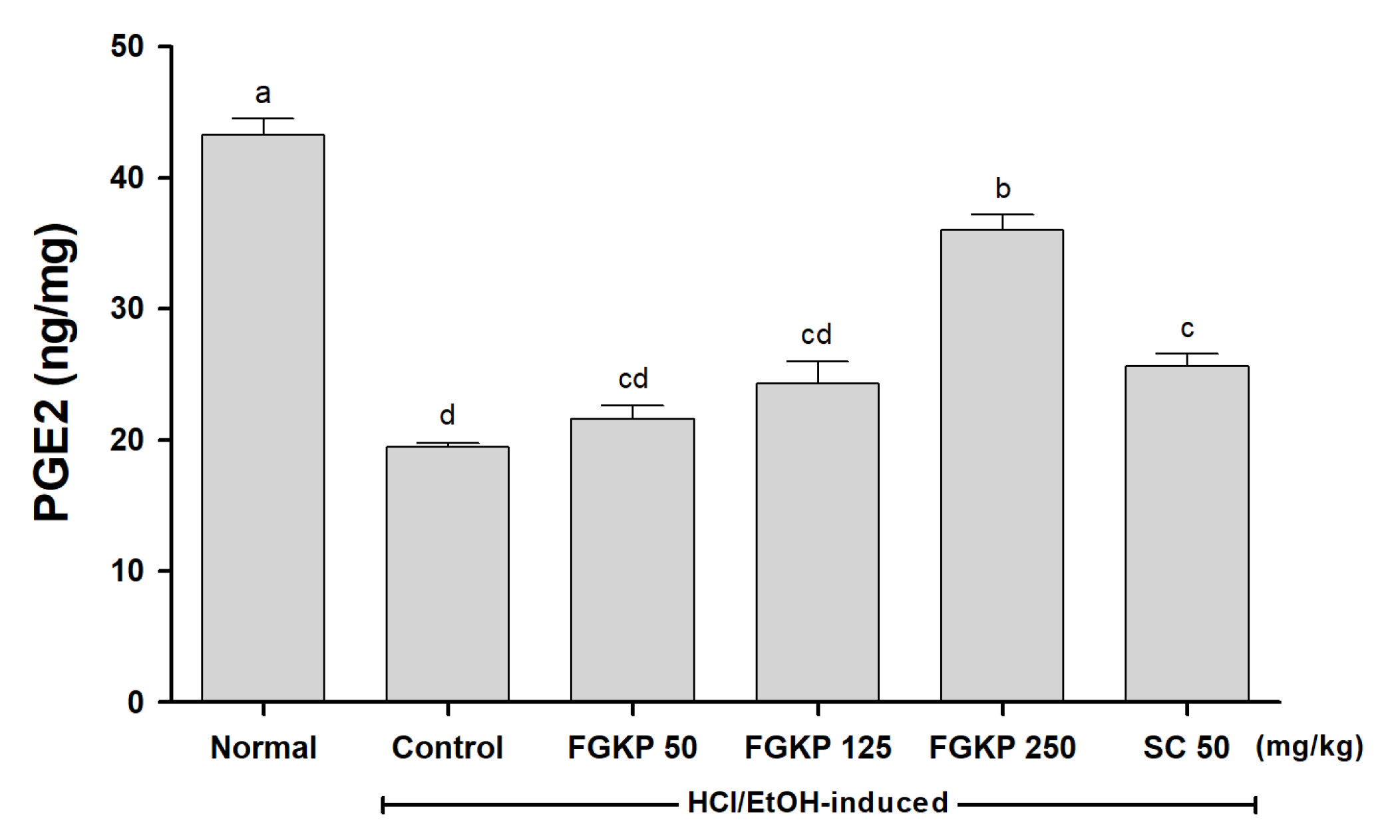
3.7. Effect of FGK on PGE2 Expression in Gastric Tissue

PGE2 has been shown to exert protective effects against various gastric injuries [32]. It is also known to play an important role in the regulation of gastric mucus secretion and can block damage to the mucous membrane caused by external stimuli [33]. The PGE2 level was 19.42 ± 0.64 ng/mg in the Control group, representing a significant reduction compared to the Normal group (43.26 ± 2.12 ng/mg) (Figure 8). However, rats that received 50, 125, or 250 mg/kg FGK showed a significant increase in PGE2 level to 21.56 ± 1.85, 24.34 ± 2.81, and 36.01 ± 2.02 ng/mg, respectively, compared to the Control group.

3.8. Effect of FGK on Gastric Secretion by Pyloric Ligation

Excessive gastric acid secretion stimulates the release of pepsin, which damages the gastric mucosa. This causes gastric damage by destroying the mucosa or increasing mucosal sensitivity to acid, congestion of microvessels, and vascular permeability [34]. Using a pyloric ligation model, we investigated the effect of FGK on gastric secretions. As a result of pyloric ligation, the Control group showed increased gastric secretions, with increased gastric juice volume, pepsin activity, free acidity, and total acidity. FGK-treated groups showed a dose-dependent decrease in these parameters. These results indicate that FGK can regulate gastric secretion in response to pyloric ligation, likely by maintaining the integrity of the gastric wall and reducing gastric ulcer formation and growth (Table 3).

4. Discussion

The gastric mucosa lining acts as a barrier against toxins such as Helicobacter infection, HCl in digestive juices, nonsteroidal anti-inflammatory agents, and alcohol [35]. Any cracks in this barrier can lead to gastric ulcers, gastritis, and even stomach cancer. If a rift occurs, the gastric acid secretion and accumulation of hydrochloric acid in the gastric mucosa are increased, the gastric mucosa itself is digested, and the gastric mucosa barrier breaks down [26,35]. Accordingly, a gastric mucosa protection effect is present if a substance suppresses or reduces in the HCl/EtOH-induced model gastric acid secretion [26].
Various fermentation processes enhance kiwi preservation, increase bioactive compound content, and increase the pharmacological activity of various antioxidant bases [36]. Lactic acid fermentation by bacteria can improve the bioavailability and the antioxidant and anti-inflammatory effects of the substance being fermented [37]. Fermentation of kiwi fruit puree powders produced using these bacteria is no exception. Gold kiwi was inoculated and fermented with two lactic acid bacteria, Lactococcus lactis VI-01 and Lactobacillus paracase VI-02, extracted, freeze-dried, and powdered for use in the study. As a result of analyzing the organic acid, total flavonoid, and carotenoid content of fermented gold kiwi (FGK) and general gold kiwi (GK), the active compounds of kiwi increased after fermentation (Table 3). In this study, three different doses of FGK (50, 125, and 250 mg/kg) were orally administered 1 h before the 150 mM HCl/60% EtOH solution treatment. The rats were measured for clinical symptoms after 30 min of HCl/EtOH solution treatment. After 1 h, changes in total hemorrhagic disease score, histopathology, and inflammatory markers were observed and all results were compared with 50 mg/kg of sucralfate.
HCl/EtOH-induced and pyloric ligation models are widely used to induce gastritis or gastric ulcers, as damage to the gastric mucosa and gastric wall can be observed by the unaided eye [38,39]. In the HCl/EtOH-induced gastric ulcer model, HCl causes severe damage to the gastric mucosa. Ethanol causes direct necrosis in the gastric mucosa and wall, resulting in a characteristic necrosis lesion due to reduced mucus formation and bicarbonate secretion [40]. Therefore, we adopted these two models to assess the gastric protective effects of FGK. Oral administration of FGK significantly reduced gastric lesions induced by HCl/EtOH and pyloric ligation. Rats treated with HCl/EtOH only showed a significant increase in redness and damage to the gastric mucosa (Figure 3, Figure 4 and Figure 5). FGK administration significantly reduced gastric juice volume, pepsin activity, free acidity, and total acidity in pyloric ligation rats (Table 3). These results demonstrate that FGK improved gastritis and protected against gastric ulcers.
Although the precise pathogenesis of gastritis has not been elucidated, the formation of reactive oxygen species (ROS) from ethanol has been shown to contribute to gastritis [40,41]. Excessive production of ROS causes oxidative stress, which causes various diseases [42,43]. A small amount of ROS generated in the stomach is quickly removed through the actions of various antioxidant enzymes and antioxidants. However, excess ROS can cause oxidative stress and damage to the gastric mucosa by overwhelming the antioxidant defenses [44,45]. Scavenging of free radicals plays a significant role in reducing or curing ulcers [46]. Kiwi fruit contains antioxidants such as ascorbic acid, carotenoids, lutein, and flavonoids that protect against oxidative stress by removing free radicals [47]. We demonstrated in this study that FGK showed intense radical scavenging activity in a dose-dependent manner, consistent with a previous study (Figure 1) [48]. This indicates that FGK can help to protect tissues from oxidative damage.
Our study also evaluated that pre-treatment with FGK prevented inflammatory conditions induced by HCl/EtOH acute gastritis. Injection of HCl/EtOH induces mucosal damage to the stomach, recognizes the damage-associated molecular patterns, and causes the subsequent stimulation of inflammatory cytokines such as IL-6 and TNF-α [49]. This activation of inflammatory cytokines appears as an activation of NF-κB. When NF-κB is activated in response to oxidative stress, it migrates into the nucleus and activates inflammatory factors and cytokines such as iNOS, COX-2, TNF-α, and IL-6 [30]. In addition, iNOS-derived NO and PGE2 synthesized by COX-2 play a pivotal role in acute and chronic gastric inflammation [31]. As expected, FGK treatment reduces the inflammatory response and prevents gastric mucosa protection at a similar level to the standard gastric ulcer treatment, sucralfate. These inflammatory cytokines promote gastric mucosal damage and delay the healing of gastric ulcers and gastritis by downregulating the enzymatic antioxidant defenses of the gastric mucosa [50]. Our results confirmed that the levels of iNOS, COX-2 overexpressed by gastritis induction, and increased TNF-α and IL-6 were adjusted from FGK 250 levels to levels similar to the Control groups.
PGE2, which is strongly associated with gastritis, has been reported to play a role in gastric homeostasis by regulating blood circulation and the epithelial cell motility of the gastric mucosa and inhibiting gastric mucus and gastric acid secretion [51,52]. Ethanol reduced the mucosa PGE concentration, and PGE2 is the most abundant gastrointestinal prostaglandin, regulating its function in internal organs, including motility and secretion [52]. For this reason, PGE2 is commonly used as a biomarker to evaluate the preventive effects of various compounds on gastritis [53,54]. In this study, the gastric protective effect of FGK was confirmed by observing the increased production of PGE2 in the gastric mucosa, suggesting that PGE2 partially mediates the gastric protective effect of FGK. Prostaglandin affects almost all elements of mucosal protection, including promoting mucus and bicarbonate secretion, maintaining mucus blood flow, improving epithelial cell resistance to cytotoxic damage, and inhibiting leukocyte recruitment [53,55].
Finally, we used a pyloric ligation model to evaluate the effects of FGK on gastric acid secretion. Pyloric ligation effectively increases gastric secretions and hydrochloric acid accumulation in the gastric mucosa [56]. Excessive gastric acid and pepsin secretion cause mucosal breakdown and disrupt the gastric mucosal barrier [57]. In this study, FGK administration significantly and dose-dependently reduced the gastric juice volume, pepsin activity, free acidity, and total acidity in pyloric ligation rats (Table 3). These results suggest that FGK protects the gastric mucosa by inhibiting gastric acid secretion.

5. Conclusions

In the present study, we showed that FGK had a gastroprotective effect in rats with gastric disease lesions. FGK increased antioxidant activity, and secretion of the defense factor PGE2 decreased the expression of inflammatory factors (iNOS, COX-2, TNF-α, and IL-6) and inhibited gastric acid secretion. In addition, FGK effectively suppressed clinical symptoms, gastric mucosal damage, and the reduction in mucosal thickness induced by acid/ethanol treatment. These results demonstrate that FGK helps to protect the gastric mucosa and is a promising candidate for the prevention and treatment of gastritis and gastric ulcers. The increase in the physiological activity of the extract by bacterial fermentation should be verified through additional research.

Author Contributions

J.K. conceived and designed the study. E.-J.J., J.-H.C., N.-Y.L., H.-J.O., H.-S.K. and J.K. conducted the experiments and analyzed the data. E.-J.J., J.-H.C. and J.K. wrote the manuscript. All authors have read and agreed to the published version of the manuscript.

Funding

This research received no external funding.

Institutional Review Board Statement

This study was approved by the Jeonbuk National University Institutional Animal Care and Use Committee (JBNU 2021-064) and conducted according to the guidelines for animal experiments in the protocol.

Informed Consent Statement

Not applicable.

Data Availability Statement

Not applicable.

Acknowledgments

This research was supported by Vitech Co., Ltd.

Conflicts of Interest

The authors declare no conflict of interest.

References

  1. Shay, H. A simple method for the uniform production of gastric ulceration in the rat. Gastroenterology 1945, 5, 43–45. [Google Scholar]
  2. Banks, W.J. Applied Veterinary Histology, 2nd ed.; William& Winkins Baltimore: Philadelphia, PA, USA, 1986; pp. 393–396. [Google Scholar]
  3. Gürsan, N. Effects of Momordica charantia L. (Cucurbitaceae) on indomethacin-induced ulcer model in rats. Turk. J. Gastroenterol. 2005, 16, 85–88. [Google Scholar]
  4. Feldman, M.; Burton, M.E. Histamin 2-receptor antagonists: Standard therapy for acid peptic diseases. N. Engl. J. Med. 1990, 323, 1672–1680. [Google Scholar]
  5. Johnson, D.A.; Oldfield, E.C. Reported side effects and complications of long-term proton pump inhibitor use: Dissecting the evidence. Clin. Gastroenterol. Hepatol. 2013, 11, 458–464. [Google Scholar] [CrossRef] [PubMed]
  6. Szabo, S.; Bynum, T.E. Alternatives to the acid-oriented approach to ulcer disease: Dose ‘Cytoprotection’ exist in man?: A new classification of antiulcer agents. Scand. J. Gastroenterol. 1988, 23, 1–6. [Google Scholar] [CrossRef] [PubMed]
  7. Haga, Y.; Nakatsura, T.; Shibata, Y.; Sameshima, H. Human gastric carcinoid detected during long-term antiulcer therapy of H2 receptor antagonist and proton pump inhibitor. Dig. Dis. Sci. 1998, 43, 253. [Google Scholar] [CrossRef] [PubMed]
  8. Karunakaran, K.; Thabrew, M.I.; Thammitiyagodage, G.M.; Galhena, B.P.; Arawwawala, L.M. The gastroprotective effect of ethyl acetate fraction of hot water extract of Trichosanthes cucumerina Linn and its underlying mechanisms. BMC Complement. Altern. Med. 2017, 17, 312. [Google Scholar] [CrossRef] [Green Version]
  9. Montefiori, M.; McGhie, T.K.; Costa, G.; Ferguson, A.R. Pigments in the fruit of red-fleshed kiwifruit (Actinidia chinensis and Actinidia deliciosa). J. Agric. Food Chem. 2005, 53, 9526–9530. [Google Scholar] [CrossRef]
  10. Jeong, C.H.; Lee, W.J.; Bae, S.H.; Choi, S.G. Chemical components and antioxidative activity of Korean glod kiwifruit. J. Korean Soc. Food Sci. Nutr. 2007, 36, 859–865. [Google Scholar] [CrossRef]
  11. Motohashi, N.; Shirataki, Y.; Kawase, M.; Tani, S.; Sakagami, H.; Satoh, K.; Kurihara, T.; Nakashima, H.; Mucsi, I.; Varga, A.; et al. Cancer prevention and therapy with kiwifruit in Chinese folklore medicine: A study of kiwifruit extracts. J. Ethnopharmacol. 2002, 81, 357–364. [Google Scholar] [CrossRef]
  12. Rush, E.C.; Patel, M.; Plank, L.D.; Ferguson, L.R. Kiwifruit promotes laxation in the elderly. Asia Pac. J. Clin. Nutr. 2002, 11, 164–168. [Google Scholar] [CrossRef] [PubMed]
  13. Park, Y.S.; Namiesnik, J.; Vearasilp, K.; Leontowicz, H.; Leontowicz, M.; Barasch, D.; Nemirovski, A.; Trakhtenberg, S.; Gorinstein, S. Bioactive compounds and the antioxidant capacity in new kiwi fruit cultivars. Food Chem. 2014, 165, 354–361. [Google Scholar] [CrossRef] [PubMed]
  14. Hussein, J.; El-Matty, D.A.; El-Khayat, Z.; Latif, Y.A.; Saleh, S.; Farrag, A.R.; Abd-El-Ghany, W. Kiwifruit extract attenuates DNA damage and vitamins reduction in indomethacin-induced experimental gastric ulcer. Jokull J. 2015, 65, 2–16. [Google Scholar]
  15. Park, K.L.; Hong, S.W.; Kim, Y.J.; Kim, S.J.; Chung, K.S. Manufacturing and physicochemical of wine using hardy kiwi fruit (Actinidia argute). Korean J. Microbiol. Biotechnol. 2013, 41, 327–334. [Google Scholar] [CrossRef]
  16. Ryu, J.Y.; Park, H.J.; Moon, J.Y.; Kim, C.S.; Somi, K. Lactic fermentation enhances the antioxidant activity of gold kiwifruit. Korean J. Food Preserv. 2018, 25, 255–262. [Google Scholar] [CrossRef]
  17. Chen, A.J.; Fu, Y.Y.; Jiang, C.; Zhao, J.L.; Liu, X.P.; Liu, L.; Zhang, Z.Q. Effect of mixed fermentation (Jinqu and Saccharomyces cerevisiae EC1118) on the quality improvement of kiwi wine. CyTA—J. Food 2019, 17, 967–975. [Google Scholar] [CrossRef] [Green Version]
  18. Bhat, R.; Suryanarayana, L.C.; Chandrashekara, K.A.; Krishnan, P.; Kush, A.; Ravikumar, P. Lactobacillus plantarum mediated fermentation of Psidium huajava L. fruit extract. J. Biosci. Bioeng. 2015, 119, 430–432. [Google Scholar] [CrossRef]
  19. Ferssard, A.; Kapoor, A.; Patche, J.; Assemat, S.; Hoarau, M.; Bourdon, E.; Bahorun, T.; Remize, F. Lactic fermentation as an efficient tool to enhance the antioxidants and fuctional foods: Impact on human health. Pharmacogn. Rev. 2010, 4, 118–126. [Google Scholar]
  20. Drummong, L. The composition and nutritional value of kiwifruit. Adv. Food Nutr. Res. 2013, 68, 33–57. [Google Scholar]
  21. Huang, J.; Wang, Y.; Ren, Y.; Wang, X.; Li, H.; Liu, Z.; Yue, T.; Gao, Z. Effect of inoculation method on the quality and nutritional characteristics of low-alcohol kiwi wine. LWT 2022, 156, 113049. [Google Scholar] [CrossRef]
  22. Sharma, O.P.; Bhat, T.K. DPPH antioxidant assay revisited. Food Chem. 2009, 113, 1202–1205. [Google Scholar] [CrossRef]
  23. Huang, D.; Ou, B.; Prior, R.L. The chemistry behind antioxidant capacity assay. J. Agric. Food Chem. 2005, 53, 1841–1856. [Google Scholar] [CrossRef] [PubMed]
  24. Hong, S.; Lee, H.A.; Lee, Y.S.; Kim, D.W.; Oh, G.W.; Woo, J.; Cho, Y.; Jeong, J.H.; Kim, O. Protective effect of halophyte Salsola komarovi Iljin against gastric ulcer induced by alcohol treatment in rats. J. Biomed. Res. 2014, 15, 170–175. [Google Scholar] [CrossRef]
  25. Cantarella, G.; Martinez, G.; Cutuli, V.M.; Loreto, C.; D’Alcamo, M.; Prato, A.; Amico-Roxas, M.; Bernardini, R.; Clementi, G. Adrenomedullin modulates COX-2 and HGF expression in reserpine-injuried gastric mucosa in the rat. Eur. J. Pharmacol. 2005, 518, 221–226. [Google Scholar] [CrossRef] [PubMed]
  26. Kim, Y.S.; Park, H.J.; Kim, H.; Song, J.; Lee, D. Gastroprotective effects of paeonia extract mixture HT074 against experimental gastric ulcers in rats. Evid. Based Complement. Alternat. Med. 2019, 2019, 3546258. [Google Scholar] [CrossRef] [PubMed]
  27. Anson, M.L. The estimation of pepsin, trypsin, papain, and cathepsin with hemoglobin. J. Gen. Physiol. 1938, 22, 79–89. [Google Scholar] [CrossRef]
  28. Lobo, V.; Patil, A.; Phatak, A.; Chandran, N. Free radicals, antioxidants and functional foods: Impact on human health. Pharmacogn. Rev. 2010, 4, 118–126. [Google Scholar] [CrossRef] [Green Version]
  29. Ito, M.; Shii, D.; Segami, T.; Kojima, R.; Suzuki, Y. Preventive actions of N-(3-aminopropionyl)-l-histidinato zinc (Z-103) through increases in the activities of oxygen-derived free radical scavenging enzymes in the gastric mucosa on ethanol-induced gastric mucosal damage in rats. Japan J. Pharmacol. 1992, 59, 267–274. [Google Scholar] [CrossRef]
  30. Yamamoto, Y.; Gaynor, R.B. Role of the NF-κB pathway in the pathogenesis of human disease states. Curr. Mol. Med. 2001, 1, 287–296. [Google Scholar] [CrossRef]
  31. Han, M.; Wen, J.K.; Zheng, B.; Zhang, D.Q. Acetylbritannilatone suppresses NO and PGE2 synthesis in RAW 264.7 macrophages through the inhibition of iNOS and COX-2 gene expression. Life Sci. 2004, 75, 675–684. [Google Scholar] [CrossRef]
  32. Brzozowski, T.; Konturek, P.C.; Konturek, S.J.; Pawlik, T. Role of prostaglandins in gastroprotection and gastric adaptation. J. Physiol. Pharmacol. 2005, 56, 53–55. [Google Scholar]
  33. Hoshino, T.; Tsutsumi, S.; Tomisato, W.; Hwang, H.J.; Tsuchiya, T.; Mizushima, T. Prostaglandin E2 protects gastric mucosal cells from apoptosis via EP2 and EP4 receptor activation. J. Biol. Chem. 2003, 278, 12752–12758. [Google Scholar] [CrossRef] [PubMed] [Green Version]
  34. Wang, X.Y.; Yin, J.Y.; Zhao, M.M.; Liu, S.Y.; Nie, S.P.; Xie, M.Y. Gastroprotective activity of polysaccharide from Hericium erinaceus against ethanol-induced gastric mucosal lesion and pylorus ligation-induced gastric ulcer, and its antioxidant activities. Carbohydr. Polym. 2018, 186, 100–109. [Google Scholar] [CrossRef] [PubMed]
  35. Zhang, Y.; Sun, L.; Lai, X.; Peng, X.; Wen, S.; Zhang, Z.; Xie, Y.; Li, Q.; Chen, R.; Zheng, X.; et al. Gastroprotective effects of extract of Jasmunum grandiflorum L. flower in HCl/EtOH-induced gastric mucosal ulceration mice. Biomed. Pharmacother. 2021, 144, 112268. [Google Scholar] [CrossRef] [PubMed]
  36. Lim, J.M.; Song, C.H.; Park, S.J.; Park, D.C.; Cho, H.R.; Jung, G.W.; Bashir, K.M.B.; Ku, S.K.; Choi, J.S. Protective effects of a triple-fermented barley extract (FBe) against HCl/EtOH-induced gastric mucosa damage in mice. Food Sci. Nutr. 2018, 6, 2036–2046. [Google Scholar] [CrossRef]
  37. Parvez, S.; Malik, K.A.; Kang, S.A.; Kim, H.Y. Probiotics and their fermented food products are beneficial for health. J. Appl. Microbiol. 2006, 100, 1171–1185. [Google Scholar] [CrossRef]
  38. Hariprasath, L.; Raman, J.; Nanjian, R. Gastroprotective effect of Senecio candicans DC on experimental ulcer models. J. Ethnopharmacol. 2012, 140, 145–150. [Google Scholar] [CrossRef]
  39. Berté, P.E.; Lopes, J.S.; Comandulli, N.G.; Rangel, D.W.; Monache, F.D.; Filho, V.C.; Siero, R.; Andrade, S.F. Evaluation of the gastroprotective activity of the extracts, fractions, and pure compounds obtained from aerial parts of Rubus imperialis in different experimental models. Naunyn. Schmiedeberg Arch. Pharmacol. 2014, 387, 313–319. [Google Scholar] [CrossRef]
  40. Bagchi, D.; Carryl, O.R.; Tran, M.X.; Krohn, R.L.; Bagchi, D.J.; Garg, A.; Bagchi, M.; Mitra, S.; Stohs, S.J. Stress, diet and alcohol-induced oxidative gastrointestinal mucosal injury in rats and protection by bismuth subsalicylate. J. Appl. Toxicol. 1998, 18, 3–13. [Google Scholar] [CrossRef]
  41. Raish, M.; Ahmad, A.; Ansari, M.A.; Alkharfy, K.; Alijenoobi, F.; Jan, B.L.; Al-Mohizea, A.M.; Khan, A.; Ali, N. Momordica charantia polysaccharide ameliorate oxidative stress, inflammation, and apoptosis in ethanol-induced gastritis in mucosa through NF-κB signaling pathway inhibition. Int. J. Biol. Macromol. 2018, 111, 193–199. [Google Scholar] [CrossRef]
  42. Halliwell, B.; Aeschbach, R.; Löliger, J.; Aruoma, O.I. The characterization of antioxidants. Food Chem Toxicol 1995, 33, 601–617. [Google Scholar] [CrossRef]
  43. Suzuki, H.; Nishizawa, T.; Tsugawa, H.; Mogami, S.; Hibi, T. Roles of oxidative stress in stomach disorders. J. Clin. Biochem. Nutr. 2015, 50, 35–39. [Google Scholar] [CrossRef] [PubMed] [Green Version]
  44. Pérez, S.; Taléns-Visconti, R.; Rius-Pérez, S.; Finamor, I.; Sastre, J. Redox signaling in the gastrointestinal tract. Free Radic. Biol. Med. 2017, 104, 75–103. [Google Scholar] [CrossRef] [PubMed]
  45. Dordević, S.; Petrović, S.; Bobrić, S.; Milenković, M.; Vucićević, D.; Zizić, S.; Kukić, J. Antimicrobial, anti-inflammatory, anti-ulcer and antioxidant activities of Carlina acanthifolia root essential oil. J. Ethnopharmacol. 2007, 109, 458–463. [Google Scholar] [CrossRef]
  46. Jayakumari, S.; Anbu, J.; Ravichandiran, V.; Anjana, A.; Kumar, G.M.; Singh, M. Antiulcerogenic and free radical scavenging activity of flavonoid fraction of Psidium guajava Linn leaves. Int. J. Pharm. Pharm. Sci. 2012, 4, 170–174. [Google Scholar]
  47. Manal, M.S.M.M.; Soltan, S.S. Effects of bioactive component of kiwi fruit and avocado (fruit and seed on hypercholesterolemic rats. World J. Dairy Food Sci. 2013, 8, 82–93. [Google Scholar]
  48. Park, Y.S.; Im, M.H.; Ham, K.S.; Kang, S.G.; Park, Y.K.; Namiesnik, J.; Leontowicz, H.; Leontowicz, M.; Trankhtenberg, S.; Gorinstein, S. Quantitative assessment of the main antioxidant compounds, antioxidant activities and FTIR spectra from commonly consumed fruits, compared to standard kiwi fruit. LWT-Food Sci. Technol. 2015, 63, 346–352. [Google Scholar] [CrossRef]
  49. Rahmawati, L.; Aziz, N.; Oh, J.; Hong, Y.H.; Woo, B.Y.; Hong, Y.D.; Manilack, P.; Souladeth, P.; Jung, J.H.; Lee, W.S.; et al. Cissus subtetragona Planch. Ameliorates inflammatory response in LPS-induced macrophages, HCl/EtOH-induced gastritis, and LPS-induced lug injury via attenuation of Src and TAK1. Molecules 2021, 26, 6073. [Google Scholar] [CrossRef]
  50. Yang, Y.; Yin, B.; Lv, L.; Wang, Z.; He, J.; Chen, Z.; Wen, X.; Zhang, Y.; Sun, W.; Li, Y.; et al. Gastroprotective effect of aucubin against ethanol-induced gastric mucosal injury in mice. Life Sci. 2017, 189, 44–51. [Google Scholar] [CrossRef]
  51. Ricciotti, E.; FitzGerald, G.A. Prostaglandins and inflammation. Arterioscler. Thromb. Vasc. Biol. 2011, 31, 986–1000. [Google Scholar] [CrossRef]
  52. Wallace, J.L. Prostaglandins, NSAIDs, and cytoprotection. Gastroenterol. Clin. N. Am. 1992, 21, 631–641. [Google Scholar] [CrossRef]
  53. Alrashdi, A.S.; Salama, S.M.; Alkiyumi, S.S.; Abdulla, M.A.; Hadi, A.H.A.; Abdelwahab, S.I.; Taga, M.M.; Hussiani, J.; Asykin, N. Mechanisms of gastroprotective effects of ethanolic leaf extracts of Jasminum sambac against HCl/Ethanol-induced gastric mucosal injury in rats. Evid. Based Complement. Alternat. Med. 2012, 2012, 786426. [Google Scholar] [CrossRef] [PubMed] [Green Version]
  54. Jeon, W.Y.; Lee, M.Y.; Shin, I.S.; Jin, S.E.; Ha, H. Curcuma aromatic water extract attenuates ethanol-induced gastritis via enhancement of antioxidant status. Evid.-Based Complement. Alternat. Med. 2015, 2015, 582496. [Google Scholar] [CrossRef] [PubMed] [Green Version]
  55. Ishihara, K.; Kuwata, H.; Ohara, S.; Okabe, H.; Hotta, K. Changes of rat gastric mucus glycoproteins in cytoprotection: Influences of prostaglandin derivatives. Digestion 1988, 39, 162–171. [Google Scholar] [CrossRef]
  56. Blair, D.W.; Williams, M.J.; Carr, A.J.; Kilpatrick, S.J. Effect of L-thyroxine on gastric secretion in the pylorus-ligated rat. Gut 1965, 6, 343–348. [Google Scholar] [CrossRef] [Green Version]
  57. Sahoo, S.K.; Sahoo, H.B.; Priyadarshini, D.; Soundarya, G.; Kuma, C.K.; Rani, K.U. Antiulcer activity ethanolic extracts of Salvadora indica (W.) leaves on Albino rats. J. Clin. Diagn Res. 2016, 10, FF07–FF10. [Google Scholar] [CrossRef]
Figure 1. Antioxidant effects of fermented gold kiwi (FGK) extracts in 2,2-diphenyl-1-picrylhydrazyl (DPPH) and 2,2′-azino-di-(3-ethylbenzothiazoline)-6-sulfonic acid (ABTS) assays. (a) DPPH free radical scavenging activity; (b) ABTS free radical scavenging activity. Ascorbic acid (A.A) was used as a standard. All results were acquired with three replicates to derive an average value. Results are expressed as mean ± SEM. a–e Different labels were significantly different at p < 0.05, as determined by Tukey’s analysis.
Applsci 12 05271 g001
Figure 2. Effect of fermented gold kiwi (FGK) extract on clinical symptoms in HCl/EtOH-induced rats. Normal, rats treated with only DW; Control, rats treated with HCl/EtOH; SC 50, rats treated with HCl/EtOH and sucralfate 50 mg/kg; FGK, rats treated with HCl/EtOH and FGK (50, 125, and 250 mg/kg). Results are expressed as mean ± SEM (N = 8). a–c Different labels were significantly different at p < 0.05 between treatments, as determined by Tukey’s analysis.
Applsci 12 05271 g002
Figure 3. Effects of fermented gold kiwi (FGK) on gastric surface protection in HCl/EtOH-induced gastric injury models. (a) Gross appearance of stomach tissues in HCl/EtOH-induced rats; (b) ratios of hemorrhagic lesion area to total area. Normal, rats treated with only DW; Control, rats treated with HCl/EtOH; SC 50, rats treated with HCl/EtOH and sucralfate; FGK, rats treated with HCl/EtOH and FGK (50, 125, and 250 mg/kg). Results are expressed as mean ± SEM (N = 8). a–d Different labels were significantly different at p < 0.05 between treatments, as determined by Tukey’s analysis.
Applsci 12 05271 g003
Figure 4. Effects of fermented gold kiwi (FGK) on gastric surface protection in HCl/EtOH-induced gastric injury models. Size of the ulcers that formed in the gastric mucosa was measured, and the sum of sizes was converted into a macroscopic damage score. Normal, rats treated with only DW; Control, rats treated with HCl/EtOH; SC 50, rats treated with HCl/EtOH and sucralfate; FGK, rats treated with HCl/EtOH and FGK (50, 125, and 250 mg/kg). Results are expressed as mean ± SEM (N = 8). a–d Different labels were significantly different at p < 0.05 between treatments, as determined by Tukey’s analysis.
Applsci 12 05271 g004
Figure 5. Effects of fermented gold kiwi (FGK) on HCl/EtOH-induced gastric lesion histology. (a) Histological sections of gastric mucosa in HCl/EtOH-induced rats; (b) effects of FGKP on periulcerative mucosal thickness in HCl/EtOH rats. Normal, rats treated with only DW; Control, rats treated with HCl/EtOH; SC 50, rats treated with HCl/EtOH and sucralfate; FGK, rats treated with HCl/EtOH and FGK (50, 125, and 250 mg/kg). Results are expressed as mean ± SEM (N = 8). a–c Different labels were significantly different at p < 0.05 between treatments, as determined by Tukey’s analysis.
Applsci 12 05271 g005
Figure 6. Effect of fermented gold kiwi (FGK) on protein expression in stomach tissues of HCl/EtOH-induced rats. (a) Protein expression; (b) iNOS/β-actin percentage of normal; (c) COX-2/β-actin percentage of normal. Normal, rats treated with only DW; Control, rats treated with HCl/EtOH; SC 50, rats treated with HCl/EtOH and sucralfate; FGK, rats treated with HCl/EtOH and FGK (50, 125, and 250 mg/kg). Results are expressed as mean ± SEM (N = 8). a–d Different labels were significantly different at p < 0.05 between treatments, as determined by Tukey’s analysis.
Applsci 12 05271 g006
Figure 7. Effects of fermented gold kiwi (FGK) on pro-inflammatory cytokine concentrations in the stomach tissues of HCl/EtOH-induced rats: (a) TNF-α and (b) IL-6. Normal, rats treated with only DW; Control, rats treated with HCl/EtOH; SC 50, rats treated with HCl/EtOH and sucralfate; FGK, rats treated with HCl/EtOH and FGK (50, 125, and 250 mg/kg). Results are expressed as mean ± SEM (N = 8). a–d Different labels were significantly different at p < 0.05 between treatments, as determined by Tukey’s analysis.
Applsci 12 05271 g007
Figure 8. Effect of fermented gold kiwi (FGK) on PGE2 expression in the stomach tissues of HCl/EtOH-induced rats. Normal, rats treated with only DW; Control, rats treated with HCl/EtOH; SC 50, rats treated with HCl/EtOH and sucralfate; FGK, rats treated with HCl/EtOH and FGK (50, 125, and 250 mg/kg). Results are expressed as mean ± SEM (N = 8). a–d Different labels were significantly different at p < 0.05 between treatments, as determined by Tukey’s analysis.
Applsci 12 05271 g008
Table 1. Macroscopic damage score.
Score (Points)Symptom
0No lesion
0.5Diffuse hyperemia
1.51 to 2 small erosions
23 to 6 small erosions
2.510 or more small erosions
31 marked erosion plus 0 to 4 small erosions
3.51 marked erosion plus 5 or more erosions
42 marked erosions plus 0 to 4 small erosions
4.52 marked erosions plus 5 or more small erosions
53 or more marked erosions
Table 2. Content of organic acids and various active compounds of FGK.
NameGKFGK
Citric acid (mg/g)4.90 ± 0.03 b5.35 ± 0.01 a
Malic acid (mg/g)1.34 ± 0.02 b5.42 ± 0.03 a
Quinic acid (mg/g)12.4 ± 0.03 b16.8 ± 0.02 a
Total flavonoids (μg/g)0.86 ± 0.03 b1.25 ± 0.05 a
Total quercetin derivatives (μg/g)0.24 ± 0.04 b0.37 ± 0.02 a
β-Carotene (μg/g)1.05 ± 0.01 NS0.99 ± 0.01
Lutein (μg/g)0.12 ± 0.01 NS0.15 ± 0.01
Gold kiwi (GK); Fermented gold kiwi (FGK). The results are expressed as mean ± SEM. a,b Different labels were significantly different at p < 0.05 between the treatments, determined by Tukey’s analysis. NS Not significantly different among results.
Table 3. Effect of FGK extract on gastric secretions in pyloric ligation rats.
GroupsGastric Juice Volume (mL/6 h)pHFree Acidity (µEq/mL)Total Acidity (µEq/6 h)Pepsin Activity (U/mL)
Control10.13 ± 1.22 a1.01 ± 0.08 c157.50 ± 0.40 b1599.20 ± 23.05 a5.02 ± 0.22 a
FGK 508.30 ± 1.13 ab1.09 ± 0.11 bc117.25 ± 3.22 ab991.38 ± 39.97 ab4.51 ± 0.10 b
FGK 1256.63 ± 1.02 b1.23 ± 0.06 bc108.50 ± 1.76 ab722.63 ± 17.16 b4.26 ± 0.30 b
FGK 2504.53 ± 0.95 c1.60 ± 0.23 a69.17 ± 3.17 a332.75 ± 21.74 bc2.26 ± 0.05 d
SC 505.63 ± 0.51 c1.38 ± 0.09 bc97.00 ± 3.60 ab558.32 ± 26.08 bc2.89 ± 0.04 c
Control, rats treated with HCl/EtOH; SC 50, rats treated with HCl/EtOH and sucralfate; fermented gold kiwi (FGK); rats treated with HCl/EtOH and FGK (50, 125, and 250 mg/kg). The results are expressed as mean ± SEM. (N = 8). a–d Different labels were significantly different at p < 0.05 between the treatments, as determined by Tukey’s analysis.
Publisher’s Note: MDPI stays neutral with regard to jurisdictional claims in published maps and institutional affiliations.

