Article

Synthesis and Critical View on the Structure-Activity Relationships of N-(Substituted phenyl)-/N-Diphenylmethyl-piperazine-Based Conjugates as Antimycobacterial Agents

1
Department of Pharmaceutical Chemistry, Faculty of Pharmacy, Comenius University Bratislava, Odbojárov 10, SK-832 32 Bratislava, Slovakia
2
Department of Chemical Drugs, Faculty of Pharmacy, Masaryk University, Palackého třída 1946/1, CZ-612 00 Brno, Czech Republic
3
Department of Pharmaceutical Analysis and Nuclear Pharmacy, Faculty of Pharmacy, Comenius University Bratislava, Odbojárov 10, SK-832 32 Bratislava, Slovakia
4
Czech Advanced Technology and Research Institute, Palacky University, Slechtitelu 27, CZ-783 71 Olomouc, Czech Republic
5
Department of Chemical Biology, Faculty of Science, Palacky University, Slechtitelu 27, CZ-783 71 Olomouc, Czech Republic
6
Department of Cell and Molecular Biology of Drugs, Faculty of Pharmacy, Comenius University Bratislava, Odbojárov 10, SK-832 32 Bratislava, Slovakia
7
Institute of Chemistry, Clinical Biochemistry and Laboratory Medicine, Faculty of Medicine, Slovak Medical University in Bratislava, Limbová 12, SK-833 03 Bratislava, Slovakia
*
Author to whom correspondence should be addressed.
Appl. Sci. 2022, 12(1), 300; https://doi.org/10.3390/app12010300
Submission received: 28 November 2021 / Revised: 21 December 2021 / Accepted: 26 December 2021 / Published: 29 December 2021
(This article belongs to the Special Issue Antitubercular Drugs: Synthesis, Mechanism and Application)

Abstract

:
This research focused on a three-step synthesis, analytical, physicochemical, and biological evaluation of hybrid molecules 6ag, containing a lipophilic 3-trifluoromethylphenyl moiety, polar carbamoyloxy bridge, 2-hydroxypropan-1,3-diyl chain and 4-(substituted phenyl)-/4-diphenylmethylpiperazin-1-ium-1-yl fragment. The estimation of analytical and physicochemical descriptors (m/zmeasured via HPLC-UV/HR-MS, log ε2 (Ch–T) from UV/Vis spectrophotometry and log kw via RP-HPLC) as well as in vitro antimycobacterial and cytotoxic screening of given compounds were carried out (i.e., determination of MIC and IC50 values). These highly lipophilic molecules (log kw = 4.1170–5.2184) were tested against Mycobacterium tuberculosis H37Ra ATCC 25177 (Mtb H37Ra), M. kansasii DSM 44162 (MK), M. smegmatis ATCC 700084 (MS), and M. marinum CAMP 5644 (MM). The impact of the 6ag set on the viability of human liver hepatocellular carcinoma (HepG2) cells was also investigated. 1-[2-Hydroxypropyl-{(3-trifluoromethyl)- phenyl}carbamoyloxy]-4-(3,4-dichlorophenyl)piperazin-1-ium chloride (6e) and 1-[2-hydroxy- propyl-{(3-trifluoromethyl)phenyl}carbamoyloxy]-4-(4-diphenylmethyl)piperazin-1-ium chloride (6g) most effectively inhibited the growth of Mtb H37Ra (MIC < 3.80 μM). The substance 6g also showed interesting activity against MM (MIC = 8.09 μM). All obtained data served as input values for structure-activity relationship evaluations using statistical principal component analysis. In fact, the toxicity of both 6e (IC50 = 29.39 μM) and 6g (IC50 = 22.18 μM) in HepG2 cells as well as selectivity index (SI) values (SI < 10.00) prevented to consider these promising antimycobacterials safe.

1. Introduction

N-containing heterocycles are exceptionally valuable structures that hold unique positions in the design and development of biologically active compounds. N-Arylpiperazines have been considered privileged scaffolds because they interact with multiple pharmacological targets with (much) higher affinity and selectivity than other fragments [1,2,3,4,5]. In addition, biologically effective molecules containing these privileged scaffolds usually provide convenient drug-like properties [6]. The N-arylpiperazine ligands can also bind to specific effector structures of mycobacteria affecting various biochemical processes within these pathogens. Overall, given scaffolds might serve as a very suitable structural platform to develop compounds with multiple pharmacological activities, including the antimycobacterial ones, or they might be involved in the construction of compound libraries [7,8]. The search for effective antimycobacterials with favorable pharmacokinetic and toxicological properties is still challenging, because infections caused by tuberculous and non-tuberculous mycobacterial strains are responsible for significant morbidity and mortality worldwide. Moreover, current antibiotic treatment options often result in suboptimal outcomes [9].
Girase et al. (2021) reported that quite a considerable number of the compounds containing an N-arylpiperazine structural motif showed remarkable activity against Mycobacterium tuberculosis H37Rv (Mtb H37Rv), H37Ra (Mtb H37Ra), M. smegmatis (MS), and other mycobacterial strains. The authors also provided structure-activity relationships (SARs) for particular classes of these molecules divided according to their chemical structure. However, there is still room for the development of novel, fast-acting, potent, and safe antimycobacterial agents [10].
The design of antimycobacterial drugs via conjugation of efficacious drug fragments is well-known. Resulting compounds might be able to simultaneously affect different biological targets, exerting multiple biological actions, or one part of this molecule can offset adverse effects caused by the other one [11,12,13,14]. Upadhayaya et al. (2010) structurally combined a highly lipophilic 2-naphthyl(phenyl)methyl moiety (A) with a polar etheric bridge (B), 2-hydroxypropan-1,3-diyl chain (C) and N-aryl-/N-(substituted)phenyl-/N-benzyl-/N-diphenylmethylpiperazine group (D) [15] providing a series of RU compounds (Figure 1).
The authors preliminary explored SARs and concluded that electron-withdrawing (EW) lipophilic substituents (i.e., R substituent or disubstitution), attached to a phenyl ring of these molecules, decreased in vitro efficiency notably or even caused its loss against Mtb H37Rv. Paradoxically, bulky lipophilic moieties at a piperazin-1,4-diyl heterocycle improved the activity. The most effective compounds containing a benzyl fragment (W = benzyl) or diphenylmethyl group (W = diphenylmethyl) inhibited the growth of Mtb H37Rv (the inhibition of 91% and 98%, respectively) showing the minimum inhibitory concentration (MIC) value of 13.41 and 11.53 μM, respectively. The decrease in electron density on ring C atoms due to the replacement of phenyl with 2-pyridyl (Figure 1) caused the loss of the ability to inhibit the growth of given slow-growing pathogens [15].
The proper selection of these substituents is essential for interactions between resulting derivatives and particular amino acids localized at a binding site of adenosine-5’-triphosphate (ATP) synthase [15]. The enzyme is strongly evolutionarily conserved among prokaryotes and eukaryotes. Oxidative phosphorylation, as a crucial process for the existence of Mtb H37Rv obligate aerobic pathogens, leads to the ATP production necessary for growth and survival. Apart from replicating mycobacteria, ATP synthase is also essential in the dormant state, because it carries specific features facilitating survival under nonreplicating conditions [16,17]. When replacing a benzyl group (W = benzyl) with the bulkier diphenylmethyl (W = dimethylphenyl) one, an extended geometry is provided at the binding site. This modification increases the compound’s interactions with this site, as a docking study predicted, thus improving in vitro anti-Mtb activity [15].
Other researchers suggested very precise modifications of fragment A, which can be made without the loss of in vitro antimycobacterial efficacy. For example, its replacement with a 3-/4-alkoxyphenyl group (alkoxy = methoxy to butoxy) was acceptable assuming simultaneous convenient structural changes within both B and D compartments. If such structural arrangement is established, the introduction of lipophilic substituents (3’-CF3 or 4’-F) at a 4-(substituted)phenylpiperazin-1-ium-1-yl moiety improved in vitro activity against Mtb H37Rv, M. kansasii DSM 44162 (MK DSM), and several other mycobacteria as well [18,19]. In addition, the positive impact of increased lipophilicity on anti-tuberculosis (anti-TB) activity was also supported with the conclusions published in [20,21,22].
The replacement of a piperazin-1,4-diyl heterocycle with a piperidin-1,4-diyl moiety together with the introduction of a 4-OH group retained high in vitro anti-TB efficiency of the resulting compound SD (Figure 2) if both 4’-Cl-3’-CF3-phenyl and 4-Cl-phenyl groups were present. Interestingly, the replacement of the latter with phenyl, 3-CF3-phenyl, or a 3,4-diCl-phenyl fragment decreased anti-TB activity [23].
The OH group within compartment C (Figure 1) is important for the activity of the series of RU compounds against Mtb H37Rv [15]. Docking studies indicated hydrogen bonding interaction(s) between a given moiety and highly conserved and charged residues of particular amino acids localized at the pathogen’s ATP synthase binding site. The synthase is an enzyme consisting of two functional domains, i.e., ATP-synthesizing F1 unit and proton-translocating F0 unit [24]. The research in [25] also confirmed that such structural arrangement of molecules, i.e., the combination of a lipophilic group, polar fragment (O-bridge), 2-hydroxypropan-1,3-diyl chain and 4-arylpiperazin-1-yl moiety, was favorable in order to interact with the synthase.
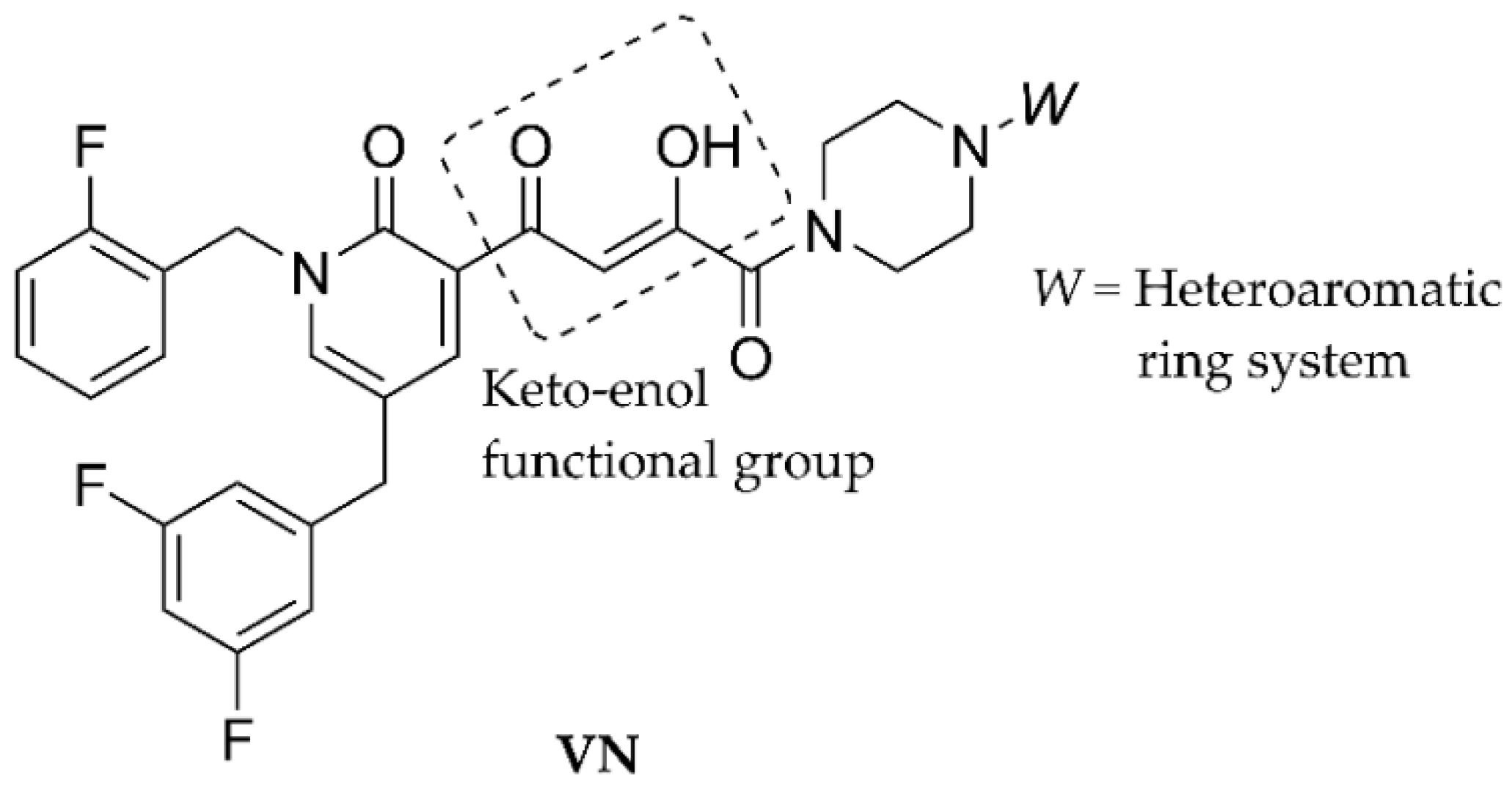
The linker might also be modified precisely (Figure 3) to afford effective molecules from a series of VN compounds against multidrug-resistant TB (MDR-TB). The modification provided different mechanisms of anti-MDR-TB action, as the designed derivatives selectively targeted and inhibited DNA-dependent RNA polymerase enzymes of the pathogens. The keto-enol functional moiety (Figure 3) is responsible for chelating Mg2+ ions located in the β’-subunit of a given enzyme [26].
The current research aimed at the synthesis of highly lipophilic racemates (Figure 4) structurally similar to those characterized in the literature [15,18,19] and estimation of the parameters describing their spectral, electronic, and lipohydrophilic properties. The structural framework of designed 6ag allowed for the assumption of their noticeable in vitro activity against some of the tested mycobacteria, i.e., an attenuated Mtb H37Ra ATCC 25177, MK DSM, M. smegmatis ATCC 700084 (MS), and M. marinum CAMP 5644 (MM).
The impact of 6ag on the viability of human liver hepatocellular carcinoma (HepG2) cells was also investigated in vitro. The cell line shows high proliferation rates and epithelial-like morphology [27], thus being used most commonly in drug metabolism and hepatotoxicity studies.
Despite the fact that a stereogenic center can be found in the structure of proposed compounds 6ag, the synthesis of racemic molecules has been a rational and convenient strategy, especially in the early phases of conceptual development of (not only) antimycobacterial drugs to confirm the relevance of the chosen structural motifs [15,18,19,28]. The replacement of an O-bridge (B; Figure 1) with an NHCOO moiety might be advantageous. The derivatives 6ag (Figure 4) would not passively cross the blood–brain barrier because of the increase in number of polar O and N atoms [29].

2. Materials and Methods

2.1. Chemistry

2.1.1. General Information

All information related to reagents used for syntheses, drying of solvents, and thin-layer chromatography (TLC) for reactions monitoring and preliminary purity determination for all synthesized or analyzed compounds (i.e., 3, 5ag, and 6ag) can be found in [30]. The details regarding yields, determinations of melting point (Mp) values, and estimations of Rf parameters (TLC) for those molecules (Figure 1) are already published [30] and are also available in the Supplementary Materials. General procedures for the preparation of the molecules (3, 5ag, and 6ag) can be found in a research paper by Pospisilova et al. (2020) [30].
The equipment, employed software tools, and particular methods to measure and interpret properly infrared (IR) spectra, nuclear magnetic resonance (NMR) spectra (1H NMR, 13C NMR, and 19F NMR), high-performance liquid chromatography high-resolution mass spectra (HPLC-UV/HR-MS) as well as ultraviolet/visible (UV/Vis) spectra for all synthesized or analyzed molecules (i.e., 3, 5ag, and 6ag) can be found in [30]; in addition, they are also provided in the Supplementary Materials.
The m/z values of observed [M+H]+ adducts for all investigated molecules were obtained (m/zmeasured) from current HPLC-UV/HR-MS analyses and compared to the theoretical ones (m/ztheoretical). The difference parameter (mass accuracy; in ppm units) was calculated following Equation (1):
Difference = [(m/ztheoreticalm/zmeasured)/m/ztheoretical] × 106
The purity of prepared or analyzed compounds (3, 5ag, and 6ag) was verified by HPLC hyphenated with ultraviolet spectrophotometry and high-resolution mass spectrometry (LC-UV/HR-MS) as listed in the Supplementary Materials.

2.1.2. Synthesis of Compounds

  • Preparation of (±)-(oxiran-2-yl)methyl-[1-(3-trifluoromethyl)phenyl]carbamate (3)
The synthesis of the compound (3) was published in [30], and the procedure is also mentioned in the Supplementary Materials. The yield, Mr, Mp, and Rf as well as 1H NMR, 13C NMR, 19F NMR, and HPLC-UV/HR-MS (ESI) analyses for a given off-white solid (Figure 4) are also available [30]. Thus, some missing data were estimated within the current research, i.e., IR spectral analysis and purity. The complete characterization of this intermediate is provided in the Supplementary Materials.
  • General preparation of carbamate compounds (5ag)
The general synthetic procedures, according to which the proposed carbamate compounds (5ag) were prepared, can be found in [30] and are also included in the Supplementary Materials together with the characteristics of employed basic amines (4ag). The oily compounds 5ad and 5f are newly synthesized within the scope of current research, and their complete spectral investigation and analytical data are provided in the Supplementary Materials.
The synthesis and majority of the spectral as well as analytical characteristics of the molecules 5e and 5g are published in [30] and, in addition, they are also included in the Supplementary Materials. Some missing parameters for these derivatives were estimated currently (i.e., Rf values (TLC), IR spectral assignments, and purity) and are provided in the Supplementary Materials as well.
  • General preparation of piperazinium chloride salts (6ag)
The general synthetic procedures, according to which the desired piperazinium chloride salts (6ag) were synthesized, can be found in [30], and the details are also given in the Supplementary Materials. The off-white solids 6ad and 6f (Figure 4) are newly prepared within the scope of current research, and their complete spectral analyses, analytical and physicochemical data are provided in the Supplementary Materials as well as in the next sections of this article.
The other off-white solids, 6e and 6g (Figure 4), were synthesized and described mostly within the research in [30], and their yields, Mr, Mp, 1H NMR, 13C NMR, 19F NMR, and HPLC-UV/HR-MS (ESI) are available; therefore, the missing Rf values (TLC) and IR spectral analyses and purity were able to be estimated within the present study. All required descriptors are listed in the Supplementary Materials. In addition, 1H NMR, 13C NMR, and 19F NMR spectra as well as HPLC-UV/HR-MS analyses of the compounds 6ag are provided in the Supplementary Materials (Figures S1–S28).

2.1.3. Determination of Some Physicochemical Properties

  • Estimation of electronic properties
The electronic properties of 6ag were defined by the values of the logarithms of molar absorption coefficients (log ε) following Lambert–Beer’s law (Equation (S1)). The details regarding employed experimental procedures published in [31,32] are also listed in the Supplementary Materials. The log ε values for the presently observed absorption maxima (λ) of 6ag (i.e., λ1 = 203.50–206.00, λ2 (Ch–T) = 238.50–243.00 (charge-transfer absorption maximum), and λ3 = 279.50–286.50 nm) are provided in Table 1.
  • Estimation of lipophilic properties
Lipohydrophilic characteristics of 6ag were estimated by reversed-phase high-performance liquid chromatography (RP-HPLC). Details on the measurements and calculation of retention factors (capacity factors; k) according to Equation (S2) can be found in the Supplementary Materials as well as in [31]. The calculations for the extrapolated retention (capacity) factor (k) logarithms for 100% water (isocratic RP-HPLC)—log kw parameters (Table 2)—were made from intercepts of linear plots between log k and φM following Equation (2):
log k = log kwS × φM
where the S parameter is the slope of a regression curve describing the solvent strength of a pure organic solvent, and φM is the volume fraction of MeOH [33,34].
The experimental details considering methods for the purity verification of 6ag are provided in [31] and the Supplementary Materials (see Tables S1 and S2).

2.2. Biological Assays

2.2.1. In Vitro Antimycobacterial Evaluation

The activity of 6ag and isoniazid (INH) standard drug (Sigma-Aldrich, Darmstadt, Germany) was inspected in vitro against an attenuated M. tuberculosis H37Ra ATCC 25177 (Mtb H37Ra), M. kansasii DSM 44162 (MK DSM), M. smegmatis ATCC 700084 (MS), and M. marinum CAMP 5644 (MM). The experimental procedures for susceptibility testing of given tuberculous and non-tuberculous mycobacteria are published in [35,36,37,38,39,40,41,42], and the details are also listed in the Supplementary Materials. The estimated MIC values (in μM units), i.e., the lowest concentrations of a compound at which no visible growth of a particular bacteria was observed, can be found in Table 3.

2.2.2. In Vitro Cytotoxicity Screening

The potential of 6ag to reduce the in vitro viability of the human Caucasian hepatocyte carcinoma (HepG2) cell line population to 50% of its maximum viability was described with inhibitory concentrations (IC50 values, in μM units). The methods for in vitro estimation of IC50 parameters are published in [43,44] and, in addition, are also provided in the Supplementary Materials. The IC50 value for a particular molecule (Table 3) was acquired from a nonlinear regression analysis based on the dependence between a semi-logarithmic plot of incubation concentration and percentage of absorbance (A) value relative to untreated controls [43,44]. These IC50 descriptors were calculated using Microsoft Excel software, version 2016 (Microsoft Corp., Redmont, WA, USA).

2.3. In Silico Evaluation of Synthesized Bases

The PASS Online (prediction of activity spectra for substances) program (Department of Bioinformatics, Laboratory of Structure–Function Based Drug Design, Institute of Biomedical Chemistry, Moscow, the Russian Federation) was used to predict a spectrum of biological activities [45,46] for synthesized bases 5ag. The probability that the analyzed compounds might be biologically active was considered and expressed by values of relevant indices (I). These I values varied from 0.000 to 1.000. The probability that analyzed bases might act as general pump inhibitors (IGPI) and membrane permeability inhibitors (IMPI) as well as drugs for cancer-associated disease treatments (ICADT) was predicted. In addition, the probability of their possible toxic effects was also predicted (Table S3), i.e., the probability of causing bradycardia (IB), palpitation (IP), and tachycardia (IT) as well as prolongation of a QT interval (IQTp).
Due to the fact that the PASS Online predictor processes so-called small molecules (i.e., compounds with Mr ˂ 900–1000) and the predictor’s limitation is Mr ˂ 1250, it was not possible to perform analyses for the corresponding final compounds (6ag), because no correct simplified molecular-input line-entry system (SMILES) codes were generated. In other words, the interactive environment of this freeware program did not allow for the processing of charged compounds or for the prediction of required descriptors [45,46]. Moreover, INH was not included in the in silico analyses because not all required indices were generated for a given reference drug.

2.4. Calculations and Statistical Analyses

The OriginPro 2019b software, version 9.6.5.169 (OriginLab Corporation, Northampton, MA, USA), was used to calculate particular statistical descriptors of linear regression analyses (log kw, S, χ2red, RSS, R, Adj. R2, and RMSE) and analyses of variance (ANOVA; F and Prob > F), both related to current RP-HPLC experiments (Table 2). Brief definitions of all calculated parameters and significance levels (indicated by stars) as well as several fundamental terms (i.e., principal components (PCs), pretreatment methods, scree plots, eigenvalues (λe), circle of correlation, and Pearson’s correlation coefficient (r), which are valid for linear regression analyses, ANOVA, and principal component analysis (PCA)) can be found in References [47,48,49,50,51] and the Supplementary Materials.
The XLSTAT software, version 2016.02.28451 (Addinsoft, New York, NY, USA) was employed for PCA [49] to explore properly the relationships between values of estimated analytical and physicochemical descriptors (m/zmeasured, log ε2 (Ch–T), and log kw) as well as log (1/MIC (M)) and log (1/IC50 (M)) parameters derived from both the MIC and IC50 data sets. These parameters resulted from in vitro biological (antimycobacterial and cytotoxic) evaluation of 6ag (Table 3). In addition, the in silico descriptors (IGPI, IMPI, ICADT, IB, IP, IT and IQTp; Table S3) of corresponding bases (5ag) were also involved in these analyses.

3. Results and Discussion

3.1. Chemistry

3.1.1. Synthesis of Compounds

In regard to the objectives set, the molecules were synthesized as follows (Figure 4). Lipophilic 3-(trifluoromethyl)phenyl isocyanate (1), as a convenient starting compound, was treated with (±)-(oxiran-2-yl)methanol (2) providing white crystalline (±)-(oxiran-2-yl)methyl-[1-(3-trifluoromethyl)phenyl]carbamate (3). The yield of given nucleophilic addition was satisfactory (81%). The opening of an (±)-(oxiran-2-yl)methyl ring of 3 was made via nucleophilic addition with particular 1-(substituted phenyl)-/1-(dimethylphenyl)piperazines (4ag) available commercially [30,52]. The desired oily carbamates (5ag) were obtained with good yields from 75% (5f) to 85% (5g).
The bases dissolved in chloroform were converted into required piperazinium chloride salts (6ag). In order to obtain pure target compounds for further analytical, physicochemical, and in vitro biological evaluations, they were crystallized from propan-2-ol twice. Thus, final substances were achieved with moderate to low yields only, i.e., from 55% (6g) to 31% (6c).
The molecules 6ag were defined comprehensively with appropriate descriptors from IR, 1H NMR, 13C NMR, and 19F NMR as well as HPLC-UV/HR-MS (ESI) spectral analyses, and these data confirmed the proposed structures. Considering the IR spectra interpretation (Supplementary Materials), the bands from 1740 (6f) to 1722 cm1 (6c) were observed as typical ones for stretching vibrations, υ (C=O). The phenyl moieties were indicated with υ (C=C) at approximately 1605 cm1. The vibrations at approximately 1512 cm1 were observed as δ (N–H) was present. The bands between 1245 and 1242 cm1 corresponded to asymmetric stretching of the C–O–C arrangement of bonds. The =C–H group afforded in-plane deformation vibrations (δip) approximately at 1020 cm1 and, in addition, its out-of-plane deformation vibrations (δoop) approximately at 845 cm1 were also noticed. In 1H NMR (Supplementary Materials), the proton signal of a protonated cyclic piperazin-1-ium-1,4-diyl moiety was observed as a singlet in the δ interval from 10.95 (6b) to 10.46 ppm (6g). The proton signal of an NHCOO group was observed as a singlet from 10.24 (6a) to 9.99 ppm (6c). Following the 13C NMR spectral data (Supplementary Materials), all carbons were confirmed in agreement with the proposed structures. The signal of fluorine within a CF3 group was observed from –60.14 (6b) to –60.20 ppm (6c) in 19F NMR. Heteronuclear coupling of fluorine atoms to 1H NMR and 13C NMR was also observed (Supplementary Materials). The protonation of only one N atom of piperazin-1-ium-1,4-diyl was observed within the structure of 6ag (Figure 4). The finding was supported by HPLC-UV/HR-MS, where particular [M+H]+ molecular peaks for these solid molecules were found (Supplementary Materials).
The purity evaluation of all salts was carried out using the LC-UV/HR-MS method. Their satisfactory purity, varying from 97.34% (6a) to 98.85% (6g; Supplementary Materials), was achieved by repeated crystallization as listed in previous sections.

3.1.2. Determination of Some Physicochemical Properties

  • Determination of electronic properties
The UV/Vis absorption spectra of methanolic solutions (c = 5.0 × 10⁻⁵ M) of 6ag were measured in the λ interval of 190–820 nm. Three absorption maxima (Table 1) were found for these solutions as follows: λ1 = 203.50–206.00, λ2 (Ch–T) = 238.50–243.00, and λ3 = 279.50–286.50 nm. This observation was in full agreement with [53], as the positions of particular absorption bands were typical for (substituted) phenylcarbamic acid derivatives. The observed bands were labeled properly as first local excitation (λ3), charge-transfer (λ2 (Ch–T)), and second local excitation (λ1) absorption maxima, respectively.
Almost all substituents attached to the aromate of a 4-(substituted phenyl)piperazin-1-ium-1-yl moiety, which carried non-bonding electrons, caused shifts in the λ1 and λ2 as well as λ3 absorption bands to longer wavelengths. Thus, bathochromic shifts at these absorption maxima were found for the compounds 6bg compared to those of 6a, a molecule containing a 4-phenylpiperazin-1-ium-1-yl group. This trend was in agreement with conclusions already published [54]. In fact, exceptions were observed only for the molecule 6b and its λ1 absorption band (equal values were find out) as well as 6g and its λ3 absorption band, for which a slight hypsochromic shift was observed (Table 1).
The log ε2 (Ch–T) parameter was the most sensitive to the differences in length, position, electronic, and steric properties of substituent(s) attached to a piperazin-1-ium-1,4-diyl moiety. Thus, when discussing SARs, special consideration was paid to log ε2 (Ch–T). Absorption intensity at this maximum increased slightly for the compounds containing bulky substituents (auxochromes) with strong EW properties (6e, 6f) and molecules with a bulky diphenylmethyl moiety (6g). This behavior is called a hyperchromic shift [55]. On the other hand, a hypochromic shift [55] (i.e., decreased values of a given parameter) was observed for mono-substituted derivatives (Table 1) containing a 4’-CH3, 4’-F or 4’-Cl substituent (6bd).
  • Determination of lipohydrophilic properties
Pathogenic Mtb strains possess an external cell wall composed mainly of a covalently attached complex of a branched polysaccharide peptidoglycan, lipopolysaccharides (arabinogalactan, lipoarabinomannan, and lipomannan), and highly lipophilic mycolic acids. These structures have important roles in the growth and integrity maintenance of the cell wall. In addition, Mtb has an outer layer composed of an array of glycolipids, lipoglycans, and lipids associated with this wall [56,57,58]. Thus, the lipophilicity is an important parameter that should definitely be taken into consideration in the design of novel anti-TB compounds to ensure their permeation via the cell wall of Mtb [59].
Computational methods (i.e., CLOGP, ALOGP, XLOGP2, or XLOGP3) are frequently used to calculate or predict the lipophilicity of various synthetic or natural antimycobacterial compounds [60,61,62,63,64,65]. However, it would be questionable if such in silico approaches might be considered accurate in predicting the lipophilic behavior of the compounds 6ag. The structure of the molecules investigated in [60,61,62,63,64,65] allowed for such predictions; however, particular predictors were not designed to evaluate salts, i.e., charged compounds as discussed in [31].
Taking into strong consideration this aspect, the lipohydrophilic features of 6ag were investigated by an experimental RP-HPLC technique. The retention behavior of a molecule in a reversed-phase (RP) chromatographic system is in very close connection with its lipophilicity commonly derived from log k [66]. The C18-functionalized silica gel (stationary phase; SPh) and liquid binary mixtures of methanol (MeOH) in different volume concentrations (60–90%; v/v) with 5% increments and water [66] were employed as suitable mobile phases (MPhs) in order to estimate these log k parameters (Tables S1 and S2).
The highest tr and log k data were determined for the most lipophilic compounds 6f (tr = 26.5828 min, log k = 1.7516), 6e (tr = 24.7159 min, log k = 1.7372), and 6g (tr = 23.1377 min, log k = 1.6913) in the MPh with a 60% proportion (v/v) of the organic modifier. The increase in the amount of MeOH (v/v) lowered the values of both tr and log k parameters as expected (Tables S1 and S2).
The estimated log k descriptors for 6ag, however, might not be considered universal parameters to adequately express their lipophilicity because of their being very notably influenced by particular chromatographic conditions [67]. The extrapolation of estimated solute retention (log k) to the elution with 100% water (log kw) was regarded as a much more adequate procedure (Table 2). The impact of lipophilicity, expressed with log kw, on the in vitro antimycobacterial activity of these compounds might be expected [31,47].
The linear relationships between estimated log k and φM related to particular molecules were statistically extremely significant, as all calculated probabilities of obtaining F-ratio (Prob > F) values belonged to the interval from 0.0000 to <0.0010 (Table 2). Very acceptable quality, which characterized all suggested linear relationships providing desired extrapolated parameters (log kws), was adjusted with the reduced chi-square (χ2red) descriptor varying from 0.0004 (6e) to 0.0040 (6c). Next, the linearity of all observed models was defined with minimal RSS (residual sum of squares) outputs from 0.0016 (6e) to 0.0201 (6c) as well as with the root mean square error (RMSE; Table 2) found between 0.0176 (6e) and 0.0635 (6c).
The log kw values of 6ag ranged from 4.1170 (6a) to 5.2184 (6g), considering the variably substituted piperazin-1-ium-1,4-diyl moiety. The lipophilicity of a 4’-CH3 substituted molecule (6b; log kw = 4.8419) was higher than that containing a 4’-F atom (6c; log kw = 4.2672). This finding was in agreement with the calculated values of the hydrophobic substituent constant (π) of both substituents, which were as follows [68]: π(4’-CH3) = 0.53 and π(4’-F) = 0.14.

3.2. Biological Assays

3.2.1. In Vitro Antimycobacterial Evaluation

The efficiency of 6ag and the INH reference drug was inspected in vitro against Mtb H37Ra, MK DSM, MS as well as M. marinum (Table 3) and described with the MIC parameter (in μM units) [39,40]. Tuberculous Mtb H37Rv and H37Ra were used to confirm (or refuse) if the newly synthesized and traditional anti-TB drugs showed in vitro activity. The question of whether H37Ra might “substitute in some manner” a highly pathogenic H37Rv or clinical tuberculous mycobacterial strains was answered clearly by Heinrichs et al. (2018). The authors concluded that the H37Ra strain was as equally useful as the H37Rv one regarding responses to in vitro screened anti-TB drugs. Moreover, the biosafety requirements concerning H37Ra were much more convenient compared to those of H37Rv [69].
The most powerful antimycobacterials were 1-[2-hydroxypropyl-{(3-trifluoromethyl)-phenyl}carbamoyloxy]-4-(3,4-dichlorophenyl)piperazin-1-ium chloride (6e) and 1-[2-hydroxypropyl-{(3-trifluoromethyl)phenyl}carbamoyloxy]-4-(4-diphenylmethyl)piperazin-1-ium chloride (6g) showing MIC < 3.80 μM against Mtb H37Ra (Table 3). These compounds were more active than INH (MIC = 58.33 μM).
Considering previous conclusions [70,71,72], it might be stated that a 3-CF3-phenylcarbamoyloxy fragment notably improved the in vitro activity of such substituted compounds against a given mycobacterium compared to the efficiency of the derivatives bearing an 2-/3-/4-alkoxyphenylcarbamoxyloxy moiety (alkoxy = methoxy or ethoxy).
In addition, both 1-[2-hydroxypropyl-{(3-trifluoromethyl)phenyl}carbamoyloxy]-4-(4-chlorophenyl)piperazin-1-ium chloride (6d) and 6e were the most effective against MK DSM with MIC = 8.09 μM (6d) and 15.13 μM (6e), respectively. Their potency was higher than that of INH (MIC = 29.16 μM; Table 3). The molecule 6g was the most active against both MS (MIC = 14.54 μM) and MM (MIC = 8.09 μM). This substance was even more promising than INH (MIC = 43.75 and 29.16 μM, respectively) as listed (Table 3).
The presence of an EW (6cf) or even an electron-donating (ED; 6b) substituent attached to a phenyl ring seemed to be more beneficial in structural variation in order to potentiate in vitro antimycobacterial activity than classical bioisosteric replacement of this substituent with hydrogen (6a). Bulky lipophilic diphenylmethyl moiety (6g) improved efficiency, especially against Mtb H37Ra and MM (Table 3).
The compounds 6ag fought in applsci-12-00300vitro tested non-tuberculous mycobacteria more effectively than the substances containing an 2-/3-/4-alkoxyphenylcarbamoxyloxy moiety (alkoxy = methoxy or ethoxy) despite the presence of a lipophilic 4’-F atom or 3’-CF3 group within their salt-forming part [19].

3.2.2. In Vitro Cytotoxicity Screening

Another goal of this research was to set in vitro the impact of 6ag on a liver cancer HepG2 cell line. The model was chosen since antimycobacterial agents have been included within a group of the top five pharmacodynamic classes associated with the highest reporting odds ratio (ROR) score. In attempt to set this ROR score, the frequency of hepatotoxicity was estimated at both the group and individual drug levels. The ROR score index describes the measure of such undesirable phenomenon [73]. The highest ROR scores with the highest risk of drug-induced liver injury were observed for the compounds belonging to antineoplastic, antiviral, analgesic, antibiotic, and antimycobacterial classes.
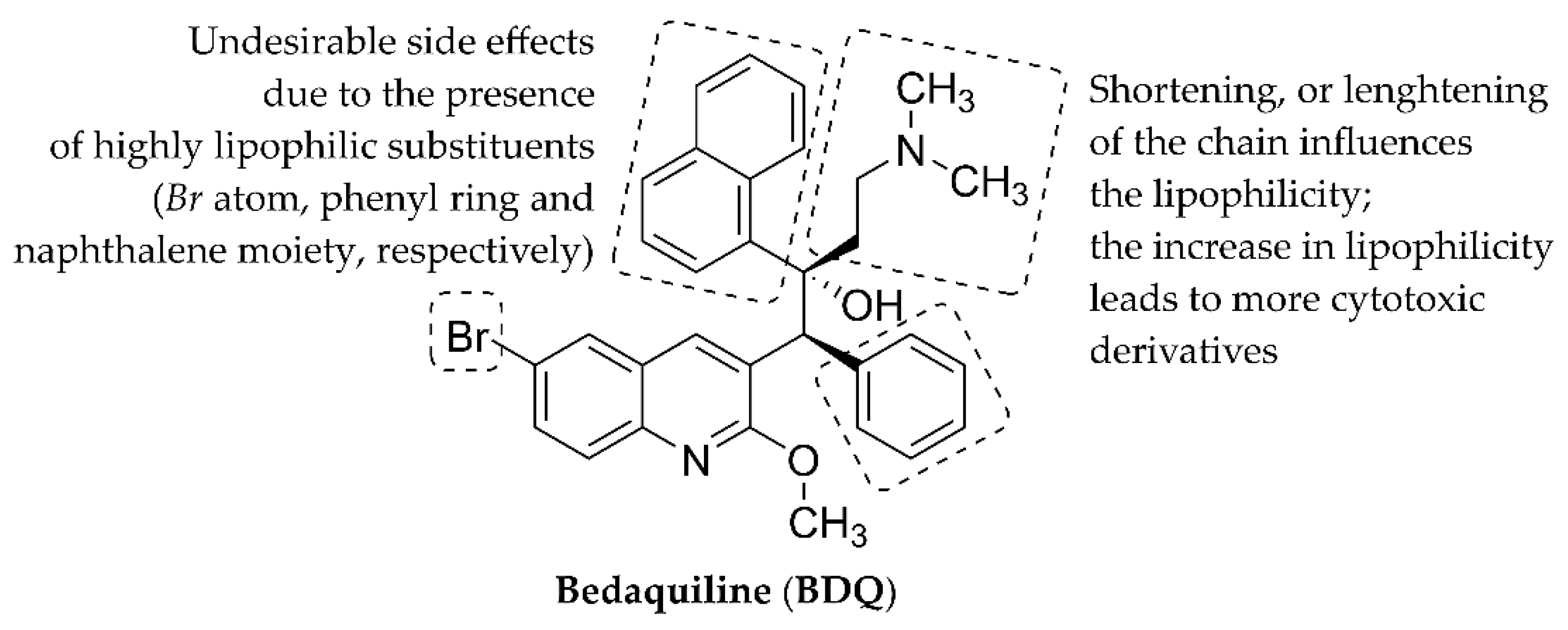
The molecules 6ag were highly lipophilic as their log kw values from 4.1170 (6a) to 5.2184 (6g) indicated (Table 2). Potential disadvantages of highly lipophilic bedaquiline (BDQ), a very effective antimycobacterial agent (Figure 5), include the inhibition of a human Ether-à-go-go-related gene (hERG) potassium channel due to the concurrent risk of cardiac toxicity, possible phospholipidosis, or hepatic toxicity. The shortening or lengthening of its dimethylaminoethyl sidechain did not improve antimycobacterial activity, but increased cytotoxicity was observed as the lipophilicity of synthesized BDQ derivatives increased [74,75].
The current nonlinear regression analyses explored the relationships between a semi-logarithmic plot of incubation concentration and percentages of absorbance relative to untreated controls, thus providing IC50 values [63]. These IC50 parameters (in μM units) describing the cytotoxic properties of 6ag varied from 22.18 (6g) to 83.08 μM (6a) as listed in Table 3. The most effective compounds (6g and 6e) against Mtb H37Ra showed IC50 = 22.18 (6g) and 29.39 μM (6e). If accepting IC50 > 25.00 μM as a “very mild” IC50 criterion characterizing low toxic derivatives [76], the cytotoxicity of 6e might be “theoretically acceptable”. However, Ambrożkiewicz et al. (2020) stated that the molecules with IC50 ≤ 100.00 μM were relatively cytotoxic [77]. The INH standard drug showed a very limited ability to reduce the population of HepG2 liver cancer cells (IC50 > 1000 μM) [63].
The limited safety of effective antimycobacterial agents due to the relatively low IC50 values was the reason for not progressing further. Poce et al. (2019) stated that molecules with IC50 ≥ 40.00 μM were safe [78]. Following the conclusions published in [76,78], both 6d (IC50 = 43.35 μM) and 1-[hydroxypropyl-{(3-trifluoromethyl)phenyl}carbamoyloxy]-4-(3-trifluoromethylphenyl)piperazin-1-ium chloride (6f; IC50 = 54.71 μM) might be “theoretically safe”. In regard to confirming or refusing the validity of statements about the cytotoxic properties of the compounds 6dg based on the conclusions published in [76,78], previous in vitro investigations [30,79,80] also contributed to the current quest.
Bouz et al. (2021) evaluated the in vitro cytotoxic properties of a tamoxifen (TMX) reference drug against the HepG2 cell line. The authors described the efficiency of TMX with IC50 = 19.56 μM [63]. Other anticancer drugs, such as afatinib, 5-fluorouracil, and sorafenib, showed IC50 = 5.40, 7.91, and 9.18 μM [79,80], respectively.
The compound 6g also efficiently inhibited the proliferation in vitro of a human monocytic leukemia THP-1 cell line showing IC50 = 5.91 μM as the concentration, which inhibited 50% of the proliferation. Moreover, lower cytotoxic activity of a cisplatin standard drug was even observed (IC50 = 13.65 µM) in those experiments [30]. Thus, both the molecules 6e and 6g were suggested as suitable structural platforms for the development of promising anticancer agents, but their eventual use in pharmacotherapeutic interventions as anti-TB drugs has to be considered.
The current “definitive” conclusion was supported by calculations of selectivity index (SI) parameters. The ratio between particular IC50 values (in μM units) of the molecules 6ag and their MIC parameters (in μM units) estimated against Mtb H37Ra, MK DSM, MS, and MM [81], respectively, was defined as SI (Table S4). The highest SI values were calculated for 6e (SI = 7.78) and 6g (SI = 6.11) as the ratio of their activity against HepG2 liver cancer cells and efficiency against Mtb H37Ra as well as for 6d (SI = 5.36) as the ratio of its activity against these cancer cells and efficiency against MK DSM.
Very thin line was found between in vitro cytotoxic and antimycobacterial properties of both 6b and 6c (Table 3). The calculated SI values preliminarily indicated that the anti-MS activity of these compounds might be the consequence of their cytotoxicity, as SI = 0.49 (6b) and 0.37 (6c) as shown in Table S4. Secrist et al. (2001) determined that if SI > 10.00, a drug candidate might be regarded as safe granting sufficient difference between (antimycobacterially) efficient and cytotoxic concentrations [81]. Overall, current experiments clearly indicate safety concerns connected with the 6ag set.

3.3. In Silico Evaluation of Synthesized Bases

Regarding the analyses of prepared bases (5ag), the PASS Online program [45,46] predicted the probability of their acting as general pump inhibitors (IGPI) and membrane permeability inhibitors (IMPI) as well as drugs for the treatment of cancer-associated diseases (ICADT). In addition, the possible toxic effects of these bases on a cardiovascular system (i.e., probability of causing bradycardia (IB), palpitation (IP), and tachycardia (IT) as well as prolongation of a QT interval in the heart (IQTp)) were also predicted (Table S3). The criteria for prediction of a specific activity are provided in the Supplementary Materials.
BDQ inhibits the synthesis of ATP in mycobacteria. The substance interferes with the activity of an F1F0 ATP synthase enzyme by inhibiting its proton pump, which is critical for the generation of membrane potential. This synthase is one of the enzymes considered essential for meeting the energy requirements of both the proliferating aerobic and hypoxic dormant stages of the life cycle for a variety of mycobacterial strains. ATP levels are highly reduced within bacterial cells upon the action of BDQ [82,83].
The highest probability to act as general pump inhibitors was calculated for the compounds containing one or two phenyl rings substituted with hydrogens only (Table S3), i.e., 5g (IGPI = 0.707) and 5a (IGPI = 0.613). The presence of other substituents provided lower values regardless of their ED (5b) or EW (5cf) properties.
The pathogenic properties of Mtb are based on thwarting the innate defense systems of a macrophage host cell. Virulence factors of Mtb disrupt physiological host cell signaling. The destruction of macrophages starts when the pathogen enters a cell, affecting (destroying) a variety of host signal transduction pathways. Moreover, the surface of these cells is remodulated as a consequence of Mtb action via altering the expression of the molecules, which are crucial for full activation of an adaptive immune response [84].
Membrane permeability inhibitors could more or less block this process. The predicted IMPI parameter for synthesized bases varied from 0.457 (5g) to 0.555 (5d). It seemed that the probability of their blocking activity was related to a precise selection of a substituent owning specific electronic and steric properties. The 4’-Cl substitution was the most favorable (5d), the lipophilicity of a given atom might be described by π4’-Cl = 0.71 and the EW effect with a Hammett substituent constant σ (σ4’-Cl = 0.23). The values set for hydrogen were as follows [68]: πH = 0.00, σH = 0.00.
Special attention should be paid to the molecule 5g (as well as its antimycobacterially effective salt 6g), as IT = 0.605 and IQTp = 0.640. The issue of polymorphic ventricular tachycardia and QT interval prolongation was also observed for BDQ. If the interval is prolonged, polymorphic ventricular tachycardia and/or “torsades de pointes” can occur. These dysfunctions may precipitate ventricular fibrillation resulting in sudden death. Fortunately, severe QT interval prolongation is an infrequent cause of BDQ interruption despite the frequent use of concomitant drugs that also prolong the QT interval [85,86].

3.4. Structure-Activity Relationships

The PCA approach was employed to explore the relationships between observed values of analytical and physicochemical parameters for the 6ag set (m/zmeasured, log ε2 (Ch–T), and log kw), in silico descriptors (IGPI, IMPI, ICADT, IB, IP, IT, and IQTp) related to 5ag, and the calculated data based on in vitro biological screening of the compounds 6ag, i.e., log (1/MIC (M)) and log (1/IC50 (M)), respectively. Firstly, all analyzed indices were precisely transformed using several data pretreatment techniques [87,88,89]. Secondly, a scree plot was constructed to correctly set a number of relevant principal components (PCs). The scree plot described adequately the relationships between calculated eigenvalues (λe) and number of PCs. The Kaiser–Guttman rule [50], as the most common stopping rule in PCA, aimed to limit the number of PCs—only PCs with λe > 1.00 were considered.
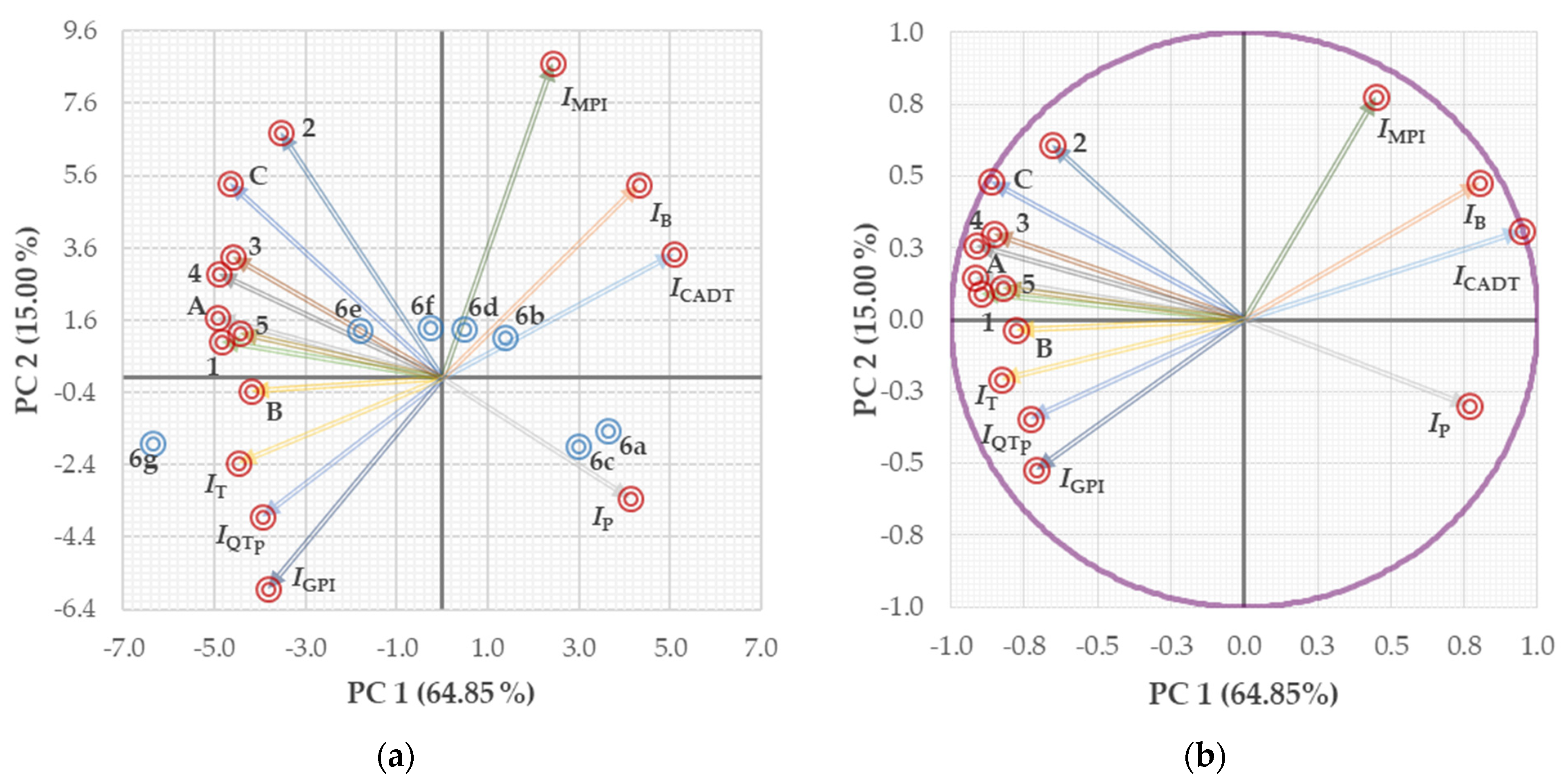
The PC 1, PC 2, and PC 3, which indicated the mathematical possibility of the existence of “real” parameters, accounted for 86.84% of the total variance, and their contributions were as follows: PC 1 (64.85%, λe = 9.73), PC 2 (15.00%, λe = 2.25), and PC 3 (6.99%, λe = 1.05). The relationship between PC 1 and PC 2 allowed for the distribution of 6ag into several subcategories. The sum of these PCs expressed the majority of the data variability (79.85% in total), as shown in Figure 6a,b.
The PC 1 served as a suitable component to separate quite clearly the antimycobacterially active and inactive derivatives. The subgroup defined by PC 1 ≤ −0.286 included 6eg, which were relatively effective (MIC ≤ 30.36 μM) against all in vitro screened mycobacteria. The substances with MIC ≥ 64.73 μM against at least one mycobacterial strain (6ad) were described with PC 1 ≥ 0.481. The lower PC 1 descriptor indicated more active antimycobacterial compound. On the other hand, higher PC 1 parameter was connected with lower activity of a derivative. The combination of both negative PC 1 and PC 2 (Figure 6a,b) can be used to characterize the most active molecule, 6g (PC 1 = −6.365 and PC 2 = −1.817). The PC 1 ≥ 2.988 together with PC 2 ≥ −1.468 defined antimycobacterially inactive compounds (6a and 6c). Other differences in the compounds’ properties mirrored in both PC 1 and PC 2 as well as other PCs are provided in the Supplementary Materials (see Table S5).
The distance between particular variables onto a plane and circle of correlation (Figure 6a,b) expressed the quality of their 2D representation. The loadings of given variables (i.e., observed and extrapolated analytical and physicochemical descriptors, the data resulted from in vitro biological evaluations as well as parameters from the PASS Online predictor) defined the contribution (“measure”) of each original variable to the chosen PCs [51]. These loadings are represented by color-coded vectors labeled following their position in a 2D score plot (Figure 6a).
The color-coded vectors were also labeled according to their position in a circle of correlation (Figure 6b). The proper indication of variables with letters A–C or digits 1–5 is provided in the Supplementary Materials. The cycle was characterized with a radius = 1 (absolute value). The visualization based on such relationships provided the sharpest angles between some pairs of vectors, i.e., A and 5, C and 3, 1 and 5, 3 and 4, IQTp and IGPI as well as IB and ICADT.
Pearson’s correlation coefficient (r) values indicated relatively satisfactory correlation between m/zmeasured (vector 1) of the compounds 6ag and their log kw (C; r = 0.8450), log ε2 (Ch–T) (B; r = 0.7959), in vitro activity against MS (3; r = 0.9431), Mtb H37Ra (1; r = 0.9240) as well as MM (4; r = 0.8141). The moderate correlation was observed between m/zmeasured and toxicity in the HepG2 cell line (5; r = 0.6681).
Absorption intensity increased slightly for the compounds containing bulky substituents with strong EW properties (6e and 6f) and molecules with a bulky diphenylmethyl moiety (6g). This behavior was connected with increased efficiency, especially against Mtb H37Ra (1; r = 0.7909) and MS (3; r = 0.7798). The correlation between log ε2 (Ch–T) (B) and log kw was weaker (C; r = 0.5896). The increased log ε2 (Ch–T) parameters of analyzed salts increased the probability for their bases to cause tachycardia (r = 0.7182). The prolongation of a QT interval was less probable (r = 0.5260).
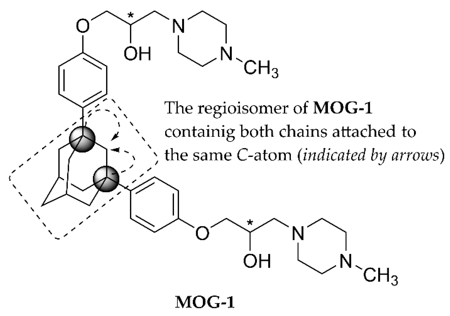
The activity of 6ag against all in vitro tested mycobacteria was quite strongly associated with their lipohydrophilic properties. In more detail, the increase in lipophilicity provided higher efficiency against MM (r = 0.9128), MS (r = 0.8650), and MK DSM (r = 0.8411) as well as Mtb H37Ra (r = 0.8199). It should be stated that both highly lipophilic adamantyl and phenyl moieties in a structure of “similar” compounds did not guarantee particularly high in vitro antimycobacterial activity. The molecule MOG-1 (Figure 7) was more than two-fold less efficient against Mtb H37Rv compared to its regioisomer [90].
The good news was that a higher log kw of 6ag might be connected with a notably lower probability of palpitation issues, which could eventually be caused by their bases (5ag; r = −0.8623). In addition, the salts would cause neither significant tachycardia (r = 0.6040) nor prolongation of a QT interval of the heart (r = 0.4478).
The increase in lipophilicity of the tested compounds provided a stronger potential to reduce the in vitro viability of the HepG2 cell population to 50% of its maximum viability (r = 0.8320). Efficient anti-MM and anti-Mtb H37Ra compounds were effective cytotoxic agents (r = 0.8138 and 0.7998, respectively). The correlation between their anti-MS or anti-MK DSM activity and cytotoxicity was weaker (r = 0.5820 and 0.5209, respectively).
The performed PCA also indicated that similar factors might influence mechanism(s) of action of analyzed molecules (6ag) against particular pairs of mycobacteria as follows: Mtb H37Ra and MS (r = 0.8098), Mtb H37Ra and MM (r = 0.7980), MK DSM and MM (r = 0.8589) as well as MS and MM (r = 0.7458).

4. Conclusions

Various substituents and, thus, different analytical and physicochemical properties of hybrid molecules containing an N-(substituted phenyl)-/N-diphenylmethylpiperazine privileged scaffold allowed for the modulation of their in vitro activity against several tuberculous and nontuberculous mycobacteria as well as a particular cancer cell line (HepG2).
The absorption intensity increased slightly for the compounds containing bulky substituents with strong EW properties (6e and 6f) as well as the molecule with a bulky diphenylmethyl moiety (6g). This behavior strongly influenced the increase in efficiency against both Mtb H37Ra (r = 0.7909) and MS (r = 0.7798). The increased lipophilicity of analyzed salts led to higher efficiency against MM (r = 0.9128), MS (r = 0.8650), and MK DSM (r = 0.8411) as well as Mtb H37Ra (r = 0.8199). Higher lipophilicity of 6ag provided their stronger ability to reduce the in vitro viability of HepG2 cell populations to 50% of its maximum viability (r = 0.8320). The efficient anti-Mtb H37Ra and anti-MM compounds (6e and 6g) reduced in vitro the viability of given cells most effectively (IC50 = 22.18 and 29.39 μM, respectively). The cytotoxic properties of all tested hybrid molecules were not connected significantly with their activity against MS (r = 0.5820) or MK DSM (r = 0.5209).
The calculated selectivity index (SI) values for the entire 6ag set (SI < 10.00) might be considered direct proof that eventual treatment of patients suffering from mycobacterial infections with the analyzed compounds or structurally similar drugs has to be conducted with care. These SI parameters clearly prevented the conclusion that the synthesized molecules, 6ag, were safe. In addition, current calculations indicated that risk of tachycardia or QT interval prolongation due to the activity of a basic form of 6g could not be completely excluded. The proper selection of the substituents attached to the fundamental structural framework of screened substances will be decisive for the development of effective antimycobacterial derivatives with a (notably) weakened ability to reduce the survival and growth of cancer cells or efficient anticancer agents.

Supplementary Materials

The following are available online at https://www.mdpi.com/article/10.3390/app12010300/s1, Table S1: Retention times tr and lipophilicity indices log k (RP-HPLC) of 6ag determined in the mobile phases with a various volume ratio (v/v) of a methanol (MeOH) organic modifier and water, i.e., from 60:40 to 70:30; Table S2: Retention times tr and lipophilicity indices log k (RP-HPLC) of 6ag determined in the mobile phases with a various volume ratio (v/v) of a methanol (MeOH) organic modifier and water, i.e., from 75:25 to 90:10; Table S3: The prediction of probability for the bases (5ag) to act as general pump inhibitors (IGPI) and membrane permeability inhibitors (IMPI) as well as drugs for the treatment of cancer-associated diseases (ICADT). The calculation of probability that these molecules might cause bradycardia (IB), palpitation (IP), tachycardia (IT), and prolongation of a QT interval (IQTp); Table S4: The selectivity index (SI) values of in vitro evaluated compounds 6ag, which were calculated as SI = IC50 (in μM units)/MIC (in μM units). The IC50s indicated toxicity in human liver hepatocellular carcinoma (HepG2) cells, the MICs were determined against M. tuberculosis H37Ra ATCC 25177 (Mtb H37Ra), M. kansasii DSM 44162 (MK DSM), M. smegmatis ATCC 700084 (MS), and M. marinum CAMP 5644 (MM); Table S5: The contribution of evaluated variables (in percentages) to particular principal components (PCs; PC 1–6); Figures S1–S28. 1H NMR, 13C NMR, and 19F NMR spectra as well as HPLC-UV/HR-MS analyses of the compounds 6ag.

Author Contributions

Design and synthesis of the compounds, J.Č., M.P., and I.M.; spectral characterization, T.P. and D.P.; determination of electronic and lipohydrophilic properties, J.Č., M.P., and D.P.; in vitro antimycobacterial screening and data evaluation, Š.M. and J.J.; in vitro cytotoxicity screening and data evaluation, Ľ.P. and F.B.; principal component analysis, G.K. and I.M.; data interpretation, conceptualization, writing—original draft preparation, and revision of the manuscript, I.M. All authors have read and agreed to the published version of the manuscript.

Funding

The authors very gratefully acknowledge the financial support received from the Faculty of Pharmacy, Comenius University Bratislava (Slovakia).

Institutional Review Board Statement

Not applicable.

Informed Consent Statement

Not applicable.

Data Availability Statement

Not applicable.

Conflicts of Interest

The authors declare no conflict of interest.

References

  1. Christopher, J.A.; Brown, J.; Doré, A.S.; Errey, J.C.; Koglin, M.; Marshall, F.H.; Myszka, D.G.; Rich, R.L.; Tate, C.G.; Tehan, B.; et al. Biophysical fragment screening of the β1-adrenergic receptor: Identification of high affinity arylpiperazine leads using structure-based drug design. J. Med. Chem. 2013, 56, 3446–3455. [Google Scholar] [CrossRef]
  2. Soskic, V.; Sukalovic, V.; Kostic-Rajacic, S. Exploration of N-arylpiperazine binding sites of D2 dopaminergic receptor. Mini Rev. Med. Chem. 2015, 15, 988–1001. [Google Scholar] [CrossRef]
  3. Mazzotta, S.; Cebrero-Cangueiro, T.; Frattaruolo, L.; Vega-Holm, M.; Carretero-Ledesma, M.; Sánchez-Céspedes, J.; Cappello, A.R.; Aiello, F.; Pachón, J.; Vega-Pérez, J.M.; et al. Exploration of piperazine-derived thioureas as antibacterial and anti-inflammatory agents. In vitro evaluation against clinical isolates of colistin-resistant Acinetobacter baumannii. Bioorg. Med. Chem. Lett. 2020, 30, 127411. [Google Scholar] [CrossRef]
  4. Lacivita, E.; Niso, M.; Stama, M.L.; Arzuaga, A.; Altamura, C.; Costa, L.; Desaphy, J.-F.; Ragozzino, M.E.; Ciranna, L.; Leopoldo, M. Privileged scaffold-based design to identify a novel drug-like 5-HT7 receptor-preferring agonist to target Fragile X syndrome. Eur. J. Med. Chem. 2020, 199, 112395. [Google Scholar] [CrossRef] [PubMed]
  5. Gao, L.; Hao, C.; Ma, R.; Chen, J.; Zhang, G.; Chen, Y. Synthesis and biological evaluation of a new class of multi-target heterocycle piperazine derivatives as potential antipsychotics. RSC Adv. 2021, 11, 16931–16941. [Google Scholar] [CrossRef]
  6. Skoreński, M.; Sieńczyk, M. The fellowship of privileged scaffolds—One structure to inhibit them all. Pharmaceuticals 2021, 14, 1164. [Google Scholar] [CrossRef]
  7. Atukuri, D.; Gunjal, R.; Holagundi, N.; Korlahalli, B.; Gangannavar, S.; Akkasali, K. Contribution of N-heterocycles towards anti-tubercular drug discovery (2014–2019); predicted and reengineered molecular frameworks. Drug Dev. Res. 2021, 82, 767–783. [Google Scholar] [CrossRef] [PubMed]
  8. Zhao, H.; Dietrich, J. Privileged scaffolds in lead generation. Expert Opin. Drug Discov. 2015, 10, 781–790. [Google Scholar] [CrossRef] [PubMed]
  9. Daley, C.L. The global fight against tuberculosis. Thorac. Surg. Clin. 2019, 29, 19–25. [Google Scholar] [CrossRef] [PubMed]
  10. Girase, P.S.; Dhawan, S.; Kumar, V.; Shinde, S.R.; Palkar, M.B.; Karpoormath, R. An appraisal of anti-mycobacterial activity with structure-activity relationship of piperazine and its analogues: A review. Eur. J. Med. Chem. 2021, 210, 112967. [Google Scholar] [CrossRef] [PubMed]
  11. Verma, S.K.; Verma, R.; Verma, S.; Vaishnav, Y.; Tiwari, S.P.; Rakesh, K.P. Anti-tuberculosis activity and its structure-activity relationship (SAR) studies of oxadiazole derivatives: A key review. Eur. J. Med. Chem. 2021, 209, 112886. [Google Scholar] [CrossRef] [PubMed]
  12. Murthy, V.S.; Tamboli, Y.; Krishna, V.S.; Sriram, D.; Ansari, S.A.; Alarfaj, A.A.; Hirad, A.H.; Vijayakumar, V. Design and characterisation of piperazine-benzofuran integrated dinitrobenzenesulfonamide as Mycobacterium tuberculosis H37Rv strain inhibitors. J. Enzyme Inhib. Med. Chem. 2021, 36, 1751–1759. [Google Scholar] [CrossRef]
  13. Singh, P.; Jaiyeola, B.; Kerru, N.; Ebenezer, O.; Bissessur, A. A review of recent advancements in anti-tubercular molecular hybrids. Curr. Med. Chem. 2017, 24, 4180–4212. [Google Scholar] [CrossRef] [PubMed]
  14. Alsayed, S.S.R.; Lun, S.; Payne, A.; Bishai, W.R.; Gunosewoyo, H. Facile synthesis and antimycobacterial activity of isoniazid, pyrazinamide and ciprofloxacin derivatives. Chem. Biol. Drug. Des. 2021, 97, 1137–1150. [Google Scholar] [CrossRef] [PubMed]
  15. Upadhayaya, R.S.; Vandavasi, J.K.; Kardile, R.A.; Lahore, S.V.; Dixit, S.S.; Deokar, H.S.; Shinde, P.D.; Sarman, M.P.; Chattopadhyaya, J. Novel quinoline and naphthalene derivatives as potent antimycobacterial agents. Eur. J. Med. Chem. 2010, 45, 1854–1867. [Google Scholar] [CrossRef]
  16. Urban, M.; Šlachtová, V.; Brulíková, M. Small organic molecules targeting the energy metabolism of Mycobacterium tuberculosis. Eur. J. Med. Chem. 2021, 212, 113139. [Google Scholar] [CrossRef]
  17. Koul, A.; Vranckx, L.; Dendouga, N.; Balemans, W.; Van den Wyngaert, I.; Vergauwen, K.; Göhlmann, H.W.H.; Willebrords, R.; Poncelet, A.; Guillemont, J.; et al. Diarylquinolines are bactericidal for dormant mycobacteria as a result of disturbed ATP homeostasis. J. Biol. Chem. 2008, 283, 25273–25280. [Google Scholar] [CrossRef] [Green Version]
  18. Waisser, K.; Doležal, R.; Čižmárik, J.; Malík, I.; Kaustová, J. The potential antituberculotics of the series of 2-hydroxy-3-(4-phenylpiperazin-1-yl)-propylphenylcarbamates. Folia Pharm. Univ. Carol. 2007, 35–36, 45–48. [Google Scholar]
  19. Malík, I.; Csöllei, J.; Jampílek, J.; Stanzel, L.; Zadražilová, I.; Hošek, J.; Pospíšilová, Š.; Čížek, A.; Coffey, A.; O’Mahony, J. The structure–antimicrobial activity relationships of a promising class of the compounds containing the N-arylpiperazine scaffold. Molecules 2016, 21, 1274. [Google Scholar] [CrossRef]
  20. Waisser, K.; Dražková, K.; Čižmárik, J.; Kaustová, J. Influence of lipophilicity on the antimycobacterial activity of the hydrochlorides of piperidinylethyl esters of ortho-substituted phenylcarbamic acids. Sci. Pharm. 2004, 72, 43–49. [Google Scholar] [CrossRef] [Green Version]
  21. Waisser, K.; Doležal, R.; Palát, K., Jr.; Čižmárik, J.; Kaustová, J. QSAR Study of antimycobacterial activity of quaternary ammonium salts of piperidinylethyl esters of alkoxysubstituted phenylcarbamic acids. Folia Microbiol. 2006, 51, 21–24. [Google Scholar] [CrossRef]
  22. Waisser, K.; Dražková, K.; Čižmárik, J.; Kaustová, J. Antimycobacterial activity of basic ethyl esters of alkoxy-substituted phenylcarbamic acids. Folia Microbiol. 2003, 48, 585–587. [Google Scholar] [CrossRef] [PubMed]
  23. Sun, D.; Scherman, M.S.; Jones, V.; Hurdle, J.G.; Woolhiser, L.K.; Knudson, S.E.; Lenaerts, A.J.; Slayden, R.A.; McNeil, M.R.; Lee, R.E. Discovery, synthesis, and biological evaluation of piperidinol analogs with anti-tuberculosis activity. Bioorg. Med. Chem. 2009, 17, 3588–3594. [Google Scholar] [CrossRef] [PubMed] [Green Version]
  24. Müller, V.; Lingl, A.; Lewalter, K.; Fritz, M. ATP Synthases with novel rotor subunits: New insights into structure, function and evolution of ATPases. J. Bioenerg. Biomembr. 2005, 37, 455–460. [Google Scholar] [CrossRef] [PubMed]
  25. Munnaluri, R.K.; Manga, V. In silico quest guided by physico-chemical descriptors of bedaquiline for new scaffolds with potential inhibitory capacity against homology model of Mycobacterium F1F0 ATP synthase. Asian J. Chem. 2018, 30, 904–912. [Google Scholar] [CrossRef]
  26. Nair, V.; Okello, M.O.; Mangu, N.K.; Seo, B.Y.; Gund, M.G. A novel molecule with notable activity against multi-drug resistant tuberculosis. Bioorg. Med. Chem. Lett. 2015, 25, 1269–1273. [Google Scholar] [CrossRef] [Green Version]
  27. Donato, M.T.; Lahoz, A.; Castell, J.V.; Gómez-Lechón, M.J. Cell lines: A tool for in vitro drug metabolism studies. Curr. Drug Metab. 2008, 9, 1–11. [Google Scholar]
  28. Parai, M.K.; Panda, G.; Chaturvedi, V.; Manju, Y.K.; Sinha, S. Thiophene containing triarylmethanes as antitubercular agents. Bioorg. Med. Chem. Lett. 2008, 18, 289–292. [Google Scholar] [CrossRef]
  29. Pajouhesh, H.; Lenz, G.R. Medicinal chemical properties of successful central nervous system drugs. NeuroRx 2005, 2, 541–553. [Google Scholar] [CrossRef] [Green Version]
  30. Pospisilova, S.; Malik, I.; Curillova, J.; Michnova, H.; Cerna, L.; Padrtova, T.; Hosek, J.; Pecher, D.; Cizek, A.; Jampilek, J. Insight into antimicrobial activity of substituted phenylcarbamoyloxypiperazinylpropanols. Bioorg. Chem. 2020, 102, 104060. [Google Scholar] [CrossRef]
  31. Malík, I.; Csöllei, J.; Solovič, I.; Pospíšilová, Š.; Michnová, H.; Jampílek, J.; Čížek, A.; Kapustíková, I.; Čurillová, J.; Pecháčová, M.; et al. Dibasic derivatives of phenylcarbamic acid against mycobacterial strains: Old drugs and new tricks? Molecules 2018, 23, 2493. [Google Scholar] [CrossRef] [Green Version]
  32. Rodger, A.; Sanders, K. Biomacromolecular applications of UV-visible absorption spectroscopy. In Encyclopedia of Spectroscopy and Spectrometry, 2nd ed.; Lindon, J.C., Tranter, G.E., Koppenaal, D.W., Eds.; Elsevier Academic Press: San Diego, CA, USA, 2010; pp. 166–173. [Google Scholar]
  33. Valkó, K.; Snyder, L.R.; Glajch, J.L. Retention in reversed-phase liquid chromatography as a function of mobile-phase composition. J. Chromatogr. A 1993, 656, 501–520. [Google Scholar] [CrossRef]
  34. Soczewiński, E. Mechanistic molecular model of liquid–solid chromatography: Retention–eluent composition relationships. J. Chromatogr. A 2002, 965, 109–116. [Google Scholar] [CrossRef]
  35. De Voss, J.J.; Rutter, K.; Schroeder, B.G.; Su, H.; Zhu, Y.; Barry, C.E., III. The salicylate-derived mycobactin siderophores of Mycobacterium tuberculosis are essential for growth in macrophages. Proc. Natl. Acad. Sci. USA 2000, 97, 1252–1257. [Google Scholar] [CrossRef] [Green Version]
  36. Schwartz, B.D.; De Voss, J.J. Structure and absolute configuration of mycobactin J. Tetrahedron Lett. 2001, 42, 3653–3655. [Google Scholar] [CrossRef]
  37. Atlas, R.; Snyder, J. Reagents, stains, and media: Bacteriology. In Manual of Clinical Microbiology (2-Volume Set), 11th ed.; Jorgensen, J.H., Pfaller, M.A., Carroll, K.C., Funke, G., Landry, M.L., Richter, S.S., Warnock, D.W., Eds.; ASM Press: Washington, DC, USA, 2015; Volume 1, pp. 316–349. [Google Scholar]
  38. Franzblau, S.G.; Witzig, R.S.; McLaughlin, J.C.; Torres, P.; Madico, G.; Hernandez, A.; Degnan, M.T.; Cook, M.B.; Quenzer, V.K.; Ferguson, R.M.; et al. Rapid, low-technology MIC determination with clinical Mycobacterium tuberculosis isolates by using the microplate Alamar Blue assay. J. Clin. Microbiol. 1998, 36, 362–366. [Google Scholar] [CrossRef] [PubMed] [Green Version]
  39. Clinical and Laboratory Standards Institute (CLSI). Methods for Antimicrobial Susceptibility Testing of Anaerobic Bacteria; Approved Standard, 8th ed.; CLSI Document M11-A8; CLSI: Wayne, NJ, USA, 2012; pp. 10–56. [Google Scholar]
  40. Clinical and Laboratory Standards Institute (CLSI). Performance Standards for Antimicrobial Susceptibility Testing, 24th ed.; Informational Supplement M100-S24; CLSI: Wayne, NJ, USA, 2014; pp. 106–211. [Google Scholar]
  41. Long, J.E.; DeJesus, M.; Ward, D.; Baker, R.E.; Ioerger, T.; Sassetti, C.M. Identifying essential genes in Mycobacterium tuberculosis by global phenotypic profiling. In Gene Essentiality. Methods and Protocols; Lu, L.J., Ed.; Humana Press (Springer): New York, NY, USA, 2015; pp. 79–98. [Google Scholar]
  42. Brown-Elliott, B.A.; Cohen, S.; Wallace, R.J., Jr. Susceptibility testing of mycobacteria. In Antimicrobial Susceptibility Testing Protocols; Schwalbe, R., Steele-Moore, L., Goodwin, A.C., Eds.; CRC Press (Taylor and Francis Group): Boca Raton, FL, USA, 2007; pp. 243–274. [Google Scholar]
  43. Raspotnig, G.; Fauler, G.; Jantscher, A.; Windischhofer, W.; Schachl, K.; Leis, H.J. Colorimetric determination of cell numbers by Janus green staining. Anal. Biochem. 1999, 275, 74–83. [Google Scholar] [CrossRef] [PubMed]
  44. Habala, L.; Pašková, L.; Bilková, A.; Bilka, F.; Oboňová, B.; Valentová, J. Antimicrobial activity and cytotoxicity of transition metal carboxylates derived from agaric acid. Eur. Pharm. J. 2021, 68, 46–53. [Google Scholar] [CrossRef]
  45. Poroikov, V.V.; Filimonov, D.A.; Ihlenfeldt, W.-D.; Gloriozova, T.A.; Lagunin, A.A.; Borodina, Y.V.; Stepanchikova, A.V.; Nicklaus, M.C. PASS biological activity spectrum predictions in the enhanced open NCI database browser. J. Chem. Inf. Comput. Sci. 2003, 43, 228–236. [Google Scholar] [CrossRef]
  46. Poroikov, V.; Filimonov, D.; Lagunin, A.; Gloriozova, T.; Zakharov, A. PASS: Identification of probable targets and mechanisms of toxicity. SAR QSAR Environ. Res. 2007, 18, 101–110. [Google Scholar] [CrossRef]
  47. Goněc, T.; Malík, I.; Csöllei, J.; Jampílek, J.; Stolaříková, J.; Solovič, I.; Mikuš, P.; Keltošová, S.; Kollár, P.; O’Mahony, J.; et al. Synthesis and in vitro antimycobacterial activity of novel N-arylpiperazines containing an ethane-1,2-diyl connecting chain. Molecules 2017, 22, 2100. [Google Scholar] [CrossRef] [Green Version]
  48. Granato, D.; Santos, J.S.; Escher, G.B.; Ferreira, B.L.; Maggio, R.M. Use of principal component analysis (PCA) and hierarchical cluster analysis (HCA) for multivariate association between bioactive compounds and functional properties in foods: A critical perspective. Trends Food Sci. Technol. 2018, 72, 83–90. [Google Scholar] [CrossRef]
  49. Pospíšilová, Š.; Malík, I.; Bezouskova, K.; Kauerova, T.; Kollar, P.; Csöllei, J.; Oravec, M.; Cizek, A.; Jampilek, J. Dibasic derivatives of phenylcarbamic acid as prospective antibacterial agents interacting with cytoplasmic membrane. Antibiotics 2020, 9, 64. [Google Scholar] [CrossRef] [Green Version]
  50. Jackson, D.A. Stopping rules in principal components analysis: A comparison of heuristical and statistical approaches. Ecology 1993, 74, 2204–2214. [Google Scholar] [CrossRef]
  51. Abdi, H.; Williams, L.J. Principal component analysis. WIREs Comp. Stat. 2010, 2, 433–459. [Google Scholar] [CrossRef]
  52. Morini, G.; Pozzoli, C.; Menozzi, A.; Comini, M.; Poli, E. Synthesis of 1,2-benzisothiazolyloxypropanolamine derivatives and investigation of their activity at β-adrenoceptors. Farmaco 2005, 60, 810–817. [Google Scholar] [CrossRef] [PubMed]
  53. Čižmárik, J.; Borovanský, A. Synthesis, u.v. and i.r. spectra of 2-piperidnoethyl esters of alkyloxyphenylcarbamic acids. Chem. Zvesti 1975, 29, 119–123. [Google Scholar]
  54. Ušćumlić, G.S.; Petrović, S.D. Reversal of substituent effect on electronic absorption spectra of N-(4-substituted phenyl)-benzamides in different solvents. Indian J. Chem. 2002, 41B, 206–210. [Google Scholar]
  55. Fleming, I.; Williams, D. Ultraviolet and visible spectra. In Spectroscopic Methods in Organic Chemistry, 7th ed.; Fleming, I., Williams, D., Eds.; Springer Nature: Cham, Switzerland, 2019; pp. 55–84. [Google Scholar]
  56. Vilchèze, C. Mycobacterial cell wall: A source of successful targets for old and new drugs. Appl. Sci. 2020, 10, 2278. [Google Scholar] [CrossRef] [Green Version]
  57. Batt, S.M.; Minnikin, D.E.; Besra, G.S. The thick waxy coat of mycobacteria, a protective layer against antibiotics and the host’s immune system. Biochem. J. 2020, 477, 1983–2006. [Google Scholar] [CrossRef]
  58. Dulberger, C.L.; Rubin, E.J.; Boutte, C.C. The mycobacterial cell envelope—A moving target. Nat. Rev. Microbiol. 2020, 18, 47–59. [Google Scholar] [CrossRef] [PubMed]
  59. Alsayed, S.R.R.; Lun, S.; Payne, A.; Bishai, W.R.; Gunosewoyo, H. Design, synthesis and antimycobacterial evaluation of novel adamantane and adamantanol analogues effective against drug-resistant tuberculosis. Bioorg. Chem. 2021, 106, 104486. [Google Scholar] [CrossRef]
  60. Marvadi, S.K.; Krishna, V.S.; Sriram, D.; Kantevari, S. Synthesis of novel morpholine, thiomorpholine and N-substituted piperazine coupled 2-(thiophen-2-yl)dihydroquinolines as potent inhibitors of Mycobacterium tuberculosis. Eur. J. Med. Chem. 2019, 164, 171–178. [Google Scholar] [CrossRef]
  61. Bvumbi, M.V.; van der Westhuyzen, C.; Mmutlane, E.M.; Ngwane, A. Riminophenazine derivatives as potential antituberculosis agents: Synthesis, biological, and electrochemical evaluations. Molecules 2021, 26, 4200. [Google Scholar] [CrossRef]
  62. Baptista, R.; Bhowmick, S.; Shen, J.; Mur, L.A.J. Molecular docking suggests the targets of anti-mycobacterial natural products. Molecules 2021, 26, 475. [Google Scholar] [CrossRef]
  63. Bouz, G.; Bouz, S.; Janďourek, O.; Konečná, K.; Bárta, P.; Vinšová, J.; Doležal, M.; Zitko, J. Synthesis, biological evaluation, and in silico modeling of N-substituted quinoxaline-2-carboxamides. Pharmaceuticals 2021, 14, 768. [Google Scholar] [CrossRef] [PubMed]
  64. Nemeček, P.; Mocák, J.; Lehotay, J.; Waisser, K. Prediction of HPLC retention factor of potential antituberculotics by QSRR. J. Liq. Chromatogr. Relat.Technol. 2011, 34, 168–181. [Google Scholar] [CrossRef]
  65. Nemeček, P.; Mocák, J.; Lehotay, J.; Waisser, K. Prediction of anti-tuberculosis activity of 3-phenyl-2H-1,3-benzoxazine-2,4(3H)-dione derivatives. Chem. Pap. 2013, 67, 305–312. [Google Scholar] [CrossRef]
  66. Nasal, A.; Siluk, D.; Kaliszan, R. Chromatographic retention parameters in medicinal chemistry and molecular pharmacology. Curr. Med. Chem. 2003, 10, 381–426. [Google Scholar] [CrossRef]
  67. Ranušová, P.; Nemeček, P.; Lehotay, J.; Čižmárik, J. QSRR modelling aimed on the HPLC retention prediction of dimethylamino- and pyrrolidino-substitued esters of alkoxyphenylcarbamic acid. Chem. Pap. 2021, 75, 2525–2535. [Google Scholar] [CrossRef]
  68. Kubinyi, H. Parameters. In Methods and Principles in Medicinal Chemistry. QSAR: Hansch Analysis and Related Approaches; Mannhold, R., Krogsgaard-Larsen, P., Timmerman, H., Eds.; VCH Verlagsgesellschaft: Weinheim, Germany, 1993; Volume 1, pp. 21–56. [Google Scholar]
  69. Heinrichs, M.T.; May, R.J.; Heider, F.; Reimers, T.; Sy, S.K.B.; Peloquin, C.A.; Derendorf, H. Mycobacterium tuberculosis strains H37ra and H37rv have equivalent minimum inhibitory concentrations to most antituberculosis drugs. Int. J. Mycobacteriol. 2018, 7, 156–161. [Google Scholar] [CrossRef] [PubMed]
  70. Malík, I.; Govender, R.; Sedlárová, E.; Csöllei, J.; Jampílek, J.; Coffey, A.; O′Mahony, J. The in vitro evaluation of ortho-/meta-/para-alkoxyphenylcarbamic acid esters bearing 4-(2-fluoro-/2-methylphenyl)piperazin-1-yl moiety against Mycobacterium tuberculosis H37Ra strain. JMBFS J. Microbiol. Biotechnol. Food Sci. 2014, 3, 286–288. [Google Scholar]
  71. Malík, I.; Govender, R.; Sedlárová, E.; Csöllei, J.; Jampílek, J.; Coffey, A.; O′Mahony, J. The in vitro evaluation of ortho-/meta-/para-alkoxyphenylcarbamic acid esters containing 4-(4-fluorophenyl)piperazin-1-yl moiety against Mycobacterium tuberculosis H37Ra strain. JMBFS J. Microbiol. Biotechnol. Food Sci. 2014, 3, 491–493. [Google Scholar]
  72. Malík, I.; Govender, R.; Sedlárová, E.; Csöllei, J.; Jampílek, J.; Coffey, A.; O′Mahony, J.; Stanzel, L. The in vitro investigation of ortho-/meta-/para-alkoxyphenylcarbamic acid esters containing substituted N-phenylpiperazin-1-yl fragment against Mycobacterium tuberculosis H37Ra strain. JMBFS J. Microbiol. Biotechnol. Food Sci. 2014, 3, 395–397. [Google Scholar]
  73. Rana, P.; Aleo, M.D.; Wen, X.; Kugut, S. Hepatotoxicity reports in the FDA adverse event reporting system database: A comparison of drugs that cause injury via mitochondrial or other mechanisms. Acta Pharm. Sin. B 2021. [Google Scholar] [CrossRef]
  74. Kakkar, A.S.; Dahiya, N. Bedaquiline for the treatment of resistant tuberculosis: Promises and pitfalls. Tuberculosis 2014, 94, 357–362. [Google Scholar] [CrossRef] [PubMed]
  75. Patel, H.; Pawara, R.; Pawara, K.; Ahmed, F.; Shirkhedkar, A.; Surana, S. A structural insight of bedaquiline for the cardiotoxicity and hepatotoxicity. Tuberculosis 2019, 117, 79–84. [Google Scholar] [CrossRef] [PubMed]
  76. Ballell, L.; Bates, R.H.; Young, R.J.; Alvarez-Gomez, D.; Alvarez-Ruiz, E.; Barroso, V.; Blanco, D.; Crespo, B.; Escribano, J.; González, R.; et al. Fueling open-source drug discovery: 177 small-molecule leads against tuberculosis. ChemMedChem 2013, 8, 313–321. [Google Scholar] [CrossRef] [Green Version]
  77. Ambrożkiewicz, W.; Kučerová-Chlupáčová, M.; Janďourek, O.; Konečná, K.; Paterová, P.; Bárta, P.; Vinšová, J.; Doležal, M.; Zitko, J. 5-Alkylamino-N-phenylpyrazine-2-carboxamides: Design, preparation, and antimycobacterial evaluation. Molecules 2020, 25, 1561. [Google Scholar] [CrossRef] [Green Version]
  78. Poce, G.; Consalvi, S.; Venditti, G.; Alfonso, S.; Desideri, N.; Fernandez-Menendez, R.; Bates, R.H.; Ballell, L.; Barros Aguirre, D.; Rullas, J.; et al. Novel pyrazole-containing compounds active against Mycobacterium tuberculosis. ACS Med. Chem. Lett. 2019, 10, 1423–1429. [Google Scholar] [CrossRef]
  79. El-Azab, A.S.; Abdel-Aziz, A.A.-M.; Abou-Zeid, L.A.; El-Husseiny, W.M.; El Morsy, A.M.; El-Gendy, M.A.; El-Sayed, M.A.-A. Synthesis, antitumour activities and molecular docking of thiocarboxylic acid ester-based NSAID scaffolds: COX-2 inhibition and mechanistic studies. J. Enzyme Inhib. Med. Chem. 2018, 33, 989–998. [Google Scholar] [CrossRef] [PubMed]
  80. El-Adl, K.; Sakr, H.; El-Hddad, S.S.A.; El-Helby, A.-G.A.; Nasser, M.; Abulkhair, H.S. Design, synthesis, docking, ADMET profile, and anticancer evaluations of novel thiazolidine-2,4-dione derivatives as VEGFR-2 inhibitors. Arch. Pharm. 2021, 354, e2000491. [Google Scholar] [CrossRef] [PubMed]
  81. Secrist, J.; Anathan, S.; Kwong, C.; Maddry, J.; Reynolds, R.; Poffenberger, A.; Michael, M.; Miller, L.; Krahenbuhl, J.; Adams, L.; et al. Search for new drugs for treatment of tuberculosis. Antimicrob. Agents Chemother. 2001, 45, 1943–1946. [Google Scholar]
  82. Andries, K.; Verhasselt, P.; Guillemont, J.; Göhlmann, H.W.H.; Neefs, J.-M.; Winkler, H.; Van Gestel, J.; Timmerman, P.; Zhu, M.; Lee, E.; et al. A diarylquinoline drug active on the ATP synthase of Mycobacterium tuberculosis. Science 2005, 307, 223–227. [Google Scholar] [CrossRef] [PubMed]
  83. Ignatius, E.H.; Dooley, K.E. New drugs for the treatment of tuberculosis. Clin. Chest Med. 2019, 40, 811–827. [Google Scholar] [CrossRef]
  84. Stutz, M.D.; Clark, M.P.; Doerflinger, M.; Pellegrini, M. Mycobacterium tuberculosis: Rewiring host cell signaling to promote infection. J. Leukoc. Biol. 2018, 103, 259–268. [Google Scholar] [CrossRef] [Green Version]
  85. Esposito, S.; Bianchini, S.; Blasi, F. Bedaquiline and delamanid in tuberculosis. Expert Opin. Pharmacother. 2015, 16, 2319–2330. [Google Scholar] [CrossRef]
  86. Cohen, K.; Maartens, G. A safety evaluation of bedaquiline for the treatment of multi-drug resistant tuberculosis. Expert Opin. Drug Saf. 2019, 18, 875–882. [Google Scholar] [CrossRef]
  87. van den Berg, R.A.; Hoefsloot, H.C.; Westerhuis, J.A.; Smilde, A.K.; van der Werf, M.J. Centering, scaling, and transformations: Improving the biological information content of metabolomics data. BMC Genom. 2006, 7, 142. [Google Scholar] [CrossRef] [Green Version]
  88. Bro, R.; Smilde, A.K. Principal component analysis. Anal. Methods 2014, 6, 2812–2831. [Google Scholar] [CrossRef] [Green Version]
  89. Boulet, J.C.; Roger, J.M. Pretreatments by means of orthogonal projections. Chemometr. Intell. Lab. Syst. 2012, 117, 61–69. [Google Scholar] [CrossRef] [Green Version]
  90. Georgiadis, M.-O.; Kourbeli, V.; Ioannidou, V.; Karakitsios, E.; Papanastasiou, I.; Tsotinis, A.; Komiotis, D.; Vocat, A.; Cole, S.T.; Taylor, M.C.; et al. Synthesis of diphenoxyadamantane alkylamines with pharmacological interest. Bioorg. Med. Chem. Lett. 2019, 29, 1278–1281. [Google Scholar] [CrossRef] [PubMed] [Green Version]
Figure 1. General structure of naphthalene-based RU derivatives investigated in vitro as well as in silico against Mycobacterium tuberculosis H37Rv. (* = chiral center).
Applsci 12 00300 g001
Figure 2. The highly lipophilic compound SD was efficient in vitro against Mycobacterium tuberculosis H37Rv. (* = chiral center).
Applsci 12 00300 g002
Figure 3. General structure of highly lipophilic anti-MDR-TB compounds (series VN) containing a keto-enol functional group as a part of their connecting chain.
Applsci 12 00300 g003
Figure 4. Synthesis of the intermediates (i.e., 3 and 5ag) and in vitro biologically screened compounds (i.e., 6ag). Reagents and conditions: (i) anhydrous toluene, continuous stirring at 70 °C for 25 h; (ii) anhydrous propan-2-ol, continuous stirring at reflux for 20 h; (iii) saturated solution of hydrogen chloride in diethyl ether. (* = chiral center).
Applsci 12 00300 g004
Figure 5. Chemical structure of bedaquiline (BDQ) and its structural modifications that lead to more cytotoxic derivatives or to molecules showing undesirable side effects.
Applsci 12 00300 g005
Figure 6. Graphical presentation of SARs for the 6ag set: (a) two-dimensional (2D) score plot, which shows the (i) principal component 1 (PC 1) and 2 (PC 2) scores; (ii) loadings of particular variables; (b) 2D mapping of loadings of variables, which indicated their (i) positions within a circle of correlation; (ii) relationships with both PC 1 and PC 2. The labeling of analyzed variables was as follows: A (vector related to the m/zmeasured variable), B (log ε2 (Ch–T)), C (log kw), IGPI (connected with the IGPI variable), IMPI (IMPI), ICADT (ICADT), IB (IB), IP (IP), IT (IT), IQTp (IQTp), and 1 (vector constructed on the log (1/MIC (M)) values. These MIC parameters resulted from the in vitro screening of 6ag against Mtb H37Ra), 2 (MK DSM), 3 (MS), 4 (MM), and 5 (vector constructed on the log (1/IC50 (M)) values. These IC50 values resulted from the in vitro screening of 6ag against the HepG2 cell line).
Applsci 12 00300 g006
Figure 7. The adamantyl group containing compound MOG-1 was less efficient in vitro against Mycobacterium tuberculosis H37Rv compared to its regioisomer. (* = chiral center).
Applsci 12 00300 g007
Table 1. The absorption maxima (λ1, λ2 (Ch–T), and λ3, in nm) observed in the UV/Vis absorption spectrum from 190 to 820 nm and logarithms of molar absorption coefficients (log ε1, log ε2 (Ch–T) and log ε3) of target compounds’ methanolic solutions (c = 5.0 × 10⁻⁵ M).
Entryλ1 (nm)log ε1λ2 (Ch–T) (nm)log ε2 (Ch–T)λ3 (nm)log ε3
6a203.504.53238.504.30280.003.25
6b203.504.52239.004.28280.503.21
6c204.004.24239.004.27281.003.24
6d204.004.48241.504.28280.503.25
6e206.004.55240.004.32286.503.34
6f205.004.51241.004.31286.503.57
6g204.004.47243.004.33279.503.07
Table 2. Extrapolated log kw parameters and S (slope) data (RP-HPLC) of the 6ag set together with the calculated values of statistical descriptors (χ2red, RSS, R, Adj. R2, RMSE, F, and Prob > F), which defined the linearity between log k and φM for a particular molecule. The indication of a significance level of the F-ratio (Prob > F parameter): extremely significant in all cases.
Entrylog kwS1 χ2red2 RSS3 R4 Adj. R25 RMSE6 F7 Prob > F
6a4.11704.77820.00360.01790.99450.98670.0598447.634.37 × 10−6
6b4.84195.58480.00170.00660.99760.99390.0408821.648.82 × 10−6
6c4.26724.98260.00400.02010.99430.98630.0635431.664.78 × 10−6
6d4.86315.50620.00200.00970.99770.99450.04411089.804.79 × 10−7
6e5.08825.58400.00040.00160.99970.99920.01767051.834.54 × 10−9
6f4.92605.35040.00230.01140.99720.99320.0478876.968.23 × 10−7
6g5.21845.90420.00110.00560.99890.99730.03352180.528.51 × 10−8
1 χ2red, reduced chi-square; 2 RSS, residual sum of squares; 3 R, correlation coefficient; 4 Adj. R2, adjusted coefficient of determination; 5 RMSE, root mean square error (standard deviation); 6 F, Fisher’s significance ratio (Fisher’s F-test); 7 Prob > F, probability of obtaining the F-ratio (significance of a whole model).
Table 3. The in vitro biological evaluation of the 6ag set and isoniazid (INH) reference drug including their activity (MIC values, in μM units) against M. tuberculosis H37Ra ATCC 25177 (Mtb H37Ra), M. kansasii DSM 44162 (MK DSM), M. smegmatis ATCC 700084 (MS), and M. marinum CAMP 5644 (MM) as well as toxicity in HepG2 cells (IC50 values, in μM units).
EntryMIC (μM)IC50 (μM)
HepG2
Mtb H37RaMK DSMMSMM
6a69.5869.58139.1669.5883.08
6b33.7633.7667.5233.7633.10
6c33.4866.96133.9266.9649.36
6d32.368.0964.7315.1243.35
6e3.7815.1330.2515.1229.39
6f15.1515.1515.1530.3654.71
6g3.6314.5414.548.0922.18
INH58.3329.1643.7529.16nd
nd = not determined.
Publisher’s Note: MDPI stays neutral with regard to jurisdictional claims in published maps and institutional affiliations.